NEUROLOGICAL FOUNDATIONS OF COGNITIVE NEUROSCIENCE - PART 8 docx
Bạn đang xem bản rút gọn của tài liệu. Xem và tải ngay bản đầy đủ của tài liệu tại đây (496.5 KB, 30 trang )
Jeffrey R. Binder
node for this phoneme is transiently suppressed.
The target phoneme, which had not been selected
because of the anticipation error, then achieves
an activation level higher than the previously
selected, now suppressed phoneme, resulting in an
exchange.
Other aspects of the paraphasic errors made by
fluent aphasics can also be accommodated by the
model if certain assumptions are accepted. For
example, as mentioned earlier, contextual phoneme
errors usually involve pairs of phonemes that
occupy the same position in their respective syllables (e.g., onset, vowel, or final position). This can
be explained by assuming that phoneme nodes are
position specific. Thus, an exchange such as “spy
fled” Ỉ “fly sped” is possible, but the exchange
“spy fled” Ỉ “dye flesp” is highly unlikely because
the /sp/ target node of the first word is represented
in the network specifically as an onset phoneme.
An analogous phenomenon at the lemma level is
the observation that contextual errors nearly always
occur between words of the same grammatical
class. For example, an exchange involving two
nouns, such as “writing a mother to my letter,” is
possible, whereas exchange of a noun for a possessive pronoun, such as “writing a my to letter
mother,” is highly unlikely. This preservation of
grammatical class follows from the assumption that
lemmas contain information about grammatical
class, which constrains the set of lemmas that are
candidates for selection at any given position in an
utterance.
What kinds of “lesions” in the network lead to an
increased incidence of paraphasic errors, and do different kinds of lesions produce different error patterns? Do such lesions have any meaning in terms
of real brain lesions? These questions are just beginning to be addressed, but preliminary reports are
interesting (Dell et al., 1997; Hillis, Boatman, Hart,
& Gordon, 1999; Martin et al., 1994; Schwartz
et al., 1994). Martin et al. (1994) proposed the idea
of modeling their patient’s paraphasic errors by
increasing the decay parameter of the network. This
produces an overall dampening effect on activation
levels, essentially weakening the ability of the
network to maintain a given pattern of activation.
198
The target lemma and its semantic neighbors, which
are activated early during the selection process
by direct input from semantic nodes, experience
abnormally large activation decay prior to lemma
selection. In contrast, lemmas that are activated at a
later stage, primarily by feedback from phoneme
nodes (i.e., phonological neighbors and mixed
phonological-semantic neighbors of the target) have
less time to be affected by the decay and so end up
with more activation relative to the target at the time
of lemma selection. The result is an increase in the
incidence of formal and mixed paraphasias relative
to other types. This class of lesion has been referred
to as a representational defect because the network
nodes themselves, which represent the lemmas,
phonemes, and phonetic features, have difficulty
remaining activated and so are unable to faithfully
represent the pattern of information being retrieved.
A similar kind of defect could as well be modeled
by randomly removing a proportion of the nodes, or
by adding random noise to the activation values.
A qualitatively different kind of lesion, referred
to as a transmission defect, results from decreasing
the connection weights between nodes (Dell et al.,
1997). This impairs the spread of activation back
and forth between adjacent levels, decreasing interactivity. As a result, selection at the lemma level is
less guided by phoneme-to-lemma feedback, producing a lower incidence of formal and mixed
errors, and selection at the phoneme level is less
governed by lemma input, resulting in a relatively
higher proportion of nonword and unrelated errors.
For both types of lesions, the overall accuracy
rate and the proportion of errors that are nonwords
increase as the parameter being manipulated (decay
or connectivity) is moved further from the normal
value. This reflects the fact that defects in either
representational integrity or connectivity, if severe
enough, can interfere with the proper spread of
activation through the network, allowing random
noise to have a larger effect on phoneme selection.
Because there are many more nonwords than words
that can result from random combinations of
phonemes, an increase in the randomness of selection necessarily produces an increase in the rate of
nonwords. This natural consequence of the model
Wernicke Aphasia
is consistent with the general correlation between
severity of paraphasia and the rate of nonword
errors observed in many studies (Butterworth, 1979;
Dell et al., 1997; Kertesz & Benson, 1970; Kohn &
Smith, 1994; Mitchum, Ritgert, Sandson, & Berndt,
1990; Moerman, Corluy, & Meersman, 1983).
Dell et al. (1997) used these two kinds of lesions
to individually model the pattern of paraphasic
errors produced by twenty-one fluent aphasic
patients (seven Wernicke, five conduction, eight
anomic, and one transcortical sensory) during a
picture-naming task. Naming was simulated in the
model by activating a set of semantic features associated with the pictured object from each trial and
recording the string of phonemes selected by the
network. Errors produced by the patients and by the
network were categorized as semantic, formal,
mixed, unrelated words, and nonwords. The decay
and connection weight parameters were altered until
the best fit was obtained for each patient between
the error pattern produced by the patient and by the
network. Good fits were obtained, and patients fell
into distinct groups based on whether the decay
parameter or the connection weight parameter was
most affected.
Patients with representational lesions (increases
in the decay rate parameter) showed relatively more
formal and mixed errors, while patients with transmission lesions (decreases in the connection weight
parameter) showed relatively more nonword and
unrelated word errors. Particularly interesting was
the finding that the formal paraphasias made by the
decay lesion group were much more likely to be
nouns (the target grammatical class) than were the
formal errors made by the connection lesion group.
This suggests that the formal errors made by the
decay group were more likely to be errors of lemma
selection, as the model predicts, while those made
by the connection lesion group were more likely to
have resulted from selection errors at the phoneme
level that happened by chance to form real words.
An important aspect of the simulation by Dell
et al. is that the “lesions” to the decay rate and
connection weight parameters were made globally,
i.e., uniformly to every node in every layer of the
network. Consequently, the simulation does not
199
attempt to model lesions that might be more localized, affecting, for example, the connections
between lemma and phoneme levels. Despite this
simplification, it is notable that all five of the conduction aphasics were modeled best using transmission lesions, while the Wernicke and anomic
groups included both representational and transmission types. A tempting conclusion is that the conduction syndrome, which features a high incidence
of nonwords relative to formal and mixed errors,
may represent a transmission defect that weakens
the connections between lemma and phoneme
levels.
Another interesting aspect of the Dell et al.
results is that anomic patients often showed a lower
incidence of nonword errors than that predicted by
the model and a lower incidence than would be
expected on the basis of the severity of their naming
deficits. Instead, these patients tended to make more
semantic errors than predicted. Other patients have
been reported who make almost exclusively semantic errors on naming tasks, without nonwords or
other phonological errors (Caramazza & Hillis,
1990; Hillis & Caramazza, 1995). This pattern is
difficult to explain on the basis of a global lesion,
but might be accounted for using a representational
lesion localized to the semantic level or a transmission lesion affecting connections between semantic
and lemma levels.
In Wernicke’s original model, the center for
word-sound images was thought to play a role in
both comprehension and production of words. It is
therefore noteworthy that the interactive, bidirectional nature of the connections in the production
model just described permits information to flow
in either direction, from semantics to phonemes or
phonemes to semantics. An ongoing debate among
language scientists is the extent to which reception
and production systems overlap, particularly with
regard to transformations between phonemes and
semantics. Psychological models of language that
employ discrete processing modules often include
a “phonological lexicon” that stores representations
of individual words in a kind of auditory format.
Early versions of the theory assumed that a single
phonological lexicon was used for both input
Jeffrey R. Binder
(comprehension) and output (production) tasks
(Allport & Funnell, 1981). It is clear, however, that
some aphasic patients have markedly disparate
input and output abilities. For example, conduction
aphasia is characterized by frequent phonemic paraphasias in all speech output tasks, whereas speech
comprehension is intact (table 9.1), indicating a
lesion localized at some point in the production
pathway but sparing the input pathway. Conversely,
patients with pure word deafness typically have
only minimal paraphasia in spontaneous speech and
naming tasks (repetition is paraphasic in pure word
deafness owing to the input deficit; see table 9.1),
indicating relative sparing of the production pathway. A variety of evidence from patients and normal
subjects supports the general notion of some degree
of independence between speech perception and
production processes (Allport, MacKay, & Prinz,
1987; Allport, 1984; Kirschner & Webb, 1982;
Nickels & Howard, 1995).
These and other observations led to proposals
that there are separate input and output phonological lexicons, i.e., distinct input and output pathways
linking phonology with semantics (Allport, 1984;
Caramazza, 1988; Monsell, 1987; Morton &
Patterson, 1980). Preliminary data from neural
network simulations also support this thesis. For
example, Dell et al. (1997) were unable to predict
the performance levels of their patients in a repetition task, which involves both input and output,
using model parameters derived from performance
in a naming (output) task. Scores for repetition were
consistently better than would have been predicted
if the same (lesioned) network was used for both
input and output, whereas the repetition performances were generally well accounted for by assuming a separate, intact, speech perceptual system.
The main objection to the idea of separate systems is the apparently needless duplication of the
phonological lexicon that it entails. The lexicon is
presumably a huge database that includes structural
and grammatical information about the entire stored
vocabulary, so this duplication seems like an inefficient use of neural resources. The model in figure
9.6, however, contains no phonological lexicon; in
200
its place are the interconnected lemma, phoneme,
and phonetic feature levels. Such an arrangement
permits an even larger set of possible relationships
between input and output speech pathways, some
of which would avoid duplication of word-level
information. For example, it may be that the pathways share only a common lemma level, or share
common lemma and phoneme levels, but use separate phoneme feature levels. Further careful study
of patients with isolated speech perception or production syndromes will be needed to more clearly
define the relationships between input and output
speech pathways.
Dissociated Oral and Written Language
Deficits
Although most Wernicke aphasics have impairments of reading and writing that roughly parallel
those observed with auditory comprehension and
speech, many show disparate abilities on tasks
performed in the auditory and visual modalities.
Because Wernicke’s aphasia is classically considered to involve deficits in both modalities
(Goodglass & Kaplan, 1972), such patients strain
the definition of the syndrome and the classification
scheme on which it is based. For example, many
patients described as having “atypical Wernicke’s
aphasia” with superior comprehension of written
compared with spoken language (Caramazza,
Berndt, & Basili, 1983; Ellis et al., 1983; Heilman,
Rothi, Campanella, & Wolfson, 1979; Hier & Mohr,
1977; Kirschner et al., 1981; Marshall, Rappaport,
& Garcia-Bunuel, 1985; Sevush, Roeltgen,
Campanella, & Heilman, 1983) could as readily
be classified as variants of pure word deafness
(Alexander & Benson, 1993; Metz-Lutz & Dahl,
1984). On the other hand, these patients exhibited
aphasic signs such as neologistic paraphasia,
anomia, or mild reading comprehension deficits
that are atypical of pure word deafness. Similarly,
patients with relatively intact auditory comprehension together with severe reading and writing
disturbances have been considered to be atypical
Wernicke cases by some (Kirschner & Webb, 1982),
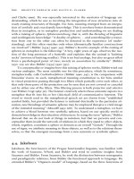
Wernicke Aphasia
201
Output
Phoneme
Input
Phoneme
A
Output
Grapheme
Semantic
B
Object
Feature
Input
Grapheme
Figure 9.7
Theoretical lesion loci underlying modality-specific language deficits. Lesion A impairs tasks involving input and output
phonemes, including auditory verbal comprehension, repetition, propositional speech, naming, reading aloud, and writing
to dictation. Lesion B impairs tasks involving input and output graphemes, including reading comprehension, propositional writing, written naming, reading aloud, and writing to dictation.
but as having “alexia and agraphia with conduction aphasia” by others (Selnes & Niccum, 1983).
Regardless of how these patients are categorized
within the traditional aphasiology nomenclature,
their deficit patterns provide additional information
about how language perception and production
systems might be organized according to the modality of stimulus or response.
Patients with superior written compared with
spoken language processing can be explained by
postulating damage to phoneme systems or pathways between phoneme and semantic representations (lesion A in figure 9.7). Such damage would
disrupt not only speech comprehension, but any
task dependent on recognition of speech sounds (repetition and writing to dictation) and any task involving production of speech (spontaneous speech,
reading aloud, naming objects, and repetition). Because pathways from visual input to semantics are
spared, such patients retain the ability to comprehend written words, match written words with
pictures, and name objects using written responses
(Caramazza et al., 1983; Ellis et al., 1983; Heilman
et al., 1979; Hier & Mohr, 1977; Hillis et al., 1999;
Howard & Franklin, 1987; Ingles, Mate-Kole, &
Connolly, 1996; Kirschner et al., 1981; Marshall
et al., 1985; Semenza, Cipolotti, & Denes, 1992;
Sevush et al., 1983). The preserved written naming
ability shown by these patients despite severely
impaired auditory comprehension and paraphasic
speech is very clearly at odds with Wernicke’s belief
that word-sound images are essential for writing.5
Errors of speech comprehension in these patients
reflect problems with phonemes rather than with
words or word meanings. For example, in writing
to dictation, patients make phonemic errors (e.g.,
they write “cap” after hearing “cat”), and in matching spoken words with pictures, they select incorrect items with names that sound similar to the
target. Such errors could result either from damage
to the input phoneme system or to the pathway
between phoneme and semantic levels. The patient
studied in detail by Hillis et al. (1999) made typical
errors of this kind on dictation and word–picture
Jeffrey R. Binder
matching tasks, but could readily discriminate
between similar-sounding spoken words like cap
and cat on a same-different decision task. This
pattern suggests that the patient was able to analyze
the constituent phonemes and to compare a sequence of phonemes with another sequence, but was
unable to translate correctly from the phoneme to
the semantic level.
Similarly, the errors of speech production
made by these patients are overwhelmingly of the
phonemic type, including phonemic paraphasias,
neologisms, and formal paraphasias, with only
infrequent semantic or mixed errors. Hillis et al.
(1999) modeled their patient’s neologistic speech
by lesioning Dell’s spreading activation speech
production network. Unlike the global lesions used
by Dell et al. (1997), Hillis et al. postulated a local
transmission lesion affecting connections between
the lemma (intermediate) and output phoneme
levels. When the lemma–phoneme connection
strength was lowered sufficiently to produce the
same overall error rate as that made by the patient
during object naming, the model network reproduced the patient’s pattern of errors with remarkable
precision, including high proportions of phonologically related nonwords (patient 53%, model 52.5%),
a smaller number of formal errors (patient 6%,
model 6.5%), and infrequent semantic or mixed
errors (patient 3%, model 2.7%). These results
provide further evidence not only for the processing locus of the lesion causing superior written
over oral language processing in this patient but
also for the concept that a focal transmission lesion
can cause a characteristic error pattern that depends
on the lesion’s locus.
Patients with this auditory variant of Wernicke
aphasia vary in terms of the extent to which speech
output is impaired. Most patients had severely paraphasic speech (Caramazza et al., 1983; Ellis et al.,
1983; Hier & Mohr, 1977; Hillis et al., 1999; Ingles
et al., 1996; Kirschner et al., 1981; Marshall et al.,
1985), but others made relatively few errors in
reading aloud (Heilman et al., 1979; Howard &
Franklin, 1987; Semenza et al., 1992; Sevush et al.,
1983). Even among the severely paraphasic patients,
reading aloud was generally less paraphasic than
202
spontaneous speech or object naming (Caramazza et
al., 1983; Ellis et al., 1983; Hillis et al., 1999).
The fact that some patients showed relatively
spared reading aloud despite severe auditory comprehension disturbance provides further evidence
for the existence of at least partially independent
input and output phoneme systems, as depicted in
the model presented here. This observation also provides evidence for a direct grapheme-to-phoneme
translation mechanism that bypasses the presumably lesioned semantic-to-phoneme output pathway.
Because patients with this pattern are relying on the
grapheme-to-phoneme pathway for reading aloud,
we might expect worse performance on exception
words, which depend relatively more on input from
the semantic pathway, and better reading of nonwords (see chapter 6 in this volume). These predictions have yet to be fully tested, although the patient
described by Hillis et al. (1999) clearly showed
superior reading of nonwords.
Patients with superior oral over written language
processing have also been reported (Déjerine, 1891;
Kirschner & Webb, 1982). A processing lesion
affecting input and output grapheme levels or their
connections (lesion B in figure 9.7) would produce
a modality-specific impairment of reading comprehension and written output directly analogous to the
oral language impairments discussed earlier. Such a
lesion would not, however, affect speech output or
speech comprehension. It is perhaps because a
disturbance in auditory-verbal comprehension is
considered the sine qua non of Wernicke aphasia
that patients with relatively isolated reading and
writing impairments of this kind have usually been
referred to as having “alexia with agraphia” rather
than a visual variant of Wernicke aphasia (Benson
& Geschwind, 1969; Déjerine, 1891; Goodglass &
Kaplan, 1972; Nielsen, 1946).
These dissociations between oral and written
language processes also offer important clues
concerning the neuroanatomical organization of
language comprehension and production systems.
For example, they suggest that input and output
phoneme systems are segregated anatomically from
input and output grapheme systems. The observation that input and output phoneme systems are
Wernicke Aphasia
often involved together, but that output may be relatively spared, suggests that these systems lie
close together in the brain, but are not entirely
overlapping. The co-occurrence, in a few patients,
of paraphasic speech output with reading and
writing disturbance and spared speech comprehension (Kirschner & Webb, 1982) suggests a smaller
anatomical distance between speech output and
grapheme systems than between speech input and
grapheme systems. These and other data regarding
lesion localization in Wernicke aphasia are taken up
in the next section.
Neuroanatomical Correlates of Wernicke
Aphasia
Wernicke’s aphasia has been recognized for well
over a century and has been a subject of great interest to neurologists and neuropsychologists, so it is
not surprising that the lesion correlation literature
concerning this syndrome is vast. The neuroanatomical basis of sensory aphasia was a central
issue for many German-speaking neurologists of
the late nineteenth and early twentieth century
who followed after Wernicke, including Lichtheim,
Bonhoefer, Liepmann, Heilbronner, Pick, Pötzl,
Henschen, Goldstein, and Kleist. French neurologists of the time who presented data on the topic
included Charcot, Pitres, Dejerine, Marie, and
others. Early contributions in English were made by
Bastian, Mills, Bramwell, Head, Wilson, Nielsen,
and others. In the last half of the twentieth century,
important investigations were reported by Penfield,
Russell, Hécaen, Luria, Goodglass, Benson, Naeser,
Kertesz, Selnes, Warrington, Damasio, and many
others. It is well beyond the scope of this chapter to
review even a small portion of this information in
detail. Our aim here is rather to sketch the origins
of some of the neuroanatomical models that have
been proposed and to evaluate, admittedly briefly,
their relation to the actual data.
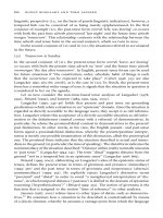
Patients with Wernicke aphasia have lesions in
the lateral temporal and parietal lobes, so a review
of the anatomy of this region is a useful starting
point for discussion (figure 9.8). The lesions involve
203
brain tissue on the lateral convex surface of these
lobes and almost never involve areas on the ventral
or medial surfaces. The lesion area typically includes cortex in and around the posterior sylvian
(lateral) fissure, giving rise to the term posterior
perisylvian to describe their general location. These
predictable locations result from the fact that in
most cases the lesions are due to arterial occlusion,
and that the vascular supply to the affected region–
the lower division of the middle cerebral artery–
follows a similar, characteristic pattern across
individuals (Mohr, Gautier, & Hier, 1992).
Temporal lobe structures within this vascular
territory include the superior temporal gyrus
(Brodmann areas 41, 42, and 22), the middle
temporal gyrus (Brodmann areas 21 and 37), and
variable (usually small) portions of the inferior
temporal gyrus (ITG; Brodmann areas 20 and 37).
Parietal lobe structures within the territory include
the angular gyrus (Brodmann area 39) and variable
portions of the supramarginal gyrus (Brodmann
area 40). In addition, the lesion almost always
damages the posterior third of the insula (the cortex
buried at the fundus of the sylvian fissure) and may
extend back to involve anterior aspects of the lateral
occipital lobe (figure 9.8).
Near the origin of this large vascular territory
is the posterior half of the STG, which studies
in human and nonhuman primates have shown to
contain portions of the cortical auditory system.
The superior surface of the STG in humans includes
a small, anterolaterally oriented convolution called
“Heschl’s gyrus” and, behind HG, the posterior
superior temporal plane or planum temporale. These
structures, located at the posterior-medial aspect
of the dorsal STG and buried in the sylvian
fissure, receive auditory projections from the medial
geniculate body and are believed to represent the
primary auditory cortex (Galaburda & Sanides,
1980; Liègeois-Chauvel, Musolino, & Chauvel,
1991; Mesulam & Pandya, 1973; Rademacher,
Caviness, Steinmetz, & Galaburda, 1993).
Studies in nonhuman primates of the anatomical
connections and unit activity of neurons in the STG
suggest that these primary areas then relay auditory
information to cortical association areas located
Jeffrey R. Binder
204
supramarginal
gyrus
Sylvian (lateral)
fissure
angular
gyrus
superior
temporal
gyrus
superior
temporal
sulcus
middle
temporal
gyrus
Figure 9.8
Gross anatomy of the lateral temporal and parietal lobes. Gyri are indicated as follows: superior temporal = vertical lines;
middle temporal = unmarked; inferior temporal = horizontal lines; angular = dots; supramarginal = horizontal waves; and
lateral occipital lobe = vertical waves. The approximate vascular territory of the lower division of the middle cerebral
artery is indicated with a dashed line.
more laterally on the superior surface and on the
outer surface of the STG (Galaburda & Pandya,
1983; Kaas & Hackett, 1998; Morel, Garraghty,
& Kaas, 1993; Rauschecker, 1998). It thus appears,
on the basis of these comparative studies, that the
superior and lateral surfaces of the STG contain
unimodal auditory cortex (Baylis, Rolls, &
Leonard, 1987; Creutzfeld, Ojemann, & Lettich,
1989; Galaburda & Sanides, 1980; Kaas & Hackett,
1998; Leinonen, Hyvärinen, & Sovijärvi, 1980;
Rauschecker, 1998), whereas the superior temporal
sulcus and more caudal-ventral structures (MTG,
ITG, AG) contain polymodal cortex that receives
input from auditory, visual, and somatosensory
sources (Baylis et al., 1987; Desimone & Gross,
1979; Hikosawa, Iwai, Saito, & Tanaka, 1988; Jones
& Powell, 1970; Seltzer & Pandya, 1978, 1994). For
regions caudal and ventral to the STG and STS,
however, inference about function in humans on the
basis of nonhuman primate data is perilous owing
to a lack of structural similarity across species. The
MTG and AG, in particular, appear to have developed much more extensively in humans than in
monkeys, so it is difficult to say whether data from
comparative studies shed much direct light on the
function of these areas in humans.
Like the STG and MTG, the AG is frequently
damaged in patients with Wernicke aphasia.
Although its borders are somewhat indistinct, the
AG consists of cortex surrounding the posterior
parietal extension of the STS and is approximately
the region Brodmann designated area 39. The SMG
(Brodmann area 40) lies just anterior to the AG
within the inferior parietal lobe and surrounds
the parietal extension of the sylvian fissure. The
SMG is frequently damaged in Wernicke aphasia,
Wernicke Aphasia
although its anterior aspect is often spared because
of blood supply from more anterior sources.
It hardly needs mentioning that Wernicke attributed his sensory aphasia syndrome to a lesion of
the STG (Wernicke, 1874, 1881), but the actual
motivations behind this view are less than obvious.
Wernicke’s case material was rather slim: ten
patients in all, only three of whom showed a
combination of auditory comprehension disturbance and paraphasic speech (reading comprehension was not mentioned). Two of these patients,
Rother and Funke, came to autopsy. In these two
cases there were large left hemisphere lesions reaching well beyond the STG, including in the patient
Rother (who also had shown signs of advanced
dementia clinically and had diffuse cerebral atrophy
at autopsy), the posterior MTG and the AG
(described as “the anastomosis of the first and
second temporal convolution”) and in Funke including the inferior frontal lobe, SMG, AG, MTG, and
inferior temporal lobe.
In emphasizing the STG component of these
large lesions, Wernicke was influenced in part by
the views of his mentor, Theodor Meynert, who
had described the subcortical auditory pathway as
leading to the general region of the sylvian fissure.
Even more important, however, was Wernicke’s
concept of the STG as the lower branch of a single
gyrus supporting speech functions (his “first primitive gyrus”), which encircles the sylvian fissure and
includes Broca’s area in the inferior frontal lobe.
Inferring from Meynert’s view that the frontal lobe
is involved in motor functions and the temporal
lobe in sensory functions, Wernicke assumed that
the STG must be the sensory analog of Broca’s
motor speech area.
Although subsequent researchers were strongly
influenced by Wernicke’s model, views regarding
the exact lesion correlate of Wernicke’s aphasia
have varied considerably (Bogen & Bogen, 1976).
As early as 1888, Charcot and his student Marie
included the left AG and MTG in the region associated with Wernicke’s aphasia (Marie, 1888/
1971). Marie later included the SMG as well (Marie
& Foix, 1917). In 1889, Starr reviewed fifty cases
205
of sensory aphasia published in the literature with
autopsy correlation, twenty-seven of whom had
Wernicke’s aphasia (Starr, 1889). None of these
patients had lesions restricted to the STG, and
Starr concluded that “in these cases the lesion was
wide in extent, involving the temporal, parietal
and occipital convolutions” (Starr, 1889, p. 87).
Similar views were expressed by Henschen,
Nielsen, and Goldstein, among others (Goldstein,
1948; Henschen, 1920–1922; Nielsen, 1946).
Much of modern thinking on this topic is influenced by the work of Geschwind, who followed
Wernicke, Liepmann, Pick, Kleist, and others in
emphasizing the role of the left STG in Wernicke’s
aphasia (Geschwind, 1971). Geschwind and his
students drew attention to left-right asymmetries
in the size of the planum temporale, that is, the
cortex posterior to Heschl’s gyrus on the dorsal
STG. This cortical region is larger on the left
side in approximately two-thirds of right-handed
people (Geschwind & Levitsky, 1968; Steinmetz,
Volkmann, Jäncke, & Freund, 1991; Wada, Clarke,
& Hamm, 1975). Recent studies have made it clear
that this asymmetry is due to interhemispheric differences in the shape of the posterior sylvian fissure,
which angles upward into the parietal lobe more
anteriorly in the right hemisphere (Binder, Frost,
Hammeke, Rao, & Cox, 1996; Rubens, Mahowald,
& Hutton, 1976; Steinmetz et al., 1990; Westbury,
Zatorre, & Evans, 1999). Geschwind and others
interpreted this asymmetry as confirming a central
role for the PT and the posterior half of the STG in
language functions (Foundas, Leonard, Gilmore,
Fennell, & Heilman, 1994; Galaburda, LeMay,
Kemper, & Geschwind, 1978; Witelson & Kigar,
1992) and argued that lesions in this area are responsible for Wernicke aphasia. Many late twentiethcentury textbooks and review articles thus equate
the posterior STG with “Wernicke’s area” (Benson,
1979; Geschwind, 1971; Mayeux & Kandel, 1985;
Mesulam, 1990).
The advent of brain imaging using computed
tomography and magnetic resonance imaging allowed aphasia localization to be investigated with
much larger subject samples and systematic,
Jeffrey R. Binder
standardized protocols (Caplan, Gow, & Makris,
1995; Damasio, 1981; Damasio, 1989; Damasio &
Damasio, 1989; Kertesz, Harlock, & Coates, 1979;
Kertesz, Lau, & Polk, 1993; Naeser, Hayward,
Laughlin, & Zatz, 1981; Selnes, Niccum, Knopman,
& Rubens, 1984). The aim of most of these studies
was to identify brain regions that are lesioned in
common across the majority of cases. This was
typically accomplished by drawing or tracing the
lesion on a standard brain template and finding areas
of lesion overlap across individuals. Several of
these studies showed the region of most consistent
overlap in Wernicke aphasia to be the posterior left
STG or STG and MTG (Damasio, 1981; Kertesz
et al., 1979), providing considerable support for
Wernicke’s original model and its refinements by
Geschwind and colleagues.
A potential problem with the lesion overlap technique is that it emphasizes overlap across individuals in the pattern of vascular supply, which may or
may not be related to the cognitive deficits in question. As already noted, Wernicke’s aphasia is due to
206
occlusion of the lower division of the middle cerebral artery. The proximal trunk of this arterial tree
lies in the posterior sylvian fissure, near the PT and
posterior STG, with its branches directed posteriorly and ventrally. The territory supplied by these
branches is somewhat variable, however, in some
cases including more or less of the anterior parietal
or ventral temporal regions shown in figure 9.8.
Because of this variability, and because retrograde
collateral flow arising from other major arteries
commonly causes variable sparing of the territory
supplied by the more distal branches, regions supplied by the trunk and proximal branches (i.e., the
STG and PT) are the most likely to be consistently
damaged (Mohr et al., 1992). Thus the region of
maximal overlap is determined largely by the
vascular anatomy pattern and is not necessarily the
region in which damage leads to Wernicke’s aphasia
(figure 9.9).
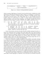
Given the critical role assigned by Wernicke and
others to the STG, it is reasonable to ask whether
lesions confined solely to the left STG actually cause
Figure 9.9
Diagram of three hypothetical ischemic lesions in the lower division of the middle cerebral artery territory, illustrating
typical patterns of lesion overlap (dark shading). Because the vascular tree in question arises from a trunk overlying the
posterior STG, this region is the most consistently damaged. Wernicke aphasia, on the other hand, might result from injury
to a more distributed system that includes middle temporal, angular, and supramarginal gyri, which are outside the area
of common overlap.
Wernicke Aphasia
Wernicke’s aphasia. Henschen was perhaps the first
to seriously test this prediction and offer evidence to
the contrary (Henschen, 1920–1922). In his meticulous review of 109 autopsied cases with temporal
lobe lesions reported in the literature, 19 cases had
damage confined to the left STG. None of these
patients had the syndrome of Wernicke’s aphasia; 5
were reported to have some degree of disturbance in
auditory comprehension, but all had intact reading
comprehension and writing. Henschen pointed out
that this pattern was inconsistent with Wernicke’s
model of the STG as a center for language comprehension and concluded that the STG is involved in
perception of spoken sounds.
Some later authors similarly disputed the claim
that lesions restricted to the posterior left STG
ever cause Wernicke’s aphasia (Foix, 1928; Mohr
et al., 1992), while several others have emphasized
that large lesions involving the STG, MTG, SMG,
and AG are typical (Damasio, 1989; Henschen,
1920–1922; Starr, 1889). Nielsen (1938) reviewed
several cases that purportedly had Wernicke’s
aphasia from an isolated posterior STG injury. Of
these, however, most had lesions clearly extending
into the MTG and the inferior parietal lobe, and
several cases were most likely caused by hematomas, which are known to produce relatively
nonlocalized neural dysfunction owing to pressure
effects from the hematoma mass.
Perhaps the best-documented case was Kleist’s
patient Papp, who presented with impaired auditory
comprehension and paraphasia (Kleist, 1962).
Reading comprehension was, unfortunately, not
tested. At autopsy there was a lesion centered in the
posterior left STG, with only minimal involvement
of the posterior MTG. Unfortunately, there was also
a large right perisylvian lesion that would, in conjunction with the left STG lesion, explain the case
as one of pure word deafness caused by bilateral
STG lesions. Kleist dismissed the importance of the
right hemisphere lesion, however, relating it to the
appearance of left hemiparesis well after the onset
of aphasia.
In contrast to this rather scant evidence in support
of the original Wernicke model, many instances of
207
isolated left STG lesion with completely normal
auditory and written comprehension have been
documented (Basso, Lecours, Moraschini, &
Vanier, 1985; Benson et al., 1973; Boller, 1973;
Damasio & Damasio, 1980; Henschen, 1920–
1922; Hoeft, 1957; Kleist, 1962; Liepmann &
Pappenheim, 1914; Stengel, 1933). Most of these
were extensive lesions that involved Heschl’s gyrus,
the PT, the posterior lateral STG, and underlying
white matter. Many of these patients had the syndrome of conduction aphasia, consisting of paraphasia (with primarily phonemic errors) during
speech, repetition, and naming; variable degrees of
anomia; and otherwise normal language functions,
including normal auditory and reading comprehension. Kleist’s patients are particularly clear examples because of the meticulous detail with which
they were studied at autopsy (Kleist, 1962). Believing as he did that the posterior left STG (and
particularly the PT) was critical for auditory comprehension, Kleist viewed these patients’ preserved
comprehension as evidence that they must have had
comprehension functions in the right STG, even
though two of the three were right-handed. Others
have echoed this view (Boller, 1973), although the
explanation seems quite unlikely given the rarity
of aphasic deficits after right hemisphere injury
(Faglia, Rottoli, & Vignolo, 1990; Gloning,
Gloning, Haub, & Quatember, 1969) and recent
functional imaging studies showing that right hemisphere language dominance is exceedingly rare in
healthy right-handed people (Pujol, Deus, Losilla,
& Capdevila, 1999; Springer et al., 1999). Recognizing this problem, Benson et al. postulated instead
that “the right hemisphere can rapidly assume the
functions of comprehension after destruction of the
Wernicke area” despite the fact that “comprehension of spoken language was always at a high level”
in their patient with left posterior STG infarction
(Benson et al., 1973, pp. 344–345).
A review of Kleist’s patients, however, suggests
another, much simpler explanation. The autopsy
figures and brief clinical descriptions provided
by Kleist make it clear that the patients’ comprehension deficits tended to increase as the lesion
Jeffrey R. Binder
extended beyond the STG, either ventrally into the
MTG or posteriorly into the AG. Subsequent CT
correlation studies provide other evidence for
a critical role of the MTG and AG in auditory
comprehension. Investigators in these studies rated
the degree of damage in selected brain regions
and correlated this information with patterns of
recovery.
Several studies showed a correspondence between poor recovery of auditory comprehension
and greater damage to the MTG, the AG, or both
(Dronkers, Redfern, & Ludy, 1995; Kertesz et al.,
1993; Naeser et al., 1987; Selnes et al., 1983). Total
infarct size was predictive of both degree of recovery and initial severity (Kertesz et al., 1993; Naeser
et al., 1987; Selnes et al., 1983; Selnes et al., 1984).
Moreover, even extensive damage to the STG did
not preclude a good recovery in some patients
(Kertesz et al., 1993; Naeser et al., 1987; Selnes et
al., 1984). One interpretation of these findings is
that they indicate a reorganization process by which
neighboring regions take over functions originally
performed by the STG (Kertesz et al., 1993). On
the other hand, Dronkers et al. (1995) presented
evidence that patients with lesions centered in the
MTG have more lasting deficits, even when the
STG is relatively spared, implying a primary
rather than a secondary role for the MTG in
comprehension.
Given the lack of reported cases with comprehension deficits from isolated STG damage, a parsimonious account of these data is that the MTG
and other areas surrounding the STG play a more
critical role in auditory comprehension than the
STG does itself, and that both initial severity and
degree of recovery are determined by the extent
of acute dysfunction in these neighboring regions.
In general, the data suggest that lesions centered in
the STG tend to produce either no comprehension
disturbance or a transient deficit that improves,
whereas MTG and AG lesions tend to produce
a more permanent deficit, with or without STG
involvement.
Further supporting this model is evidence that the
MTG and more ventral areas of the left temporal
208
lobe play a critical role in accessing and storing
semantic representations. For example, the syndrome of transcortical sensory aphasia, which is
characterized by impairments of spoken and written
language comprehension without phonemic paraphasia, has been consistently linked to lesions in
the ventral and ventrolateral temporal lobe that
involve the fusiform gyrus and the ITG, and to
posterior convexity lesions that involve the
posterior MTG and the temporo-occipital junction (Alexander, Hiltbrunner, & Fischer, 1989;
Damasio, 1989; Kertesz, Sheppard, & MacKenzie,
1982; Rapcsak & Rubens, 1994).
Many aphasic patients (most of whom fit the
classic syndromes of anomic aphasia or transcortical sensory aphasia) have now been described who
show comprehension or naming deficits that are
relatively restricted to particular object categories
(Forde & Humphreys, 1999). Such patients may
make more errors with living than nonliving items,
more errors with animals than tools, more errors
with fruits and vegetables than other objects, and so
on. The category-specific nature of these deficits
suggests damage at the level of semantic representations, and nearly all the cases have been
associated with lesions involving left temporal
lobe regions outside the STG. Perhaps the first such
patient was Nielsen’s case, C.H.C., who developed
severe impairment of auditory comprehension
after focal infarction of the left MTG and ITG
(Nielsen, 1946). C.H.C. had marked anomia, but
was able to recognize and name living things much
better than nonliving objects. Similar cases have
been associated with focal infarctions of the left
MTG or ITG (Hart & Gordon, 1990; Hillis &
Caramazza, 1991) or with herpes encephalitis
that caused anterior ventral temporal lobe damage
(Laiacona, Capitani, & Barbarotto, 1997; Silveri &
Gainotti, 1988; Sirigu, Duhamel, & Poncet, 1991;
Warrington & Shallice, 1984).
Other evidence for the importance of the left MTG
in semantic processing comes from a report by
Chertkow and colleagues (Chertkow, Bub, Deaudon,
& Whitehead, 1997), who studied eight aphasic
patients with comprehension deficits following
Wernicke Aphasia
posterior perisylvian lesions (two Wernicke’s
aphasia, six global aphasia). Five of the patients
showed comprehension deficits in associative
matching tasks, even when the test materials consisted entirely of pictures, which suggested damage
to semantic information stores. In these patients, the
lesions extended further ventrally than in the other
three patients, with the largest area of overlap in the
middle and posterior MTG.
Finally, several studies show that aphasic patients
who make primarily semantic paraphasias have
lesions restricted to ventral temporal regions,
particularly the posterior MTG and ITG (Cappa,
Cavallotti, & Vignolo, 1981; Gainotti, Silveri, &
Villa, 1986). In contrast, patients who make primarily phonemic paraphasias have posterior STG,
insula, or inferior parietal lesions (Benson et al.,
1973; Cappa et al., 1981; Damasio & Damasio,
1980; Palumbo, Alexander, & Naeser, 1992). A
similar dorsal-ventral dissociation between areas
associated with phonemic and semantic paraphasia
has been observed during electrical interference
stimulation studies (Ojemann, 1983).
Some authors have disputed the importance of
the left MTG in word comprehension. In particular, a case reported by Pick in 1909 (Pick, 1909)
and later cited by Nielsen and others (Henschen,
1920–1922; Hickok & Poeppel, 2000; Nielsen,
1946) has been used as evidence to the contrary. At
autopsy the patient had cysts in the white matter of
both temporal lobes, the remnants of intracerebral
hemorrhages, which affected much of the middle
portion of the MTG bilaterally, and on the left also
involved the white matter of the posterior MTG,
portions of the STG, and a small amount of the
angular gyrus. The patient was apparently able to
understand spoken words, although his own speech
was paraphasic and unintelligible, consisting of
“disconnected nonsense,” and he was completely
unable to write. The case provides some negative
evidence, although this is tempered by the knowledge that subcortical hematomas are known to
produce rather unpredictable deficits relative to cortical lesions, and by the fact that the patient was not
examined until 3 weeks after the onset of the stroke,
209
during which time considerable recovery may have
occurred.
Against this single case are several examples,
from the same time period, of patients with small
left MTG cortical lesions who showed profound
comprehension disturbances (Henschen, 1920–
1922). The patient of Hammond, for example, had
complete loss of comprehension for spoken and
written material as a result of a focal lesion that involved the midportion of the left MTG (Hammond,
1900). Nielsen’s patient, C.H.C., who developed
severe comprehension disturbance after a posterior
MTG and ITG lesion, has already been mentioned
(Nielsen, 1946). Although ischemic lesions restricted to the MTG are rather rare owing to the
anatomical characteristics of the vascular supply,
the modern literature also contains several examples
(Chertkow et al., 1997; Dronkers et al., 1995;
Hart & Gordon, 1990). These patients uniformly
demonstrated deficits in spoken and written word
comprehension.
If the STG and PT do not play a primary role in
language comprehension, damage to these regions
almost certainly contributes to the paraphasic component of Wernicke’s aphasia. As noted earlier, isolated posterior STG lesions have frequently been
observed in association with phonemic paraphasia
(Benson et al., 1973; Damasio & Damasio, 1980;
Kleist, 1962; Liepmann & Pappenheim, 1914), as
have lesions in nearby posterior perisylvian areas
also frequently damaged in Wernicke’s aphasia,
such as the SMG and posterior insula (Benson et al.,
1973; Damasio & Damasio, 1980; Palumbo et al.,
1992). This functional–anatomical correlation has
been further corroborated by cortical stimulation
studies demonstrating the appearance of phonemic
paraphasia and other speech errors during electrical
interference stimulation of the posterior STG
(Anderson et al., 1999; Quigg & Fountain, 1999).
It thus appears that the posterior STG (including
the PT), the SMG, and the posterior insula play
a critical role in the selection and production of
ordered phoneme sequences. In addition to the
selection of output phonemes, this complex process requires mapping from output phoneme to
Jeffrey R. Binder
articulatory codes, sensory feedback mechanisms
that help guide movements of the vocal tract, and
short-term memory mechanisms for maintaining a
phoneme sequence as it is being produced (Caplan
& Waters, 1992).
To summarize some of this extensive material,
there seems to be little evidence that lesions of the
STG and/or PT produce the profound, multimodal
comprehension disturbance typical of Wernicke’s
aphasia, but such lesions do regularly cause paraphasic production, particularly phonemic paraphasia. In contrast to the effects of isolated STG lesions,
lesions in more ventral areas of the temporal lobe
and in the angular gyrus may produce profound
disturbances in comprehension. The clear double
dissociation between phonemic paraphasia and
comprehension impairment observed in patients
with posterior STG lesions and in patients with
lesions beyond the STG, respectively, is strong evidence that these two components of Wernicke’s
aphasia syndrome have no necessary functional or
anatomical link. Their co-occurrence in Wernicke’s
aphasia, according to the model being developed
here, results from the fact that the typical lesion
in Wernicke’s aphasia includes the STG but
spreads beyond it into surrounding areas ventral and
posterior to the STG that are critical for word
comprehension.
As discussed earlier, patients with fluent aphasia
do not always have equivalent impairment in comprehending spoken and written words. This is to
be expected given the very different pathways to
semantic representations that are engaged as a result
of phonemic versus graphemic input. The available
anatomical data suggest that patients with relatively
worse speech comprehension and better reading
comprehension characteristically have lesions in the
left temporal lobe (Hier & Mohr, 1977; Hillis et al.,
1999; Ingles et al., 1996; Kirschner et al., 1981;
Roeltgen, Sevush, & Heilman, 1983). It is important to note that when the lesions are unilateral, the
deficits nearly always involve both modalities, i.e.,
the differences between spoken and written comprehension are relative rather than absolute. Relative sparing of reading comprehension seems to be
210
most pronounced when the lesion is restricted to the
dorsal temporal lobe, involving only the STG and
MTG (Kirschner et al., 1981), or to the anterior
aspect of the temporal lobe.
The patient of Hillis et al. (1999), who presented
with speech comprehension deficit and phonemic
paraphasia after a small hemorrhage in the posterior
left sylvian fissure, is an extreme example in that
reading comprehension (as assessed by word–
picture matching and synonym matching) was
entirely normal. This patient, however, had encephalomalacia in the contralateral anterior perisylvian region, the result of a previous meningioma
resection, and so probably had disturbed speech
comprehension as a result of bilateral superior
temporal lobe damage, as occurs in the syndrome
of pure word deafness (Barrett, 1910; Buchman,
Garron, Trost-Cardamone, Wichter, & Schwartz,
1986; Goldstein, 1974; Henschen, 1918–1919;
Tanaka, Yamadori, & Mori, 1987).
Two similar recent cases are well documented,
both of whom had severe disturbance of speech
comprehension, phonemic paraphasia, sparing of
reading comprehension, and bilateral perisylvian
lesions sparing the MTG and more ventral temporal
areas (Marshall et al., 1985; Semenza et al., 1992).
It is notable that the patient of Semenza et al.
presented with language deficits only after a right
hemisphere lesion, an earlier left unilateral lesion
having caused no comprehension or production
deficits. These three patients are by no means
unique: many, if not most, of the reported cases of
pure word deafness from bilateral superior temporal lesions also had varying degrees of phonemic
paraphasia, sometimes with mild anomia (Buchman
et al., 1986; Goldstein, 1974).
Thus there appear to be two distinct syndromes
of preserved comprehension for written over spoken
language. In cases with multimodal deficits and
relative sparing of reading, the lesion is unilateral
and affects multiple regions in the left temporal
lobe. This lesion damages some part of the pathway
leading from input phoneme representations to
semantics, with relatively less involvement of the
grapheme-to-semantics pathway. In patients with
Wernicke Aphasia
complete sparing of reading comprehension, the
lesion affects the STG bilaterally, affecting only the
phoneme pathway. The complete sparing of reading
comprehension in the latter syndrome suggests that
the functional impairment lies at a relatively early
stage in the phoneme-to-semantics pathway, such as
at the input phoneme level or its connections to the
intermediate level (Hillis et al., 1999). The anatomical data, then, suggest that this early component is
bilaterally organized in the STG, in contrast to later
components of the phoneme-to-semantics pathway,
such as the intermediate level or its connections
to the semantic level, which are more unilaterally
represented and partially overlap the grapheme-tosemantics pathway.
Patients with this auditory variant of Wernicke
aphasia also have relatively greater impairment
of speech production compared with writing (Hier
& Mohr, 1977; Hillis et al., 1999; Kirschner et al.,
1981; Marshall et al., 1985; Roeltgen et al., 1983;
Semenza et al., 1992). In keeping with the studies
cited previously, the mix of speech errors depends
on the location of the lesion along the dorsal-ventral
axis of the temporal lobe. Lesions involving ventral
temporal regions produce empty speech with few
phonemic errors (Hier & Mohr, 1977), while temporal lobe lesions confined to the STG or involving
the STG and SMG produce marked phonemic paraphasia with frequent neologisms (Hillis et al., 1999;
Semenza et al., 1992). Naming errors consist primarily of omissions (inability to produce a word)
in the larger lesions and phonemic paraphasia or
neologism in the STG and SMG cases. Analogous
to reading comprehension, writing performance in
these patients is impaired but relatively better than
speaking if the lesion is large (Hier & Mohr, 1977;
Kirschner et al., 1981; Roeltgen et al., 1983) and is
almost completely preserved if the lesion is confined to the STG and SMG (Hillis et al., 1999;
Marshall et al., 1985; Semenza et al., 1992). These
data indicate that, as with the input pathways, the
phoneme and grapheme production pathways are
to some extent functionally and anatomically independent. In particular, the phoneme output pathway
is strongly associated with the left STG and SMG,
211
which appear not to be involved much at all in
the grapheme ouput pathway. Although large left
temporal lobe lesions produce impairments in
both modalities, writing production is relatively
less dependent on the temporal lobe than is speech
production.
The converse syndrome involves relative impairment of reading comprehension and writing
compared with speech comprehension. Evidence
exists in the early aphasia literature (Déjerine, 1892;
Henschen, 1920–1922; Nielsen, 1946) as well as in
more recent studies (Basso, Taborelli, & Vignolo,
1978; Kirschner & Webb, 1982) localizing this syndrome to the posterior parietal lobe or parietotemporo-occipital junction, including the angular gyrus.
Such cases further illustrate the relative independence of grapheme input from phoneme input pathways as well as writing from speech production
mechanisms.
It should be noted that cases exist of patients with
speech comprehension deficits from lesions in the
vicinity of the angular gyrus (Chertkow et al., 1997;
Henschen, 1920–1922), so it remains unclear why
some patients with lesions in this region have relatively preserved speech comprehension. It may
be that speech comprehension is more likely to be
preserved as the lesion focus moves posteriorly
in the parietal lobe, or that the variability from case
to case merely reflects individual variability in the
functional anatomy of this region. The patients
described by Kirschner and Webb (1982) are somewhat intermediate in this regard, in that they presented initially with speech comprehension deficits
that later cleared, leaving predominantly reading
comprehension and writing impairments. These
patients also showed persistent paraphasic errors
in speech, as well as naming difficulty, prompting
Kirschner and Webb to classify them as atypical
cases of Wernicke’s aphasia rather than “alexia with
agraphia.”
From the point of view of the model developed
here, the paraphasic speech of the patients described
by Kirschner and Webb can be attributed to involvement of the posterior STG and/or the SMG, which
was documented in two of the three cases (the third
Jeffrey R. Binder
patient was not scanned). Thus, the co-occurrence
of alexia, agraphia, and paraphasic speech in these
patients may simply reflect the anatomical proximity of the angular gyrus, which appears to be
critical to both the grapheme-to-semantics pathway activated during reading and the semanticsto-grapheme pathway activated during writing,
to the output phoneme pathway in the STG and
SMG.
More detailed studies of agraphia have uncovered
patients in whom there appear to be writing deficits
related specifically to damage in the phoneme-tographeme pathway. This syndrome, known as
phonological agraphia, is characterized by particular difficulty writing or spelling nonwords (e.g.,
slithy) compared with real words. The spelling of
nonwords is thought to depend particularly on a
direct translation from output phonemes to output
graphemes because these items have no representation at the semantic level. The spelling of actual
words, in contrast, can be accomplished by either
the phoneme-to-grapheme pathway or by a less
direct phoneme-to-semantic-to-grapheme route.
A
212
One functional lesion that could produce phonological agraphia would be damage to the output
phoneme level, which would be expected to produce co-occurring phonemic paraphasia. This prediction is well supported by the available lesion
data, which show that most patients with phonological agraphia have SMG lesions, often with
accompanying posterior STG damage, and are
also severely paraphasic (Alexander, Friedman,
Loverso, & Fischer, 1992; Roeltgen et al., 1983).
The phoneme-to-grapheme mapping process is
certain to be rather complex, however, probably
involving an intermediate representational level as
well as short-term memory systems to keep both the
phoneme string and the grapheme string available
while the writing process unfolds. At present it is
unclear precisely which process or combination of
processes is impaired by the posterior perisylvian
lesions producing phonological agraphia.
Figure 9.10 summarizes some of the functional–
anatomical correlations observed in patients with
lateral convexity temporal and/or parietal lobe
lesions. Such correlations can only be approximate
B
Figure 9.10
Summary of some lesion-deficit correlations in fluent aphasia. The figures are approximations only and represent the
author’s interpretation of a large body of published data. (A) Patterns of paraphasia. Triangles mark areas in which damage
produces phonemic errors, and circles mark areas associated with verbal errors. (B) Comprehension deficits. Triangles
indicate regions in which bilateral lesions cause an auditory verbal comprehension deficit without impairment of reading
comprehension. Squares indicate regions associated with auditory verbal deficit, and circles indicate areas associated with
impaired reading comprehension. Auditory verbal and reading areas overlap through much of the posterior temporal lobe
and segregate to some degree in anterior temporal and posterior parietal regions.
Wernicke Aphasia
owing to the great variability present in naturally
occurring lesions, the often incomplete anatomical and/or behavioral descriptions of the data, and
the underlying intersubject variability in functional
organization. Clinical signs also depend greatly on
the amount of time elapsed since the initial injury.
As mentioned, for example, the mixture of phonemic and verbal paraphasias observed in Wernicke
aphasia evolves to some extent over time, so part A
of the figure is nothing more than a general outline.
Other data concerning the functional anatomy of
Wernicke’s aphasia and related syndromes come
from functional neuroimaging studies of normal
language processing, which are summarized in the
next section.
Functional Neuroimaging Studies
As should be clear from the previous section, studies
of lesion location are performed with two general
aims in mind. The first of these is the more modest:
to describe the lesion that produces a clinical syndrome. Like the other aphasias, Wernicke aphasia
can be viewed simply as a syndrome—a collection
of deficits that tend to occur together—without
reference to an underlying theoretical model of
how damage produces the syndrome. Research
along these lines has focused, for example, on defining the average lesion characteristics associated with
the syndrome and how variations from the average
are associated with variations in the syndrome.
The second aim, a natural outgrowth of the first,
involves formulation and testing of an underlying
processing model that describes the functional role
of each brain region involved in the lesion area.
Such models are interesting in their own right and,
more important, can lead to a deeper understanding of the syndrome, permitting predictions to be
made about the location of a lesion in newly encountered patients, factors that produce variations
in the syndrome, and the manner and time course of
recovery.
Although much has been learned about underlying brain processes from studying lesions, this
approach also has important limitations. The overall
213
size and exact location of lesions vary considerably
across individuals, creating a large number of lesion variables that may or may not be related to the
behavioral deficits. As noted earlier, commonly
shared features of the vascular supply result in areas
of lesion overlap across subjects, independently of
any shared deficits. The detection of deficits varies
with the method and timing of testing, and with the
a priori aims of the researcher. Finally, damage to
one subsystem in a distributed processing network
may interfere with a wide assortment of behaviors,
leading to overlocalization through false attribution
of these behaviors to the lesioned area.
Functional imaging of intact human brains
provides useful complementary information for
the development of neuroanatomically oriented processing models. In contrast to lesion techniques,
these methods provide a picture of the full, intact
system at work. By experimentally manipulating
aspects of the task performed during scanning and
recording the regional changes in activation correlated with these manipulations, inferences can be
made about the processes carried out in each brain
region. By integrating this information with that
obtained from lesion studies, it is hoped that a more
complete and explicit theory will emerge to account
for how damage in specific regions or combinations
of regions leads to specific deficits. This section
presents a brief overview of PET and fMRI studies
of speech and language processing that are relevant
to an account of Wernicke aphasia. Where possible,
the data are compared and contrasted with information from lesion-deficit correlation studies.
Perception of Speech Sounds
Many PET and functional MRI (fMRI) studies have
focused on the neural basis of processing speech
sounds. In most such studies, brain activation states
were measured during the presentation of speech
sounds in contrast to no sounds, a comparison that
consistently and robustly activates the STG bilaterally (Binder et al., 2000; Binder et al., 1994b;
Dhankhar et al., 1997; Fiez, Raichle, Balota, Tallal,
& Petersen, 1996a; Fiez et al., 1995; Hirano et al.,
1997; Howard et al., 1992; Jäncke, Shah, Posse,
Jeffrey R. Binder
Grosse-Ryuken, & Müller-Gärtner, 1998; Mazoyer
et al., 1993; O’Leary et al., 1996; Petersen, Fox,
Posner, Mintun, & Raichle, 1988; Price et al.,
1996b; Warburton et al., 1996; Wise et al., 1991).
The stimuli used in these experiments included
syllables, single words, pseudowords, reversed
speech, foreign words, and sentences. Activated
areas included Heschl’s gyrus, the PT, the dorsal
STG anterior to HG (the planum polare and the
dorsal temporal pole), the lateral STG, and the superior temporal sulcus. These results fit very well in
the long tradition linking speech comprehension
with the STG, and many investigators have simply
viewed these experiments as revealing activation of
“Wernicke’s area.”
What has sometimes been forgotten in interpreting such results is that speech is a very complex
and nuanced acoustic signal, containing a variety of
simultaneous and sequential auditory patterns that
must be analyzed prior to phoneme or word recognition (Klatt, 1989; Liberman et al., 1967; Oden
& Massaro, 1978; Stevens & Blumstein, 1981).
These auditory operations include not only the
well-known spectral analysis performed by the
cochlea and reflected in tonotopic organization of
the primary auditory cortex, but also analysis of
static spectral shapes and changes in spectral
configurations over time, and analysis of temporal asynchronies (see the section on comprehension disturbance). The possibility that considerable
neural activity might be required for analysis of
these acoustic features has often been overlooked
in neuroimaging studies of speech perception,
although such neural activity could explain much of
the STG activation observed in such studies. More
important, it seems likely that such prephonemic
auditory analysis constitutes an important and conceptually distinct processing level between primary
auditory and word recognition levels. A proposal of
this kind was first put forward clearly by Henschen
in 1918, although he has received almost no credit
for it.6
In addition to these purely theoretical concerns,
there are aspects of the STG activation results themselves that suggest a prelinguistic, auditory basis
214
for at least some of the activation. For example,
although language functions are believed to be
lateralized to the left hemisphere in most people,
STG activation by speech sounds occurs bilaterally.
Many investigators reported no asymmetry in the
degree of left versus right STG activation (Fiez
et al., 1995; Hirano et al., 1997; Howard et al.,
1992; Jäncke et al., 1998; O’Leary et al., 1996;
Warburton et al., 1996; Wise et al., 1991). Others
found slightly stronger activation on the left side,
although the degree of asymmetry was small
(Binder et al., 2000; Mazoyer et al., 1993). Many
of the studies examined only passive listening,
which might not be expected to fully engage the
language system and therefore might explain the
lack of leftward lateralization. However, in several
studies, adding a language task did not produce
greater asymmetry than passive listening (Fiez
et al., 1995; Grady et al., 1997; Wise et al., 1991).
The consistent finding of bilateral, symmetrical
activation is consistent with an account based on
general auditory processing, which would be expected to occur bilaterally. Another observation
consistent with this view is that the degree of
STG activation is very closely correlated with the
amount of auditory information presented, i.e., the
number of sounds presented per unit of time (Binder
et al., 1994a; Dhankhar et al., 1997; Mummery,
Ashburner, Scott, & Wise, 1999; Price et al., 1992;
Price et al., 1996b; Wise et al., 1991) and is usually
neglible during silent language tasks involving
purely visual stimulation (e.g., silent word reading)
(Howard et al., 1992; Petersen et al., 1988; Price
et al., 1994; Rumsey et al., 1997).
Finally, anatomical studies (Flechsig, 1908;
Galaburda & Pandya, 1983; Jones & Burton, 1976;
Kaas & Hackett, 1998; Mesulam & Pandya, 1973;
Rademacher et al., 1993; von Economo & Horn,
1930) and electrophysiological data from human
and nonhuman primates (Baylis et al., 1987;
Creutzfeld et al., 1989; Leinonen et al., 1980;
Liègeois-Chauvel et al., 1991; Merzenich & Brugge,
1973; Morel et al., 1993; Rauschecker, 1998) are
consistent with a unimodal, auditory processing
role for most of the STG, particularly the dorsal (HG
Wernicke Aphasia
and PT) and lateral aspects of the gyrus. These
observations suggest that much of the STG activation observed during auditory presentation of speech
arises from processing the complex auditory information present in these stimuli rather than from
engagement of linguistic (phonemic, lexical, or
semantic) processes.
In an effort to directly assess the contribution of
early auditory processes to STG activation, several
research groups have compared activation of the
STG by speech sounds with activation by simpler,
nonspeech sounds such as noise and tones. These
experiments included both passive listening and
active, target detection tasks. The consistent finding is that speech and nonspeech sounds produce
roughly equivalent activation of the dorsal STG,
including HG and PT, in both hemispheres
(Belin, Zatorre, Lafaille, Ahad, & Pike, 2000;
Binder et al., 2000; Binder et al., 1997; Binder
et al., 1996; Démonet et al., 1992; Mummery et al.,
1999; Zatorre, Evans, Meyer, & Gjedde, 1992).
Indeed, in several studies, tones produced stronger
215
activation of the PT than speech sounds, particularly
when active decision tasks were performed (Binder
et al., 1997; Binder et al., 1996; Démonet et al.,
1992). These data strongly support the idea that
neural activity in the dorsal STG (HG and PT) has
more to do with processing acoustic information
than linguistic information. Confirmatory support
comes from a recent fMRI study of acoustic complexity, in which it was shown that the PT responds
more strongly to frequency-modulated tones than
to unorganized noise, suggesting that this region
plays a role in the analysis of temporally organized
acoustic patterns (Binder et al., 2000).
In contrast to these findings for the dorsal STG,
more ventral areas, located on the anterolateral STG
and within the adjacent superior temporal sulcus,
are preferentially activated by speech sounds (figure
9.11). Although bilateral, this activation shows a
modest degree of leftward lateralization (Binder et
al., 2000; Binder et al., 1997; Démonet et al., 1992;
Mummery et al., 1999; Zatorre et al., 1992). The relatively anterior and ventral location of this “speech
Figure 9.11
Brain locations associated with stronger activation to speech sounds than to non-speech sounds (tones or noise) in five
imaging studies (Binder, Frost, Hammeke, Bellgowan, Springer, Kaufman, Possing, 2000; Binder, Frost, Hammeke, Cox,
Rao, Prieto, 1997; Demonet et al., 1992; Mummery, Ashbumer, Scott, & Wise, 1999; Zatorre, Evans, Meyer, & Gjedde,
1992). The squares represent activation peaks in standard stereotaxic space. The anterior-posterior (y) and inferiorsuperior (z) axes of the stereotaxic grid are shown with tick marks at 20-mm intervals. All left and right peaks have been
collapsed onto common left and right sagittal planes at x = ±55.
Jeffrey R. Binder
sound region” was initially surprising given the
traditional emphasis on the PT and posterior STG
as centers for speech comprehension. In contrast
to this traditional model, the functional imaging
data thus suggest that projections from primary to
secondary auditory cortex enabling speech recognition follow an anteroventral rather than a posterior course. Recent anatomical studies in monkeys
provide further support for this model by showing
two distinct projection systems within the auditory
system, one anteriorly directed and presumably supporting the recognition of complex sounds, and the
other posteriorly directed and presumably involved
in sound localization (Romanski et al., 1999). Also
of note, the STS location of these speech soundprocessing areas neatly explains several previously
documented cases of pure word deafness in which
the lesion involved the STS bilaterally while
sparing the dorsal STG (Barrett, 1910; Henschen,
1918–1919).
The nature of the processes carried out by this
speech sound region, however, remains somewhat
uncertain. The fact that speech sounds activate the
region more than tones or noise does not necessarily mean that this activation is related to language
processing. Because the tone and noise stimuli used
in these studies were much less complex from
an acoustic standpoint than the speech stimuli, it
may be that the increased activation for speech
sounds simply represents a more complex level of
auditory pattern analysis. This is underscored by
the fact that stronger activation is observed in the
STS for speech sounds irrespective of whether the
sounds are words or nonwords (Binder et al., 2000;
Démonet et al., 1992). In fact, activation in this
region is not even different for speech and reversed
speech (Binder et al., 2000; Dehaene et al., 1997;
Hirano et al., 1997; Perani et al., 1996; Price et al.,
1996b). Scott et al. addressed this issue by contrasting speech sounds with spectrally rotated
speech (Scott, Blank, Rosen, & Wise, 2000). The
latter is produced by inverting speech sounds in the
frequency domain, thus maintaining their acoustic
complexity but rendering the original phonemes
mostly unintelligible (Blesser, 1972). The results
216
show what appears to be a further subdivision
within the speech sound region. On the lateral STG,
anterolateral to the primary auditory cortex, the responses were as strong for spectrally rotated speech
as for normal speech, suggesting processing at an
auditory level. Further ventrally, in the STS, the
responses were stronger for speech than for spectrally rotated speech, suggesting neural activity
related to phoneme recognition.
These findings indicate the existence of a hierarchical processing stream concerned with speech
perception that is composed of at least three stages
located within the STG and STS. In accord with
anatomical and neurophysiological studies of
the auditory cortex, the earliest stage involves
sensory processors located in primary and belt
auditory regions on the superior temporal plane,
including the PT, which respond to relatively simple
frequency and intensity information (Galaburda
& Pandya, 1983; Mendelson & Cynader, 1985;
Merzenich & Brugge, 1973; Morel et al., 1993;
Phillips & Irvine, 1981; Rauschecker, Tian, Pons,
& Mishkin, 1997). Further anterolaterally, on the
lateral surface of the STG, are areas that respond
to more complex and combinatorial acoustic phenomena, such as configurations of spectral peaks
and dynamic spectral and intensity modulations
(Rauschecker, 1998; Rauschecker et al., 1997; Tian,
Reser, Durham, Kustov, & Rauschecker, 2001).
Still further ventrally, within the STS, are cortical
regions that appear to respond selectively in the
presence of intelligible phonemes (Scott et al.,
2000). The anterior and ventral course of this processing stream has been remarked on already.
What is perhaps most strikingly different about
this model in comparison with the conventional
view of Wernicke’s area, however, is that none of
these processing stages involve access to words
or word meanings. That is, all of the processes so
far discussed pertain specifically to recognition of
speech sounds rather than comprehension of words.
This model thus agrees well with neurolinguistic
descriptions of patients with pure word deafness
who have bilateral lesions in the STG and/or the
STS. These patients have disturbed perception of
Wernicke Aphasia
speech phonemes, but do not have difficulty comprehending word meaning (when tested with visually presented words) or accessing words during
speech production.
Processing Word Forms
According to the processing model described earlier and illustrated in schematic form in figure 9.2,
comprehension of heard or seen words requires
mapping from unimodal sensory representations,
such as phonemes or graphemes, to semantic representations. As discussed at points throughout this
chapter and illustrated in figure 9.3, the arbitrary
and nonlinear nature of these mappings suggests
the need for intermediate processing levels that represent combinations of phonemes or graphemes.
Theories that envision these combinatorial representations as localized and equivalent to whole
words describe them as the “phonological lexicon”
and “orthographic lexicon.”
In other theories, intermediate levels represent
phoneme and letter combinations in a distributed
manner with no one-to-one relationship between
words and representational units. Common to both
of these theoretical positions is the idea that the
intermediate levels enable mapping from phoneme
or grapheme information to semantics, and that the
intermediate levels represent information pertaining
to the (phonological or orthographic) structure of
words. The neutral expression “word-form processing” captures these commonalities and so will be
used to refer to intermediate levels of processing.
Many functional imaging studies have addressed
word-form processing using either spoken or printed
stimuli. The studies summarized here are those in
which brain activation from word or wordlike
stimuli was compared with activation from stimuli
that were not wordlike. One issue complicating the
interpretation of these data is that stimuli can have
varying degrees of “wordlikeness” (reflecting, for
example, such factors as the frequency of letter combinations, number of orthographic or phonological
neighbors, frequency of neighbors, and pronounceability), and many imaging studies do not incorpo-
217
rate any clear metric for this crucial variable. For the
most part, however, the contrasting conditions in
these studies have involved extremely different
stimuli in order to create clear distinctions between
stimuli with or without word form.
Another issue complicating many of these experiments is that activation of word-form information may be accompanied by activation of semantic
information, particularly when real words are used
as stimuli and when subjects are encouraged to
process the words for meaning. To avoid this confound, the following discussion focuses on studies
in which either (1) stimuli used in the word-form
condition were wordlike but were not real words
(i.e., were pseudowords), or (2) semantic processing requirements were matched in the word-form
and baseline tasks.
In phonological word-form studies, the usual
contrast is between spoken words and reversed
words (i.e., recordings of spoken words played
backward). Although reversed playback of spoken
words makes them unrecognizeable as meaningful
words, this manipulation does not completely remove phonological structure since subjects reliably
report phonemes on hearing such stimuli and there
is even a degree of consistency across subjects in
the particular phoneme sequences heard (Binder
et al., 2000). Indeed, several studies have shown
no differences in brain activation by words and
reversed words (Binder et al., 2000; Hirano et al.,
1997). Other investigators, however, have observed
activation differences favoring words (Howard
et al., 1992; Perani et al., 1996; Price et al., 1996b).
The peak activation foci observed in these word
versus reversed speech contrasts are distinctly separate from those in the STG and STS described
earlier in association with speech versus nonspeech
contrasts. As shown in figure 9.12, the word versus
reversed speech peak activations lie in the middle
temporal and posterior inferior temporal gyri,
areas adjacent to but distinct from the superior
temporal auditory cortex. Unlike the speech sound
activations observed in the STG and STS, activation in these areas is strongly lateralized to the left
hemisphere.
Jeffrey R. Binder
218
Orthographic Word Form
Phonologic Word Form
Speech > Nonspeech
All Word Form
Figure 9.12
Activation sites associated with word-form processing, almost all of which have been found in the left hemisphere. The
top panel shows left hemisphere activation peaks from seven word form experiments (Perani, Dehaene, Grassi, Cohen,
Cappa, Dupouz, Fazio, Mehler, 1996; Price, Wise, Warburton et al., 1996; Price et al., 1994; Price, Wise, & Frackowiak,
1996; Tagamets, Novick, Chalmers, & Friedman, 2000). The bottom panel illustrates segregation of these word-form
activation foci (circles) from speech perception areas (squares); the latter are also found in the right hemisphere (see
figure 9.11).
In orthographic word-form studies, the usual contrast is between words or pseudowords (pronounceable nonwords that look like words, e.g., tweal)
and consonant letter strings (Bavelier et al., 1997;
Herbster, Mintun, Nebes, & Becker, 1997; Howard
et al., 1992; Indefrey et al., 1997; Petersen, Fox,
Snyder, & Raichle, 1990; Price et al., 1994; Price,
Wise, & Frackowiak, 1996c; Small et al., 1996;
Tagamets, Novick, Chalmers, & Friedman, 2000).
Consonant strings (e.g., mpfjc) differ from wordlike
stimuli in two ways. First, they tend to contain letter
combinations that do not occur or occur only infrequently in the language (e.g., mp at the initial
position or jc at the final position of mpfjc). These
stimuli thus do not have a familiar orthographic
structure and presumably produce only weak activation at the orthographic word-form level. Second,
consonant strings in English are typically unpronounceable (except by an effortful insertion of
schwa sounds between consonants) and should thus
produce only weak activation of phonological word
form and output phoneme representations. These
two factors are, of course, inextricably linked to
some degree. Because of the quasi-regular relationship between graphemes and phonemes, increasing the degree of orthographic structure tends to
increase the degree of phonological structure,
leading to increased pronounceability.
As shown in figure 9.12, the peak activation foci
in studies contrasting orthographically wordlike
Wernicke Aphasia
stimuli with consonant strings have tended to cluster in the posterior MTG, the posterior STS, and
the posterior ITG (Bavelier et al., 1997; Herbster
et al., 1997; Howard et al., 1992; Indefrey et al.,
1997; Price et al., 1994; Price et al., 1996c; Small
et al., 1996; Tagamets et al., 2000). Similar activation peaks were observed in these studies whether
the word-form stimuli used were real words or
meaningless pseudowords, a finding that lends
credence to the notion that the processing level or
levels being identified are presemantic in nature.
Like the activation sites observed in spoken wordform studies, these foci have almost all been in the
left hemisphere.
One striking aspect of these results is the considerable overlap between regions identified in spoken
and printed word-form studies (figure 9.12). This
suggests that the phonological word-form system
used to map input phonemes to semantics and the
orthographic word-form system used to map input
graphemes to semantics are at least partially overlapping in the posterior MTG and ITG. Another
possible explanation for this overlap is that both the
spoken and written word-form conditions activate
representations of output phonemes. These representations are activated explicitly in tasks requiring
the repetition of heard speech or reading aloud of
orthographic stimuli, but are probably also engaged
automatically whenever the brain is presented with
stimuli that have a phonological structure (Macleod,
1991; Van Orden, 1987). Thus, some of the overlap
in figure 9.12 could be due to activation of output
phoneme representations or intermediate levels that
lead to output phonemes.
Semantic Processing
Semantic processes are those concerned with
storing, retrieving, and using knowledge about the
world, and are a key component of such ubiquitous behaviors as naming, comprehending and formulating language, problem solving, planning, and
thinking. Our focus here is on tasks involving
comprehension of word meaning. As should be
clear by now, understanding the meaning of words
219
is a complex process that engages multiple representational stages and nonlinear transformations.
The following review summarizes functional
imaging studies that attempted to isolate the final
stage of this processing sequence, in which semantic representations are activated (Binder et al.,
1999; Chee, O’Craven, Bergida, Rosen, & Savoy,
1999; Démonet et al., 1992; Mummery, Patterson,
Hodges, & Price, 1998; Poldrack et al., 1999; Price,
Moore, Humphreys, & Wise, 1997; Pugh et al.,
1996). The semantic tasks used in these studies
required that meaning-based judgments be made
about words. These tasks included deciding if a
word represented a concept from a particular category (e.g., living or nonliving, foreign or domestic, and abstract or concrete), deciding whether two
words were related in meaning, or deciding which
of two words was closer in meaning to a third word.
The identification of brain activation related to
semantic access during such tasks requires the same
sort of subtraction strategy employed in the speech
perception and word-form experiments just reviewed. For tasks engaging semantic access, the
appropriate control condition is one in which identical sensory, phoneme or grapheme, and word-form
processing occurs, but without activation of (or with
less activation of) semantic information.
Two types of experimental design have been
used. In the first, control stimuli are pseudowords
(either spoken or written), and the control task
involves a judgment about the phonological structure (word form) of the pseudowords. These control
tasks have included deciding whether pseudowords
contain a target phoneme (Binder et al., 1999;
Démonet et al., 1992), whether two written pseudowords rhyme (Pugh et al., 1996), and whether a
written pseudoword has two syllables (Poldrack
et al., 1999). Because the words and pseudowords
are matched on low-level sensory and word-form
characteristics, differences in the activation level
between conditions are likely to be related to
semantic processes. Activated areas in these studies
(i.e., those in which activation was greater for the
semantic condition than for the control condition)
are shown, for those studies that reported activation
Jeffrey R. Binder
peaks, in figure 9.13. These areas included the left
angular gyrus, the left superior frontal gyrus, the left
inferior frontal gyrus, the left fusiform gyrus and
parahippocampus, and the left posterior cingulate
cortex.
The second type of experiment is similar, except
that the control task involves a judgment about the
phonological structure of words rather than pseudowords. This design provides a tighter control for
word-form processing because even carefully constructed pseudowords may not be as wordlike in
structure as real words. For theorists who embrace
the idea of localized whole-word representations
that are accessed only in the presence of real words,
using real words as control stimuli is necessary in
order to “subtract” activation due to lexical (as
opposed to semantic) processing. A potential disadvantage of this design is the possibility that using
real words in the control condition may result in
some degree of automatic activation of semantic
information, even when the task being performed is
not semantic (Binder & Price, 2001). In all of these
studies, the control task required the subjects to
judge whether the word contained a particular
number of syllables (Chee et al., 1999; Mummery
et al., 1998; Poldrack et al., 1999; Price et al.,
1997).
220
As shown in figure 9.13, the activations in these
studies were nearly identical to those observed in
the experiments using pseudoword control stimuli,
and included the angular gyrus, the superior frontal
gyrus, the inferior frontal gyrus, the ventral temporal cortex, the MTG and ITG, and the posterior cingulate cortex in the left hemisphere. It should be
noted that in two of these studies only the frontal
lobe was imaged (Demb et al., 1995; Poldrack et al.,
1999).
Although they are not perfectly consistent, these
results indicate a distributed group of left hemisphere brain regions engaged specifically during
activation and retrieval of semantic information.
One of the more consistently identified areas (in
four of the five studies in which it was imaged) is
the angular gyrus (Brodmann area 39). Brodmann
area 39 is a phylogenetically recent brain area
that is greatly expanded in the human relative to the
nonhuman primate brain (Geschwind, 1965). It
is situated strategically between visual, auditory,
and somatosensory centers, making it one of the
more reasonable candidates for a multimodal convergence area involved in storing or processing
very abstract representations of sensory experience
and word meaning.
Figure 9.13
Activation peaks where a semantic task produced stronger activation than a phonological task in seven imaging studies
of semantic processing (Binder, Frost, Hammeke, Bellgowan, Rao, Cox, 1999; Chee, O’Craven, Bergida, Rosen, & Savoy,
1999; Demonet et al., 1992; Mummery, Patterson, Hodges, & Price, 1998; Poldrack et al., 1999 (two studies); Price,
Moore, Humphreys, & Wise, 1997). Squares indicate experiments using pseudowords in the phonological task; sites
marked by circles are from experiments using words in the phonological task.
Wernicke Aphasia
Other areas frequently identified in these semantic studies include the dorsal prefrontal cortex in the
superior frontal gyrus and sulcus (seven of seven
studies), the ventral temporal cortex in the fusiform
and parahippocampal gyri (four of five studies), the
inferior frontal gyrus (four of seven studies), and the
posterior cingulate cortex and adjacent ventral precuneus (three of five studies). These regions are well
outside the area damaged in Wernicke’s aphasia and
so are not discussed further here (see Binder and
Price, 2001, for a discussion of these and related
results).
In a few studies, activation foci were observed in
the left MTG and ITG (Chee et al., 1999; Mummery
et al., 1998; Price et al., 1997), suggesting that a
subset of this ventrolateral temporal region may
subserve semantic-level processes in addition to
word-form processes. Several functional imaging
studies have demonstrated enhanced activation of
the posterior MTG when subjects identify objects
in the tool category compared with objects from
other categories, and when subjects generate verbs
relative to generating nouns (Martin, 2001). The
proximity of these activation sites to the visual
motion-processing region (human “area MT”) has
led to speculation that the posterior MTG may store
semantic representations related to visual motion,
which are particularly salient semantic features for
manipulable objects and verbs (Martin, 2001).
Phonological Production
The functional imaging studies discussed to this
point have concerned transformations from spoken
or visual word input to semantics, that is, the pathways engaged during comprehension of speech
and written text. Speech production, another language process impaired in Wernicke’s aphasia, has
received some attention in functional imaging
studies. As discussed earlier, deficits of ordered
phoneme selection, which result in phonemic paraphasia, are the hallmark of posterior perisylvian
lesions damaging the posterior STG and STS, the
posterior insula, or the ventral supramarginal gyrus.
On the basis of this correlation, a reasonable pre-
221
diction is that these regions should show activation
under task conditions that engage output phoneme
selection relative to conditions that do not activate
output phonemes.
One source of information on this question has
already been mentioned: studies contrasting pronounceable with unpronounceable letter strings.
As shown in figure 9.12, activation peaks in these
studies were found in the posterior left MTG and
ITG, but also involved the posterior left STS. In
fact, some studies have shown particularly strong
effects in the posterior STS (Bavelier et al., 1997;
Howard et al., 1992; Indefrey et al., 1997; Price
et al., 1994; Small et al., 1996). As noted earlier,
however, it is difficult to attribute the posterior
STS activation specifically to processing of output phonemes because the pronounceable and
unpronounceable items in these studies also differed
along orthographic dimensions. Findings from the
auditory word-form comparisons, however, provide
indirect support for such an interpretation. These
studies, in which spoken words were contrasted
with reversed forms of the words, reveal activation
of the left MTG and ITG, but do not generally show
differential activation in the posterior STS. If
we assume that both normal and reversed speech
input produce some degree of activation of output
phonemes (i.e., that isolated phonemes may be perceived in these stimuli even if they do not have word
form), a contrast between these stimuli would not
be expected to show activation of output phoneme
systems.
Other evidence corroborating a specific role
for the posterior left STS in processing output
phonemes comes from a study by Wise et al. (2001).
These authors reported common activation of the
posterior left STS in three experiments. In the first
of these, passive listening to words was contrasted
with passive listening to signal-modulated noise
(white noise that was amplitude modulated using
the amplitude contours of speech sounds). Regions
selectively activated by words included the anterior
STS and the anterolateral STG bilaterally, which
is consistent with other speech-nonspeech comparisons (see figure 9.11). In the left hemisphere,
Jeffrey R. Binder
this activation spread posteriorly to involve the posterior STS. In the second experiment, posterior left
STS activation was observed during a silent wordgeneration task (“think of as many words as possible that are related to a cue word”) relative to a
resting state. Other temporoparietal regions activated in this contrast included the adjacent posterior
left STG, the posterior left MTG, and the left supramarginal gyrus. These results are consistent with
several other studies that showed posterior STG
and/or STS activation during word generation contrasted with rest (Fiez et al., 1996a; Hickok et al.,
2000). In the final and most compelling experiment,
the subjects generated words either aloud or silently
at various rates that were controlled by varying
the rate of presentation of cue words. The analysis
searched for brain regions in which the activation
level was correlated with the combined rate of
hearing and internally generating words. Only the
posterior left STS showed such a correlation.
The selection of output phonemes may lead to
overt speech production by movement of the vocal
tract, or to some form of “internal speech” without
articulation or phonation. If output phonemes are
represented in the posterior left STS, then overt
speech production must involve an interface
between this brain region and speech articulation
mechanisms located in the inferior frontal lobe. The
lesion literature on phonemic paraphasia suggests
that this interface exists within the cortical and subcortical pathways lying between the posterior STS
and the inferior frontal lobe, i.e., in the posterior
STG, supramarginal gyrus, and posterior insula.
It is likely that this interface also involves proprioceptive and other somatosensory input from
the adjacent inferior parietal cortex, which provides
dynamic feedback concerning position and movement of the vocal tract (Luria, 1966). For longer
utterances, it may also be necessary to maintain a
short-term record of the phoneme sequence to
be uttered so that this information does not fade
while articulation is in progress (Caplan & Waters,
1992). This “phonological buffer” is particularly
implicated in internal speech and in tasks in which
the phoneme sequence must be maintained in con-
222
sciousness for an extended period without overt
articulation.
Although little convergent data regarding this
phoneme-to-articulation pathway are yet available,
a few imaging results are suggestive. Paus et al.
(Paus, Perry, Zatorre, Worsley, & Evans, 1996) had
subjects whisper two syllables repeatedly at varying
rates. Auditory input was held constant across conditions by presenting continuous white noise that
masked any perception of speech. The investigators
searched for brain regions in which the activation
level was correlated with the rate of speech articulation. One area showing this pattern was a small
focus in the left planum temporale. Activation in
the left precentral gyrus, a motor area associated
with speech production, also varied with rate. The
authors suggested that the left planum temporale
and left premotor cortex function together during
speech production, possibly as an interactive feedforward and feedback system.
Wise et al. (2001) also searched for brain regions that are activated during speech production
independent from auditory input. Their subjects
were given a phrase (“buy Bobby a poppy”) and
asked to (1) say the phrase repeatedly aloud; (2)
mouth the phrase with lip movement but no sound
production; (3) sound out the phrase by substituting
the syllable “uh” for the original syllables, thereby
activating diaphragm, vocal cord, and glottal components of production without lip or tongue articulators; and (4) internally vocalize the phrase
repeatedly without movement or sound. The authors
contrasted the first three conditions, all of which
involved overt motor production, with the internal
vocalization condition. Similar to the study by Paus
et al., activated regions included the left ventral
motor cortex and a small focus in the posterior left
sylvian fissure (coordinates -42, -40, +20). This
focus is at the most posterior and medial aspect of
the sylvian fissure, at the junction of the planum
temporale, the supramarginal gyrus, and the posterior insula. It is worth noting that the posterior left
STS, which we have suggested may be involved in
representation of output phonemes, was not identified in this study, a result predicted by the fact that