Báo cáo y học: "Analyzing and minimizing PCR amplification bias in Illumina sequencing libraries" pps
Bạn đang xem bản rút gọn của tài liệu. Xem và tải ngay bản đầy đủ của tài liệu tại đây (464.72 KB, 14 trang )
METH O D Open Access
Analyzing and minimizing PCR amplification bias
in Illumina sequencing libraries
Daniel Aird
1
, Michael G Ross
1
, Wei-Sheng Chen
2
, Maxwell Danielsson
2
, Timothy Fennell
3
, Carsten Russ
1
,
David B Jaffe
1
, Chad Nusbaum
1
, Andreas Gnirke
1*
Abstract
Despite the ever-increasing output of Illumina sequencing data, loci with extreme base compositions are often
under-represented or absent. To evaluate sources of base-composition bias, we traced genomic sequences ranging
from 6% to 90% GC through the process by quantitative PCR. We identified PCR during library preparation as a
principal source of bias and optimized the conditions. Our improved protocol significantly reduces amplification
bias and minimizes the previously severe effects of PCR instrument and temperature ramp rate.
Background
The Illumina sequencing plat form [1], like other mas-
sively parallel sequencing platforms [2,3], continues to
produce ever-increasing amounts of data, yet suffers
from under-representation and reduced quality at loci
with extreme base compositions that are recalcitrant to
thetechnology[1,4-6].Unevencoverageduetobase
composition necessitates sequencing to excessively high
mean coverage for de novo genome assembly [7] and for
sensitive polymorphism discovery [8,9]. Although loci
with extreme base composition constitute only a small
fraction of the human genome, they include biologically
and medically relevant re-sequencing target s. For exam-
ple, 104 of the first 136 coding bases of the retinoblas-
toma tumor suppressor gene RB1 are G or C.
Traditional Sanger sequencing has long been known
to suffer from problem s related to the base composition
of sequencing templates. GC-rich stretches led to com-
pression artifacts. Po lymerase slippage in poly(A) runs
and AT dinucleotide repeats caused mixed sequencing
ladders and poor read quality. Processes upstream of the
actual sequencing, such as cloning, introduced bias
against inverted repeats, extreme base-co mpositio ns or
genes not tolerated by t he bacterial cloning host. Gaps
due to unclonable sequences had to be recovered and
finished by PCR [10], or, in some cases, by re sorting to
alternative hosts [11]. Cloning bias hindered efforts to
sequence the AT-rich genomes of Dictyostelium [12]
and Plasmodium [13]andexcludedtheGC-richfirst
exons of about 10% of protein-coding genes in the dog
(K Lindblad-Toh, personal communication) from an
otherwise high-quality reference genome assembly [14].
New genome sequencing technologies [1-3,15-17] no
longer rely on cloning in a microbial host. Instead of
ligating DNA fragments to cloning vectors, the three
major platforms currently on the market (454, Illumina
and SOLiD) involve ligation of DNA fragments to spe-
cial adapters for clonal amplification in vitro rather than
in vivo. Due to the massively parallel nature of the pro-
cess, standardized reaction conditions must be applied
to amplify and sequence complex libraries of fragments
thatcompriseawidespectrumofsequencecomposi-
tions. All three platforms display systematic biases and
unevenness as the observed coverage distributions are
significantly wider than the Poisson distribution
expected from unbiased, random sampling [18].
The Illumina sequencing process consists of i) library
preparation on the lab bench, ii) cluster amplification,
sequencing-by-synthesis and image analysis on proprie-
tary instruments, followed by iii) post-sequencing data
processing. Bias can be introduced at all three stages.
For example, high cluster densities on the Illumina flow-
cell suppress GC-rich reads. Changes to sequencing kits,
protocols and instrument firmware can affect the base
composition of sequencing data. Moreover, bias is
known to vary between laboratories, from run to run or
even from lane to lane on the same flowcell. Such varia-
bility and instability in the system confound comparative
* Correspondence:
1
Genome Sequencing and Analysis Program, Broad Institute of MIT and
Harvard, 320 Charles Street, Cambridge, MA 02141, USA
Full list of author information is available at the end of the article
Aird et al. Genome Biology 2011, 12:R18
/>© 2011 Aird et al.; licensee BioMed Central Ltd. This is an open access article distri buted under the terms of the Creative Commons
Attribution License ( which permits unre stricted use, distribution, and reproduction in
any medium, provided the original work is properly cited.
studies [19,20] and render systematic bias investi gatio ns
difficult.
Here, we set out t o evaluate sources of bias during
Illumina library preparation and to ameliorate the
effects. We undertook a systematic dissection of the
process, using quantitative PCR (qPCR) instead of Illu-
mina sequencing as a quick and system-independent
read-out for base-composition bias. We identified library
amplification by PCR as by far the most discriminatory
step. We examined hidden factors such as make and
model of thermocyclers and modified the thermocy cling
protocol. We tested alternative PCR enzymes and che-
mical ingredients in amplific ation reactions. F inally, we
validated the qPCR results by Illumina sequencing. Our
optimized protocol amplifies sequencing libraries more
evenly than the standard protocol and minimizes the
previously severe effects of PCR instrument and tem-
perature ramp rate.
Results
Following a diverse panel of loci through the Illumina
library preparation
The Illumina library preparation protocol is a multi-step
process consisting of shearing of the input DNA, enzy-
matic end repair, 5’ -phosphorylation and 3’-single-dA
extension of the resulting fragments, adapter ligation,
size fractionation on an agarose gel and PCR amplifica-
tion of adapter-ligated fragments. Bias can potentially be
introduced at any step, including the physical clean-up
steps that remove proteins, nucleotides and small DNA
fragments.
Since virtually all genomes have their base composi-
tion in a narrow %GC range, we used a composite geno-
mic DNA sample with a range of base composition
spanning almost the entire spectrum as a test substrate
throughout our investigation of sources of bias. We
started with an equimolar mixture of DNA prepared
from Plasmodium falciparum (genome size 23 Mb; GC
content 19%), Escherichia coli (4.6 Mb; 51% GC) and
Rhodobacter sphaeroides (4.6 Mb; 69% GC). The com-
posite 32-Mb ‘PER’ genome is abo ut 100 tim es smaller
than a typical mammalian genome, making it a more
tractable size for our analyses. A histogram of the %GC
distribution of 50-bp windows in the three genomes is
shown in Figure S1 in Additional file 1.
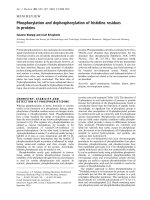
We next developed a panel of qPCR assays that define
amplicons ranging from 6% to 90% GC (Table S1 in
Additional file 2). The amplicons were very short (50 to
69 bp) and thus allowed us to perform qPCR assays on
sheared ‘PER’ DNA and on aliquots drawn at various
points along the protocol (Figure 1). We determined the
abundance of each locus relative to a standard curve of
input ‘PER’ DNA. To adjust for differences in DNA con-
centration, we normalized the calculated quantities
relative to the average quantity of the 48% GC and 52%
GC amplicons in each sample.
The input ‘PER’ genomic DNA is unbiased per defini-
tion. As expected, a scatter plot of the normalized quan-
tity of each amplicon over its GC content was
essentially flat from 6% to 90% GC when plotted on a
log scale, validating the qPCR-based bias assay (Figure
1a). Shearing the DNA did not lead to any obvious
skewing of the base composition (Figure 1b), nor did
the subsequent three enzymatic reaction steps up to the
adapter ligation (Figure 1c). This is not surprising since
up to this point no explicit DNA-fractio nation step had
taken place other than the clean-up steps. Analyzing the
ligation mixture of adapter-ligated fragments by qPCR
would not reveal potential bias during any of the enzy-
matic reactions necessary for ligating the adapter to the
sheared DNA fragments because the mixture presum-
ably includes some adapter-less fragments.
To perform a bias assay exclusively on the adapter-
ligated fraction, we set up a ligation with non-
phosphorylated biot inylated adapters, isolated the adap-
ter-ligated DNA fragment s by streptavidin capture and
released the captured insert fragments by denaturation
for analysis by qPCR. We saw very little, if any, systema-
tic GC bias in the adapter-ligated fraction (Figure 1f,g),
and thus no evidence for strong discrim ination based on
base composition during any of the preceding enzymatic
reactions and clean-up steps.
Excising a narrow size range (corresponding to
approximately 170- to 190-bp genomic fragments) from
a preparative agarose gel did not skew the base compo-
sition (Figure 1d). However, as few as ten PCR cycles
using the enzyme formulation (Phusion HF DNA poly-
merase) and thermocycling conditions prescribed in the
standard Illumina protocol depleted loci with a GC con-
tent > 65% to about a hundredth of the mid-GC refer-
ence loci (Figure 1e). Amplicons < 12% GC were
diminished to approximately one-tenth of their pre-
amplification level. Between the steep flanks on either
side, the GC-bias plot was essentially flat. Its plateau
phase (defined as the segment on the %GC axis with no
more than one data point b elow a relative abundance of
0.7) ranged from 11% to 56% GC.
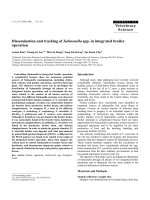
Comparing three thermocyclers at their default ramp
speeds
PCR protocols published by kit manufacturers or in the
scientific literature usually specify the t emperature and
duration time of each thermocycling step (for example,
10 s at 98°C for the denaturation step during each cycle
for the PCR enrichment of Illumina libraries) but rarely
the temperature ramping speed or the make and model of
the thermocycler. For the experiment shown in Figure 1
(and for a replic ate experiment shown in Figur e 2, bright
Aird et al. Genome Biology 2011, 12:R18
/>Page 2 of 14
(
a
)
(b)
(
c)
(
d)
(
e)
(f)
(g)
100
10
1
0.1
Relative
abundance (%)
GC content of amplicon (%)
100
10
1
0.1
100
10
1
0.1
100
10
1
0.1
100
10
1
0.1
100
10
1
0.1
100
10
1
0.1
10
0
500
100500
100500
100500100500
100500
100500
Genomic DNA
Sheared DNA
Adapter
ligation
Gel size
selected
After
PCR
Biotinylated adapter
ligation
Adapter-ligated
fragments
GC content of amplicon (%)
Relative
abundance (%)
Relative
abundance (%)
Relative
abundance (%)
Relative
abundance (%)
Relative
abundance (%)
Relative
abundance (%)
Figure 1 Tracing a diverse panel of loci through the Illumina library preparation. (a-e) At five steps in the standard protocol aliquots were
removed and analyzed for base-composition bias by qPCR. (f,g) To isolate and analyze the ligation-competent population of DNA fragments, a
separate ligation reaction with biotinylated adapters was performed followed by streptavidin capture of fragments carrying at least one adapter.
The quantity of each amplicon in a given sample was divided by the mean quantity of the two amplicons closest to 50% GC. The resulting
relative abundances of amplicons were plotted on a log
10
scale over their respective GC contents.
Aird et al. Genome Biology 2011, 12:R18
/>Page 3 of 14
red line), we used the default heating and cooling rates (6°
C/s and 4.5°C/s, respectively) on thermocycler 1 (see
Materials and methods for make and model).
Running the PCR protoc ol on thermocyler 2 (at its
defaultheatingandcoolingratesof4°C/sand3°C/s,
respectively) extended the plateau to 76% GC (Figure 2,
purple). Thermocyler 3 had the slowest default ramp
speed (2.2°C/s). Its bias plot was fla t from 13% to 84%
GC before dropping down to one-tenth the level for the
two most GC-rich loci (Figure 2, dark red). These results
are consistent with the notion that an overly steep ther-
moprofile does not leave sufficient time above a critical
threshold temperature, causing incomplete denaturation
and poor amplification of the GC-rich fraction.
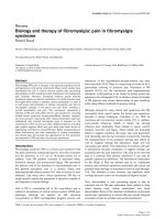
Optimizing the PCR conditions
To develop a robust protocol that produces consistent
results across a wide range of ramp speeds and thermo-
cyclers, we chose to optimize the reaction conditions on
thermocycler 1, t he worst performer, at its fast default
ramp speed. We reasoned that a protocol that works
well on this machine would also work on a slower-
ramping thermocycler.
Simply extending the initial denaturation step (from
30 s to 3 minutes) and the denaturation step during
each cycle (from 10 s to 80 s) overcame the detrimental
effects of the overly fast ramp rate, albeit without fully
restoring the extremely high-GC fraction (Figure 3a,
dark red squares). Long denaturation produced a library
of similar quality as the shorter denaturation on the
slow-ramping thermocyler 3 (Figure 2, dark red).
Adding 2M betaine without changing the thermopro-
file had an equivalent effect on moderately high-GC
fragments but led to a slight depression of loci in the
10% to 40% GC range (Figure 3a, black triangles). Add-
ing 2M betaine and extending the denaturation times
rescued - in fact slightly over-represented - loci at the
extreme high end of the GC spectrum at the expense of
low-GC fragments (Figure 3b, black triangles), shifting
the plateau to the right (23 to 90% GC).
By substituting Phusion HF with the AccuPrime Taq
HiFi blend of DNA polymerases and fine-tuning the
thermoprofile, specifically by prolonging the den atura-
tion step and lowering the temperature for primer
annealing and extension from 72°C to 65°C, we obtained
the GC-bias profile shown in Figure 3b (blue diamonds).
These conditions restored extremely high-GC loci
almost fully while avoiding the suppression of moder-
ately low-GC amplicons seen with Phusion HF and 2M
betaine (black triangles). The plateau ranged from 11%
to 84% GC with only a very slight drop above. Lowering
the temperature for the extension even further (to 60°C)
shifted the balance slightly in favor of AT-rich loci at
the expense of GC-rich ones (see below).
We performed a side-by-side comparison of the Accu-
Prime Taq HiFi PCR protocol on the fastest-ramping
thermocycler 1 and on the slowest-ramping thermocycler
3 and found few, if any, differences in th e GC-bias curves
80 10
0
0 20 40 60
GC content of amplicon (%)
0.1
1
10
100
Relative abundance (%)
Figure 2 Effect of temp erature ramp rates. The standard PCR protocol with Phusion HF DNA polymerase and short initial (30 s) and in-cycle
(10 s) denaturation times was performed on three different thermocyclers at their respective default temperature ramp settings. Heating and
cooling rates were 6°C/s and 4.5°C/s on thermocycler 1 (bright red line), 4°C/s and 3°C/s on thermocycler 2 (purple line) and 2.2°C/s and 2.2°C/s
on thermocycler 3 (dark red line).
Aird et al. Genome Biology 2011, 12:R18
/>Page 4 of 14
(Figure S2a in Additional file 1). We also tested it on
adapter-ligated fragment libraries that had been sheared
and size-selected to approximately 360-bp instead of
180-bp inserts . The GC profiles of PCR-amplified larger-
insert libraries were almost as flat as that of a small-insert
control library amplified in par allel, with a slightly
rounder shoulder, reaching the flat phase at 17% instead
of 13% GC (Figure S2b in Additional file 1).
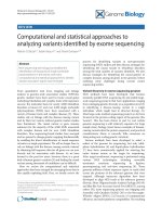
Direct comparison of fragment library and sequencing reads
The qPCR assay measures the composition of the PCR-
ampli fied library. It is likely that downstream steps such
as cluster amplification, sequencing-by-synthesis, image
analysis and o ff-instrument data processing also intro-
duce bias. T o directly compare input libraries and the
final output data, that is, the quality-filtered and aligned
Illumina reads, we sequenced four 400-bp fragment
libraries for which we also had qPCR data and counted
the sequencing reads covering the very same loci.
As shown in Figure 4, for a library amplified with
AccuPrime Taq HiFi using 60°C for the primer exten-
sion step, sequencing and qPCR GC profiles closely
track each other, including some of the pronounced ups
and downs that may reflect amplification traits of indivi-
dual loci, such as sequence context or potential for hair-
pin formation, not captured in their average GC content
0.1
1
10
100
Relative abundance (%)
(a)
80 10
0
0 20 40 60
GC content of amplicon (%)
0.1
1
10
100
Relative abundance (%)
(b)
Figure 3 Optimizing the PCR conditions. (a) Neither extending the denaturation times (dark red squares) nor adding 2M betaine (black
triangles) is sufficient to recover extremely GC-rich DNA fragments by PCR with Phusion HF. (b) Combining long denaturation and 2M betaine is
effective for the high-GC fraction (black triangles) but the profile is not as even over the entire GC spectrum as after PCR with AccuPrime Taq
HiFi (blue diamonds) using extended denaturation times and a lower temperature (65°C) for primer annealing and extension.
Aird et al. Genome Biology 2011, 12:R18
/>Page 5 of 14
indicated on the x-axis. A superimpos ition of qPCR and
sequencing data for three differently amplified libraries
is available in Figure S3 in Additional file 1.
We noted some outliers. For example, amplicons with
approximately 70% or 80% GC received less sequence
coverage than their neighbors in %GC space, despite
relatively high abundance in the library. Close examina-
tion of amplicons > 50% GC suggested an effect of
sequence context. We found the %GC of a 250-bp win-
dow centered on the amplicons a better predictor of
under-coverage than the %GC of the amplicons proper
(Figure S4 in Additional f ile 1). The systematic drop in
sequence coverage with increasing GC content wa s not
caused by a proportionate under-representati on of high-
GC loci in the library, indicating that there is bias
downstream of library preparation.
Genome-wide sequence coverage
Our test loci, which had been selected in part based on
their ability to be amplified by PCR, may or may not be
true representatives of their respective base composi-
tions at large. To measure sequencing bias genome-
wide, we calculated the average ratio of observed to
expected (unbiased) coverage for 50-bp sliding windows.
Superimposing genome -wide and loci-specific bias data,
each normalized relative to the mid-GC (48 to 52%)
fraction, showed that the selected loci were, by and
large, good proxies for their respective %GC categories -
despite the distinct amplification behavior of individual
loci (Figure S5 in Additional file 1).
The standard Phusion HF PCR (short denaturation
and fast ramp) depleted sequences > 70% GC to less
than a hundredth of the mid-GC reference windows
(Figure 5, red squares). Adding betaine and prolonging
the denaturation step rescued the hi gh-GC fraction effi-
ciently and thoroughly (Figure 5, black triangles): 50-bp
windows with up to 94% GC still received more than
half the mean coverage of those with approximately 50%
GC, demonstrating that stretches of 50 bases consisting
almost entirely of Gs and Cs can be sequenced, provided
they are present in the library. H owever, this gain of
high-GC sequences came at the expense of high-AT
sequences, which suffered a significant loss compared to
the standard Phusion HF library.
Consistent with the qPCR data, libraries amplified
with AccuPrime Taq HiFi were less skewed than
libraries amplified with Phusion . Extending the annealed
primer with AccuPrime Taq HiFi at 65°C (Figure 5, blue
diamonds) outperformed both Phusion reactions at the
low-GC end while retaining the high-GC fraction almost
as well as Phusion with betaine (Figure 5, black trian-
gles). Lowering the extension temperature to 60°C
(Figure 5, purple diamonds) returned even more low-
GC sequences while diminishing t he yield of GC-rich
reads somewhat. Extension at 60°C produced an ampli-
fied library wherein all bins of 50-bp windows between
2% and 96% GC received at least one-tenth the average
coverage of the mid-GC reference.
No single PCR protocol was ideal. The best protocol
for high GC, Phusion H F with b etaine, led to poor
80 10
0
0 20 40 60
GC content of amplicon (%)
0.1
1
10
100
Relative abundance (%)
Figure 4 Comparing input library and output sequencing data. Shown is the relative abundance of loci in t he library as determined by
qPCR (purple) and the relative abundance of Illumina sequencing reads covering these loci in one lane of Hi-Seq data (black). Both data sets
were normalized to the average of the two loci closest to 50% GC.
Aird et al. Genome Biology 2011, 12:R18
/>Page 6 of 14
representation of high-AT loci. The protocol that
worked best for high AT, AccuPrime Taq HiFi with pri-
mer ext ension at 60°C, compromised the high-GC frac-
tion. A pool of two differently amplified libraries would
be more complex than either l ibrary alone, but would
also add cost by doubling the amount of library con-
struction required. It would still be biased and, when
sequenced, produce an intermediate GC-bias profile
similar to those shown in Figure S6 in Additional file 1
that were generated by pooling sequencing reads.
We also calculated the fraction of the genome that
received less than one-tenth the mean genome-wide cov-
erage (Table 1). By this measure, AccuPrime Taq HiFi
PCR with primer extension at 60°C was clearly the best
amplification condition for the AT-rich P. falciparum
genome, and overall, for the composite ‘PER’ genome,
71% of which consists of P. falciparum DNA. This
method was slightly worse than the 65°C extension pro-
tocol for the GC-rich R. sphaeroides genome, for which
long-denaturation PCR with Phusio n in the presence of
0.1
1
10
100
Relative coverage
(%, log scale)
0
20
40
60
80
100
0 20 40 60 80 100
Relative coverage
(%, linear scale)
GC content of 50-base window (%)
(
a
)
(b)
Figure 5 ’PER’ genome-wide base composition bias curves. (a,b) Shown is the GC bias in Illumina reads from a 400-bp fragment library
amplified using the standard PCR protocol (Phusion HF, short denaturation) on a fast-ramping thermocycler (red squares), Phusion HF with long
denaturation and 2M betaine (black triangles), AccuPrime Taq HiFi with long denaturation and primer extension at 65°C (blue diamonds) or 60°C
(purple diamonds). To calculate the observed to expected (unbiased) read coverage, the number of reads aligning to 50-bp windows at a given
%GC was divided by the number of 50-bp windows that fall in this %GC category. This value was then normalized relative to the average value
from 48% through 52% GC and plotted on a log
10
scale (a) or linear scale (b).
Aird et al. Genome Biology 2011, 12:R18
/>Page 7 of 14
betaine c ame out o n top. Th e E. coli genome was very
evenly covered by three condition s. Only the standard
PCR protocol with Phusion HF and short denaturation,
when performed with an overly fast temperature ramp,
left more than 0.5% of the E. coli genome under-covered.
Rescuing GC-rich loci in the human genome
To test if our optimized conditions improve the repre-
sentation of biologically relevant loci in the human gen-
ome, we developed qPCR assays for eight GC-rich loci
near gene promoters and four size-matched control loci.
All eight test loci had been under-represented in pre-
vioussequencingrunswithstandardPCR-amplified
libraries. We amplified a fragment library of human
DNA on the fast-ramping thermocycler 1 using the
standard Phusion and the AccuPrime Taq HiFi (exten-
sion at 65°C) protocols. The first protein-coding exon of
the tumor suppressor gene RB1 was below the detection
limit in the standard library (Figure 6a) and near unity
(109% of the average of the four control loci) in the
improved library (Figure 6b). The mean relative a bun-
danceofalleighttestlocirosefrom3%(range0to
11%) to 116% (range 60 to 153%).
Comparison of PCR-amplified and PCR-free Illumina
libraries
Kozarewa et al. [21] developed a protocol for Illumina
sequencing without PCR to amplify and enrich adapt er-
ligated DNA fragments. We sequenced a P CR-amplified
and a PCR-free human 180-bp fragment library side-by-
side on an Illumina Hi-Seq flowcell and calculated the
mean coverage (relative to the mean genome-wide cov-
erage) of a larger set of GC-rich loci (Table S3 in Addi-
tional file 2). The 100 test loci were 200 bp in length,
located on or near annotated transcription start sites,
had a mean GC content of 80% (standard deviation 5%)
and were known to be poorly covered in previous
whole-genome sequencing runs. By this measure, the
PCR-amplified library (AccuPrime Taq HiFi with exten-
sion at 65°C) and the PCR-free library performed
equally: the mean coverage of the test loci was 28% in
both data sets, a 3.6-fold under-representation.
By sequencing the PCR-amplified library, 50-bp win-
dows from 12% to 92% received at least half t he mean
coverage of those with 50% GC (Figure 7a,b). Only
about 0.2% of 50-bp windows in the human reference
genome - and less than 0.02% of 50-bp windows that
overlap with the human exome - fall outside this range.
With the PCR-free library, the mean relative coverage of
GC-rich loci stayed near or above unity all the way to
100% GC. The PCR-free library was also slightly better
for AT-rich loci, with up to 1.4-fold better coverage of
50-bp stretches containing only one G or C. From 8% to
88% GC, the fold increase by sequencing an unamplified
fragment was less than 1.25 (Figure 7c). More than
99.9% of all 50-bp windows in the human genome fall
in this category.
We note that skipping the PCR step during library
preparation does not necessarily yield unbiased Illumina
sequencing reads, presumably due to bias introduced
further downstream in the sequencing process.
Discussion
In this study, we traced a diverse panel of qPCR ampli-
cons through the standard Illumina library constructi on
process to define sources of bias in the Illumina sequen-
cing process and to enable us to develop p rotocols that
ameliorate bias. We identified the enrichment PCR step
as the primary source of base-composition bias in frag-
ment libraries and developed an optimized PCR proto-
col that produces libraries that are far less skewed than
standard PCR-amplified Illumina libraries . We note that
substantial bias is added at downstream steps on the
Illumina instrument. Two of these steps, cluster amplifi-
cation and sequencing-by-synthesis, also involve primer
extension by DNA polymerases. Nonetheless, the benefit
of a more evenly amplified fragment library carries
through to the very end of the process with sequencing
reads covering GC-rich and AT-rich loci that had little
if any coverage before.
We found that hidden factors in the protocol, in parti-
cular the thermocycler and temperature ramp rate, can
play a surprisingly big role in introducing bias. We rea-
soned that it would be impractical to standardize the
make and model of PCR machines across the Illumina
sequencing community. It would be similarly difficult to
universally calibrate machine performance by adjusting
the temperature ramp rates of d ifferent types of instru-
ments. We therefore optimized the reaction conditions
on the PCR machine with the fastest heating and cool-
ing rate - the machine that performed most poorly with
the standard protocol. We extended the denaturation
Table 1 Percentage of bases covered at less than
one-tenth of the mean ‘PER’-wide coverage
PCR condition P. falciparum E. coli R. sphaeroides ’PER’
Phusion HF short
(standard)
denaturation, fast
ramp
41% 0.59% 95% 42%
Phusion HF long
denaturation, 2M
betaine
45% 0.00011% 0.0096% 33%
AccuPrime Taq HiFi
long denaturation,
extension at 65°C
20% 0.00015% 0.032% 14%
AccuPrime Taq HiFi
long denaturation,
extension at 60°C
8.8% 0.00017% 0.085% 6.4%
Aird et al. Genome Biology 2011, 12:R18
/>Page 8 of 14
step to provide sufficient time above the temperature
threshold necessary for complete denaturation of GC-
rich DNA fragments no matter how steep the
thermoprofile.
Long and, presumably, complete denaturation alone
does not rescue extremely GC-rich fragments in PCR
reactions with Phusion HF polymer ase, an enzyme with
relatively weak strand-displacement activity, potentially
limiting its ability to polymerize through hairpins on th e
template strand. Betaine may help to keep a GC-rich
template single-stranded, but it may also cause prema-
ture dissociation of the newly synthesized strand from
an AT-rich template.
AccuPrime Taq HiFi is a blend of taq polymerase,
pyrococcus polymerase and a proprietary accessory pro-
tein added by the manufacturer to improve the priming
specificity. It is conceivable that this accessory protein
(which may have single-strand binding and stabilization
(a)
(b)
0.1
1
10
100
Relative abundance (%)
0.1
1
10
100
1 2 3 4 5 6 7 8 9 10 11 12
Relative abundance (%)
Locus
First exon of RB1
First exon of RB1
Figure 6 Optimized PCR conditions rescue GC-rich promoter regions in the human genome. (a,b) A 180-bp fragment library of human
DNA was amplified using (a) standard conditions (Phusion HF, short denaturation) or (b) optimized conditions (AccuPrime HiFi, long
denaturation, extension at 65°C) on the fast-ramping thermocycler 1. The amplified libraries were analyzed by qPCR. Orange bars indicate the
quantity of eight GC-rich loci near gene promoters relative to the mean quantity of four size-matched control loci (blue bars; mean set to 100%
in each graph). Error bars represent the range of two measurements averaged to calculate the quantity of each locus. Locus 7 is the first
protein-coding exon of the tumor suppressor gene RB1.
Aird et al. Genome Biology 2011, 12:R18
/>Page 9 of 14
(a)
(b)
(c)
80 10
0
0
1
2
3
4
5
0 20 40 60
Fold-increase of coverage
with PCR-free library (x)
GC content of 50-bp windows (%)
0
20
40
60
80
100
120
Relative coverage
(%, linear scale)
0.1
1
10
100
Relative coverage
(%, log scale)
Figure 7 Sequencing bias with PCR-am plified and PCR-free libraries. (a,b) Shown is the mean normalized coverage of 50-bp windows in
the human genome having the GC-content indicated on the x-axis for a PCR-free (orange dots) and a PCR-amplified (blue diamonds) Illumina
sequencing library. Both fragment libraries had approximately 180-bp inserts. The PCR amplification was performed with AccuPrime Taq HiFi
(long denat., primer extension at 65°C). The coverage was plotted on a log
10
(a) and a linear scale (b). The data points at extremely high GC,
where the reads from the PCR-free library had a mean base quality of less than Q20 (open symbols), were omitted in the middle panel (b).
(c) The ratios of the two curves in (a,b), that is, the fold-increase in mean coverage by sequencing a PCR-free library instead of a PCR-amplified
library. The shaded histogram is the %GC distribution of 50-bp windows in the human genome. More than 99.9% of all 50-bp windows in the
genome contain 8% to 88% GC and received a less than 1.25-fold increase in coverage. Less than 0.01% of all 50-bp windows contain 90% or
more GC. The open circles at 96% and 98% GC denote data for which the mean base quality of the reads from the PCR-free library was
below Q20.
Aird et al. Genome Biology 2011, 12:R18
/>Page 10 of 14
activity) also helps to displace strands and melt hairpin
structures during the polymerization. It is also possible
that the excellent perfo rmance is simply due to the
complementary strengths and synergy of two different
enzymes working together, or to the chemical com posi-
tion of the reaction buffer.
Experimental noise and imperfections in the data not-
withstanding, the qPCR-based GC-bias profiles were
reproducible and highly informative and predictive. Our
test loci appear to be good proxies for their respective
%GC bins that allow extrapolation to the genome at
large - despite distinct amplification ‘personalities’ of
individual loci.
Interestingly, PCR-induced depletion of the high-
GC fraction can largely be prevented whereas under-
representation of AT-rich fragments can only be slightly
ameliorated at best - for example, by lowering the tem-
perature during the primer extension. Testing additional
PCR enzymes, buffers and reaction conditions will be
necessary to further improve the representation of the
AT-rich fraction. Ironically, it was our inability to find
PCR conditions that work well for AT-rich fragments
that led us to posit anti-AT bias during end-repair of
adapter ligation as the root cause of the AT-deficit.
However, when we examined this hypothesis directly
using biotinylated adapters, we did not see significant
ligation bias of a magnitude that could possibly explain
the lack of AT-rich fragments in PCR-amplified frag-
ment libraries (Figure 1).
We note that none of our conditions work equally
well at rescuing, at the same time, under- representation
of regions that are either extremely GC-rich or GC-
poor. At the time of this writing, by our assay, PCR with
AccuPrime Taq HiFi at a low primer-extension tem-
perature is the best compromise. We did not screen and
test an exhaustive list of PCR enzymes and reaction
conditions. It is possible that other enzymes would per-
form as well or even better. So me of them may be
superior in other respects, such as fidelity, size bias,
cross-platform compatibility, costs or lot- to-lot variabil-
ity. However, our simple and quick qPCR bias assay will
enable a wider search for optimal PCR reagents and
amplification conditions.
Obviously, the best way to avoid bias during PCR is to
avoid library amplification by PCR altogether [6,21]. On
the other hand, PCR-free libraries require relatively large
amounts of input DNA and are thus impractical for
many sample types. Furthermore, there is no enrichment
of sequenceable fragments carrying adapters on b oth
ends, and the yield of such fragments is very sensitive to
variations in DNA quality and purity, which in turn can
affect the efficiency of end repair and adapter ligation.
We also note that preparing PCR-free l ibraries alone
does not necessarily gu arantee unbiased sequencing data
as significant bias is introduced elsewhere in the process.
Importantly, PCR-free protocols are not readily amen-
able to automation and are therefore not the best choice
as the default protocol in high-throughput sequencing
facilities such a s the Broad Institute Genome Sequen-
cing Platform, where currently about 1,000 Illumina
sequencing libraries are made per week to support a
diverse portfolio of sequencing projects. Improved PCR
conditions like the one described here will likely satisfy
the vast majority of projects. Enhancing the coverage of
high-GC loci is critical for human genome and exome
sequencing in cancer and medical genetics, the major
sequencing applications in terms of bases generated.
Solving the loss of AT-ric h loci remains a challenge, but
has less of an impact on human genome sequencing
and on the sequencing field as a whole. We expect that
PCR-free methods, which are invaluable and superior
for extreme base composit ions at both ends of the %GC
spectrum, will be reserved for projects that are most
sensitive to base-composition bias and can supply input
DNA of sufficient quality and quantity.
Conclusions
qPCR is an inexpensive and quick assay for representa-
tional bias in Illumina fragment libraries. Our optimized
PCR conditions are significantly better and more robust
than the standard protocol in that they amplify more
evenly across a wider range of base compositions and
minimize the previously detrimental effect of fast-ramp-
ing thermocyclers. By optimizing instead of eliminating
the PCR-amplification step, our protocol is easy to
impl ement in high-throughput production and does not
increase the DNA input requirements for routine Illu-
mina library construction.
Materials and methods
Genomic DNA
DNA from P. falciparum 3D7 was a gift of Dr Daniel
Neafsey (Broad Institute). DNA from E. coli K12
MG1655 and R. sphaeroides 2.4.1, kindly prepared by Dr
Louise Williams (Broad Institute), was obtained from
the Broad Institute Genomic Sequencing Sample Reposi-
tory. The equimolar composite ‘PER’ DNA sample was a
5:1:1 mixture (by mass) of the three DNAs. The human
DNA was NA12878 (Coriell Institute, Camden, NJ,
USA).
Standard Illumina fragment libraries
Illumina fragment libraries were constructed using Illu-
mina paired-end DNA sample prep kit v1 with the fol-
lowing modifications. DNA (3 μgin280μlTEbuffer)
was sheared for 6 minutes on an S2 sonicator (Covaris,
Aird et al. Genome Biology 2011, 12:R18
/>Page 11 of 14
Woburn, MA, USA). The settings for short-insert frag-
ment libraries were 5% duty cycle, intensity 10, and 200
cycles per burst. For long-insert fragment libraries, the
intensity was reduced to 5. The modes of short and
long fragment-size distributions were approximately 225
bp and 325 bp, re spectively. End-repair reactions (56 μl)
contained 1× T4 DNA ligase buffer (NEB, Ipswich, MA,
USA), 1.4 mM ATP, 0.4 mM dNTPs, 0.1 mg/ml bovine
serum albumin, 15 units T4 DNA polymerase (NEB), 50
units T4 polynucleotide kinase (NEB) and were incu-
bated stepwise for 15 minutes at 12°C and 15 minutes
at 25°C. The non-templated 3’-single-dA extension was
performed for 30 minutes at 37°C in 50 μl containing
1× Klenow buffer (NEB), 0.2 mM dATP and 15 u nits
Klenow exo
-
(NEB). Adapter ligations (50 μl) contained
1× Quickligation buffer (NEB), 3 μl annealed paired-end
adapter oligonucleotides (Illumina, San Diego, CA,
USA), 5 units T4 DNA ligase (NE B) and were carried
out for 15 minutes at 25°C. All reaction clean-ups were
performed using a MinElute PCR purification kit (Qia-
gen, Hilden, Germany). Fragment libraries were size-
selected on 3% NuSieve 3:1 (Lonza, Basel, Switzerland)
agarose gels run in 1× TAE buffer. SYBR Green (Invi-
trogen, Carlsba d, CA, USA) stained DNA was visualized
on a DarkReader (Clare Chemicals, Dolores, CO, USA).
Gel slices were excised 90 bp larger than the desired
insert size, that is, 250 to 290 bp for (180 ± 20)-bp
inserts, 410 to 490 bp for (360 ± 40) -bp inserts, and 450
to 530 bp for (400 ± 40)-bp inserts. Size-selected DNA
was purified with a Qiagen MinElute gel extraction kit
and quantified using the Quant-iT dsDNA HS assay
(Invitrogen).
Library amplification by PCR
PCR with I llumina PE 1.0 and 2.0 enrichment primers
was performed in 50-μl reactions containing 1 to 2 ng
of size-selected small-insert (approximately 180 bp) frag-
ment libraries or 2 to 4 ng of size-selected large-insert
(approximately 360 bp or 400 bp) fragment libraries.
Standard reactions contained 1× Phusion High-Fidelity
PCR master mix with HF buffer (NEB). Standard (short
denaturation) thermocycling for PCR with Phusion was
30 s at 98°C for the initial denaturation follo wed by 10
cycles of 10 s at 98°C, 30 s at 65°C and 30 s at 72°C and
a final extension for 5 minutes at 72°C. The ‘long dena-
turation’ Phusion thermocycling protocol was 3 minutes
at 98°C; 10 × (80 s at 98°C, 30 s at 65°C, 30 s at 72°C);
10 minutes at 72°C. Betaine (5 M stock solution) was
from USB (Cleveland, OH, USA). One unit of Accu-
Prime Taq DNA polymerase High Fidelity (Invitrogen)
was used in 50 μl 1× AccuPrime PCR buffer II. The
thermoprofile of AccuPrime Taq HiFi reactions included
the same long denaturat ion steps as the ‘long denatura-
tion’ Phusion protocol above: 3 minutes at 98°C; 10 ×
(80 s at 98°C, 90 s at 65°C or 60°C); 10 minut es at 65°C
or 60°C. Thermocycler 1 was a Mastercycler epgradient
S (Eppendorf, Hamburg, Germany). Thermocycler 2 was
an Eppendorf Mastercycler epgradient (no ‘S’). Thermo-
cycler 3 was a Gene Amp PCR System 9700 with gold-
plated solid silver block (Applied Biosystems, Foster
City, CA, USA) and was run in 9600 emulation mode.
PCR products were purified w ith 1.8× AmPure XP
beads (Beckman Coulter Genomics, Danvers, MA, USA)
and eluted in Qiagen’s EB buffer.
PCR-free fragment libraries for Illumina sequencing
Shearing, end-repair, single-dA extension and adapter
ligation were performed as de scribed above for standard
Illumina fragment libraries with the following excep-
tions: the genomic DNA (9 μg of NA12878 total human
DNA) was sheared in three batches of 3 μgeach;the
ligation reaction contained 120 pmol pre-annealed full-
length paired-end Illumina adapters [21]; the product of
the adapter-ligation was size-selected to an apparent
size range of 320 to 350 bp relative to a double-stranded
size marker.
qPCR
Primer pairs for ‘PER’ and human qPCR assays, their
genome of origin, sequence, length and GC-content of
amplicons are listed in Tables S1 and S2 in Additional
file 2. These tables also contain their designation as
either ‘low’, ‘mid’ or ‘high’ GC qPCR assays, indicating
which qPCR protocol was used. Low and mid-GC qPCR
reactions contained 5 μl Power SYBR Green PCR master
mix (Applied Biosystems), 3 μlofprimerpair(see
Tables S1 and S2 in Additional file 2 for the recom-
mended concentration of the primer pair), 0.6 μl of tem-
plate DNA in T
10
E
0.1
buffer (sample, standard curve or
blank) and 1.4 μlH
2
O for a final volume of 10 μl. Hi gh
GC qPCR reactions contained 6 μl Power SYBR Green
PCR master mix (Applied Biosystems), 2.4 μl5M
betaine, 3 μl of primer pair, 0.6 μl of template DNA in
T
10
E
0.1
buffer in a final volume of 12 μl. qPCR reactions
were performed on a 7900HT real-time PCR instrument
(Applied Biosystems). The thermocycling protocol was
2 minutes at 50°C; 10 minu tes at 95°C; 50 × (20 s at
95°C, 20 s at 47.5°C, 120 s at 55°C) for low GC a ssays,
2 minutes at 50°C; 10 minu tes at 95°C; 50 × (20 s at
95°C, 20 s at 55°C, 40 s at 60°C) for mid-GC assays and
2 minutes at 50°C; 10 minu tes at 95°C; 50 × (20 s at
95°C, 60 s at 60°C) for high GC assays. The standard
curve for absolute quantification of ‘PER’ amplicons was
prepared from the very same mixture of ‘PER’ DNA that
was used for preparing the Illumina fragment libraries.
It consisted of a five-fold dilution series ranging from
2.6 ng down to 170 fg (nominally 75,125 down to 5 hap-
loid ‘ PER’ genome equivalents) and a non-template
Aird et al. Genome Biology 2011, 12:R18
/>Page 12 of 14
control. Approximately 100 pg ‘ PER’ sample libraries
was added per reaction as determined by a Quant-iT
dsDNA HS assay of the sample. The known standards
for quantification of human loci were a five-fold dilution
series of human female genomic DNA (Promega, Madi-
son, WI, USA) ranging from 33 ng down to 2.1 pg per
reaction . Approximately 1 ng of human fragment library
was added per reaction. All reactions were performed in
duplicate. Data points that were equal to or less than
three-fold above background (no template) amplification
were omitted. Duplicate qPCR measurements were aver-
aged. To normalize for differences in the DNA concen-
tration between ‘ PE R’ -derived samples, the average
quantity of each amplicon in a given sample was divided
by the mean average quantities of the 48% and 52% GC
amplicons in the same sample. Hence, all GC-bias plots
meet at 50% GC. For human-derived samples, we
divided the average quantity of each amplicon by the
mean quantity of four control loci (9 to 12 in Table S2
in Additional file 2).
Isolation of biotin-adapter-ligated DNA fragments by
streptavidin capture
Non-phosporylated biotinylated Illumina adapters were
prepared by annealing 5’-ACACTCTTTCCCTACAC-
GACGCTCTTCCGATCxT and 5’-GATCGGAAGAGC
GGTTCAGCAGGAATGCCGAG-3BioTEG (IDT, Cor al-
ville, IA, USA) where ‘x’ denotes a nuclease-resistant phos-
phorothioate linkage and ‘3BioTEG’ a biotin attached via a
15-atom linker at the 3’ end. Ligation to end-repaired and
3’-dA extende d genomic DNA fragments was carried out
as described above for regular adapters. Ligations were
cleaned up by Qiagen MinElute and 1× AmPure XP beads
(Beckman Coulte r Genomics). M-280 streptavidin Dyna-
beads (50 μl; Invitrogen) were washed three times and
resuspended in 400 μl of 1 M NaCl, 10 mM Tris-HCl, pH
7.5, and 1 mM EDTA. After adding the ligation reactions
(26 μl), the beads were kept in suspension for 30 minutes
at room temperature on a rotary mix er, pulled down and
washed once for 15 minutes at room temperature with
0.5 ml 1× SSC/0.1% SDS. Ligation products were eluted by
resuspending the beads in 50 μl0.1MNaOH.After
15 minutes at room temperature, the beads w ere pulled
down, and the supernatant (containing melted-off single-
stranded genome fragments with the non-biotinylate d
adapter oligo attached) was transferred to a tube containing
70 μ l 1 M Tris-HCl, pH 7.5. The neutralized eluate was
desalted and c oncen trated on a Qiagen MinElute column.
Illumina sequencing and sequence analysis
Sequencing was performed in paired-end mode with
Illum ina HiSeq 2000 chemistry using Illumina data ana-
lysis pipeline version 1.8. ‘PER’ fragment libraries were
sequenced at densities of 2.9 to 3.2 million clusters
per tile. Paired-end reads (median insert size 39 8 to 405
bp) were aligned by BWA [22] version 0.5.7-6 (r1399) to
the ‘PER’ reference, which was constructed by concate-
nating the references of the component species. Each
PCR condition was represented by a single lane of data
consisting of 124 to 142 million mapped reads, each 101
bases in length. PCR-amplified and PCR-free human
libraries were sequenced at 3.1 and 3.9 million clusters
per tile, respectively. Paired-end 101-base reads (median
fragment insert size 172 bp and 166 bp, respectively)
were aligned by BWA to human reference sequence
GRCh37/hg19. Relative representations of the qPCR
amplicon loci in sequencing data were determined by
locating each amplicon in the ‘PER’ reference genome
and comparing the average number of reads per base in
each amplicon to the average number of reads per base
across the entire ‘PER’ genome (excluding ambiguo us/
unknown bases) . Since one lane of Hi- Seq data per
library provided only five- to six-fold coverage of the
human genome - not sufficient to calculate a meaningful
coverage statistics for any given single locus - we calcu-
lated the mean read coverage per base for all 100 loci at
once and divided this number by the genome-wide aver-
age. The mean sequence coverage of 50-bp windows at
a given %GC bin was the number of observed reads that
aligned to the 50-bp windows divided by the number of
read alignments one would expect for perfectly even
coverage given the number of 50-bp windows with this
%GC. To calculate the relative coverage, the sequence
coverage of each category was divided by the mean cov-
erage of 50-bp windows from 48% to 52% GC. The
number of ‘PER’ bases covered at less than 10% of the
mean coverage was similarly determined by examining
the number of reads overlapping each non-ambiguous
base and comparin g that to the ‘PER’-wide average. The
‘PER’ sequencing data used for this study are available
at the NCBI Sequence Read Archive under study num-
ber SRP004833 [NCBI:SRX033223, NCBI:SRX033224,
NCBI:SRX033225, NCBI:SRX033226]. The human
sequencing data are available under study number
SRP005622 [NCBI:SRX040660, NCBI:SRX040661].
Additional material
Additional file 1: Supplementary Figures 1 to 6.
Additional file 2: Supplementary Tables 1 to 3.
Abbreviations
bp: base pair; ‘PER’: pool of genomic DNA prepared from Plasmodium
falciparum, Escherichia coli and Rhodobacter sphaeroides; qPCR: quantitative
PCR.
Acknowledgements
We thank the staff of the Broad Institute Sequencing Platform and the
Educational Outreach Program. We also thank Lesley Gaffney for last-minute
Aird et al. Genome Biology 2011, 12:R18
/>Page 13 of 14
help with the artwork. This project has been funded in part with funds from
the National Institute of Allergy and Infectious Diseases under Contract No.
HHSN27220090018C, and from the National Human Genome Research
Institute (HG03067-05).
Author details
1
Genome Sequencing and Analysis Program, Broad Institute of MIT and
Harvard, 320 Charles Street, Cambridge, MA 02141, USA.
2
Learning
Community C, Cambridge Rindge and Latin School, 459 Broadway,
Cambridge, MA 02138, USA.
3
Genome Sequencing Platform, Broad Institute
of MIT and Harvard, 320 Charles Street, Cambridge, MA 02141, USA.
Authors’ contributions
DA, WSC and MD carried out research in the lab and analyzed qPCR data.
MGR, TF and DBJ analyzed sequencing data. CR and CN coordinated the
research. AG conceived the project and wrote the paper.
Competing interests
The authors declare that they have no competing interests.
Received: 20 October 2010 Revised: 23 December 2010
Accepted: 21 February 2011 Published: 21 February 2011
References
1. Bentley DR, Balasubramanian S, Swerdlow HP, Smith GP, Milton J,
Brown CG, Hall KP, Evers DJ, Barnes CL, Bignell HR, Boutell JM, Bryant J,
Carter RJ, Keira Cheetham R, Cox AJ, Ellis DJ, Flatbush MR, Gormley NA,
Humphray SJ, Irving LJ, Karbelashvili MS, Kirk SM, Li H, Liu X, Maisinger KS,
Murray LJ, Obradovic B, Ost T, Parkinson ML, Pratt MR, et al: Accurate
whole human genome sequencing using reversible terminator
chemistry. Nature 2008, 456:53-59.
2. Margulies M, Egholm M, Altman WE, Attiya S, Bader JS, Bemben LA, Berka J,
Braverman MS, Chen YJ, Chen Z, Dewell SB, Du L, Fierro JM, Gomes XV,
Godwin BC, He W, Helgesen S, Ho CH, Irzyk GP, Jando SC, Alenquer ML,
Jarvie TP, Jirage KB, Kim JB, Knight JR, Lanza JR, Leamon JH, Lefkowitz SM,
Lei M, Li J, et al: Genome sequencing in microfabricated high-density
picolitre reactors. Nature 2005, 437:376-380.
3. McKernan KJ, Peckham HE, Costa GL, McLaughlin SF, Fu Y, Tsung EF,
Clouser CR, Duncan C, Ichikawa JK, Lee CC, Zhang Z, Ranade SS,
Dimalanta ET, Hyland FC, Sokolsky TD, Zhang L, Sheridan A, Fu H,
Hendrickson CL, Li B, Kotler L, Stuart JR, Malek JA, Manning JM,
Antipova AA, Perez DS, Moore MP, Hayashibara KC, Lyons MR, Beaudoin RE,
et al: Sequence and structural variation in a human genome uncovered
by short-read, massively parallel ligation sequencing using two-base
encoding. Genome Res 2009, 19:1527-1541.
4. Dohm JC, Lottaz C, Borodina T, Himmelbauer H: Substantial biases in
ultra-short read data sets from high-throughput DNA sequencing.
Nucleic Acids Res 2008, 36:e105.
5. Hillier LW, Marth GT, Quinlan AR, Dooling D, Fewell G, Barnett D, Fox P,
Glasscock JI, Hickenbotham M, Huang W, Magrini VJ, Richt RJ, Sander SN,
Stewart DA, Stromberg M, Tsung EF, Wylie T, Schedl T, Wilson RK,
Mardis ER: Whole-genome sequencing and variant discovery in C.
elegans. Nat Methods 2008, 5:183-188.
6. Quail MA, Kozarewa I, Smith F, Scally A, Stephens PJ, Durbin R, Swerdlow H,
Turner DJ: A large genome center’s improvements to the Illumina
sequencing system. Nat Methods 2008, 5:1005-1010.
7. Maccallum I, Przybylski D, Gnerre S, Burton J, Shlyakhter I, Gnirke A, Malek J,
McKernan K, Ranade S, Shea TP, Williams L, Young S, Nusbaum C, Jaffe DB:
ALLPATHS 2: small genomes assembled accurately and with high
continuity from short paired reads. Genome Biol 2009, 10:R103.
8. Ioerger TR, Koo S, No EG, Chen X, Larsen MH, Jacobs WR Jr, Pillay M,
Sturm AW, Sacchettini JC: Genome analysis of multi- and extensively-
drug-resistant tuberculosis from KwaZulu-Natal, South Africa. PLoS One
2009, 4:e7778.
9. Nusbaum C, Ohsumi TK, Gomez J, Aquadro J, Victor TC, Warren RM,
Hung DT, Birren BW, Lander ES, Jaffe DB: Sensitive, specific polymorphism
discovery in bacteria using massively parallel sequencing. Nat Methods
2009, 6:67-69.
10. Garber M, Zody MC, Arachchi HM, Berlin A, Gnerre S, Green LM, Lennon N,
Nusbaum C: Closing gaps in the human genome using sequencing by
synthesis. Genome Biol 2009, 10:R60.
11. Leem SH, Kouprina N, Grimwood J, Kim JH, Mullokandov M, Yoon YH,
Chae JY, Morgan J, Lucas S, Richardson P, Detter C, Glavina T, Rubin E,
Barrett JC, Larionov V: Closing the gaps on human chromosome 19
revealed genes with a high density of repetitive tandemly arrayed
elements. Genome Res 2004, 14:239-246.
12. Eichinger L, Pachebat JA, Glockner G, Rajandream MA, Sucgang R,
Berriman M, Song J, Olsen R, Szafranski K, Xu Q, Tunggal B, Kummerfeld S,
Madera M, Konfortov BA, Rivero F, Bankier AT, Lehmann R, Hamlin N,
Davies R, Gaudet P, Fey P, Pilcher K, Chen G, Saunders D, Sodergren E,
Davis P, Kerhornou A, Nie X, Hall N, Anjard C, et al: The genome of the
social amoeba Dictyostelium discoideum. Nature 2005, 435:43-57.
13. Gardner MJ, Hall N, Fung E, White O, Berriman M, Hyman RW, Carlton JM,
Pain A, Nelson KE, Bowman S, Paulsen IT, James K, Eisen JA, Rutherford K,
Salzberg SL, Craig A, Kyes S, Chan MS, Nene V, Shallom SJ, Suh B,
Peterson J, Angiuoli S, Pertea M, Allen J, Selengut J, Haft D, Mather MW,
Vaidya AB, Martin DM, et al: Genome sequence of the human malaria
parasite Plasmodium falciparum. Nature 2002, 419:498-511.
14. Lindblad-Toh K, Wade CM, Mikkelsen TS, Karlsson EK, Jaffe DB, Kamal M,
Clamp M, Chang JL, Kulbokas EJ, Zody MC, Mauceli E, Xie X, Breen M,
Wayne RK, Ostrander EA, Ponting CP, Galibert F, Smith DR, DeJong PJ,
Kirkness E, Alvarez P, Biagi T, Brockman W, Butler J, Chin CW, Cook A, Cuff J,
Daly MJ, DeCaprio D, Gnerre S, et al: Genome sequence, comparative
analysis and haplotype structure of the domestic dog. Nature 2005,
438:803-819.
15. Drmanac R, Sparks AB, Callow MJ, Halpern AL, Burns NL, Kermani BG,
Carnevali P, Nazarenko I, Nilsen GB, Yeung G, Dahl F, Fernandez A, Staker B,
Pant KP, Baccash J, Borcherding AP, Brownley A, Cedeno R, Chen L,
Chernikoff D, Cheung A, Chirita R, Curson B, Ebert JC, Hacker CR, Hartlage R,
Hauser B, Huang S, Jiang Y, Karpinchyk V, et al: Human genome
sequencing using unchained base reads on self-assembling DNA
nanoarrays. Science 2010, 327:78-81.
16. Eid J, Fehr A, Gray J, Luong K, Lyle J, Otto G, Peluso P, Rank D, Baybayan P,
Bettman B, Bibillo A, Bjornson K, Chaudhuri B, Christians F, Cicero R, Clark S,
Dalal R, Dewinter A, Dixon J, Foquet M, Gaertner A, Hardenbol P, Heiner C,
Hester K, Holden D, Kearns G, Kong X, Kuse R, Lacroix Y, Lin S, et al: Real-
time DNA sequencing from single polymerase molecules. Science 2009,
323:133-138.
17. Pushkarev D, Neff NF, Quake SR: Single-molecule sequencing of an
individual human genome. Nat Biotechnol 2009, 27:847-852.
18. Smith DR, Quinlan AR, Peckham HE, Makowsky K, Tao W, Woolf B, Shen L,
Donahue WF, Tusneem N, Stromberg MP, Stewart DA, Zhang L, Ranade SS,
Warner JB, Lee CC, Coleman BE, Zhang Z, McLaughlin SF, Malek JA,
Sorenson JM, Blanchard AP, Chapman J, Hillman D, Chen F, Rokhsar DS,
McKernan KJ, Jeffries TW, Marth GT, Richardson PM: Rapid whole-genome
mutational profiling using next-generation sequencing technologies.
Genome Res 2008, 18:1638-1642.
19. Chiang DY, Getz G, Jaffe DB, O’Kelly MJ, Zhao X, Carter SL, Russ C,
Nusbaum C, Meyerson M, Lander ES: High-resolution mapping of copy-
number alterations with massively parallel sequencing. Nat Methods
2009, 6:99-103.
20. He S, Wurtzel O, Singh K, Froula JL, Yilmaz S, Tringe SG, Wang Z, Chen F,
Lindquist EA, Sorek R, Hugenholtz P: Validation of two ribosomal RNA
removal methods for microbial metatranscriptomics. Nat Methods 2010,
7:807-812.
21. Kozarewa I, Ning Z, Quail MA, Sanders MJ, Berriman M, Turner DJ:
Amplification-free Illumina sequencing-library preparation facilitates
improved mapping and assembly of (G+C)-biased genomes. Nat
Methods 2009, 6:291-295.
22. Li H, Durbin R: Fast and accurate short read alignment with Burrows-
Wheeler transform. Bioinformatics 2009, 25:1754-1760.
doi:10.1186/gb-2011-12-2-r18
Cite this article as: Aird et al.: Analyzing and minimizing PCR
amplification bias in Illumina sequencing libraries. Genome Biology 2011
12:R18.
Aird et al. Genome Biology 2011, 12:R18
/>Page 14 of 14