Báo cáo y học: "Modulated contact frequencies at gene-rich loci support a statistical helix model for mammalian chromatin organizatio" ppsx
Bạn đang xem bản rút gọn của tài liệu. Xem và tải ngay bản đầy đủ của tài liệu tại đây (751.41 KB, 13 trang )
RESEARC H Open Access
Modulated contact frequencies at gene-rich loci
support a statistical helix model for mammalian
chromatin organization
Franck Court
1
, Julie Miro
2
, Caroline Braem
1
, Marie-Noëlle Lelay-Taha
1
, Audrey Brisebarre
1
, Florian Atger
1
,
Thierry Gostan
1
, Michaël Weber
1
, Guy Cathala
1
and Thierry Forné
1*
Abstract
Background: Despite its critical role for mammalian gene regulation, the basic structural landscape of chromatin in
living cells remains largely unknown within chromo somal territories below the megabase scale.
Results: Here, using the 3C-qPCR method, we investigate cont act frequencies at high resolution within interphase
chromatin at several mouse loci. We find that, at several gene-rich loci, contact frequencies undergo a periodical
modulation (every 90 to 100 kb) that affects chromatin dynamics over large genomic distances (a few hundred
kilobases). Interestingly, this modulation appears to be conserved in human cells, and bioinformatic analyses of
locus-specific, long-range cis-interactions suggest that it may underlie the dynamics of a significant number of
gene-rich domains in mammals, thus contributing to genome evolution. Finally, using an original model derived
from polymer physics, we show that this modulation can be understood as a fundamental helix shape that
chromatin tends to adopt in gene-rich domains when no significant locus-specific interaction takes place.
Conclusions: Altogether, our work unveils a fundamental aspect of chromatin dynamics in mammals and
contributes to a better understanding of genome organization within chromosomal territories.
Background
Within the interphasic cell nucleus, the mammalian gen-
ome, packed into the chromatin, is s patially restrained
into specific chromosomal territories [1,2] and is distrib-
uted in at least two spatial compartments: one enriched
in active genes and open chromatin [3-7] and the other
containing inactive and closed chromatin [4,7,8]. It was
recently proposed that, at the megabase (Mb) scale, chro-
mosome territories consist of a series of fractal globules
[4]. However, below that scale, a nd beyond the simple
nucleosomal array, the basic structural landscape of the
chromatin in living cells remains enigmatic.
At the supranucleosomal level (approximately 10 to 500
kb), it is largely accepted that one essential determinant in
relation to gene expression and other chromosomal activ-
ities is chromatin looping [9]. However, because of
technological limitations, access to this level of chromatin
organization remains problematic [10]. From this perspec-
tive, the advent of the Chromosome Conformation Cap-
ture (3C) assay [11,12] represents a decisive technological
and scientific breakthrough since it permits the identifica-
tion of long-range cis and trans chromatin interactions in
their native genomic context. Subsequently, several 3C-
based methods have been developed that allow the
unbiased large-scale identific ation of such interactions
[4,7,13-16]. Noticeably, theuseofapopulation-based
approach like the 3C-real-time quantitative PCR (qPCR)
protocol [17,18], combined with appropriate algorithms
for accurate data normalization [19], provides a powerful
quantitative method that allows high-resolution analysis
(on the kilobase scale) of the average contact frequencies
between distant g enomic regio ns within a locus. Th is
information is particularly interesting as contact frequen-
cies essentially depend on constraints that the chromatin
may undergo at that scale. Constraints resulting from
locus-specific interactions are easily identified in 3C-qPCR
experiments since they appear as local peaks where the
* Correspondence:
1
Institut de Génétique Moléculaire de Montpellier (IGMM), UMR5535 CNRS,
Universités Montpellier 1 et Montpellier 2. 1919, Route de Mende, 34293
Montpellier Cedex 5, France
Full list of author information is available at the end of the article
Court et al. Genome Biology 2011, 12:R42
/>© 2011 Court et al.; licensee BioMed Central Ltd. This i s an open access article d istributed under the terms of the Creative Co mmons
Attribution License ( which permits unrestricted use, di stribution, and reproduct ion in
any medium, provided the original work is properly cited.
interaction frequency is at least four to five times higher
than the surrounding collision levels [17,19]. Furthermore,
they are detected only in some experiments targeting spe-
cific regulatory sequences within a given locus. On the
contrary, intrinsic constraints, resulting from fundamental
characteristics of the chromatin (compaction, flexibility,
basic non-linear shape), areexpectedtohaveasimilar
impact on contact frequ encies at many sites and numer-
ous loci.
Here, using a 3C-qPCR approach [17], we determined
random collision frequencies within interphase chromatin
at several mouse loci. We demonstrate that, in the absence
of significant locus-specific interactions, several gene-rich
domains of the chromatin display modulated contact fre-
quencies in both mouse and human, thus revealing the
existence of an unexpected intrinsic constraint. We pro-
pose that this constraint results from a preferential non-
linea r shape that the chromatin tends to adopt and show
that the observed modulations can be described by poly-
mer models as if, at these loci, the chromatin was statisti-
cally shaped into a helix.
Results
Several mouse gene-rich loci display modulation of
contact frequencies
To focus on the interphase chromatin, we worked on pre-
parations of cell nuclei from postnatal mouse livers
[20,21], and to minimize potential interference of locus-
specific long-range interactions, we restricted our analysis
to mouse loci where no significant local peaks could be
detected in 3C-qPCR experiments. As previously
suggested for its human ortholog [22], the mouse Usp22
(Ubiquitin carboxyl-terminal hydrolase 22) locus, on chro-
mosome 11, displays such characteristics . Two intergenic
HindIII sites (F1 and F7 in Figure 1a) were separately used
as anchors to determine interaction frequencies with other
HindIII sites found throughout this locus. As expected, for
site separations lower than 35 kb, rand om collision fre-
quencies decrease with increasing site separations (Figure
1b, upper-left panel). However, a floating mean analysis of
these data (red squares in Figure 1b) indicated a stabiliza-
tion of random collision frequencies around 60 kb and a
surprising increase for higher site separations, reaching a
maximum for distances around 100 kb. Indeed, between
these two positions, the mean interacti on frequency (0.85
versus 1.37, respectivel y) increases very significantly (P =
0.007, Ma nn-Whitney U-test). We then investigated four
additional gene-rich loci that displaye d no evidence for
long-range specif ic interaction s in the po stnatal m ouse
liver: the Dlk1 (Delta-like 1 homologue) locus on chromo-
some 12 [19,23], the Ln p (Limb and neural patterns/Luna-
park) and Mtx2 (Metaxine 2) loci on chromosome 2, and
the Emb (Embigin) locus on chromosome 13 (Figure 1a).
Interestingly, similar modulation in random collision
frequencies was shown at all four loci (Figure 1b). In con-
clusion, for eleven intergenic sites (anchors) distributed in
five loci and four distinct mouse chromosomes, one can
always observe that random collision frequencies increase
for site separations around 80 to 110 kb. Therefore, this
modulation reflects s ome intri nsic constraints resulting
from fundamental properties of the chromatin (co mpac-
tion, flexibility, basic non-linear shape) rather than a
locus-specific interaction.
Since this modulation was similar at all loci investigated,
we plotted all the data into a single graph (Figure 2a). Sta-
tistical analyses indicated a significant increase of random
collision frequencies for site separations around 100 kb
compared to those around 60 kb (P = 0.005, Mann-Whit-
ney U-test), followed by a very significant decrease
between 100 and 140 kb (P = 0.0002, Mann-Whitney
U-test). Very interestingly, random collision frequencies
stabilized between 140 and 180 kb before finally dropping
for distances above 180 kb (P = 0.099, Mann-Whitney
U-test; Figure 2a). This observation suggests that a second
significant modulation for separation distances may occur
around 180 kb and raise the possibility that these modula-
tions occur with a periodicity of approximately 90 kb.
To assess this periodicity, we needed to examine ran-
dom collision frequencies for larger site separations. This
wasmadepossiblebyaddingaprimerextensionstepto
the 3C protocol (see Materials and methods). We then
repeated experiments at the anchor site F1 of the Usp22
locus and investigated a novel genomic site (F-28) located
one potential modulation away (91.3 kb upstream from
site F1 and 109.9 kb from site F7) (Figure 1a). These
experiments validated our observations in two separate
biological samples (embryonic day 16.5 and adult mouse
liver) for site separation distances as far as 340 kb, reveal-
ing three consecutive modulations with a periodicity of
about 90 to 100 kb (Additional file 1 ). Noticeably, as
expected, site F-28, located 90 to 100 kb (one modula-
tion) upstream of sites F1 and F7, displays a similar mod-
ulation in contact frequencies, confirming, once again,
that this phenomenon is unlikely to r esult from site-
specific interactions.
We conclude that several gene-rich mouse loci display
an unexpected 90-kb modulation that affects contact
frequenc ies over large genomic distances. To simplify
further statistical analyses, we decided to describe this
90-kb modulation as consecutive supran ucleosomal
domains encompassing separation distances where ran-
dom collision frequencies alternate between high and
low values (Figure 2a).
Contact frequencies at a mouse gene-desert locus
Previous 3C studies in yeast [11] and human [14] indi-
cated strong differences for chromatin dynamics
between GC-rich a nd AT-rich/gene-poor loci [24]. To
Court et al. Genome Biology 2011, 12:R42
/>Page 2 of 13
Chr11qA5
72 805bp
R9
10kb
F25 F35 F48
Gtl2 Dlk1
10kb
F3
F14
F5
Dlk1 Locus- Chr12
Emb
10kb
R7
R4
Emb Locus- Chr13
Lnp
10kb
R46
R41
Lnp Locus- Chr2
Mtx2
10kb
R56
R2
Mtx2 Locus- Chr2
91 344 bp
F- 2 8
Gtlf3b
F1
Usp22 Chr11
Aldh3a1
Usp22
Tnfrsf13bKcnj12
F7
10kb
(a)
(b)
0 50 100 150
02468
Site separation (kb)
Rel. crosslinking frequency
p=0.005
p=0.031
**
***
0.89
n=19
1.30
n=17
0.83
n=10
R2
R56
Mtx2
0
50 100 150
Site separation (kb)
0 20 40 60 80 100 120 140
02468
Site separation (kb)
Rel. crosslinking frequency
p= 0.03
***
Emb
1.03
n=10
1.59
n=13
0.84
n=8
R4
R7
p= 0.054
Site separation (kb)
0 40 12080
0 20 40 60 80 100 120 140
02468
Site separation (kb)
Rel. crosslinking frequency
p= 0.018
p= 0.111
**
Dlk1
0.75
n=10
1.30
n=7
0.51
n=2
F3
F5
F14
Site separation (kb)
0 40 12080
0
2
4
6
8
0 50 100 150
02468
Site separation (kb)
Rel. crosslinking frequency
p= 0.007 p= 0.267
0.85
n=11
1.37
n=11
0.83
n=5
F1
F7
Usp22
***
Site separation (kb)
0 50 100 150
20 40 60 80 100 120
02468
Site separation (kb)
Rel. crosslinking frequency
p=0.057
p=0.071
*
*
Lnp
1.14
n=11
1.58
n=7
1.08
n=7
R41
R46
0
2
4
6
8
Rel. crosslinking frequency
Site separation (kb)
0 40 12080
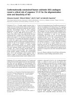
Figure 1 Random collision frequencies at five mouse gene-rich loci. (a) Maps of mouse loci investigated. Genes are indicated by full boxes
and promoters by thick black arrows. The scale bar indicates the size of 10 kb of sequence. The names of the loci and chromosomal location
are indicated above each map. The HindIII (Usp22, Emb, Lnp, Mtx2 and 11qA5 gene-desert loci) or EcoRI (Dlk1 locus) sites investigated are
indicated on the maps. Arrows indicate the positions of the primers used as anchors in 3C-qPCR experiments. (b) Random collision frequencies
at five mouse gene-rich loci. Locus names are indicated above each graph. Random collision frequencies were determined by 3C-qPCR in the
30-day-old mouse liver at the indicated anchor sites (for further details see Materials and methods). They were determined in three independent
3C assays each quantified at least in triplicate and the data were normalized as previously described [19]. Error bars are standard error of the
mean of three independent 3C assays. Grey circles, triangles or squares are data points obtained from distinct genomic sites as indicated on the
graphs. In each graph, red squares represent the floating mean (20-kb windows, shift of 10 kb). P-values (Mann-Whitney U-test) account for the
significance of the differences observed between the higher and the lower points of the floating mean. They were calculated from the values of
the average random collision frequencies in a window of 30 kb around these points (values indicated in the figure) (One asterisk indicates a P-
value < 0.1 and > 0.05; double asterisks a P-value < 0.05 and > 0.01 and triple asterisks a P-value < 0.01).
Court et al. Genome Biology 2011, 12:R42
/>Page 3 of 13
assess whether such differences also exist in the mouse,
we investigated four genomic sites (anchors) located
within a gene-desert/AT-rich region of the 11qA5 chro-
mosomal band (Figure 2b). Consistent with previous
work in human [14], we found that random collision
frequencies decrease dramatically for short site separa-
tions, reaching very low basal random collision levels for
sites separated by only 5 to 6 kb. Opposite to gene-rich
regions, however, no significant increase was observed
for large site separations. We conc lude that chromatin
dynamics in gene-desert domains is radically different
from that observed in intergenic portions of gene-rich
domains, with random collisions frequencies noticeably
decreasing much more rapidly for shorter genomic
distances.
Modulated contact frequencies at gene-rich loci are
conserved in human chromatin
To assess whether modulated contact frequencies of gene-
rich domains could be detected in human chromat in, we
used published ‘Chromosome Conformation Capture Car-
bon Copy’ (5C) data obtained at the human b-globin locus
[13] from experiments where only residual (very weak)
locus-specific interactions were detected. Statistical analy-
sis revealed a significant increase of random collision fre-
quencies for site separa tions around 100 kb (P = 0.022,
Mann-Whitney U-test) followed by a very significant
decrease for larger site separations (P = 0.0003, Mann-
Whitney U-test) (Additi onal file 2). Therefore, the 90-kb
modulation observed for random collision frequencies at
several mouse gene-rich loci appears to be conserved at
the human b-globin locus.
Genomic consequences of modulated contact frequencies
Modulations in contact frequencies, as observed here for
gene-rich regions, should have fundamental implications
for gene regulation and mammalian genome evolution.
Indeed, if, as demonstrated in this work, the frequency
of random collisions does not regularly decrease accord-
ing to geno mic distances but displays a perio dica l mod-
ulation, then cis-regulatory sequences that (for
mechanistic reasons) should interact together over long
distances will tend to accumulate at preferred relative
separation distances where the collision dynamics is fun-
damentally the most prone to such contacts. According
to this propo sal, cis-interacting sequences should posi-
tion into supranucleosomal domain I (less than 35 kb)
or domain III (around 90 kb), and eventually in domain
V (around 180 kb), since the higher basal collision levels
are found in these domains. Using the READ Riken
Expression Array Database [25], we identified 130
mouse genes that display strong co-expression patterns
with at least one other gene located less than 400 kb
away in cis (see Materials and methods) and showed
that, around such co-expressed genes, conserved
sequences are significantly over-represented in both
domain III (+7.9%) and domain V (+6.6%) (P =4×10
-5
and 1 × 10
-3
, respectively, t-tests from randomizations)
(Figure 3a). The number of conserved sequences is close
to a random distribution in domains I and II but shows
a significant under-representation (-8.6%; P =4×10
-6
,
t-test) in domain IV (between the first and second mod-
ulations) where the lower random collisions frequencies
(a)
(b)
0 50 100 150 200
0246
Site separation (kb)
D.I
D.II D.III D.IV D.V D.VI
8
6
4
2
0
0 50 100 150
200
250
02040
Crosslinking fre
q
Rel. Crosslinking
frequency
40
0
20
50 150 0 100
1.04 1.31 0.80 0.91 0.49
p=0.005 p=0.0002 p = 0.28 p = 0.099
*** *** *
(n =64) (n =67) (n =21) (n =11) (n =4)
35kb 70kb 115kb 160kb 205kb
Site separation (kb)
Rel. crosslinking frequency
Site separation (kb)
Figure 2 Random collision frequencies in gene-rich and gene-
desert regions. (a) Experimental data obtained for mouse gene-
rich regions (shown in separate graphs in Figure 1b) have been
plotted into a single graph. A few data points at separation
distances above 150 kb, which were omitted in Figure 1b, are
included. Statistical analyses were performed on the floating mean
(red squares) as explained in Figure 1b. The dashed lines delimit
supranucleosomal domains (D.I to D.VI) that encompass separation
distances where random collision frequencies are alternatively lower
and higher: 0 to 35 kb (domain I), 35 to 70 kb (domain II), 70 to 115
kb (domain III), 115 to 160 kb (domain IV), 160 to 205 kb (domain V)
and 205 to 250 kb (domain VI). (b) Random collision frequencies
were determined by 3C-qPCR at four sites (R9, F25, F35 and F48;
Figure 1) located in an AT-rich/gene-desert region located on
mouse chromosome 11. Red squares represent the floating mean
(20-kb windows, shift of 10 kb). Error bars are standard error of the
mean (the triple asterisks indicate a P-value < 0.01).
Court et al. Genome Biology 2011, 12:R42
/>Page 4 of 13
were observed. We conclude that, as a predicte d conse-
quence of our findings, conserved intergenic sequences
of clustered co-expressed genes are signif icantly over-
represented within supranucleosomal domains III and V
corresponding to the first and second modulations of
random collision frequencies.
Interestingly, recent genome-wide mapping of chro-
mosomal interactions in human by Hi-C experiments
also provides direct experimental validation of our pro-
posal. Indeed, these data confirm that long-range inter-
actions in Giemsa-ne gative bands, containing gene-rich
regions, are favored for site separations around 90 kb
(domain III) relative to Giemsa-positive bands, which
are gene-poor regions (Figure 3b). Therefore, both
bioinformatic analyses and genome-wide Hi-C experi-
ments support the predicted consequences of a 90-kb
modulation and suggest that this phenomenon underlies
the chromatin dynamics of a significant number of
gene-rich loci in mammals.
The statistical helix model
We reasoned that the modulations of contacts frequencies
observed at several gene-rich loci may reflect a preferential
statistical shape that the chromatin tends to adopt when
no strong locus-specific interactions take place. Since this
constraint appears to be independent of the genomic posi-
tion at all five gene-rich loci investigated, this preferential
non-linear shape should possess a long-range translational
symmetry. This led us to postulate that this statistical
shape may correspond to a simple helix organization.
The dynamics of chromatin has been successfully
modeled in yeast [11,24] using a Freely Jointed Chain/
Kratky-Porod worm-like chain model [26]. This model
is given in Equation 1 [24], which expresses the relation-
ship between crosslinking frequency X(s) (in mol × liter
-
1
×nm
3
) and site separation s (in kb):
X(s)=
k × 0.53 × β
−
3
/
2
× exp
−
2
β
2
×
(
L × S
)
−3
(1)
The b term represents the number of Kuhn’s statisti-
cal segments and depends on polymer shape. Equations
2a and 2b (see Materials and methods) provide the b
terms used for linear and circular polymers, respectively.
For a polymer folded into a circular helix, we developed
the following b term (see Materials and methods):
β =
D
2
× sin
2
π × L × s
√
π
2
× D
2
+ P
2
+
P
2
× L
2
× s
2
π
2
× D
2
+ P
2
L ×
S
(5)
where D is the diameter of the helix (in nm) and P its
step (in nm). In the above equations, S is the length of
the Kuhn’s statistical segment in kb, which is a measure
of the flexibility of the chromatin, and k is the crosslink-
ing efficiency, which reflects experimental variations.
The linear mass density L is the length of the chromatin
in nm that contains 1 kb of genomic DNA.
Using Equation 1 and the appropriate b terms, we
fitted our experimental data to three polymer models.
The linear model fits appropriately only for site
(a)
(b)
0
0,05
0,1
0,15
0,2
0,25
0,3
0,35
0,4
0,45
50 à 70
70 à 115
115 à
160
160 à
205
205 à
250
g
n…
p =0.007
D.II D.III D.IV D.V D.VI
***
G -neg (gene -rich)
G -pos (gene -poor)
Rel. Count of interactions
205- 250 160 -205 115-160
70
-
115 50-70
Domains of separation distances (kb)
0,08
0,1
0,12
0,14
0,16
0,18
0,2
0,22
0,24
0 50 100 150 200 250
0.10
0.12
0.14
0.16
0.18
0.20
0.22
0.24
Rel. Count of
TSS/cs distances
35 205 250
TSS/cs distances (kb)
0
0.08
D.I D.II D.III D.IV D.V D.VI
+7.9% -8.6% +6.6%
p = 4.10
-5
p = 4.10
-6
p = 10
-3
***
***
**
115 16070
D.II D.III D.IV D.V D.VID.I
Figure 3 Influence of modula ted random collision frequencies
on long-range interactions and mammalian genome evolution.
(a) Separation distances between conserved sequences (cs) and
transcription start sites (TSS) of co-expressed mouse genes were
determined as explained in the Materials and methods section.
Black triangles depict the relative count of separation distances
obtained for each supranucleosomal domain. Black squares indicate
the mean of relative counts obtained from 30 random samples of
genes. Error bars represent the 95% confidence intervals for
randomization. Separation distances are significantly over-
represented in domains III and V (+7.9% and +6.6%, respectively)
while they are significantly under-represented in domain IV (-8.6%)
(P-values of t-tests are indicated on the graph). (b) Histogram
depicting the relative counts of cis-interactions in human GM06990
or K562 cells (Hi-C experiments from [4]) occurring in Giemsa-
negative (gene-rich regions, white bars) or Giemsa-positive (gene-
poor regions, gray bars) bands. For each set, the number of
interactions was counted in each supranucleosomal domain (as
defined in Figure 2a). Counts in each domain were normalized
against the total number of sequence-tags counted over all
domains (D.I to D.VI). Error bars represent standard error of the
mean of two Hi-C experiments. The P-value indicated on the figure
was obtained from a t-test (double asterisks indicate a P-value <
0.05 and >0.01, and triple asterisks a P-value < 0.01).
Court et al. Genome Biology 2011, 12:R42
/>Page 5 of 13
separations lower than 35 kb (domain I; black line in Fig-
ure 4, lower panel). By setting an apparent circular con-
straint (c = 110.515 ± 2.028 kb), the circular polymer
model [11] better fits the experimental data but only for
site separations lower than this apparent circular con-
straint c (that is, below 110kb) (Additional file 3). Finally,
the statistical helix model provides a valid description
over the entire range of genomic distances investigated (0
to 340 kb; R
2
= 0.38; red line in Figure 4). Importantly,
this finding shows that modulated contact frequencies
observed at mammalian gene-rich loci can be described
as if the chromatin was statistically shaped into a helix
350300250200150100500
Site separation (kb)
5
2
1
0.5
0.2
0.1
0.05
0.02
Rel. crosslinking frequency
R
2
= 0.09
R
2
= 0.38
Rel. crosslinking frequency
0kb
2
4
6
5
1
3
7
8
0
p=0.0015 p=0.0016
1.22
(0.84)
n=85
1.58
(1.73)
n=122
1.13
(0.73)
n=61
1.52
(0.76)
n=48
0.63
(0.53)
n=30
p=0.010
D.II D.III D.IVD.I D.V D.VI
***
***
***
35kb 70kb 115kb 160kb 205kb 250kb
295kb 340kb
1;30
(0.43)
n=8
D.VII
0.66
(0.34)
n=18
D.VIII
Usp22
Usp22 PE
Dlk1
Lnp
Mtx
Emb
**
**
K =932,677±70,254 S =2.709±0.081 kb
‹D›=292.03±4.80 nm ‹P› = 162.13±8.75 nm
Sh=94.090 +/- 1.599 kb
**
p<2.10
-5
p=0.028p=0.016
Figure 4 Fitting the statistical helix polymer model to random collision frequencies quantified at mouse gene-rich loci. 3C-qPCR data
shown in Figure 2a and Additional file 1 (Usp22PE) were compiled into a single graph (upper panel). Error bars are standard error of the mean.
The dashed lines delimit supranucleosomal domains as defined in Figure 2a. The graph shows the best fit analyses obtained with the linear
polymer model (Equations 1 and 2a; black curve) or the statistical helix model (Equations 1 and 5; red curve). Correlation coefficients (R
2
) are
indicated in the lower panel, which shows the same graph where collision frequencies are represented in a logarithmic scale. Best fit parameters
for the statistical helix model are indicated within the graph (lower panel) and have been used to calculate the expected theoretical means of
random collision frequencies for each supranucleosomal domain (numbers in brackets in upper panel), which are in good agreement with the
means obtained from the experimental data (values indicated above the expected means). P-values (Mann-Whitney U-test) account for the
significance of the differences observed between the experimental means of two adjacent domains. One can note, amongst the experimental
points, a few outliers. To minimize the weight of these data points, we chose a non-parametric statistical test (double asterisks indicate a P-value
< 0.05 and > 0.01 and triple asterisks a P-value < 0.01).
Court et al. Genome Biology 2011, 12:R42
/>Page 6 of 13
for which we estimated the structural parameters: dia-
meter D = 292.03 ± 4.80 nm and step P =162.13±8.75
nm (Figure 4). Noteworthy, the estimated length of the
statistical segment S = 2.709 ± 0.081 kb, indicate s that
the mammalian chromatin is more flexible than its yeast
counterpart, for which a value of S =4.7±0.45kbwas
obtained for GC-rich regions [24]. These parameters
allow calculation of the length of DNA folded into one
turn of this statistical helix: Sh = 94.090 ± 1.599 kb (see
Materials and methods).
It is important to stress that the shape of the chroma-
tin described by these parameters is averaged over the
whole population of c ells analyzed (5 million nuclei in
each 3C sample) and thus is more likely to represent a
statist ical shape arising from the global dynamics of the
chromatin than a fixed organization (Figure 5).
Discussion
This work reveals that some gene-rich regions of the
mouse and human genomes display modulation of their
contact frequencies. Several lines of evidence indicate that
this modulation arises from an intrinsic constrai nt rather
than from locus-specific constrain ts. F irstl y, f or a g iven
locus, a similar 90-kb modulation is observed at several
genomic sites assayed. For example, at the Dlk1 locus it
occurs at site F3 and sites F5 (9 kb away from F3) and F14
(62.7 kb away); at the Usp22 locus, it takes place at site F-
28 as well as sites F1 (91.4 kb away) and F7 (109.9 kb
away). Secondly, this 90-kb modulation was found at five
distinct gene-rich loci located on four differe nt mouse
chromosomes. Finally, using published 5C data [13], we
found a very similar modulation at the human b-globin
locus in cells where very weak interactions were found.
Interestingly, this modulation was not revealed in previous
3C experiments that we, and many others, performed in
mouse or human. There are at least two reasons why this
phenomenon went unnoticed. Firstly, the amplitude of the
modulation is very weak and could only be significantly
revealed when a relatively large number of experimental
points were obtained from a highly quantitative method
and combined together into a sing le graph after accurate
normalization of the data [19]. Secondly, at many gene-
rich loci (see, for example, [1 4]), strong locus-specific
interactions (above four times the local random collision
level) take place, which very likely perturb this modulation.
However, as observed in this work (outliers in Figure 4) or
in GM06990 cells for the human b-globin locus [13]
(Additional file 2), modulation can be perceived despite
some residual and weak locus-specific cis-ortrans-inter-
actions (below three to four times the local random colli-
sion level). Interestingly, this modulation is not a simple
consequence of gene expression per se since RT-qPCR
analysis indicated that, in the sample s investigated (3 0-
day-old mous e liver) , some loci are compl etely repressed
(Dlk1 locus), or display very low expression levels (Emb
and Lnp loci), while others contain expressed genes
(UspP22 and Mtx2 loci) (Additional file 4). However,
according to our modeling, the statistical helix would be
inaslightlymore‘open’ configuration at the expressed
loci (with a diameter D of about 303.92 ± 6.55 nm and a
step P of 177.38 ± 12.05 nm), compared to silent loci (D =
278.83 ± 7.65 nm and P = 149.20 ± 13.67 nm) (Additional
file 5). Nevertheless, these differences are minor and the
statistical helix model is valid in both situations.
To what extent does this phenomenon apply to sub-
stantial parts of mammalian genomes? Our work sug-
gests that gene-rich regions of the mammalian
chromatin display modulated contact frequencies while
no modulation could be evidenced in gene-poor regions
(Figure 2b). As previously discussed, direct experimental
detection of such modulations requires finding cellular
systems where no strong locus-specific interactions
occur. This is an important caveat that is particularly
difficult to circumvent at many gene-rich loci that we
may wish to investigate. In this work, the modulation
could be observed at only five mouse and one human
loci. Therefore, it remains difficult to speculate on
whether such a phenomenon may apply to a substantial
part of gene-rich domains, or whether it is rather lim-
ited to few loci. Clearly, however, both bioinformatic
analyses and genome-wide mapping of chromatin inter-
actions [4] indicate that this phenomenon may underlie
the dynamics of a significant number of locus-specific
interactions in gene-rich domains of mammalian chro-
matin (Figure 3).
As previously mentioned, one consequence of modu-
lated contact frequency is that long-range interacting cis-
regulatory sequences will undergo const raints that will
‹
D
›
~290 nm
‹
P
›
~160 nm
Figure 5 The statistical helix model. The statistical helix model
that we propose in this study (Equations 1 and 5) suggests that, in
the absence of strong locus-specific interactions, some gene-rich
domains of the mammalian chromatin tend to adopt a helix shape.
This helix is averaged over the whole population of cells analyzed (5
million nuclei in each 3C sample) and thus more likely represents a
statistical shape arising from the global dynamics of the chromatin
than a fixed organization. It is characterized by a mean diameter
〈D〉 and mean step 〈P〉, and it thus likely corresponds with
the place where the probability of finding the chromatin at a given
t time is the highest (black helical curve).
Court et al. Genome Biology 2011, 12:R42
/>Page 7 of 13
tend to accumulate them within specific supranucleosomal
domains where the collision dynamics is fundamentally
the most appropriate for contacts. This property may
explain the peculiar arrangements of genes and cis-regula-
tory elements observed at several important mammalian
loci, such as the ‘ global control region’ (GCR) at the
mouse Hoxd (Homeobox d) locus, which is located at one
or two modulations away from the genes that it regulates.
It was suggested that ‘the GCR would have concentrated,
in the course of evolution, several important enhancers,
due to an intrinsic property to work at a distance’ [27].
The modulation of contact frequencies revealed in this
work represents one such intrinsic property that may con-
tribute to enhancer clustering in mammals.
Our work suggests that modulated contact frequencies
arise from an intrinsic constraint that applies to the
chromatin. This led us to wonder about the nature of
this constraint and to propose that it may result from a
preferential statistical shape that the chromatin tends to
adopt in gene-rich regions when no strong locus-specific
interactions take place. This hypothesis is supported by
the finding that modulated contact frequencies can be
described by polymer models as if, in these regions, the
chromatin was sta tistically shaped into a helix (Figure
4). Interestingly, by using 3C data obtained in the yeast
Saccharomyces cerevisiae [24], we showed that the statis-
tical helix model may also be valid for GC-rich (but not
AT-rich) domains of the yeast genome (Additional files
6 and 7).
One consequence of folding the chromatin into a helix-
shaped structure is that t he volu me it occupies increases
dramatically. This increase can be estimated by calculat-
ing the volumetric mass density (Vs) of the statistical
helix. In mammals, Vs =1.02×10
5
±0.05×10
5
nm
3
/kb
(or 0.0098 ± 0.0005 bp/nm
3
; estimated from Equation 6
givenintheMaterialsandmethods section and best fit
parameter shown in Figure 4). This can be compared to
the estimated volumetric mass density V of the postu-
lated 30-nm chromatin fiber: V =6.8×10
3
nm
3
/kb (cal-
culatedfromEquation6withD =30nm;〈R〉 =9.6
nm and s = 1 kb). Therefore, the folding of a putative 30-
nm chromatin fiber into a statistical helix would result in
a 15.00 ± 0.73-fold increase (Vs/V) of the volume that it
occupies. Finally, if the entire diploid genome had a heli-
cal chromatin organization as shown in Figure 5, it
would occupy a volume of about 610 μm
3
(the volume
occupied by such a helix encompassing two times 3 ×
10
9
bp), which is higher than the volume of a regular
mammalian nucleus (approximately 520 μm
3
for a
nuclear diameter of 10 μm). Therefore , in addition to the
helix-shaped organization described above, other types of
dynamic folding should exist that achieve higher levels of
chromatin compaction. This hypothesis is supported by
our finding showing that the dynamics of random
collisions in gene-desert regions is completely different
to that observed in gene-rich domains.
The pioneering work of Ringrose et al.[28]demon-
strated that chromatin behaves like a linear polymer at
short distances. This work was based on quantitative
comparison of in vivo recombination events and was lim-
ited to short site separation distances (less than 15 kb).
Our work suggests that the upper limit for such linear
polymer model s may occur, in gene-rich regions, for
separation distances around approx imately 35 kb (supra-
nucleosomal domain I; Figure 4). For higher genomic dis-
tances, spanning at least 340 kb (Figure 4), the statistical
helix polymer model describes accurately the dynamics
of the chromatin. What is the upper limit of validity for
this model? We know that, at a larger scale, the chroma-
tin is confined within the limited space of the chromo-
some territory [2,29]. This ‘ chromosomal territory
constraint’ will necessarily imp act on the accuracy of the
statistical helix polymer model to describe chromatin
dynamics. Cell imaging techniques have suggested that
polymer models are incompatible with spatial distance
measurements obtained for genomic separations over 4
Mbp [30,31]. Therefore, the upper limit should lie some-
where between 34 0 kb and 4 Mbp. Bas ed on the biophy-
sical parameters provided in Figure 4, we calculated how,
in interphasic cells, the spatial distances should vary as a
function of genomic site separations and compared the
resulting values to those measured in fluorescence in situ
hybridization (FISH) experiments. For separation dis-
tances below 1 Mb, spatial distances predicted from the
statistical helix model (red curve in Additional file 8) are
fullycompatiblewiththedistances measured in FISH
experiments (data points in Additional file 8) [32]. How-
ever, above 1 Mb, the statistical helix model does not fit
with the experimental data and, therefore, the upper
limit of validity of this model appears to reside at separa-
tion distances around 1 Mb. This suggestion is in agree-
ment with the recent comprehensive mapping of
chromosomal interactions in the human genome (Hi-C
experiments) showing that, above the megabase scale, the
chromatin adopts a ‘fractal globule’ conformation [4]. In
line with modeling approaches pioneered by Dekker and
colleagues [11,24], our work suggests that, below the
megabase scale, chromatin dynamics within such glo-
bules can be accurately described by appropriate polymer
models. We can reasonably expect that the increasing
sensitivity of both cell imaging and 3C-derived techni-
ques will soon help us to assess the validity of this
approach, thus enlightening one of the last remaining
‘mysteries’ of mammalian genome organization.
Conclusions
In this work, we have discovered an unexpected 90- to
100-kb modulation of contact frequencies at gene-rich
Court et al. Genome Biology 2011, 12:R42
/>Page 8 of 13
loci of mammalian chromatin. We show that this modu-
lation has important implications for genome evolution
and we provide an original model that suggests that the
modulation may result from a fundamental statistical
helix shape that the chromatin tends to adopt when no
significant locus-specific interactions are taking place.
Altogether, our work contributes to a better understand-
ing of the fundamental dynamics of mammalian chro-
matin within chromosomal territories.
Materials and methods
Mouse breeding
All experimental designs and procedures were i n agree-
ment with the guidelines of the animal ethics committee
of the French ‘Ministère de l’Agriculture’.
3C-qPCR/SybGreen assays
The 3C-qPCR assays were performed as previously
described [17] with a few important modifications that
increased the efficiency of the 3C assays four-fold, thus
allowing real-time PCR quantifications of 3C products
using the SybGreen technology instead of TaqMan
probes used in previous work [17,19]. The 3C-qPCR
method [17] was modified as follows. Step 2: 5 × 10
6
nuclei were crosslinked in 1% formaldehyde. Step 8:
added 5 μl of 20% (w/v) SDS (final 0.2%). Step 10:
added 50 μl of 12% (v/v) Triton X-100 diluted in 1 ×
ligase buffer from Fermentas (40 mM Tris-HCl pH7.8,
10 mM MgCl
2
, 10 mM DTT, 5 mM ATP). Step 13:
added 450 U of restriction enzyme (EcoRI for the Dlk1
locusorHindIIIfortheotherloci).Step16:incubated
30 minutes at 37°C; shake at 900 rpm. Step 34: addi-
tional digestions were performed using BamHI for the
Dlk1 locus and StyI for the other loci. Step 39:
adjusted 3C assays with H
2
Oto25ng.μl
-1
.3Cpro-
ducts were quantified (during the linear amplification
phase) on a LighCycler 480 II apparatus (Roche, Basel,
Switzerland); 10 minutes at 95°C followed by 45 cycles
10 s at 95°C/8 s at 69°C/14 s at 72°C) using the Hot-
Start Taq Platinum Polymerase from Invitrogen (Carls-
bad, California, USA) (10966-34) and a standard 10 ×
qPCR mix [33] where the usual 300 μMdNTPwere
replaced with 1,500 μMofCleanAmpdNTP(TEBU
040N-9501-10). Standards curves for qPCR were gen-
erated from BACs (Invitrogen) as previously described
[17]: RP23 55I2 for the Usp22 locus; RP23 117C15 for
the Dlk1 locus; and a subclone derived from RP23 3D5
for the gene-desert region. For 3C-qPCR analyses of
site F-28 at Usp22 locus, a PCR product encompassing
733bparoundsiteF-28wasgeneratedfromgenomic
DNA (FA4 gccatactcagccacagggac and RA2 cctgatct-
cacgaatcaccctc). This PCR product (0.1 μg) was mixed
with 3.4 μg of the RP23 55I2 BAC before HindIII
digestion and ligation to generate standard curves.
Data obtained from these experiments a re included in
Additional file 9 (gene-rich loci) or Additional file 10
(gene-desert locus). 3C-qPCR primer sequences are
given in Additional file 11. The number of sites ana-
lyzed in each experiment were as follows: Usp22 locus,
for anchor sites F1 and F 7, 34 and 40 sites, respec-
tively; Dlk1 locus, for anchor sites F14/F5 and F3, 23/
17 and 9 sites, respectively; Emb locus, for anchor sites
R4 and R7, 31 and 30 sites, respectively; Lnp locus, for
anchor sites R41 and R46, 27 and 25 sites, respectively;
Mtx2 locus, for anchor sites R2 and R56, 52 sites for
each anchor; and for the gene-desert locus, for anchor
sites R9/F25/F35 and F48, 36/40/40 and 38 sites,
respectively.
Primer extension
For each biological sample and each exten sion primer
(1F, cagtccagtgagacacatggttg; FA1, gttaaacccacagggcaa-
gagc), six reactions were performed, pooled, purified
with a QiaQuick PCR purification kit and diluted in
H
2
O at 12.5 ng.μl
-1
. Each reaction was done as follows:
0.1 μM of extension primer was added to a 10-μl reac-
tion containing 1 × qPCR mix [33] and 1 μlofhighly
concentrated 3C assay (containing about 200 to 300 ng
of genomic DNA). Primers were extended by the Hot-
Start Taq Platinum polymerase (Invitrogen) in a Light-
Cycler apparatus (3 minutes at 95°C followed by 45
cycles 1 s at 95°C/5 s at 70°C/15 s at 72°C). Amplified
3C products were quantified by qPCR as explained
above. Data obtained from these experiments are
included in Additional file 9.
RT-qPCR quantification
Total RNA extraction and RT-qPCR quantification were
performed as previously described [20,21] using Super-
script III reverse transcriptase (Invitrogen; 150 U for 45
minutes at 50°C).
Supranucleosomal domains
Supranucleosomal domains (D.I to D.VI) were defined
from statistical analyses (Mann-Whitney U te sts) per-
formed on data shown in Figure 2a. They encompass
separation distances where random collision frequencies
are alternatively lower and higher: 0 to 35 kb (domain
I), 35 to 70 kb (domain II), 70 to 115 kb (domain III),
115 to 160 kb (domain IV), 160 to 205 kb (domain V)
and 205 to 250 kb (domain VI).
Mathematical methods
We used the Freely Jointed Chain/Kratky-Porod worm-
like chain model [26]. This model is given in Equation 1
(Equation 3 of [24]]), which expresses the relationship
between the crosslinking frequency X(s) (in mol × liter
-1
×nm
3
) and the site separation s (in kb):
Court et al. Genome Biology 2011, 12:R42
/>Page 9 of 13
X(s)=
k × 0.53 × β
−
3
/
2
× exp
−
2
β
2
×
(
L × S
)
−3
(1a)
with, for a linear polymer:
β =
s
S
(2a)
In Equation 1, S is the length of the Kuhn’sstatistical
segment in kb, which is a measure of the flexibility of
the chromatin, and k is the efficiency of crosslinking,
which reflects experimental variations. The linear mass
density L is the length of the chromatin in nm that con-
tains 1 kb of genomic DNA. For the foll owing analyses,
we used a value L = 9.6 nm/kb [26] estimated from a
packing ratio of 6 nucleo somes per 11 nm of chromatin
in solution at physiological salt concentrations, corre-
sponding to a nucleosome repeat length of about 190
bp, as found in mammal ian cell lines. By introducing
parameter c giving the ‘apparent circle size’ in kb into
the b term of Equation 2a, Dekker et al. [11] derived a
model (Equation 2b) that des cribes the dynamics of
interactions within a circular polymer:
β =
s
S
×
1 −
s
/
c
(2b)
The b term in Equation 1 corresponds to the number
n of Kuhn’s statistical segments [26], which is directly
related to the average spatial dista nce between the sites
〈R〉 in nm and the length of the statistical segment S
as given in Equation 3:
β =
R
2
L ×
S
(3)
Interestingly, by setting appropriately the 〈R〉 para-
meter in E quation 3 and using the resulting b term in
Equation 1, one can simulate spatial constraints that
‘fold’ the intrinsically linear polymer. Such modifications
help us to model the dynamics of random collisions
within a chromatin that possesses higher levels of orga-
nization. For a linear polymer, the average spatial dis-
tance 〈R〉 is directly linked to site separation s as
given in Equation 4a:
R
= s ×
L
(4a)
and thus substitution of Equation 4a in Equation 3
yields the b term given in Equation 2a. For a circular
polymer, the average spatial distance 〈R〉 can be
linked to site separation s by intr oducing the previously
described [11] appa rent circular constraint c as given in
Equation 4b:
R
= s × L ×
1 −
s
/
c
(4b)
and thus substitution of Equation 4b in Equation 3
yields the b term given in Equation 2b.
For a polymer folded into a circular helix the average
spatial distance 〈R〉 (in nm) is related to site separa-
tion s (in kb), to the mean diameter D of the helix in
nm and the mean step P in nm as given in Equation 4c:
R
=
D
2
× sin
2
π × L × s
√
π
2
× D
2
+ P
2
+
P
2
× L
2
× s
2
π
2
× D
2
+ P
2
(
nm
)
(4c)
Substitution of Equation 4c in Equation 3 yields the b
term given in Equation 5:
β =
D
2
× sin
2
π × L × s
√
π
2
× D
2
+ P
2
+
P
2
× L
2
× s
2
π
2
× D
2
+ P
2
L ×
S
(5a)
Finally, the b term given in Equation 5 can be used in
Equation 1 to provide a model that describes random
collisions within a circular helix polymer. (Note that, for
P = 0, Equation 5 describes a circularized polymer of
size D and when both P =0andD tend to infinity the
equation is able to describe a linear polymer). The
length of one turn on the statistical helix Sh was calcu-
lated from the best-fit curve (Figure 4) by applying the
second derivative method.
The volumetric mass density of the supranucleosomal
chromatin Vs was calculated from Equation 6:
Vs =
R
× π ×
D
2
2
s
nm
3
kb
(6)
where 〈R〉 corresponds to Equation 4c.
Best-fit analyses
Best-fit analyses were implemented under the R software
[34]. We used the ‘ nls object’ (package stats version
2.8.1), which determines the nonlinear (weighted) leas t-
squares estimates of the parameters of nonlinear models.
Bioinformatics and statistical analyses
Contact frequencies at the human b-globin l ocus in the
EBV-transformed lymphoblastoid cell line GM06990
were downloaded from [13] (Supplemental Tables 6 and
7). These 5C data were normalized using our previously
published algorithm [19] and compiled into a graph
(Additional file 2).
Co-expressed genes were selected from the READ
Riken Expression Array Database [25], which contains
the relative expression levels of 16,259 transcripts in 20
mouse tissues. Housekeeping genes, which tend to accu-
mulate in clusters [35] and are co-expressed but do not
necessarily share cis-acting regulatory elements, have
Court et al. Genome Biology 2011, 12:R42
/>Page 10 of 13
been excluded. According to the criteria defined by Fe r-
rari and Aitke n [36], housekeeping genes were consid-
ered as those having a P-value > 0.5. The resulting
datab ase contained 11,701 genes. We then retained only
genes for which expression data were available for at
least 15 tissues and selected gene pairs separated by l ess
than 400 kb. This database, containing 6,619 genes, was
used for identification of clustered co-expressed gene
pairs and randomizations (see below).
For each possible gene pair, co-expression levels were
determined by calculating the Pearson correlation coeffi-
cient (r) from their relative expression levels in at least
15 tissues. Co-expressed genes were defined as those
having either similar (r ≥ 0.8) or opposite (r ≤ -0.8) tis-
sue-specific expression pa tt erns. This finally provided a
set of 130 strongly co-expressed/co-reg ulated genes. We
then determined the relative site separations between
the transcriptional start sites of these co-regulated genes
and conserved intergenic sequences. Conserved
sequences were downloaded from the mouse genome
(July 2007 assembly, filter 0.9, no overlap with UCSC
Genes) on the U CSC server. We limited our analysis to
a maximal separation distance of 250 kb covering the
six previously defined supranucleosomal domains (Fig-
ure 2a). In order to obtain a database of conserved
sequences that is significantly enriched in shared regula-
tory elements, we removed conserved sequences that are
located in transcription units or promoter regions (less
than 3 kb from a transcriptional start site). Finally, we
counted site separation distances included in each
domain and each count was normalized to the total site
separat ion distan ces counted (ov er 250 kb). To evaluat e
the tendencies toward over- or under-representation of
site separations in each domain, we randomly extracted
130 genes from the initial database and calculated site
separation distances of conserved sequences, which were
counted and normalized as mentioned above. This ran-
domization was repeated 30 times. Normal distribution
was checked for counts in each domain (Shapiro-Wilk
tests). We then calculated the 95% confidence interval
(E) from the following equation:
E = μ ± t.σ
(
N
)
1/2
where t is the t-student variable as read in the Student’s
table for a degree of freedom of 29 and an alpha risk fac-
tor of 0.5 (t = 2.04), μ is the mean number of counts, s is
the standard deviation and N is the number of randomi-
zations performed (here 30). Error bars represent the
95% confidence interval for counts in each domain.
Hi-C data used in Figure 3b are from published experi-
ments [4]: Gene Expre ssion Omnibus acces sion numbers
[GSM455137] (sequencing of [GM06990] cells-lane1),
[GSM455138] (sequencing of [GM06990] cells-lane2),
[GSM455139] (sequencing of K562 cells-lane1) and
[GSM455140] (sequencing of K562 cells-lane2). For each
of the four datasets, we selected all the p airs of sequence
tags located on the same chromosome and removed those
located on distinct chromosomes (that is, we removed
trans-interact ions). Pairs of sequence-tags were classified
by chromosome. We extracted the positions of all HindIII
and NcoI sites from the human genome (hg18). Restric-
tion fragments were numbered and, for each restriction
fragment, we specified the positions of the 5’ and 3’ ends.
The downloaded positions of the tags were replaced by
the position of the corresponding restriction site. For this
operation we used the restriction fragment numbers pro-
vided in the downloaded files. Direction ‘0’ corresponds to
a restriction site located at the 3’ end of the restriction
fragment (sense reading of the sequence-tag) while direc-
tion ‘1’ corresponds to the 5’ end (antisense reading of the
sequence-tag). We then assembled datasets generated
from lanes 1 and 2 of each experiment. We extracted from
the UCSC server the positions of chromosomal bands
(Giemsa-negative and Giemsa-positive; hg18). We selected
all pairs of sequence-tags for which both partners are
located within the same chromosomal band (to remove
long-range/inter-band interactions). Data were pooled into
two separ ate sets: a first set corresponding to all pairs of
sequence-tags located in Giemsa-negative bands and a sec-
ond one corresponding to pairs of sequence tags located in
Giemsa-positive bands (threshold above 50, that is, g-pos-
100, g-pos-75 and g-pos-50; see UCSC server). For each
set, we selecte d interactions that are represented by at
least four pairs of sequence tags (multiple pairs of
sequence tags for identical interaction partners) and calcu-
lated for each interaction the separation distance between
the restriction sites (using the positions previously calcu-
lated as described a bove). In each s et (Giemsa-negative
and Giemsa-positive), the number of interactions were
counted in each supran ucleosomal domain (as defined in
Figure 2a) and this number was normalized to the to tal
number of interactions counted in all domains (D.I to D.
VI). Data are presented in a histogram (Figure 3b) that
provides, for each domain, a comparison between the
counts of interactions in Giemsa-negative and Giemsa-
positive sets.
Additional material
Additional file 1: Random collision frequencies in gene-rich regions
for large separations distances. Random collision frequencies were
determined by 3C-qPCR after a primer extension step (see Materials and
methods) at two Usp22 genomic sites (sites F1 and F-28) (Figure 1a) in
liver samples from 16.5-days-post-coitus embryos (grey data points) or
30-day-old mice (white data points). Data analysis was as described in
the legend of Figure 1b. Red squares represent the floating mean (45-kb
windows, shift of 22.5 kb). We determined the higher and the lower
points of the floating mean for site separations above 40 kb and
calculated the average random collision frequencies (values are indicated
Court et al. Genome Biology 2011, 12:R42
/>Page 11 of 13
in the figure) of sites located 40 kb around these points (horizontal black
bars). P-values (Mann-Whitney U-test) account for the significance of the
differences observed between these averages. Error bars are standard
error of the mean.
Additional file 2: Collision frequencies at the human b-globin locus.
Collision frequencies at the human b-globin locus (a gene-rich region on
chromosome 11p15.4) were obtained from several published 5C
experiments performed in GM06990 cells, an EBV-transformed
lymphoblastoid cell line where this locus is not expressed and where only a
very weak/residual interaction was detected (Supplemental Tables 6 and 7
in [13]). Data from each experiment were normalized according to a
previously published algorithm [19] and plotted into a single graph.
Statistical analyses were performed as explained in the legend of Figure 1b.
Additional file 3: Fitting the circular polymer model to mouse gene-
rich loci. The circular polymer model (Equations 1 and 2b) was fitted to
3C-qPCR data obtained at gene-rich loci. The best fit curve is shown in
red and best fit parameters are as follows: R2 = 0.50 with K = 725,785 ±
66,540; S = 2.515 ± 0.092 kb; c = 110.515 ± 2.028 kb. The black curve
depicts the best fit obtained with the linear polymer model (Equations 1
and 2a; R2 = 0.18).
Additional file 4: Gene expression at loci investigated by 3C-qPCR.
Total RNA from 30-day-old mouse liver was prepared and mRNA levels
were determined by RT-qPCR relative to Gapdh mRNA level. The Usp22,
LnP and Mtx2 genes were found to be expressed. Very low levels of
expression were found for the Gtlf3b, Aldh3a2 and Emb genes. The other
genes (Kcnj12, Tnfref13b, Gtl2, Dlk1 and HoxD13) are fully repressed.
Additional file 5: Random collisions at silent versus expressed loci.
Data points represent collision frequencies determined at silent (Dlk1/
Emb/Lnp; black circles) or expressed (Usp22/Mtx2; red circles) loci. Best fit
of the statistical helix model (Equations 1 and 5) was performed for each
dataset (black curve = silent loci; red curve = expressed loci). The values
of best fit parameters for each data set are indicated in the graph. Both
the diam eter (D) and the step (P) of the helix are larger in the expressed
loci compared to the silent ones.
Additional file 6: Fitting the statistical helix model to the yeast
Saccharomyces cerevisiae genome. In order to test whether a statistical
helix organization may be valid for other organisms, we fitted the
statistical helix polymer model to the 3C data obtained in the yeast S.
cerevisiae [24]. For both AT-rich and GC-rich regions (Additional file 7a
and 7b, respectively), correlation coefficients (R2 = 0.82 and 0.80,
respectively) were similar to those obtained from published models (R2
= 0.81 and 0.79, respectively) [24]. For AT-rich regions, consistent with
previous findings [24], the statistical helix model predicts a linear polymer
organization (Additional file 7a). However, data obtained in GC-rich
domains are fully compatible with a statistical helix organization.
Compared to mammals, chromatin dynamics in yeast can be described
as a statistical helix that would have a slightly smaller diameter (212.62 ±
31.73 nm) but a much wider step (310.94 ± 54.86) (Additional file 7b).
Finally, using these best-fit parameters and Equation 4c, we calculated
how, according to this statistical helix model, the spatial distances should
vary
as a function of genomic site separations. We found that spatial
distances calculated from the statistical helix model are in good
agreement with those measured in high-resolution FISH analyses
performed in living yeast cells (Additional file 7c) [37]. Therefore, the
statistical helix model may also be valid to describe chromatin dynamics
in GC-rich domains of the S. cerevisiae genome.
Additional file 7: Fitting the statistical helix model to the yeast
Saccharomyces cerevisiae genome. Data published by Dekker for the
yeast S. cerevisiae [24] were normalized using the previously published
algorithm [19] and the statistical helix polymer model (Equations 1 and 5
was fitted to normalized data. (a) For AT-rich regions, consistent with
previous findings [24], the statistical helix model (red curve) predicted a
linear polymer organization (black curve). In this case, the best fit values
obtained for the diameter D and the step P are not relevant, as indicated
by large standard deviations. (b) In GC-rich regions, the statistical helix
model (red curve), fits with a distended helical shape. Best-fit parameters
are indicated above the graph. They were calculated using a linear mass
density of 11.1 nm/kb [11]. The black curve depicts the best fit of the
linear polymer model and the green curve the best fit of the circular
polymer model. Note that the lengths of the statistical fragments obtained
from the statistical helix model (S = 6.060 ± 0.519 kb and 4.558 ± 0.503 kb
for AT-rich and GC-rich domains, respectively) are compatible with the
parameters previously obtained with the linear or circular polymer models
(S = 6.4 ± 0.34 kb and 4.7 ± 0.45 kb, respectively) [24]. (b) Using the best-
fit parameters obtained for the yeast S. cerevisiae (b), we calculated the
expected mean spatial distances (in nm) for increasing site separation
distances (0 to 140 kb) for both the statistical helix (Equation 4c; red curve)
and the linear polymer (Equation 4a; black curve) models. The
experimental spatial distances (in nm) obtained by Bystricky et al. (Table 1
and Supplementary Table of [37]) from high-resolution FISH experiments
were plotted into this graph (open squares, adjusted average distances;
black diamonds, average peak distances). The statistical helix model is in
good agreement with these experimental data.
Additional file 8: An upper limit of validity for the statistical helix
model. Expected spatial distances (in nm) were calculated as a function
of increasing genomic distances (in kb) using either Equation 4a (linear
polymer model, black curve, with L = 9.6 nm/kb) or Equation 4c and the
biophysical parameter given in Figure 4 (statistical helix model, red
curve). Dashed lines represent the expected deviations due to standard
errors on the measured biophysical parameters (Figure 4). Details about
mathematical equations are given in the Materials and methods section.
Data points (blue diamonds) depict spatial distances measured by FISH
experiments as reported by van den Engh et al. [32]. These data points
were obtained from a gene-rich chromosomal region containing the
Huntington disease locus.
Additional file 9: 3C-qPCR dataset for gene-rich regions.
Additional file 10: 3C-qPCR dataset for the gene-desert region.
Additional file 11: 3C-qPCR primers.
Abbreviations
3C: Chromosome Conformation Capture; BAC: bacterial artificial
chromosome; FISH: fluorescence in situ hybridization; qPCR: real-time
quantitative polymerase chain reaction.
Acknowledgements
We thank Annie Varrault, Luisa Dandolo, Laurent Journot, Georges Lutfalla,
Jean-Marc Victor, Jacques Piette and Jean-Marie Blanchard for stimulating
scientific discussions and the staff from the animal unit at the IGMM for
technical assistance. This work was supported by the Association pour la
Recherche contre le Cancer (ARC), the Centre National de la Recherche
Scientifique (PIR Interface 106245) and the Agence Nationale de la
Recherche (ANR-07-BLAN-0052-02) to TF. The CEFIC-Long-range Research
Initiative (LRI-EMSG49-CNRS-08) to MW. FC was supported by a fellowship
from the Ligue Nationale contre le cancer (Ardèche section). The funders
had no role in study design, data collection, analysis and interpretation,
decision to publish or writing of the manuscript.
Author details
1
Institut de Génétique Moléculaire de Montpellier (IGMM), UMR5535 CNRS,
Universités Montpellier 1 et Montpellier 2. 1919, Route de Mende, 34293
Montpellier Cedex 5, France.
2
Current address: INSERM U827, Laboratoire de
Génétique des Maladies Rares, IURC, 64, avenue du Doyen G Giraud, 34093
Montpellier Cedex 5, France.
Authors’ contributions
FC improved the 3C protocol, performed 3C-qPCR experiments, developed
an algorithm for 3C data processing, contributed to development of the
mathematical models and performed bio-informatics analyses. JM and CB
contributed to the design of the study and performed 3C-qPCR
experiments. MNLT performed 3C-qPCR experiments. AB performed bio-
informatics analyses. FA developed the primer extension step and
performed 3C-qPCR experiments. TG contributed to bio-informatics analyses
and performed statistical tests. MW developed best fit analyses and edited
the manuscript. GC conceived of the study, performed 3C-qPCR experiments
and edited the manuscript. TF conceived of and designed the study,
contributed to the development of the mathematical models, performed
Court et al. Genome Biology 2011, 12:R42
/>Page 12 of 13
best fit analyses and wrote the manuscript. All authors read and approved
the final manuscript.
Competing interests
The authors declare that they have no competing interests.
Received: 31 March 2011 Accepted: 10 May 2011
Published: 10 May 2011
References
1. Cremer T, Cremer M: Chromosome territories. Cold Spring Harb Perspect
Biol 2010, 2:a003889.
2. Meaburn KJ, Misteli T: Cell biology: chromosome territories. Nature 2007,
445:379-781.
3. Iborra FJ, Pombo A, Jackson DA, Cook PR: Active RNA polymerases are
localized within discrete transcription “factories’ in human nuclei. J Cell
Sci 1996, 109:1427-1436.
4. Lieberman-Aiden E, van Berkum NL, Williams L, Imakaev M, Ragoczy T,
Telling A, Amit I, Lajoie BR, Sabo PJ, Dorschner MO, Sandstrom R,
Bernstein B, Bender MA, Groudine M, Gnirke A, Stamatoyannopoulos J,
Mirny LA, Lander ES, Dekker J: Comprehensive mapping of long-range
interactions reveals folding principles of the human genome. Science
2009, 326:289-293.
5. Osborne CS, Chakalova L, Brown KE, Carter D, Horton A, Debrand E,
Goyenechea B, Mitchell JA, Lopes S, Reik W, Fraser P: Active genes
dynamically colocalize to shared sites of ongoing transcription. Nat
Genet 2004, 36:1065-1071.
6. Schoenfelder S, Sexton T, Chakalova L, Cope NF, Horton A, Andrews S,
Kurukuti S, Mitchell JA, Umlauf D, Dimitrova DS, Eskiw CH, Luo Y, Wei CL,
Ruan Y, Bieker JJ, Fraser P: Preferential associations between co-regulated
genes reveal a transcriptional interactome in erythroid cells. Nat Genet
2010, 42:53-61.
7. Simonis M, Klous P, Splinter E, Moshkin Y, Willemsen R, de Wit E, van
Steensel B, de Laat W: Nuclear organization of active and inactive
chromatin domains uncovered by chromosome conformation capture-
on-chip (4C). Nat Genet 2006, 38:1348-1354.
8. Bantignies F, Grimaud C, Lavrov S, Gabut M, Cavalli G: Inheritance of
Polycomb-dependent chromosomal interactions in Drosophila. Genes Dev
2003, 17:2406-2420.
9. Fraser P, Bickmore W: Nuclear organization of the genome and the
potential for gene regulation. Nature 2007, 447:413-417.
10. Naumova N, Dekker J: Integrating one-dimensional and three-
dimensional maps of genomes. J Cell Sci 2010, 123:1979-1988.
11. Dekker J, Rippe K, Dekker M, Kleckner N: Capturing chromosome
conformation. Science 2002, 295:1306-1311.
12. Tolhuis B, Palstra RJ, Splinter E, Grosveld F, de Laat W: Looping and
interaction between hypersensitive sites in the active beta-globin locus.
Mol Cell 2002, 10:1453-1465.
13. Dostie J, Richmond TA, Arnaout RA, Selzer RR, Lee WL, Honan TA, Rubio ED,
Krumm A, Lamb J, Nusbaum C, Green RD, Dekker J:
Chromosome
Conformation Capture Carbon Copy (5C): a massively parallel solution
for mapping interactions between genomic elements. Genome Res 2006,
16:1299-1309.
14. Fraser J, Rousseau M, Shenker S, Ferraiuolo MA, Hayashizaki Y, Blanchette M,
Dostie J: Chromatin conformation signatures of cellular differentiation.
Genome Biol 2009, 10:R37.
15. Horike S, Cai S, Miyano M, Cheng JF, Kohwi-Shigematsu T: Loss of silent-
chromatin looping and impaired imprinting of DLX5 in Rett syndrome.
Nat Genet 2005, 37:31-40.
16. Zhao Z, Tavoosidana G, Sjolinder M, Gondor A, Mariano P, Wang S,
Kanduri C, Lezcano M, Sandhu KS, Singh U, Pant V, Tiwari V, Kurukuti S,
Ohlsson R: Circular chromosome conformation capture (4C) uncovers
extensive networks of epigenetically regulated intra- and
interchromosomal interactions. Nat Genet 2006, 38:1341-1347.
17. Hagège H, Klous P, Braem C, Splinter E, Dekker J, Cathala G, de Laat W,
Forné T: Quantitative analysis of chromosome conformation capture
assays (3C-qPCR). Nat Protoc 2007, 2:1722-1733.
18. Splinter E, Heath H, Kooren J, Palstra RJ, Klous P, Grosveld F, Galjart N, d e
Laat W: CTCF mediates long-range chromatin looping and local
histone modification in the beta-globin locus. Genes Dev 2006,
20:2349-2354.
19. Braem C, Recolin B, Rancourt RC, Angiolini C, Barthes P, Branchu P, Court F,
Cathala G, Ferguson-Smith AC, Forne T: Genomic matrix attachment
region and chromosome conformation capture quantitative real time
PCR assays identify novel putative regulatory elements at the imprinted
Dlk1/Gtl2 locus. J Biol Chem 2008, 283:18612-18620.
20. Milligan L, Antoine E, Bisbal C, Weber M, Brunel C, Forné T, Cathala G: H19
gene expression is up-regulated exclusively by stabilization of the RNA
during muscle cell differentiation. Oncogene 2000, 19:5810-5816.
21. Milligan L, Forné T, Antoine E, Weber M, Hemonnot B, Dandolo L, Brunel C,
Cathala G: Turnover of primary transcripts is a major step in the
regulation of mouse H19 gene expression. EMBO Rep 2002, 3:774-779.
22. Gheldof N, Tabuchi TM, Dekker J: The active FMR1 promoter is associated
with a large domain of altered chromatin conformation with embedded
local histone modifications. Proc Natl Acad Sci USA 2006, 103:12463-12468.
23. Takada S, Tevendale M, Baker J, Georgiades P, Campbell E, Freeman T,
Johnson MH, Paulsen M, Ferguson-Smith AC: Delta-like and Gtl2 are
reciprocally expressed, differentially methylated linked imprinted genes
on mouse chromosome 12. Curr Biol 2000, 10:1135-1138.
24. Dekker J: Mapping in vivo chromatin interactions in yeast suggests an
extended chromatin fiber with regional variation in compaction.
J Biol
Chem 2008, 283:34532-34540.
25. Bono H, Kasukawa T, Hayashizaki Y, Okazaki Y: READ: RIKEN Expression
Array Database. Nucleic Acids Res 2002, 30:211-213.
26. Rippe K: Making contacts on a nucleic acid polymer. Trends Biochem Sci
2001, 26:733-740.
27. Spitz F, Gonzalez F, Duboule D: A global control region defines a
chromosomal regulatory landscape containing the HoxD cluster. Cell
2003, 113:405-417.
28. Ringrose L, Chabanis S, Angrand PO, Woodroofe C, Stewart AF:
Quantitative comparison of DNA looping in vitro and in vivo: chromatin
increases effective DNA flexibility at short distances. EMBO J 1999,
18:6630-6641.
29. Mateos-Langerak J, Bohn M, de Leeuw W, Giromus O, Manders EM,
Verschure PJ, Indemans MH, Gierman HJ, Heermann DW, van Driel R,
Goetze S: Spatially confined folding of chromatin in the interphase
nucleus. Proc Natl Acad Sci USA 2009, 106:3812-3817.
30. Jhunjhunwala S, van Zelm MC, Peak MM, Murre C: Chromatin architecture
and the generation of antigen receptor diversity. Cell 2009, 138:435-448.
31. Sachs RK, van den Engh G, Trask B, Yokota H, Hearst JE: A random-walk/
giant-loop model for interphase chromosomes. Proc Natl Acad Sci USA
1995, 92:2710-2714.
32. van den Engh G, Sachs R, Trask BJ: Estimating genomic distance from
DNA sequence location in cell nuclei by a random walk model. Science
1992, 257:1410-1412.
33. Lutfalla G, Uzé G: Performing quantitative reverse-transcribed polymerase
chain reaction experiments. Methods Enzymol 2006, 410:386-400.
34. The R Project for Statistical Computing [].
35. Lercher MJ, Urrutia AO, Hurst LD: Clustering of housekeeping genes
provides a unified model of gene order in the human genome. Nat
Genet 2002, 31:180-183.
36. De Ferrari L, Aitken S: Mining housekeeping genes with a naive Bayes
classifier. BMC Genomics 2006, 7:277.
37. Bystricky K, Heun P, Gehlen L, Langowski J, Gasser SM: Long-range
compaction and flexibility of interphase chromatin in budding yeast
analysed by high-resolution imaging techniques. Proc Natl Acad Sci USA
2004, 101:16495-16500.
doi:10.1186/gb-2011-12-5-r42
Cite this article as: Court et al.: Modulated contact frequencies at gene-
rich loci support a statistical helix model for mammalian chromatin
organization. Genome Biology 2011 12:R42.
Court et al. Genome Biology 2011, 12:R42
/>Page 13 of 13