Báo cáo y học: "ExpressionPlot: a web-based framework for analysis of RNA-Seq and microarray gene expression data" doc
Bạn đang xem bản rút gọn của tài liệu. Xem và tải ngay bản đầy đủ của tài liệu tại đây (1.89 MB, 12 trang )
ExpressionPlot: a web-based framework for
analysis of RNA-Seq and microarray gene
expression data
Friedman and Maniatis
Friedman and Maniatis Genome Biology 2011, 12:R69
(28 July 2011)
Friedman and Maniatis Genome Biology 2011, 12:R69
/>
SOFTWARE
Open Access
ExpressionPlot: a web-based framework for
analysis of RNA-Seq and microarray gene
expression data
Brad A Friedman1,2,3* and Tom Maniatis4
Abstract
RNA-Seq and microarray platforms have emerged as important tools for detecting changes in gene expression and
RNA processing in biological samples. We present ExpressionPlot, a software package consisting of a default back
end, which prepares raw sequencing or Affymetrix microarray data, and a web-based front end, which offers a
biologically centered interface to browse, visualize, and compare different data sets. Download and installation
instructions, a user’s manual, discussion group, and a prototype are available at />Rationale
RNA-Seq has emerged in recent years as the eminent
platform for analysis of gene expression and RNA processing [1-3]. However, processing the raw sequence
data to get useful and accurate information about gene
expression and RNA processing is still a daunting task,
even for computationally inclined researchers. High
quality software packages now exist to perform specific
steps in the analysis pipeline [4-10], as well as webbased systems such as Galaxy [11] and GenePattern [12]
that enable the management of data flow through these
tools. We present ExpressionPlot, an open source solution consisting of a back end pipeline, which performs
alignment and statistical analyses, and a web-based front
end, which allows users to explore and further compare
the completed analyses. Compared to Galaxy and GenePattern, ExpressionPlot’s web-based front end is novel
in the ease with which one can browse and manipulate
gene expression results: gene/isoform lists are one-click
filterable, sortable and hyperlinked to the underlying
genomic regions in the table_browser tool. Furthermore,
even with differing platforms (such as microarray versus
RNA-Seq) or organisms (such as mouse versus human),
the front end can automatically compare changes in
gene expression across different experiments using the 4
way and heatmap tools.
* Correspondence:
1
Department of Molecular and Cell Biology, Harvard University, 7 Divinity
Ave, Cambridge, MA 02138, USA
Full list of author information is available at the end of the article
ExpressionPlot can be tested as a virtual machine
(running under VirtualBox), or installed directly into an
existing web server. Input to ExpressionPlot can be raw
sequence data (FASTQ files) or Affymetrix array data
(CEL files), completed alignments (BAM files), or tables
of gene expression values and changes generated by
other back ends. Once data are pre-processed, the webbased front end allows users to easily browse measures
of quality control, plot changes in gene expression and
RNA processing, browse hyperlinked tables of changed
genes and splicing events, generate read plots from a
genomic view, compare different datasets (including
from different organisms or between microarray and
RNA-Seq), generate empirical cumulative distribution
functions (ECDFs) to look at levels or changes in a
cohort of genes, and look up levels of specific genes.
The ExpressionPlot back end can also generate BAM
and BigWig files upon request, and for downstream analysis the web-based front end can output spreadsheets
with gene and exon statistics. ExpressionPlot includes a
web-controllable user account and access control system
by which pre-published data can be shared with other
users, or, when appropriate, made public. Finally,
ExpressionPlot does not require a cluster; it can run on
any machine with sufficient memory to hold the bowtie
indexes (usually at least 3 or 4 GB) and hard drive space
to hold the sequencing data and processed files (roughly
1 to 2 GB per lane).
In short, ExpressionPlot is a unified solution for gene
expression analysis of RNA-Seq and microarray data.
© 2011 Friedman and Maniatis; licensee BioMed Central Ltd. This is an open access article distributed under the terms of the Creative
Commons Attribution License ( which permits unrestricted use, distribution, and
reproduction in any medium, provided the original work is properly cited.
Friedman and Maniatis Genome Biology 2011, 12:R69
/>
Tasks of gene expression analysis
RNA-Seq and microarray analyses begin with the following pre-processing tasks. Back end pre-processing tasks
(RNA-Seq): 1, alignment; 2, read accumulation; 3, statistical calculations. Back end pre-processing tasks (microarrays): 1, background subtraction; 2, probe
normalization; 3, probe accumulation; 4, statistical
calculations.
The pre-processing tasks are sequential and usually
performed for all analysis projects. In ExpressionPlot
they are performed by the back end, which is started
from the command line on the server. A typical RNASeq data set might take a few days to run, most of
which is spent on alignments. Using pre-aligned data
sets is possible by importing from BAM files.
Once the pre-processing tasks have been completed,
the subsequent tasks can be considered a mixture of
global (discovery-based) and specific (hypothesis-based)
tasks. In ExpressionPlot these tasks are the domain of
the web-based front end, and all run on-demand within
seconds. Global tasks: (a) quality control; (b) generation
of plots and tables of changed genes/events; (c) genomewide comparison of changes from different experiments/
data sets. Specific tasks: (a) examining reads/probe
intensities from a particular genomic region; (b) examining levels/changes of a particular gene/splicing event or
set of genes/splicing events.
ExpressionPlot provides simple mechanisms to perform all of these steps.
Back end pre-processing tasks (RNA-Seq)
Alignment
ExpressionPlot uses bowtie [9] to align reads to the genome and then a database of splice junctions. The splice
junction databases that come with ExpressionPlot were
generated by combining the known half-junctions from
each gene in every possible forward-splicing combination (exon n splices to exon m where m >n). Pre-computed junction databases can be downloaded and
installed with the EP-manage.pl script (human, mouse
and rat as of press time) or can easily be generated
using the make_junctions_database.pl script that comes
with ExpressionPlot. ExpressionPlot’s alignment strategy
is to find and use only unique best alignments either to
the genome or to the splice junction database (Figure S1
in Additional file 1). For paired-end data an additional
step is taken to try to align the single ends individually
(Figure S2 in Additional file 1).
Counting reads for genes and RNA processing events
Aligned reads are then mapped to gene models and
alternative splicing events. Users can supply their own
models and events or download and install pre-computed annotations using EP-manage.pl (currently available for human, mouse and rat). The pre-computed
Page 2 of 11
gene models are built from all exons of any transcript
(based on UCSC known genes [13] or Ensembl [14]). A
read is counted towards any gene that contains the
aligned positions, possibly split by a junction, on either
strand within its exons. Scripts and detailed instructions
to generate annotations for other genomes are included.
Pre-computed candidate skipped exon events are created from all known exons, regardless of whether or not
they are known to be skipped. For skipped exons, skipping reads are considered as splice junction-spanning
reads that both skip the exon and are additionally
anchored in known splice sites of the host genes (Figure
S3 in Additional file 1).
For intron retention, the number of reads aligning to
the intron is compared to the number aligning to locally
constitutive flanking exons (Figure S4 in Additional file
1). Locally constitutive means that, based on the underlying annotation, all transcripts flanking that intron contain those exons (Figure S5 in Additional file 1). As with
skipped exons, the pre-computed sets contain candidate
events for all known introns.
Finally, alternative terminal exon events are created
for genes with multiple transcript start sites (TSSs) or
multiple poly-adenylation/cleavage sites (PACS). These
events compare reads supporting a candidate terminal
exon with more distal (5’ of TSS or 3’ of PACS) exons.
Such events are created for all but the 5’-most TSS and
3’-most PACS (Figure S6 in Additional file 1).
Support for other types of events, including alternative
splice sites and sequence variants (due to single nucleotide polymorphisms or RNA editing), is planned for a
future release.
Statistical calculations
For changes in gene expression ExpressionPlot uses the
DESeq package [15] to model biological variation in the
calculation of P-values. This package normalizes samples using median fold change, and models the read
counts using the negative binomial distribution, including a term for both sampling and biological noise.
Alternatively, users can choose a modification of a previously described procedure [16] to detect technical differences between two lanes or groups of lanes. In a
similar spirit to DESeq and other existing packages
[17,18], total read counts are normalized using a robust
procedure that is not dominated by the mostly highly
expressed genes. In this step, the effective total number
of reads in each sample is optimized to minimize the
resultant number of significantly changed genes, a procedure we call ‘minimize significant changes’ (Methods,
Supplementary Methods in Additional file 1 and example data in Additional file 2). Finally, a binomial test is
performed on the number of reads aligning to a particular gene from the two samples to determine if the ratio
is significantly different from the ratio of total numbers
Friedman and Maniatis Genome Biology 2011, 12:R69
/>
of reads in the two samples (Supplemental Methods in
Additional file 1).
For the RNA processing events, we form two-by-two
contingency tables looking at the numbers of reads supporting the two isoforms in the different samples (for
example, Figures S3, S4, and S6 and Supplementary
Methods in Additional file 1). The P-values are then
derived from either Fisher’s exact test (which is known
to be conservative in this regime (Supplementary Methods in Additional file 1) or, if all the ‘expected values’
are greater than 5, the Chi-squared test.
By default, the ExpressionPlot back end generates Pvalues that are not adjusted for multiple testing. This
should be kept in mind when setting cutoffs on the website. We usually use a P-value cutoff of 10 4 . For example, using the UCSC genes cluster for mouse (mm9)
there are 27,389 genes, so, on average, this cutoff would
yield no more than 3 false positives. Actually, in most
RNA-Seq data sets many of the genes are not expressed
or are expressed at extremely low levels, and so the
expected number of false positives is even lower since the
small P-values are not achievable for these genes. Users
who prefer to work with Benjamini-Hochberg-corrected
P-values can choose to do so by providing the correct
switches as described in the User’s Guide.
Pre-processing tasks (microarrays)
Background subtraction and probe normalization
ExpressionPlot uses Affymetrix Power Tools [19] to
perform the background subtraction using either
Page 3 of 11
mismatch probes (3’ UTR arrays) or GC-control
probes (exon arrays), and follows this with quantile
normalization of background-subtracted probe intensities. Users can use any affymetrix array for which they
have the appropriate library files, but for the following
arrays those files can be automatically downloaded and
installed by EP-manage.pl: HG-U133 (A/B), HGU133_Plus_2, HuExon, MOE430 (A/B), MoExon and
Rat230_2.
Statistical calculations
For microarray data, gene levels are estimated first by
finding all ‘detected probes’, which are defined as probes
with positive (background-subtracted) intensities across
all arrays in the project. Once these probes are defined,
the gene level in each array is summarized as the median probe intensity. P values for gene level changes are
calculated by default using the Limma package [20], or,
optionally, the t-test. As with the RNA-Seq pipeline, the
P-values are not by corrected for multiple testing unless
specifically requested.
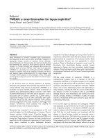
Web-based front end: global tasks
Website users are initially presented with a landing page
with links and short descriptions of all the different
tools available in ExpressionPlot (Figure 1). The navigation bar at the top, as well as the login box on the top
right, are present on every page during the website
experience for easy navigation. The ‘manual’ link opens
the page of the User’s Guide relevant to the currently
selected tool.
Figure 1 The ExpressionPlot home page. The website opens with this screen, giving a list of tools available in ExpressionPlot, and a login box
in the top right. The navigation bar on top appears on all pages, giving links to the other tools. The ‘manual’ link is context-aware: it
automatically opens the User’s Guide (in another tab) to the page explaining the current tool.
Friedman and Maniatis Genome Biology 2011, 12:R69
/>
Quality control
The ExpressionPlot front end provides several quality
control tools for RNA-Seq data. The read_types tool
graphs the number of reads in each sample of each
‘type’: non-aligning, multiply-aligning, paired-end
uniquely aligning, or single-end uniquely aligning (Figure 2a). The user can also run this tool looking at only
the uniquely aligning reads to see if they align to exons,
introns, intergenic regions or junctions (Figure 2b). The
correlation tool generates either a heatmap or a
Page 4 of 11
hierarchical clustering dendrogram showing the pairwise
correlations of gene expression profiles in the RNA-Seq
or microarray samples of your project (Figure 2c; Supplementary Methods in Additional file 1).
For paired-end data sets, the pairdist tool shows the
fraction of paired end reads for which (1) the two ends
align to different chromosomes, (2) the two ends align
to the same chromosome but on the same strand, (3)
the two ends align to the same chromosome and different strands but the minus end strand is upstream of the
(a)
(b)
(c)
(d)
Figure 2 Screen shots of ExpressionPlot quality control tools. (a) read_types tool showing all read types. Numbers of non-aligning
(Nonmatch), mulitply-aligning (Mult), unique genome-aligning (Genomic) and unique junction-aligning (Junction) reads are shown for each lane
from a mouse tissue transcriptome dataset [3]. Numbers (1/2) indicate different libraries; letters (A/B/C) indicate different lanes of the same
library. (b) read_types tool showing matching read types, normalized to 100%. (c) Pairwise correlation heatmap of gene expression profiles
generated from each lane. (d) pairdist tool shows ECDF of paired-end distances of ‘canonical’ reads (same chromosome, different strand, minus
strand read downstream of plus strand read). ‘Distance’ is defined as the genomic distance, in nucleotides, between the aligned positions of the
last sequenced bases of the two reads (can be negative if the alignments overlap). The samples have been de-identified (data in Additional file
3). Numbers in parentheses indicate median paired-end distance for each sample (add 36 for both sequences and 50 for both Illumina adaptors
(+172) to get complete library size).
Friedman and Maniatis Genome Biology 2011, 12:R69
/>
plus end strand, and (4) the two ends align to the same
chromosome, different strands, minus end downstream
of the plus end but there is at least one intron between
the two ends. The fifth category of reads, where the two
ends do not flank any known intron, can be used to
estimate the insert size, and ECDFs of the insert sizes
(defined as the length of the un-sequenced part of the
library between the paired ends) for the different lanes
are also plotted by this tool (Figure 2d; data in Additional file 3).
Generation of plots and tables of changed genes/events
The 2way tool and its associated table browser are the
basic tools to examine the relationships between gene
(a) (gene-level changes)
Page 5 of 11
levels (or RNA processing events) in two different
samples. The x-axis will correspond to one sample
(such as ‘wild type’), and the y-axis to another (such as
‘mutant’). The project and pair of samples are chosen
by the user from drop-down menus and the plots, like
all the other plots in ExpressionPlot, are generated on
demand by the web server. The 2way plot is a scattergram where points correspond to genes (or RNA processing events, for example, cassette exons), and are
colored according to whether they are significantly different in the two samples (Figure 3a,b). P-value and
fold-change cutoffs for significance can be controlled
by the user.
(b) (cassette exon splicing changes)
(c)
Figure 3 Screen shots of ExpressionPlot 2way plot and table_browser. (a) 2way plot of human tissue panel RNA-Seq data [1] showing
brain gene expression on the y-axis and average expression in all other tissues (pooled) on the x-axis. Blue points correspond to genes
significantly higher (P ≤ 10-4, fold change ≥20, 370 points) in brain relative to the other tissues; green points correspond to significantly lower.
(b) 2way plot showing cassette exon usage (inclusion:skip read ratios) instead of gene levels in the same data set. The heavy lobe above the
diagonal corresponds to exons with zero skipping reads in the brain, and the lighter lobe below the diagonal corresponds to exons with zero
skipping reads in all other tissues. Although the P-values are still valid, in these regimes the inclusion:skip ratio statistic is less precise. (c) Partial
screen shot of table browser showing brain-enriched cassette exons in the same data set. The context menu was triggered by the mouse
clicking on the row for CLTA (clathrin, light chain A) and offers the user links to open the seqview genome browser tool in a window covering
either the entire gene or just the alternative exon. In either case the exon will be automatically highlighted (Figure 5).
Friedman and Maniatis Genome Biology 2011, 12:R69
/>
After the plot is generated, action buttons are presented to the user to access the significantly changed
genes or RNA processing events in the table browser.
This screen presents the user with a dynamic table
whose rows correspond to changed genes/events (Figure
3c). The columns of the table contain identifiers for the
gene or event (like gene name, chromsome, strand and
position), as well as all the associated statistics (such as
read numbers, RPKM values (reads per kilobase gene
model per million total reads), and P-values). The table
can be sorted by clicking on the header of the desired
field, or filtered using a text string or a numeric filter.
Action buttons allow for the export of the table into
other software, such as R or OpenOffice (or Excel), for
automatic conversion of the genes into other IDs (such
as Ensembl or Entrez), and for the automatic generation
of expression-controlled background sets of similarly
expressed but unchanged genes (in terms of either
RPKM or raw read numbers - the user chooses,
although we recommend raw read numbers to avoid
transcript length biases [21]). These background sets are
appropriate for downstream gene ontology or motif
analysis.
A convenient feature of the table browser is the ability
to click on any row to be presented with a link to the
ExpressionPlot genome browser seqview. This browser
(a)
Page 6 of 11
displays both RNA-Seq reads, including those spanning
junctions, as well as array probe intensities, along with
gene annotations (described below).
Comparison of changes from different experiments/data
sets
Having examined changes in two different conditions of
a single experiment, it is natural to ask how these
changes compare to another experiment. Sometimes
this second experiment may be part of the same project,
but in other cases it could be part of another project,
and maybe even have been performed on another platform (for example, RNA-Seq versus microarray) or in
another organism (for example, human versus mouse).
The 4way tool and its associated table browser automatically match up changed genes or RNA processing
events from different experiments, presenting them in a
similar manner to its 2way cousin. After selecting two
projects, and a pairwise comparison, P-value and foldchange cutoff for each, ExpressionPlot generates a scattergram where each point corresponds to a gene (or
event). Here the x-axis shows the change in that gene/
event in the first comparison and the y-axis shows the
change in the second comparison (Figure 4). For example, points in the upper right quadrant would correspond to genes/events increased in both experiments,
whereas those in the upper left quadrant would be
(b)
Figure 4 Screen shots of ExpressionPlot 4way plots showing cross-platform and cross-species comparisons. (a) Heart-enriched gene
expression in human tissue panel exon array [28] (x-axis) and RNA-Seq [1] (y-axis) data sets. Points correspond to genes. Fold-change of
expression in heart is plotted versus all other samples in corresponding data set. Genes enriched in heart are plotted further to the right (exon
array) and/or up (RNA-Seq), and those higher in other samples are further to the left and/or down. Genes significantly different only on one
platform are colored red (exon array) or green (RNA-Seq) and those different on both platforms are colored blue. P-value cutoffs are 0.01 for
exon array and 10-4 for RNA-Seq, and fold-change cutoffs are 2 for both platforms. Colored numbers show number of genes in each category.
(b) Similar plot comparing the same x-axis (human heart-enriched gene expression by exon array) to mouse heart-enriched gene expression,
also by exon array (y-axis).
Friedman and Maniatis Genome Biology 2011, 12:R69
/>
decreased in the x-axis experiment, but increased in the
y-axis experiment. Points are colored according to
whether the gene/event is significantly changed in one
or both experiments, with blue representing those changed in both experiments.
As with the 2way tool, after the plot is generated
ExpressionPlot offers the user action buttons to select a
group of genes/events to further examine in the 4way
table browser. For example, clicking ‘Up/Up’ would
show a table of genes/events increased in both experiments. This table shows the annotation of the gene/
event (identifier, chromosome, position, strand, and so
on) as well as all the associated statistics. It has the
same fields that would be shown in the 2way browser,
but they are then repeated for both experiments. This
includes the annotation fields, since sometimes they are
from different organisms. As with the 2way browser,
there are action buttons to download, convert IDs and
generate background sets. Finally, clicking on a row of
the table opens a context menu with links that will
automatically open the genome browser to the right
part of the genome for the two experiments. In the case
of RNA processing events the correct genomic region
will be automatically highlighted within the browser, so
the user can quickly find, for example, a differentially
spliced cassette exon.
Page 7 of 11
The heatmap tool (Figure S8 in Additional file 1)
allows the user to compare larger numbers of change
profiles. Here all the different comparisons from one
project are laid out along the x-axis and all the comparisons from a second (possibly different) project are
laid out along the y-axis. The color of each square of
the heatmap indicates the similarity of the two comparisons. The user can choose from a variety of statistics to quantify similarity. This tool is a useful way to
look for relationships within larger numbers of
experiments.
Web-based front end: specific tasks
Examining reads from a particular genomic region
The seqview tool is ExpressionPlot’s genome browser
(Figure 5). With it, the user can select the project of
interest, then query either by a gene name or genomic
region. One of several annotations can be chosen, and
then a plot is generated showing either the pileup of
reads in that region (with strands separated or merged,
as requested by the user) or of the hybridization intensities of microarray probes in that region. Zooming and
scrolling is implemented, and users can also highlight
specific genomic coordinates. Barplots are automatically
generated showing levels of genes within the requested
regions.
brain-enriched exon
Figure 5 Screen shot of ExpressionPlot’s genome browser seqview. The region of the CLTA gene, which contains a brain-enriched exon
(pink), is shown. Known transcripts of CLTA are seen along the bottom (arrowheads indicate plus strand). The accumulation of RNA-Seq reads
from five human tissues is shown on the top. The heights of black bars indicate numbers of reads overlapping each genomic position, whereas
the heights of blue brackets indicate numbers of reads overlapping splice junctions. Data from RNA-Seq human tissue panel [1].
Friedman and Maniatis Genome Biology 2011, 12:R69
/>
The pairplot tool is a genome browser specifically
designed to visualize the relationship between the
aligned positions of paired ends. Only one sample can
be visualized at a time. The gene annotation of the
requested region is shown, as well as the pileup track
from the seqview tool showing total numbers of reads.
Above this a scattergram shows a point for each pairedend read aligning to the genomic region. The x-axis
gives the position of the plus-strand end and the y-axis
gives the position of the minus-strand end. The colors
and sizes of the points indicate the number of reads
aligning to each pair of coordinates. Under conditions of
constitutive splicing, the scattergram should form a series of segments above each exon and parallel to the
diagonal, with the distance to the diagonal dictated by
the paired-end insert and intron size. Alternatively
spliced regions, however, will show multiple parallel segments corresponding to the different isoforms. The relative strength of the segments corresponds to the
abundances of the two isoforms (Figure S9 in Additional
file 1).
Examining levels or changes of particular genes or events
The genelev tool generates barplots of gene levels
(RPKM) with error bars (Figure 6a). The ecdf tool
allows the user to visualize the levels or fold changes of
a set of genes by plotting the cumulative distribution of
those genes’ levels in the samples of a project or fold
changes in the pairwise comparisons of a project (Figure
6b). Instead of looking at the distribution of the whole
set, the event_heatmap tool visualizes the individual
levels or fold-change of all the genes in the set as a
heatmap (Figure 6c).
Administrative tasks
ExpressionPlot has an access-management system that
makes it easy for end users to share their data or release
it publicly. New user accounts can be made automatically through the website, including an e-mail-based
password recovery feature. When invoking the back end
for a given project one user is assigned ‘admin’ privileges. Users can then assign either ‘view’ or ‘admin’ privileges to other users on projects for which they are
‘admin’, or can add a ‘public’ flag to the project to make
it visible without login. These permissions are all controlled via a simple web interface.
Download, installation, help
Visit the ExpressionPlot website at [22] for instructions
on how to download and install the latest version.
ExpressionPlot requires an existing MySQL and Apache
web server, as well as the RApache module. The install.
pl script checks all the dependencies and tries to satisfy
or make suggestions on how to satisfy any that are
Page 8 of 11
missing. It then downloads and installs the latest version
of ExpressionPlot. Alternatively, a VirtualBox hard drive
is available running Ubuntu linux with ExpressionPlot
already installed. In either case, after installation is complete the EP-manage.pl script can be used to download
and add on bowtie indexes, annotations and microarray
library files as required. Example data sets, both unprocessed and processed, can also be installed using the
same script. The User’s Guide can be found at [23] and
contains detailed instructions on setting up and running
ExpressionPlot.
Please use the ExpressionPlot discussion group to post
technical questions or hints. This can be accessed by
visiting the ExpressionPlot Google group [24] or by
sending e-mail to
Extracting biological meaning from high
throughput data
ExpressionPlot offers the gene expression community an
easy-to-use tool for automated analysis of gene expression and RNA processing data. The back end offers a
solution to the problem of detecting significant changes
in gene expression and RNA processing, while the webbased interface offers data analysis, visualization and
browsing tools that realize the biological potential of
this new technology.
Methods
Calculating P-values for significance of changes in gene
expression
Given total numbers of reads in two samples (or two
groups of samples) n1 and n2, g1 and g2 of which align
to a particular gene of interest, we model g2 as a binomial distribution with parameters q2 and g, where q2 =
n 2 /(n 1 + n 2 ), and g = g 1 + g 2 is the total number of
reads aligning to the gene in either sample. The (twotailed) P-value is then calculated using R’s binom.test()
function.
Minimize significant changes method to estimate
effective total read numbers
To estimate the effective total number of reads n1 and
n2 in a pair of samples (or pair of groups of samples),
we estimate q2, which is the fraction of reads in the second sample, and then set n 2 = q 2 N and n 1 = N - n 2
where N is the total number of uniquely aligning reads
from either sample.
The theory of our calculation of q2 is that once a Pvalue cutoff is set, any potential choice of q2 will lead to a
certain number of significantly changed genes, say C(q2),
which could be calculated by applying the procedure
described above to every gene (for example 27,389 genes
in mouse). Thus, we have the optimization problem:
Friedman and Maniatis Genome Biology 2011, 12:R69
/>
(a) (MyD88 gene levels)
Page 9 of 11
(b) (spleen-enriched genes)
(c)
Figure 6 ExpressionPlot screen shots examining spleen-enriched genes in human exon array tissue panel data [28]. (a) Levels of Myd88,
a key signaling protein in the innate immune system [29], in human tissues using the genelev tool. (b) ecdf showing tissue enrichment (fold
change relative to all other tissues) of the 316 genes least 5-fold enriched in the spleen at a P-value cutoff of 10-4. The sharp angle at 2.3 in the
spleen curve indicates the 5-fold cutoff. The position of the cerebellum curve to the left of all the others may reflect the general depletion of
immune cells, which is characteristic of the spleen, within the nervous system. (c) event_heatmap showing the fold enrichments of the 316
spleen-enriched genes in all 11 tissues in the panel. The screen shot was edited by removing many of the genes from the middle for formatting
purposes and adding an arrow to indicate Myd88, which is part of a cluster of spleen-enriched genes also enriched in the liver. The depletion of
the spleen-enriched genes in the cerebellum is evident by the excess blue color in the cerebellum row.
min C(q2 ) : 0 ≤ q2 ≤ 1
q2
Solving the problem by convex optimization methods
would be feasible but slow due to the cost of re-calculating C(q2). Instead, we use the binconf() function from
R’s Hmisc library [25] to calculate a 95% confidence
interval for q 2 for every gene, based on the observed
number of reads. This interval corresponds to the range
of q2 for which that gene is not significantly changed.
Then the range 0 to 1 is split into windows of width
0.0001, and the number of genes whose confidence
interval overlaps each of these windows is counted. The
uncertainty introduced by using windows as point estimates is mitigated by their small radius: a difference of
0.0001 (0.01%) in the sample size estimate will have a
minute effect on resultant gene levels. The value of q2
for the window overlapped by the confidence intervals
of the most genes (or the mean of the q2 for the several
windows if there is a tie for the most intervals) is then
taken as the optimum. Empirical tests show that this
method is extremely robust to the choice of P-value cutoff (data not shown). This is implemented in a very
Friedman and Maniatis Genome Biology 2011, 12:R69
/>
short R function called minimize.significant.changes() in
BradStats.R [26].
European Nucleotide Archive accession numbers
The previously unpublished (and de-identified) data sets
used to create Figure 2d, and Figures S7 and S9 in Additional file 1 are available from the European Nucleotide
Archive under accession number ERP000619, available at
[27].
Archival copy of software
For archival purposes, version 1.3 of the software is
included as Additional file 4, but it is recommended to
use the latest version available through the website.
Additional material
Additional file 1: Supplementary figures, methods, references, and
description of other additional files.
Additional file 2: Data for Figure S7 in Additional file 1.
Additional file 3: Data for Figure 2d.
Additional file 4: Archival copy of software.
Abbreviations
ECDF: empirical cumulative distribution function; PAC: poly-adenylation/
cleavage site; RPKM: reads per kilobase gene model per million total reads;
TSS: transcription start site.
Acknowledgements
We would like to thank Y Katz, SL Ng, J Gertz and M Muratet for critical
reading of the manuscript; S O’Keeffe, M Muratet and D Quest for software
testing and technical suggestions; CB Burge for hosting our prototype
server; D Housman for scientific advice and laboratory space during the
development of this software; IK Friedman and B Lewis for administrative
support; HP Phatnani, C Lobsiger, J Cahoy, J Zamanian and other members
of the Barres Lab (Stanford), Myers Lab (HudsonAlpha Institute), Ravits Lab
(Benaroya Institute) and Maniatis Lab (Harvard/Columbia) for providing data
and/or user feedback. This work was supported by a grant from the ALS
Therapy Alliance.
Author details
1
Department of Molecular and Cell Biology, Harvard University, 7 Divinity
Ave, Cambridge, MA 02138, USA. 2The Koch Institute for Integrative Cancer
Research, Massachusetts Institute of Technology, Cambridge, MA 02139, USA.
3
Department of Bioinformatics and Computational Biology, Genentech, Inc.,
1 DNA Way, South San Francisco, CA 94080, USA. 4Department of
Biochemistry and Molecular Biophysics, Columbia University College of
Physicians and Surgeons, 701 West 168th St, New York, NY 10032, USA.
Authors’ contributions
BF conceived of and wrote the software and the manuscript. TM helped in
its design and coordination and in drafting the manuscript. Both authors
read and approved the final manuscript.
Competing interests
The authors declare that they have no competing interests.
Received: 24 December 2010 Accepted: 28 July 2011
Published: 28 July 2011
Page 10 of 11
References
1. Wang ET, Sandberg R, Luo S, Khrebtukova I, Zhang L, Mayr C, Kingsmore SF,
Schroth GP, Burge CB: Alternative isoform regulation in human tissue
transcriptomes. Nature 2008, 456:470-476.
2. Nagalakshmi U, Waern K, Snyder M: RNA-Seq: a method for
comprehensive transcriptome analysis. Curr Protoc Mol Biol 2010, Chapter
4, Unit 4.11.1-13.
3. Mortazavi A, Williams BA, McCue K, Schaeffer L, Wold B: Mapping and
quantifying mammalian transcriptomes by RNA-Seq. Nat Methods 2008,
5:621-628.
4. Trapnell C, Pachter L, Salzberg SL: TopHat: discovering splice junctions
with RNA-Seq. Bioinformatics 2009, 25:1105-1110.
5. Trapnell C, Williams BA, Pertea G, Mortazavi A, Kwan G, van Baren MJ,
Salzberg SL, Wold BJ, Pachter L: Transcript assembly and quantification by
RNA-Seq reveals unannotated transcripts and isoform switching during
cell differentiation. Nat Biotechnol 2010, 28:511-515.
6. Li H, Ruan J, Durbin R: Mapping short DNA sequencing reads and calling
variants using mapping quality scores. Genome Res 2008, 18:1851-18581.
7. Katz Y, Wang ET, Airoldi EM, Burge CB: Analysis and design of RNA
sequencing experiments for identifying isoform regulation. Nat Methods
2010, 7:1009-1015.
8. Robinson JT, Thorvaldsdóttir H, Winckler W, Guttman M, Lander ES, Getz G,
Mesirov JP: Integrative Genomics Viewer. Nat Biotechnol 2011, 29:24-26.
9. Langmead B, Trapnell C, Pop M, Salzberg SL: Ultrafast and memoryefficient alignment of short DNA sequences to the human genome.
Genome Biol 2009, 10:R25.
10. Wu Z, Jenkins B, Rynearson T, Dyhrman S, Saito M, Mercier M, Whitney L:
Empirical bayes analysis of sequencing-based transcriptional profiling
without replicates. BMC Bioinformatics 2010, 11:564.
11. Goecks J, Nekrutenko A, Taylor J: Galaxy: a comprehensive approach for
supporting accessible, reproducible, and transparent computational
research in the life sciences. Genome Biol 2010, 11:R86.
12. Reich M, Liefeld T, Gould J, Lerner J, Tamayo P, Mesirov JP: GenePattern
2.0. Nat Genet 2006, 38:500-501.
13. Fujita PA, Rhead B, Zweig AS, Hinrichs AS, Karolchik D, Cline MS,
Goldman M, Barber GP, Clawson H, Coelho A, Diekhans M, Dreszer TR,
Giardine BM, Harte RA, Hillman-Jackson J, Hsu F, Kirkup V, Kuhn RM,
Learned K, Li CH, Meyer LR, Pohl A, Raney BJ, Rosenbloom KR, Smith KE,
Haussler D, Kent WJ: The UCSC Genome Browser database: update 2011.
Nucleic Acids Res 2011, , 39 Database: D876-882.
14. Hubbard TJP, Aken BL, Ayling S, Ballester B, Beal K, Bragin E, Brent S,
Chen Y, Clapham P, Clarke L, Coates G, Fairley S, Fitzgerald S, FernandezBanet J, Gordon L, Graf S, Haider S, Hammond M, Holland R, Howe K,
Jenkinson A, Johnson N, Kahari A, Keefe D, Keenan S, Kinsella R,
Kokocinski F, Kulesha E, Lawson D, Longden I, et al: Ensembl 2009. Nucleic
Acids Res 2009, 37:D690-697.
15. Anders S, Huber W: Differential expression analysis for sequence count
data. Genome Biol 2010, 11:R106.
16. Marioni JC, Mason CE, Mane SM, Stephens M, Gilad Y: RNA-seq: an
assessment of technical reproducibility and comparison with gene
expression arrays. Genome Res 2008, 18:1509-1517.
17. Robinson MD, Oshlack A: A scaling normalization method for differential
expression analysis of RNA-seq data. Genome Biol 2010, 11:R25.
18. Bullard J, Purdom E, Hansen K, Dudoit S: Evaluation of statistical methods
for normalization and differential expression in mRNA-Seq experiments.
BMC Bioinformatics 2010, 11:94.
19. Affymetrix - Affymetrix Power Tools.. [ />partners_programs/programs/developer/tools/powertools.affx].
20. Smyth GK: Linear models and empirical bayes methods for assessing
differential expression in microarray experiments. Stat Appl Genet Mol Biol
2004, 3, Article3.
21. Oshlack A, Wakefield M: Transcript length bias in RNA-seq data
confounds systems biology. Biol Direct 2009, 4:14.
22. ExpressionPlot.. [ />23. ExpressionPlot User’s Guide.. [ />24. ExpressionPlot Google Group.. [ />expressionplot].
25. CRAN - Package Hmisc.. [ />index.html].
Friedman and Maniatis Genome Biology 2011, 12:R69
/>
Page 11 of 11
26. BradStats.R - expressionplot - Project Hosting on Google Code.. [http://
code.google.com/p/expressionplot/source/browse/trunk/lib/R/BradStats.R].
27. European Nucleotide Archive: ERP000619.. [ />data/view/ERP000619].
28. Affymetrix - Sample Data, Exon 1.0 ST Array Dataset.. [http://www.
affymetrix.com/support/technical/sample_data/exon_array_data.affx].
29. Akira S, Takeda K: Toll-like receptor signalling. Nat Rev Immunol 2004,
4:499-511.
doi:10.1186/gb-2011-12-7-r69
Cite this article as: Friedman and Maniatis: ExpressionPlot: a web-based
framework for analysis of RNA-Seq and microarray gene expression
data. Genome Biology 2011 12:R69.
Submit your next manuscript to BioMed Central
and take full advantage of:
• Convenient online submission
• Thorough peer review
• No space constraints or color figure charges
• Immediate publication on acceptance
• Inclusion in PubMed, CAS, Scopus and Google Scholar
• Research which is freely available for redistribution
Submit your manuscript at
www.biomedcentral.com/submit