Báo cáo y học: "Genome sequence of an Australian kangaroo, Macropus eugenii, provides insight into the evolution of mammalian reproduction and development" docx
Bạn đang xem bản rút gọn của tài liệu. Xem và tải ngay bản đầy đủ của tài liệu tại đây (3.53 MB, 26 trang )
Genome sequence of an Australian kangaroo,
Macropus eugenii, provides insight into the
evolution of mammalian reproduction and
development
Renfree et al.
Renfree et al. Genome Biology 2011, 12:R81
(29 August 2011)
RESEARCH Open Access
Genome sequence of an Australian kangaroo,
Macropus eugenii, provides insight into the
evolution of mammalian reproduction and
development
Marilyn B Renfree
1,2*†
, Anthony T Papenfuss
1,3,4*†
,JanineEDeakin
1,5
, James Lindsay
6
, Thomas Heider
6
,
Katherine Belov
1,7
, Willem Rens
8
,PaulDWaters
1,5
, Elizabeth A Pharo
2
,GeoffShaw
1,2
,EmilySWWong
1,7
,
Christophe M Lefèvre
9
,KevinRNicholas
9
,YokoKuroki
10
, Matthew J Wakefield
1,3
, Kyall R Zenger
1,7,11
, Chenwei Wang
1,7
,
Malcolm Ferguson-Smith
8
, Frank W Nicholas
7
, Danielle Hickford
1,2
,HongshiYu
1,2
, Kirsty R Short
12
, Hannah V Siddle
1,7
,
Stephen R Frankenberg
1,2
,KengYihChew
1,2
,BrandonRMenzies
1,2,13
, Jessica M Stringer
1,2
, Shunsuke Suzuki
1,2
,
Timothy A Hore
1,14
, Margaret L Delbridge
1,5
,AmirMohammadi
1,5
, Nanette Y Schneider
1,2,15
,YanqiuHu
1,2
,
William O’Hara
6
, Shafagh Al Nadaf
1,5
, Chen Wu
7
, Zhi-Ping Feng
3,16
,BenjaminGCocks
17
, Jianghui Wang
17
,PaulFlicek
18
,
Stephen MJ Searle
19
, Susan Fairley
19
,KathrynBeal
18
,JavierHerrero
18
, Dawn M Carone
6,20
, Yutaka Suzuki
21
,
Sumio Sugano
21
,AtsushiToyoda
22
, Yoshiyuki Sakaki
10
,ShinjiKondo
10
,YuichiroNishida
10
, Shoji Tatsumoto
10
,
Ion Mandiou
23
,ArthurHsu
3,16
, Kaighin A McColl
3
, Benjamin Lansdell
3
, George Weinstock
24
, Elizabeth Kuczek
1,25,26
,
Annette McGrath
25
,PeterWilson
25
, Artem Men
25
, Mehlika Hazar-Rethinam
25
, Allison Hall
25
,JohnDavis
25
,
David Wood
25
, Sarah Williams
25
, Yogi Sundaravadanam
25
,DonnaMMuzny
24
, Shalini N Jhangiani
24
, Lora R Lewis
24
,
Margaret B Morgan
24
, Geoffrey O Okwuonu
24
,SanJuanaRuiz
24
, Jireh Santibanez
24
, Lynne Nazareth
24
,AndrewCree
24
,
Gerald Fowler
24
, Christie L Kovar
24
, Huyen H Dinh
24
,VanditaJoshi
24
,ChynJing
24
, Fremiet Lara
24
, Rebecca Thornton
24
,
Lei Chen
24
, Jixin Deng
24
,YueLiu
24
,JoshuaYShen
24
, Xing-Zhi Song
24
, Janette Edson
25
, Carmen Troon
25
,
Daniel Thomas
25
, Amber Stephens
25
, Lankesha Yapa
25
, Tanya Levchenko
25
, Richard A Gibbs
24
,DesmondWCooper
1,28
,
Terence P Speed
1,3
, Asao Fujiyama
22,27
, Jennifer A M Graves
1,5
,RachelJO’Neill
6
, Andrew J Pask
1,2,6
, Susan M Forrest
1,25
and Kim C Worley
24
Abstract
Background: We present the genome sequence of the tammar wallaby, Macropus eugenii, which is a member of
the kangaroo family and the first representative of the iconic hopping mammals that symbolize Australia to be
sequenced. The tammar has many unusual biological characteristics, including the longest period of embryonic
diapause of any mammal, extremely synchronized seasonal breeding and prolonged and sophisticated lactation
within a well-defined pouch. Like other marsupials, it gives birth to highly altricial young, and has a small number
of very large chromosomes, making it a valuable model for genom ics, reproduction and development.
Results: The genome has been sequenced to 2 × coverage using Sanger sequencing, enhanced with additional
next generation sequencing and the integration of extensive physical and linkage maps to build the genome
assembly. We also sequenced the tammar transcriptome across many tissues and developmental time points.
* Correspondence: ;
† Contributed equally
1
The Australian Research Council Centre of Excellence in Kangaroo
Genomics, Australia
Full list of author information is available at the end of the article
Renfree et al. Genome Biology 2011, 12:R81
/>© 2011 Renfree et al.; licensee BioMed Central Ltd. This is an open access a rticle distributed under the terms of the Creative Co mmons
Attribution License ( which permits unrestricted use, distribution, and reproduction in
any medium, provided the original work is properly cited.
Our analyses of these data shed light on mammalian reproduction, development and genome evolution: there is
innovation in reproductive and lactational genes, rapid evolution of germ cell genes, and incomplete, locus-specific
X inactivation. We also observe novel retrotransposons and a highly rearranged major histocompatibility complex,
with many class I genes located outside the complex. Novel microRNAs in the tammar HOX clusters uncover new
potential mammalian HOX regulatory elements.
Conclusions: Analyses of these resources enhance our understanding of marsupial gene evolution, identify
marsupial-specific conserved non-coding elements and critical genes across a range of biological systems,
including reproduction, development and immunity, and provide new insight into marsupial and mammalian
biology and genome evolution.
Background
The tammar wallaby holds a unique place in the natural
history of Australia, for it was the first Austral ian marsu-
pial discovered, and the first in which its special mode of
reproduction was noted: ‘their manner of procreation is
exceeding strange and highly worth observing; below the
belly the fe male carries a pouch into which you may put
your hand ; inside the pouch are her nipples, and we have
fou nd that the young ones grow up in this pouch with the
nipples in their mouths. We have seen some young ones
lying there, which were on ly thesizeofabean,thoughat
the same time perfectly proportioned so that it seems cer-
tain that they grow there out of the nipples of the mam-
mae from which they draw their food, until t hey are
grown up’ [1]. These observations were made by Fran-
cisco Pelseart, Captain of the ill-fated and mutinous
Dutch East Indies ship Batavia in 1629, whilst ship-
wrec ked on the Abrolhos Islands off the coast of Gerald-
ton in Western Australia. It is therefore appropriate that
the tammar should be the first Australian marsupial sub-
ject to an in-depth genome analysis.
Marsupials are distantly related to eutherian mammals,
having shared a common ancestor between 130 and 148
million years ago [2-4]. The tammar wallaby Macropus
eugenii is a small member of the kangaroo family, the
Macropodidae, within the genus Macropus,whichcom-
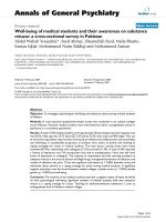
prises 14 species [5] (Figur e 1). The macropodi ds are the
most specialized of all marsupials. Mature females weigh
about 5 to 6 kg, and males up to 9 kg. The tammar is
highly abundant in its habitat on Kangaroo Island in
South Australia, and is also found on the Abrolhos
Islands, Garden Island and the Recherche Archipelago,
all in Western Australia, as well as a few small areas in
the south-west corner of the continental mainland. These
populations have been separated for at least 40,000 years.
Its size, availability and ease of handling have made it the
most intensively studied model marsupial for a wide vari-
ety of genetic, developmental, reproductive, physiological,
biochemical, neur obiological and ecological studies
[6-13].
In the wild, female Kangaroo Island tammars have a
highly synchronized breeding cycle and deliver a single
young on or about 22 January (one gestation period
after the longest day in the Southern hemisphere, 21 to
22 December) that remains in the pouch for 9 to 10
months. The mother mates within a few hours after
birth but development of the resulting embryo is
delayed during an 11 month period of suspended anima-
tion (embryonic diapause). Initially diapause is main-
tained by a lactation-mediated inhibition, and in the
second half of the year by photoperiod-me diated inhibi-
tion that is removed as day length decreases [14]. The
anatomy, physiology, e mbryology, endocrinology and
genetics of the tammar have been described in detail
throughout development [6,11-13,15].
The marsupial mode of reproduction exemplified by
the tammar with a short gestation and a long lactation
does not imply inferiorit y, nor does it represent a transi-
tory evolutionary stage , as w as originally thought. It is a
successful and adaptable lifestyle. The maternal invest-
ment is minimal during the relatively brief pregnancy
and in early lactation, allowing the mothe r to respond to
altered environmental conditions [11,12,15]. The tam-
mar, like all marsupials, has a fully func tional placenta
that makes hormones to modulate pregnancy and par-
turition, control the growth of the young, and provide
signals for the maternal recognition of pregnancy
[14,16-18]. The tammar embryo develops for only 26
days after diapause, and is born when only 16 to 17 mm
long and weighing about 440 mg at a developmental
stage roughly equivalent to a 40-day human or 15-day
mouse embryo. The kidney bean-sized newborn has well-
developed forelimbs that allow it to clim b up to the
mother’s pouch, where it attaches to one of four available
teats. It has functional , though not fully develope d, olfac-
tory, respirato ry, circulatory and digestive systems, but it
is born with a n embryonic kidney and undifferentia ted
immune, thermoregulatory and reproductive systems, all
of which become functionally differentiated during the
lengthy pouch life. Most major structures and organs,
including the hindlimbs, eyes, gonads and a significant
portion of t he brain, differentiate while the young is in
the pouch and are therefore readily available for study
[11,12,19-24 ]. They also h ave a sophisticated lactational
Renfree et al. Genome Biology 2011, 12:R81
/>Page 2 of 25
Gondwanaland
South
America
Australia
Didelphidae
Vombatidae
Phascolarctidae
Pseudocheiridae
Macropodidae
Thylacomyidae
Peramelidae
Dasyuridae
Macropodidae
P. xanthopus
T. thetis
M. rufus
M. robustus
M. antilopinus
W. bicolor
M. parma
M. rufogriseus
M. agilis
M. eugenii
0
MnoilliYsraeAog
Mesozoic Cenozoic
146
65
Tarsipedidae (1)
11
0
Figure 1 Phylogeny of the marsupials. Phy logenetic relationships of the orders of Marsupialia. Top: the plac ement of the contemporary
continents of South America and Australia within Gondwanaland and the split of the American and Australian marsupials. Relative divergence in
millions of years shown to the left in the context of geological periods. The relationship of the Macropodide within the Australian marsupial
phylogeny shown is in purple with estimated divergence dates in millions of years [5,162,163]. Representative species from each clade are
illustrated. Inset: phylogeny of the genus Macropus within the Macropodidae showing the placement of the model species M. eugenii (purple)
based on [59]. Outgroup species are Thylogale thetis and Petrogale xanthopus.
Renfree et al. Genome Biology 2011, 12:R81
/>Page 3 of 25
physiology with a milk composition that changes
throughout pouch life, ensuring that nutrient supply is
perfectly matched for each stage of development [25].
Adjacent teats in a pouch can deliver milk of differing
composition appropriate for a pouch young and a young-
at-foot [26].
Kangaroo chromosomes excited some of the e arliest
comparative cytological studies of mammals. Like other
kangaroos, the tammar has a low diploid number (2n =
16) and very large chromosomes that are easily distin-
guished by size and morphology. The low diploid number
of marsupials makes it easy to study mitosis, cell cycles
[27], DNA replication [28], radiation sensitivity [29], gen-
ome stability [30], chromosome elimination [31,32] and
chromosome evolution [33,34]. Marsupial sex chromo-
somes are particularly informative. The X and Y chromo-
somes are small; the basic X chromosome constitutes only
3% of the haploid genome (compared with 5% in euther-
ians) and the Y is tiny. Comparative studies show that the
marsupial X and Y are representative of the ancestral
mammalian X and Y chromosomes [35]. However, in the
kangaroos, a large heterochromatic nucleolus organizer
region became fused to the X and Y. Chromosome paint-
ing confirms the extreme conservation of kangaroo chro -
mosomes [36] and their close relationship with karyotypes
of more distantly related marsupials [37-40] so that gen-
ome studies are likely to be highly transferable across mar-
supial species.
The tammar is a member of the Australian marsupial
clade and, as a macropodid marsupial, is maximally diver-
gent from the only other sequenced model marsupial, the
didelphid Brazilian grey short-tailed opossum, Monodel-
phis domestica [41]. The South American and Australasian
marsupials followed independent evolutionary pathways
after the separation of Gondwana into the new continents
of South America and Australia about 80 million years
ago and after the divergence of tammar and opossum
(Figure 1) [2,4]. The Australasian marsupials have many
unique specializations. Detailed knowledge of the biology
of the tammar has informed our interpretation of its gen-
ome and highlighted many novel aspects of marsupial
evolution.
Sequencing and assembly (Meug_1)
ThegenomeofafemaletammarofKangarooIsland,
South Australia origin was sequenced using the whole-
genome shotgun (WGS) approach and Sanger sequen-
cing. DNA isolated from the lung tissue of a single tam-
mar was used to generate WGS libraries with inserts of 2
to6kb(TablesS1andS2inAdditionalfile1).Sanger
DNA sequencing was performed at the Baylor College of
Medicine Human Genome Sequencing Center (BCM-
HGSC), and the Australian Genome Research Facility
using ABI3730xl sequencers (Applied BioSystems, Foster
City, CA, USA). Approximately 10 million Sanger WGS
reads, representing about 2 × sequence coverage, were
submitted to the NCBI trace archives (NCBI BioProject
PRJNA12586; NCBI Taxonomy ID 9315). An additional
5.9 × sequence cov erage was generated on an ABI SOLiD
sequencer at BCM- HGSC. These 25-bp paired-end data
with average mate-pair distance of 1.4 kb (Table S3 in
Additional file 1) [SRA:SRX011374] were used to correc t
contigs and perform super-scaffolding. The initial tam-
mar genome assembly (Meug_1.0 ) was construct ed using
only the low coverage Sanger sequences. This was then
improved with additional scaffolding using sequences
generated with the ABI SOLiD (Meug_1.1; Table 1;
Tables S4 to S7 in Additional file 1). The Meug_1.1
assembly had a contig N50 of 2.6 kb and a scaffold N50
of 41.8 kb [GenBank:GL044074-GL172636].
The completeness of the assembly was assessed by com-
parison to the avail able cDNA d ata. Using 758,0 62 454
FLX cDNA sequences [SRA:SRX019249, S RA:SRX019250],
76% are found to some extent in the assembly and 30% are
found with more than 80% of their length represented
(Table S6 in Additional file 1). Compared to 14 ,878 San-
ger-sequenced ESTs [GenBank:EX195538-EX203564, Gen-
Bank:EX203644-EX210452], more than 85% are found in
the assembly with at least one half their length aligned
(Table S7 in Additional file 1).
Table 1 Comparison of Meug genome assemblies
Assembly version
1.0 1.1 2.0
Contigs (million) 1.211 1.174 1.111
N50 (kb) 2.5 2.6 2.91
Bases (Mb) 2546 2,536 2,574
Scaffolds 616,418 277,711 379,858
Max scaffold size NA 472,108 324,751
Gaps (Mb) NA 539 619
N50 (kb) NA 41.8 34.3
Complex scaffolds NA 128,563 124,674
Singleton scaffolds NA 149,148 255,184
Co-linear with BACs NA 87.2% (418) 93.4% (298)
Co-linear with ESTs NA 82.3% (704) 86.7% (454)
Summary statistics for the tammar genome assemblies. These statistics
indicate the extension and merging of contigs done to improve the assembly.
The larger number of scaffolds and smaller scaffold N50 is a consequence of
higher stringency in the 2.0 scaffolding workflow. The higher stringency
isolated many contigs. However, the numbe r of complex (that is, useful)
scaffolds is similar between the assemblies. For co-linear estimates, the
scaffolds were linearized and BACs and cDNA libraries were mapped against
them. The 1.1 and 2.0 assemblies were validated against 169 BAC contigs and
84,718 ESTs (that were not incorporated into either genome assembly). We
determined the percen tage of contigs where the scaffolding matched the
order and orientation when compared to BACs or ESTs (co-linear with BACs/
ESTs). Parentheses indicate the total number of contigs identified after
alignment to BAC contigs or ESTs.
Renfree et al. Genome Biology 2011, 12:R81
/>Page 4 of 25
Additional sequen cing and assembly
improvement (Meug_2)
Contig improvement
The tammar genome assembly was further improved
using additional data consisting of 0.3 × coverage by
paired and unpaired 454 GS-FLX Titanium reads [SRA:
SRX080604, SRA:SRX085177] and 5 × coverage by paired
Illumina GAIIx reads [SRA:SRX085178, SRA:SRX081248]
(Table S8 in Additional file 1). A local reassembly strat-
egy mapped the additional 454 and Illumina data against
Meug_1.1 contigs. Added data were used to improve the
accuracy of base calls and to extend and merge contigs.
The Meug_2.0 assembly [GenBan k:ABQO000000000]
(see also ‘Data availability’ section) has 1.111 million con-
tigswithanN50of2.9kb.Contigswerevalidated
directly by PCR on ten randomly selected contigs. The
assembly was also assessed by aligning 84,718 ESTs and
169 BAC sequences to the genome. The amount of
sequence aligni ng correctly to the genome assembly
showed modest improvement between Meug_1.1 and
Meug_2.0 (Table 1; Table S9 in Additional file 1).
Scaffolding and anchoring using the virtual map
Scaffolds were constructed using the previously men-
tioned Illumina paired-end libraries with insert sizes of
3.1 kb (8,301,018 reads) and 7.1 kb (12,203,204 reads),
454 paired-end library with an insert size of 6 kb and
SOLiD mate pair library. The mean insertion distances
for each library were empirically determined using paired
reads where both ends mapped within the same contig
and only those within three standard deviations from the
mean were used for scaffolding. The contigs were
ordered and orie nted using Bambus [42], through three
iterations of scaffolding to maximize the accuracy of the
assembly. The highest priority was given to the library
with the smallest standard deviation in the paired end
distances, and the remaining libraries arranged in des-
cending order. Initial scaffolding by Bambus was per-
formed using five links as a threshold [43]. Overlapping
contigs were identified and set aside before reiteration.
This step was performed twice and the overlapping con-
tigs pooled. The non-overlapping and overlapping con-
tigs were then scaffolded independently. Any scaffolds
found to still contain overlap were split apart. The result-
ing assembly has 324,751 scaffolds wit h an N50 of 34,279
bp (Table 1). Scaffolds were assigned to chromosomes by
aligning them to markers from the virtual map [44],
represen ted using sequences obtained from the opossum
and human genomes [45]. We assigned 6,9 79 non-over-
lapping scaffolds (163 Mb or 6% of the genome assembly)
to the seven autosomes. The vast majority of the genome
sequence remained unmapped.
Tammar genome size
The tammar genome size was estimated using three inde-
pendent methods: direct assessment by quantitative PCR
[46]; bivariate flow karyotyping and standard flow cytome-
try; and genome analyses based in the Sanger WGS reads,
using the Atlas -Genometer [47]. These three ap proaches
produced quite different genome size estim ates (Tables
S11 to S13 in Additional file 1) so the average size esti-
mate, 2.9 Gb, was used for the purposes of constructing
the Meug_2.0 integrated genome assembly. The smaller
genome size of tammar compared to human is unlikely to
be due to fewer genes or changes in gene s ize (Figure S1
in Additional file 2), but may be accounted for by the
greatly reduced centromere size of 450 kb/chromosome
and number (n = 8) [48] compared to the human centro-
mere size of 4 to 10 Mb/chromosome (n = 23).
Physical and linkage mapping
Novel strategies were developed for the construction of
physical and linkage maps covering the entire genome.
The physical map consists of 520 loci mapped by fluores-
cence in situ hybridization (FISH) and was constructed
by mapping the ends of gene blocks conserved between
human and opossum, thereby allowing the location of
genes within these conservedblockstobeextrapolated
from the opossum genome onto tammar chromosomes
[37] (JE Deakin, ML Delbridge , E Koina, N Ha rley, DA
McMillan, AE Alsop, C Wang, VS Patel, and JAM
Graves, unpublished results). Three different approaches
were used to generate a linkage map consisting of 148
loci spanning 1,402.4 cM or 82.6% of the genome [49].
These approaches made the most of the available tammar
sequence (genome, BACs or BAC ends) to identify
markers to increase coverage in specific regions of the
genome. Many of these markers were also physically
mapped, providing anchors for the cre ation of an inte-
grated map comprising all 553 distinct loci included in
the physical and/or linkage maps. Interpolation of seg-
ments of conserved synteny (mainly from the opossum
assembly) into the integrated map then made it possible
to predict the geno mic content and organization of the
tammar genome through the construction of a virtual
genome map comprising 14,336 markers [44].
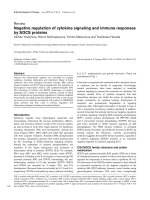
Mapping data were used to construct tammar-human
(Figure 2) and tammar-opossum comparative maps in
order to study genome evolutio n. Regions of the genome
were identified that have undergone extensive rearrange-
ment when comparisons between tammar and opossum
are made. These are in addition to previously known
rearrangements based on c hromosome-specific paints
[50]. For example, tammar chromosome 3, consisting of
genes that a re on nine human chromosomes (3, 5, 7, 9,
Renfree et al. Genome Biology 2011, 12:R81
/>Page 5 of 25
10, 12, 16, 17, 22; Figure 2) and the X have an extensive
reshuffling of the gene order. Rearrangements on the
remaining chromosomes are mostly the result of large-
scale inversions. This enabled us to predict the ancestral
marsupial karyotype, revealing that inversions and micro-
inversions have played a major role in shaping the gen-
omes of marsupials (JE Deakin, ML Delbridge, E Koina,
N Harley, DA McMillan, AE Alsop, C Wang, VS Patel,
and JAM Graves, unpublished results).
Genome annotation
The Ensembl genebuild (release 63) for the Meug_1.0
assembly identified 18,258 genes by projection from high
quality reference genomes. Of these, 15,290 are protein
coding, 1,496 are predicted pseudo-genes, 525 are micro-
RNA (miRNA) genes, and 42 are long non-coding RNA
genes, though these are composed of just 7 different
families: 7SK, human accelerated region 1F, CPEB3 ribo-
zyme, ncRNA repressor of NFAT, nuclear RNase P,
RNase MRP and Y RNA.
Since the c overage is low, many genes m ay be fragmented
in the assembly or even unsequenced. The Ensembl gene-
build pipeline scaffolds fragmented gen es using compa ra-
tive data and constructs ‘GeneScaffolds’. There are 10,257
GeneScaffolds containing 13,037 genes. The annotation
also contains 9, 454 genes interrupted by Ns. To partially
ameliorate the problems of missing genes, a number of
BACs from targeted locations have been sequenced and
annotated, including the HOX gene clusters (H Yu, Z-P
Feng, RJ O’Neill, Y Hu, AJ Pask, D Carone, J Lindsay, G
Shaw, AT Papenfuss, and MB Renfree, unpublished
results), major histocompatibility complex (MHC) [51], X
chromosome (ML Delbridge, B Landsdell, MT Ross, TP
Speed, AT Pape nfu ss, JAM Graves, unpublished results),
pluripotency genes, germ cell genes, spermatogenesis genes
[52,53] and X chromosome genes. Findings from these are
summarized in later sections of this paper.
Expansion of gene families
Many genes evolve and acquire novel function through
duplication and divergence. We identified genes that
have undergone expansions in the marsupial lineage but
remain largely unduplicated in eutherians and reptiles
(Table S15 in Additional file 1). Both the tammar and
opossum have undergone expansion of MHC class II
genes, critical in the immune recognition of extracellular
pathogens, and TAP genes that are responsible for load-
ing endogenously derived antigens onto MHC class I
proteins. Three marsupial-specific class II gene families
exist: DA, DB and DC. Class II genes have undergone
further duplications in the tammar and form two geno-
mic clusters, adjacent to the antigen-processing genes
1
7
13
8
14
3
9
15
4
11
5
2
19
16
20
10
6
12
17
18
21
22
X
Human chromosomes
X32 1
4
5 6 7
MHC class I
MHC class II
KERV
MHC cluster composition
38
19
17
32
22
21
24
39
4
40
3
2
23
5
12
27
26
36
31
14
15
16
45
11
46
24
9
26
29
28
23
45
35
32
48
Y
Olfactory receptor
Figure 2 Homology of tammar regions to the human karyotype, and location of major histocompa tibility complex, classical clas s I
genes and olfactory receptor gene. Colored blocks represent the syntenic blocks with human chromosomes as shown in the key. A map of
the locations of the tammar major histocompatibility complex (MHC) is shown on the right-hand side of each chromosome. The rearranged
MHCs are on chromosome 2 and clusters of MHC class I genes (red) near the telomeric regions of chromosomes 1, 4, 5, 6, and 7. MHC class II
genes are shown in blue, olfactory receptors are shown in orange and Kangaroo endogenous retroviral elements found within these clusters are
shown in green. The location of the conserved mammalian OR gene clusters in the tammar genome are shown on the left-hand side of each
chromosome. OR genes are found on every chromosome, except for chromosome 6 but including the X. The location of the OR gene clusters
(numbers) are shown, and their approximate size is represented by lines of different thickness.
Renfree et al. Genome Biology 2011, 12:R81
/>Page 6 of 25
[51]. The opossum has one TAP1 and two TAP2 genes,
while the tammar has expanded TAP1 (two genes) and
TAP2 (three genes) genes [51]. We also detected marsu-
pial expansions linked to apoptosis (NET1, CASP3,
TMBIM6) and sensory perception (olfactory receptors).
Genomic landscape
Sequence conservation
We next explored sequence conservation between tammar
and opossum using sequence similarity as a sensitive
model of conservation. We found that 38% of nucleotides
in the tammar genome (Meug_1.0) could be aligned to the
high-quality op ossum genome (7.3× ). Of the aligned
sequence, 72% was unannotated, reflecting a high propor-
tion of conserved non-coding regions between the marsu-
pial species. The level of conservation between opossum
and tammar varied from 36.0 to 40.9% across the different
opossum chromosomes (Table S16 in Additional file 1).
This variation seems modest and may be largely stochastic,
but it is interesting to examine further. Opossum chromo-
some 1 has 40.6% sequence conservation with the tammar.
The gene order between tammar and opossum chromo-
some 1 is also highly conserved. This may mean that
within the tamma r genome assembly scaffolds , the align-
ment is well anchored by conserved protein-coding genes,
making the intergenic sequence easier to align. Thus this
‘high’ conservation may be largely due to inherent biases
in the approach. Opossum chromosome X has the most
conserved sequence compared to tammar (40.9%), despite
the high level of rearrangement between the tammar and
opossum X. Intriguing ly, the proport ion of conserved
sequence on opossum chromosome X that is located in
unannotated regions is also the highest of any chromo-
some (28.2%; Table S16 in Additional file 1) despite the
level of rearrangement. This may indicate a significant
number of non-coding regulatory elements on the X chro-
mosome. The mechanism of X inactivation in marsupials
is not well understood. Examination of transcription
within individual nuclei shows that there is at least
regional coordinated expression of gene s on the pa rtially
inactive X [54-56]. It would be interesting to determine
whether these conserved non-coding sequences are
involved.
GC content
The average GC content based upon the assembly
Meug_2.0 is 38.8% (Table 2), while the GC content based
upon cytometry is 34%. This is lower than the GC content
for human (41%) but similar to opossum (38%). The tam-
mar X also has a GC content (34%) lower than that of the
opossum X (42%). Thus, tammar chromosomes are rela-
tively GC poor. The proportion of CpGs in the tammar
genome is higher than that of the opossum, but similar to
human (Table 2). The GC content was also calculated
from RIKEN full-length cDNA pools and varied from 44%
to 49% across tissue types (Table S17 in Additional file 1),
indicating that the lower GC content of the tammar gen-
ome is contained within non-exonic regions.
Repeats
The repeat c ontent of the tammar wallaby genome was
assessed using RepeatMasker, RepeatModeler and ab
initio repeat prediction programs. The Repbase database
of consensus repeat sequences was used to identify
repeats in the genome derived from known classes of ele-
ments [57] (Table 2). RepeatModeler uses a variety of ab
initio tools to identify repetitive sequences irrespective of
known classes [58]. After identification, the putative de
novo repeats were mapped against the Repbase repeat
annotations using BLAST. Any de novo repeat with at
least 50% identity and coverage was annotated as that
specific Repbase element. All putative de novo repeats
that could not be annotated were considered bona fide ,
de novo repeats. The results from the database and de
novo RepeatMasker annotations were combined, and any
ove rla pping annotat ion s were merged if they were of the
same class of repeat element. Overlapping repeats from
different classes were reported; therefore, each position
Table 2 Comparison of repeat landscape in tammar and other mammals
Tammar Opossum Platypus Human Mouse
Total assembly size (Gb) 2.7 3.48 2.3 2.88 2.55
Interspersed repeats (%)
Total 52.8 52.2 44.6 45.5 40.9
LINE/non-LTR retroelements 28.6 29.2 21.0 20.0 19.6
SINE 11.7 10.4 22.4 12.6 7.2
ERV 3.9 10.6 0.47 8.1 9.8
DNA transposon 2.9 1.7 1.1 2.8 0.8
C+G (%) 38.8 37.7 45.5 40.9 41.8
CpG (%) 3.5 2.3 NA 3.7 3.9
Comparative analyses of the interspersed repeat content in the tammar and other sequenced mammalian genomes. Repeat modeller combined dataset includes
ab initio annotation of de novo repeats. ERV, endogenous retroviral element; LTR, long terminal repeat; NA, not available.
Renfree et al. Genome Biology 2011, 12:R81
/>Page 7 of 25
inthegenomemayhavemorethanoneunique
annotation.
The total proportion of repetitive sequence in the tam-
mar was found to be 52.8%, although this is probably an
underestimate resulting from the low coverage. This is
similar to the repeat content of the opossum genome
(52.2%). The proportion of LINEs and SINEs was also
similar between opossum and ta mmar; however, the
overall content for lon g terminal repeat (LTR) elements
was significantly below that observed for any other mam-
mal (only 3.91%) with t he exception o f the plat ypus
(about 0.47%). Interestingly, 36 elements were identified
that were tammar-specific, including novel LTR elements
(25), SINEs (1), LINEs (4) and DNA elements (3). More-
over, analyses of the small RNA pools that emanate from
repeats (see below) allowed for identification of a novel
SINE class that is rRNA derived and shared among all
mammals (J Lindsay, DM Carone, E Murchison, G Han-
non, AJ Pask, MB R enfree, and RJ O’Neill, unpublished
results; MS Longo, LE Hall, S Trusiak, MJ O’Neill, and RJ
O’Neill, unpublished results).
Given the unique small size of the tammar centromere,
estimated to cover only 450 kb [48], the genome was
further scanned for putative pericentric regions using our
previously annotated centromere repeat elements [59].
We identified 66,256 contigs in 53,241 scaffolds as having
centromeric sequences and these were further examined
for repeat structure. Analyses of these regions confirms
the proposed punctate distribution of repeats within peri-
centromeric regions of the tammar [48,60] and indicate
the absence of monomeric satellite repeats in the centro-
meres of this species (J Lindsay, S Al Seesi, RJ O’ Neill,
unpublished results) compared w ith many others
(reviewed in [61,62]).
The tammar transcriptome
Sequencing of the tammar genome has been augmented
by extensive transcriptomic sequencing from multiple tis-
sues using both Sanger sequencing and the Roche 454
platform by a number of different groups. Transcriptome
datasets coll ect ed are summarized in Table S17 in Addi-
tional file 1 and are described in more detail in several
companion papers. Sequences from the multiple tissues
have been combined to assess the assembly and annota-
tion, and to provide a resource that supplements the low
coverage tammar genome by identifying and adding unse-
quenced and unannotated genes.
Transcriptomes of the testis [DDBJ:FY644883-
FY736474], ovary [DDBJ:FY60256 5-FY644882], mam-
mary gland [GenBank:EX195538-EX203564, GenBank:
EX203644-EX210452], gravid uterus [DDBJ:FY469875-
FY560833], hypothalamus [DDBJ:FY5 60834-FY6025 65)
and cervical and thoracic thymus [SRA:SRX019249,
SRA:SRX019250] were sequenced. Each dataset was
aligned to the assembly (Meug_1.0) using BLASTN. The
proportion of reads that mapped varied between
approxim ately 50% and 90% depending on the tissues of
origin (Figure S2a Additional file 3). Of the successfully
mapped reads, the proportion aligning to annotated
gen es (Ensembl annotation or 2 kb up- or downstream)
were more similar between libraries (Figure S2b in
Additional file 3). However, the lowest rates at which
reads mapped to annotated genes in the genome were
observed in transcripts from the two thymuses and the
mammary gland. The former is unsurprising as a large
number of immune genes are expressed in the thymus
and are likely to be more difficult to annotate by projec-
tion due to their rapid evolution. The lower rate at which
these ESTs aligned to annotated genes in mammary
gland may reflect the highly sophisticated and complex
lactation of marsupials (reviewed in [12]), a conclusion
supported by the large number of unique genes identified
with whey acidic protein and lipid domains (Figure 3).
The mammary transcriptome may also contain a large
number of immune transcripts. Together, these findings
suggest a high degree of innovation in immune and lacta-
tion genes in the tammar. Previous analyses revealed that
about 10% of transcripts in the mammary transcriptome
were marsupial-specific and up to 15% are therian-speci-
fic [63]. Conversely, the high proportion of reads map-
ping to annotated genes in the testis and ovary (> 80%)
suggest that there is significant conservation of active
genes involved in reproduction between mammalian spe-
cies (see section on ‘Reproductive genes’
The testis, ovary, hypothalamus and gravid uterus full-
length cDNA libraries were end-sequenced at RIKEN to
evaluate composition and complexity of each transcrip-
tome. We produced 360,350 Sanger reads in total (Table
S18a in Additional file 1). Reads were clustered and the
ratio of the clusters to reads was used as an estimate of
the tissue’s transcriptomic complexity. The hypothalamus
showed the highest complexity (44.3%), whereas ovary
showed the lowest (18.8%). We then looked for represen-
tative genes in each library by aligning reads to the Refseq
database using BLASTN. For example, homologues of
KLH10 and ODF1/2, both of which function in spermato-
genesis and male fertility, were found to be highly repre-
sented in the testis library (4.3% and 3.5% respectively).
The hypothalamus library was rich in tubulin family genes
(7.9% of reads), and hormone-related genes such as SST
(somatostatin; 1.8% of reads) (see Table S18b in Additional
file 1 for details).
Highly divergent or tammar-specific transcripts
Based upon stringent alignments to Kyoto Encyclopedia
of Genes and Genomes genes (E-value < 10
-30
), it was
initially estimated that up to 17% of ovary clusters, 22%
of testis clusters , 29% of gravid uterus clusters and 5 2%
Renfree et al. Genome Biology 2011, 12:R81
/>Page 8 of 25
of hypothalamus clus ters were tammar-spec ific or highl y
divergent. Unique genes were identified by clustering of
the EST libraries (to remove redundancy) followed by
alignment of the unique reads to dbEST (NCBI) with
BLASTN [64] using an E-value threshold of 10
-5
.We
identifie d 4,678 unique ESTs (6.1%) from a total of
76,171 input ESTs (following clustering) and used t hese
for further analyses. Sequences were translated using
OrfPredictor [65] and passed through PfamA [66] for
classification. Of the unique genes that could be classified
using this approach, many appear to be receptors or tran-
scriptional regulators (Figure 3). A large number of
unique ESTs contained whey acidic protein and lipid
domains, common in milk proteins, suggesting a rapid
divers ification of these genes in the tammar genome. An
EST containing a unique zona pellucida domain was also
identified. Detailed expression was examined for 32
unique genes isolated from the RIKEN testis RNA-Seq
pool. Of the initial 32, 11 were gonad-specific. Spatial
expression of five of these genes was examine d by in situ
hybridization in adult testes and ovaries. One gene was
germ cell-specific, two genes had weak signals in the
somatic tissue and the remaining two genes were not
detected.
Small RNAs
Recently, it has become clea r that small RNAs are essen-
tial regulatory molecules involved in a variety of path-
ways, including gene regulation, chromatin dynamics and
genome defense. While many small RNA classes appear
to be well conserved, such as the miRNA s, it has become
evident that small RNA classes can also evolve rapidly
and contribute to species incompatibilities [67-70]. Our
analyses of the tammar small RNAs focused on known
classes of small RNAs, miRN As, and Piwi-interacting
RNAs (piRNAs), as well as a novel class first i dentified in
the tammar wallaby, centromere repeat-associa ted short
interacting RNAs (crasiRNAs) [48] (Figure 4a).
Small RNAs in the size range 18 to 25 nucleotides,
including miRNAs, from neonatal fibroblasts, liver, ovary,
testis and brain w ere sequenced [GEO:GSE30370, SRA:
SRP007394] and annotated. Followi ng the mapping pipe-
line (Supplementary methods in Additional file 1), hairpin
predictions for the precursor sequence within the tammar
genome for each small RNA in this class were used. Those
small RNAs derived from a genomic location with a bona
fide hairpin were classified as miRNA genes and further
analyzed for both conserved and novel miRNAs. Of those
annotated in Ensembl, one was confirmed as a novel
Domain types in novel proteins
O
ther
Transmembrane
Transcription regulation
Unknown
Diverse function
Immune system
Whey acidic protein
Lipids/fatty acids
Membrane associated
Protease
Reproductive
Nervous system associate
d
Ion channel
Kinase
RNA
Cytokines
Figure 3 Classification of novel tammar genes. Summary of protein domains contained within translated novel ESTs isolated from the
tammar transcriptomes. A large proportion of unique genes contain receptor or transcriptional regulator domains. The next largest classes of
unique ESTs were immune genes, whey acidic protein and lipid domain containing genes. These findings suggest a rapid diversification of
genes associated with immune function and lactation in the tammar.
Renfree et al. Genome Biology 2011, 12:R81
/>Page 9 of 25
(
a
)
0 10000 20000
miRNA overlap
br
ain
liver ovary testis
Homo sapiens
Pan troglodytes
Rattus norvegicus
Monodelphis domestica
Ornithorhynchus anatinu
s
Gallus gallus
(b)
0
15000
30000
45000
60000
miRNA
piRNA
new small
RNA class
number of reads
nt size
11
13 15 17 19 21 23 25 27 29 31 33 35 37 39 41 43 45 47
(c)
887-1247bp
MACs peak
crasiRNA
anotated cen repeats
modeler
anti-CENP-A ChiP-seq reads
3962
0
# reads
LINE
LINE
LINE
LINE
novel repeat
LINE6-CEN
fibroblast
Figure 4 A survey of both conserved and novel small RNAs in the tammar genome. (a) Size ranges of the major classes of small RNAs.
The x-axis shows number of reads mapped to the tammar genome while the size of the read in nucleotides is on the y-axis. Boxes denote each
major class analyzed in the tammar. Classes targeted for sequencing and full annotation include the miRNAs (18 to 22 nucleotides), the piRNAs
(28 to 32 nucleotides) and the newly discovered crasiRNAs (35 to 45 nucleotides). (b) Five tammar miRNA libraries (brain, liver, fibroblast, ovary
and testis) were pooled and mapped to the tammar genome. miRNAs with a complete overlap with miRBase entries mapped to the tammar
genome were considered conserved and annotated according to species. Heat map showing the frequency of conserved mirBase entries per
tissue and per species as identified in the tammar. A high degree of overlap (that is, conservation) was observed between tammar and human
for fibroblast and testis, but a relatively low degree of overlap was observed for the brain. (c) The complex tammar centromere. Genome
browser view of chromatin immunoprecipitation-sequencing (ChIP-Seq) for DNA bound by the centromere-specific histone CENP-A mapped to a
centromeric contig (top, blue). Nucleotide position on the contig is shown on the x-axis and depth of reads shown on the y-axis. Tracks
illustrated: MACs peak (model-based analyses of Chip-Seq (black); locations for mapped reads of crasiRNAs (red); location of annotated
centromere sequences (in this example, the centromeric LINE L6; purple); modeler repeat prediction track (green). crasiRNAs co-localize to DNA
found in CENP-A-containing nucleosomes and are enriched in regions containing known centromere sequences.
Renfree et al. Genome Biology 2011, 12:R81
/>Page 10 of 25
tammar miRNA gene and a further 56 as putative miRNA
genes. Using a cross-database mapping scheme targe ting
both miRBase [71 -74] and the tam mar genome assembly
(Supplementary methods in Additional file 1), 11% of miR-
NAs in the tammar tissues analy zed were related to pre-
viously annotated miRNAs (Figure 4b). However, the
majority of miRNA alignments in the genome did not
overlap with previously identified miRNAs and are thus
considered novel. Combining these datasets with the gene
annota tions, 147 target genes were conserved with other
mammals. Of these, four were shared between mouse and
tammar and twelve were shared between human and tam-
mar, thus indicating that the tammar miRNA repository
might provide new targets for s tudy in these species.
Moreover, there were nine novel target genes in the tam-
mar genome, pointing to both tammar-specific miRNA
regulation as well as potentially novel targets in human
that were previously unknown. Small RNAs were also
identified in the HOX clusters (see ‘HOX gene patterning
in the limb’ section below).
piRNAs are predominantly found in ovaries and testes
[69,75,76]. Global comparisons to RepBase and our de
novo repeat database show that the overall composition of
tammar piRNAs in testis is similar in terms of repeat ele-
ment type (th at is, SINEs, LINEs, and so on) to that
observed for other species. In addition, there were ovary-
specific piRNAs derived from de novo tammar repeats,
which may contribute to the observed hybrid incompat-
ibility observed in this group of marsupial mammals
[60,77-79].
The first identification o f crasiRNAs (35 to 42 nucleo-
tides) found that they contain centromere repeat-derived
sequences specific to the retroelement KERV (kangaroo
endogenous retrovirus) [48,60]. Approximately 68% of
repeat-associated crasiRNAs mapped within viral-derived
repeat s (such as KERV) [80], SINE, and LINE elements (J
Lindsay, S Al Seesi, RJ O’ Neill, unpublished results).
Many of these elements mapped to centromeres using
primed in situ labeling (PRINS), and mapped to scaffolds
enriched for centromere-specific repeats and CENP-A-
containing nucleosomes (as determined by ChIP-se q)
[GEO:GSE30371, SRA:SRP007562], confirming that this
pool consists of centr omeric elements (Figure 4c). Closer
examination of this sequence pool and the progenitor
sequences within the genome uncovered a distinct motif
specific to the crasiRNAs, which may indicate novel bio-
genesis (J Lindsay, S Al Seesi, and RJ O’Neill, unpublished
results).
Immunity
The organization of the tammar MHC is vastly different
from that of other mammals [81,82]. Rather than forming
a single cluster, MHC genes are found on every chromo-
some, except the sex chromosomes (Figure 2). The MHC
itself is found on chromosome 2q and contains 132 genes
spanning 4 Mb [51]. This region was sequenced using a
BAC-based Sanger sequencing strategy as it did not
assemble well from the low-coverage sequencing. An
expansion of MHC class II genes is accompanied by dupli-
cation of antigen-processing genes. The seven classical
MHC class I genes are all found outside the core MHC
regio n. KERVs may have contributed to this re-organiza-
tion (Figure 2).
The tammar wallab y has two thymuses: a thoracic thy-
mus (typically found in all mammals) and a dominant
cervical thymus. Based on digital gene expression profiles
both thymuses appear functionally equivalent and drive
T-cell development [83]. Transcriptomic sequencing also
shows that both thymuses express genes that mediate
distinct phases of T-cell differentiation, including the
initial commitment of blood stem cells to the T lineage
(for example, IL-7R, NOTCH1, GATA3, SPI1, IKZF1), the
generation of T-cell receptor diversity and development
of the thymic environment (for example, TRAF6, TP63
and LTBR). In the thymus transcriptomes, we identified
and annotated 34 cytokines and their receptors (10 che-
mokines, 22 interleukins and 2 interferons), 22 natural
killer cell receptors (20 leukocyte receptor complex
(LRC) genes and 2 natural killer complex (NKC) genes),
3 antimicrobial peptides (2 beta-defensins and 1 catheli-
cidin), post-switch immunoglobulin isotyp es IgA and IgG
and CD4 and CD8 T-cell markers.
At birth, the altricial pouch young is exposed to a variety
of different bacterial speci es in the pouch. These include
Acinetobacter spp., Escherichia coli and Corynebacteria
spp. [84]. These bacteria remain in the pouch despite the
female tammar extensivel y cleaning the pouch by licking
before birth. To survive in this patho gen-lad en environ-
ment, the immunologically naive neonate is reliant o n
immune factors, which are transmitted from the mother
through the milk. The sequencing of the gen ome uncov-
ered a family of cathelicidin genes, which are expressed in
the mammary gland during lactation and encode powerful
antimicrobial peptides. These peptides may provide
unique opportunities to develop novel therapeutics against
emerging multidrug-resistant superbugs.
Due to the rapid evolution of immune genes, a high pro-
portion of tammar immune genes were not annotated
using automated annotation pipelines. For this reason an
Immunome Database for Marsupials and Monotremes has
been established [85]. This database contains over 5,000
marsupial and monotreme immune sequences from a vari-
ety of EST projects, as well as expert-curated gene predic-
tions. Marsupial chemokine, interleukin, natural killer cell
receptor, surface receptor and antimicrobial peptide gene
sequences are also available. Genomic evidence confirms
that the marsupial immune system is on par with the
eutherian immune system in terms of complexity.
Renfree et al. Genome Biology 2011, 12:R81
/>Page 11 of 25
Sex chromosomes
Marsupial sex chromosomes have been shown to repre-
sent the ancestral sex chromosomes, to which an autoso-
mal region was fused early in the eutherian radi ation.
Thus, the basic marsupial X shares h omology with the
long arm and pericentri c region of the human X [35,36].
The tammar Y share s only five genes with the degraded
eutherian Y [86] (Figure 5).
Marsupial sex chromosomes lack the autosomal addition
and so are expected to be smaller than those of eutherian
mammals. The opossum X is about 97 Mb (Table S12
in Additional file 1). The larger size of the tammar X
(150 Mb) reflects the a ddition of a heterochromatic arm
containing satellite repeats and the nucleolus-organizing
region [59]. Of the 451 protein coding genes on the opos-
sum X chromosome, 302 have orthologues in the tammar
Ensembl gene build. Gene mapping indicates that the gene
order within the tammar X is scrambled with respect to
both the opossum and human X chromosomes [37]. This
scrambling of the marsupial X contrasts to the eutherian X
chromosome, which is almost identical in gene content
and order between even the most dista ntly related taxa
[87,88]. The rigid conservation of the eutherian X was
hypothesized to be the result of strong purifying selection
against rearrangements that might interrupt a chromo-
some-wide mechanism to effect X-chromosome inactiva-
tion. Consistent with this h ypothes is, inactivation on the
scrambled ma rsupial X is incomplete, locus-specifi c, and
does not appear to be controlled by an inactivation center
[54,56].
In many marsupial species the Y chromosome is a min-
ute element of about 12 Mb. The tammar Y is larger, as
the result of t he addition to the X and Y in the early
macropodid radiation of a heterochromatic long arm that
contained the nucleolar organizing region (NOR) and
NOR-associated repeats [59]. Degradation of the Y
removed active rDNA genes but left repetitive seque nces
with homology to the NOR-bearing short arm of the X
[89,90]. The tammar Y chromosome bears at least ten
genes, which are all located on the tiny short arm of the
Y (reviewed in [91]) (V Murtagh, N San kovic, ML Del-
bridge,YKuroki,JJBoore,AToyoda,KSJordan,AJ
Pask, MB Renfree, A Fujiyama, JAM Graves and PD
Waters, unpublish ed results). All ten have orthol ogues
on the Y of a distantly related Australian dasyurid marsu-
pial, the Tasmanian devil, implying that the marsupial Y
chromosome is conserved (Figure 5). It has degraded
more slowly than the eutherian Y, which retains only
four (human) or five (other mammals) genes from the
ancient XY pair [91,92].
Like most genes on the human Y, all of these tammar Y
geneshaveanXpartner,fromwhichtheyclearly
diverged. Some tammar Y genes are expressed exclusively
in the testis (for example, the marsupial-specific ATRY
[93]), but mos t have widespread expression. Phylogenetic
analysis of the X and Y copies of the se ten tammar XY
genes indicate that marsupial Y genes have a complex
evolutionary history.
X chromosome inactivation
Epigenetic silencing of one X chrom osome occurs in
female mammals as a means of dosage compensation
between XX females and XY males. Classic work on
kangaroos established that X inactivation occurs in marsu-
pials, but is paternal, incomplete and tissue-specific [94]
and apparently occurs in the absence of the XIST control-
ling element [95,96]. Using tammar sequence to isolate
X-borne genes and study their expression at the level of
individual nuclei using RNA in situ hybridization, it has
been found that different genes hav e a characteristic fr e-
quency of expression from one or both loci, suggesting
that it is the probability of expression rather than the rate
of transcription that is controlled [54]. The absence of
clustering of high- or low-expressing genes has not so far
provided evidence for an inactivation center. It appears
that X inactivation in marsupials, like eutherians, uses a
repressive histone-mediated gene silencing, and although
inactive marks are not identical [55,56], they do ha ve
Tammar
Human
X
X
Y
Y
Opossum
X
Devil
Y
Figure 5 Comparative map of X and Y chromosomes.
Comparison of X/Y shared gene locations on the tammar wallaby,
grey short-tailed opossum and human X chromosomes. Blue
represents the X conserved region, which is common to all therian
X chromosomes. Green represents the X added region, which is on
the X in eutherian mammals, but autosomal in marsupial mammals.
Ten genes have been identified on the short arm of the tammar Y
chromosome, all with a partner on the X, and an orthologue on the
Tasmanian devil Y. In contrast, only four genes on the human Y
have a partner on the conserved region of the X.
Renfree et al. Genome Biology 2011, 12:R81
/>Page 12 of 25
H3K27 trimethylation and t argeting to the perinucleolar
compartment [97].
Reproductive genes
Marsupials differ from eutherian mammals primarily in
their unique mode of reproduction. In contrast to mice
and humans, in which sexual differentiation occurs in
utero, the altricial 440 mg tammar neonate has indiffer-
ent gonads on the day of birth and does not undergo
gonadal sex determination until approximately 2 days
later (testis) and 8 days later (ovary) [22]. This postnatal
differentiation of the gonads therefore provides an
unparalleled model for studying sex determination and
sexual differ entiation and enables experimental manipu-
lation not possible in eutherian species. We have shown
that almost all genes critical for testis and ovarian devel-
opment are highly conserved between the tammar,
mouse and human at the molecular level [98,99], but
their precise role in gonadogenesis may differ between
the mammalian groups.
Gonadal differentiation genes
ATRX is an ultra-conserved, X-linked gene essential for
normal testis development in humans. Marsupials are
unique among the mammals in that they have ortholo-
gues of this gene on both their X and Y chromosomes
(ATRX and ATRY, respectively). Almost all X-linked
genes once shared a partner on the Y, but the vast major-
ity of these have been lost during its progressive degen-
eration. The Y-linked ATRX orthologue was lost in the
eutherian lineage before their radiation, but was retained
in the marsupial lineage. ATRY shows functional speciali-
zation, and is exclusively expressed in the developing and
adult testis of the tammar, while tammar ATRX is
broadly expressed, but is absent in the developing testis,
unlike eutherians [93]. The distribution of ATRX mRNA
and protein i n the d evelopin g gonads is ultra-con served
between the tammar and the mouse [100], and is found
within the germ cells and somatic cells. ATRX therefore
appears to have a critical and conserved role in normal
development of the testis and ovary that has remained
unchanged for up to 148 million years of mammalian
evolution [100].
Desert hedgehog (DHH) is another essential signaling
molecule required for normal testicular patterning in
mice and humans. Members of the hedgehog family of
secreted proteins act as i ntercellular transducers that
control tissue patterning acro ss th e entire embryo. Like
other hedgehog proteins, DHH signals through the
PTCH receptors 1 and 2 [101]. DHH, PTCH1 and
PTCH2 in the tammar are highly conserved with their
eutherian orthologues. However, unlike in eutherian
mammals, DHH expression is not restricted to the
testes during tammar development, but is also detected
in the developing ovary (WA O’Hara, WJ Azar, RR Beh-
ringer, MB Renfree, and AJ Pask, unpublished results).
Furthermore, hedgehog-signaling inhibitors disrupt both
testicular and ovarian differentiation [101]. Together,
thesedataconfirmahighlyconservedroleforDHHin
the formatio n o f bo th the male and female t ammar
gonad.
Most interestingly, DHH is clearly a mammal-specific
gonadal development gene. Hedgehog orthologues that are
described as DHH in non-mammalian vertebrates actually
form a distinct lineage no more closely related to mamma-
lian DHH than they are to Sonic hedgehog (SHH) or
Indian hedgehog (IHH) orthologues (Figure 6). Thus, DHH
is the only mammal-specific gonadal development gene
other than SRY so far discovered. In the tammar PTCH2 a
novel exon (exon 21a) was detected that is not annotated
in any eutherian PTCH2 proteins (WA O’Hara, WJ Azar,
RR Behringer, MB Renfree, and AJ P ask, unpublished
results). These analyses suggest that DHH evolved recently
in vertebrates, yet acquired a critical role in mammalian
gonadal development before the e utherian-marsu pial diver-
gence. However, the role of DHH in gonadogenesis has
become more specialized to the testis in the eutherian
lineage.
Figure 6 Desert hedgehog phylogeny. A phylogenetic t ree
showing the relationship of the SHH, IHH, DHH, and fish desert-like
genes. Each group is composed of representatives from mammalian
and non-mammalian species. The mammalian DHH group (green)
clusters tightly and forms a separate linage to the fish DHH-like
genes (red), which are no more closely related to DHH than they
are to vertebrate IHH (yellow) and SHH (blue). Hs, human; Tt,
dolphin; Xt, Xenopus; Gag, chicken; Mum, mouse; Me, tammar.
Renfree et al. Genome Biology 2011, 12:R81
/>Page 13 of 25
Germ cell genes
The differentiation of the somatic cell lineages in the ovary
and testis, mediated by the pathways described above, is
critical for the subsequent development of the germ cells.
Germ cells carry the genetic information from one genera-
tion to the next, making them arguably the most impor-
tant cell lineage in the body. Comparative analyses of the
genes essential for mouse and human germ cell develop-
ment using the tammar genome presented an unexpected
parad ox. It was presumed that the genes mediating germ
cell specification and development in mammals would be
highly conserved because this cell lineage is critical for
species’ survival. However, our analyses indicate that many
genes are rapidly evolving and likely to be c ontrolled by
specific elements in each mammalian lineage.
Orthologues of genes critical for the specification and
development of eutherian germ cells, including BMP 4,
PRDM1 and PRD M14, were identified in the tammar
genome. The tammar genome also contains transcripts
for DDX4 (VASA) [102]. One transcript encodes a full
length protein and the other has e xon 4 spliced out. In
silico analysis an d 3’ RACE showed that tammar DDX4
also utilizes mo re than one polyA signal [102]. The sig-
nificance of these differentially spliced and alternatively
polyadenylated DDX4 transcripts is unknown but may
represent alternative mechanisms for controlling DDX4
expression; the 3’ untranslated region of DDX4 in many
species controls the localization, stabilization and trans-
lation of the gene [103]. Some genes expressed in mur-
ine primordial germ cells (PGCs) but not essential for
their development lack marsupial orthologues. Stella is
expressed in PGCs and in pluripotent cells but mice
lacking Stella do not have any defects in germ cell spe-
cification or development [104]. In humans, STELLA is
located on c hromosome 12p13, a region kno wn for
structural c hromosomal changes that are commonly
associated with germ cell tumor formation. This region
contains a cluster of genes, including NANOG and
GDF3 [105] , that are expressed in pluripotent cells. The
syntenic regio n in the tammar and opossum co ntains
NANOG and GDF3 but STELLA is absent, suggesting it
evolved only recently in the eutherian lineage. Similarly,
interferon inducibl e transmembrane pr otein (Ifitm)3 is
produced in cells compe tent to form PGCs in mice
[106], and both Ifitm3 and Ifitm1 are thought to med-
iate migration of PGCs from the posterior mesoderm
into the endoderm [107]. Ifitm proteins 1 and 3 are
expressed in early murine PGCs [106,108] but deletion
of the locus containing Ifitm1 and Ifitm3 has no appar-
ent effect on germ cell specification or migration [109].
The tammar genome contains several IFITM ortholo-
gues, some expressed in the early embryo, as in the
mouse. The low sequence conservation between
marsupial and eutherian IFITM orthologues suggests
that the IFITMs may not be critical for mammalian
germ cell development.
Spermatogenesis genes
The genes regulating the later differentiati on of the germ
cells into mature oocytes and spermatocytes, especially
those controlling spermatogenesis, are much more con-
served between marsupials and eutherians than the signals
that trigger their initial development. In eutherian mam-
mals, there are a disproportionately high number of genes
involved in spermatogenesis located on the X chromo-
some [110]. From the genome analyses in the tammar, it is
clear that some of these genes were originally autosomal,
and others appear to be on the ancestral X of the therian
ancestor.
AKAP4, a scaffold protein essential for fibrous sheath
assembly during spermatogenesis, is X-linked in the
tammar as it is in eutherian mammals and maintains a
highly conserved role in spermatogenesis [111]. In con-
trast, the Kallmansyndromegene1(KAL1)isX-linked
in eutherians but autosomal in the tammar, located on
chromosome 5p in a block of genes transposed to the X
chromosome in an ancestral eutherian [52]. Despite its
different chromosomal location, KAL1 is highly con-
served and expressed in neuronal tissues as well as in
the developing and adult gonads throughout spermato-
genesis. Thus KAL1 probably evolved its role in mam-
malian gametogenesis befor e its relocation to the
eutherian X [52]. Another eutherian X-linked gene,
TGIFLX is absent from the tammar genome, but its pro-
genitor, TGIF2,ispresentandappearstofunctionin
gametogenesis. Once again, this suggests that the gene
had a role in spermato genesis before its retrotrans posi-
tion to the eutherian X [53]. These genomic and func-
tional analyses not only shed light on the control of
mammalian spermatogenesis, but also on genome evolu-
tion. These data support the theory that the X chro mo-
some has selectively recruited and maintained
spermatogenesis genes during eutherian evolution.
Developmental genes
The segregation of the first cell lineages and specification
of embryonic and extra-embry onic cell lineages have
been studied e xtensively in the mouse . However, the
mouse has a highly specialized embryogenesis, quite dif-
ferent from that of other mammals. Unlike a typical
eut herian blastocyst with its inne r cell mass, the tammar
conceptus forms a unilaminar blastocyst of approxi-
mately 100 cells that lacks a readily defined pluriblast in
the form of an inner cell mass. It can undergo a pro-
longed period of diapause. Thus, these differences high-
light the developmental plasticity of mammalian embryos
Renfree et al. Genome Biology 2011, 12:R81
/>Page 14 of 25
and genome analysis may provide comparative data that
clarify the under lying contr ol mechanisms of early mam-
malian development.
Pluripotency genes
The tammar embryo develops when the embryonic disc
forms on the blastocyst surface. The difference in embryo
specification raises many interesting questions about early
marsupial and mammalian development in general. After
the differentiation of the embryonic area, t he tammar
embryo proper develops in a planar fashion on the surface
of the embryonic vesicle. This makes the study of early
embryonic events and morphogenesis easier to observe
and manipulate than in the complicated egg cylinder
formed in the mouse.
It is still unknown how the cells are specified in the
unilaminar blastocyst that will go on to form the
embryo in the tammar, but in the polyovular dasyurid
marsupials, and also in the opossum, there appears to
be cellular polarity in cleavage stages (reviewed in
[112]). Whether the signals that regulate s pecification
and induction are the same or different from those that
regulate the specificati on of the eutherian mammal
inner cell mass is under investigation. However,
POU5F1 expression is limited to pluripotent cell types
in the tammar as in eutherians. Marsupials additionally
have a POU2 orthologue that is similarly expressed in
pluripotent tissues but is also expressed in a broad
range of adult tissues, suggesting that unlike POU5F1,
the role of POU2 may function in maintaining multipo-
tency in adult stem ce lls [113]. In the tammar, opossum
and platypus genomes, but not in eutherian genomes,
POU2 is an ancient vertebrate paralogue of POU5F1
[113,114]. Tammar wallaby POU2 is co-expressed in
embryonic pluripotent tissues wit h POU5F1 but is also
expressed in a broad range of adult tissues, sugges ting it
may also additionally functio n in maintaining multip o-
tency in adult marsupial stem cells [113].
Orthologues of the vast majority of early devel opmen-
tal genes charact erized in the mouse wer e identifi ed in
the tammar genome, including those encodi ng key tran-
scription factors, such as POU5F1, SOX2, NANOG,
CDX2, EOMES, GATA4, GATA6 and BRACHYURY.
Genes encoding components of key signaling pathways
in early development are largely conserved between
tammar and mouse. One exceptio n is TDGF1 (also
called CRIPTO), which is present in eutherians but
absent from the genome in tammar (as well as in those
of opossum, platypus and non-mammalian vertebrates).
TDGF1 encodes a co-receptor of NODAL signaling,
which has a central role in early germ layer formation
and axial specification in the mouse and in self-renewal
of human embryonic stem cells [115]. Thus, TDGF1 is
eutherian-specific, while the related paralogue CFC1
(also called CR YPTIC ) is widely conserved in all verte-
brates. This suggests the evolution of partly divergent
roles for NODAL signaling in early embryonic pattern-
ing among mammals.
Embryonic patterning
Once the early embryo is formed, the b ody plan must be
established. The HOX genes are essent ial regul ators of
embryonic patterning in all animals, mediating the specifi-
cation of structures along the anterior-posterior axis. In
the tammar, as in all vertebrates, the HOX genes are
arranged in four clusters. The clusters are low in repetitive
elements compared to the rest of the genome (H Yu, Z-P
Feng, RJ O’Neill, Y Hu, AJ Pask, D Carone, J Lindsay, G
Shaw, AT Papenfuss, and MB Renfree, unpublished
results). The tammar HOX clusters have a high degree of
both conservation and innovation in the protein-coding
and non-coding functional elements relative to eutherian
mammals (Figure 7). Intronic regions are mostly divergent,
but have isolated regions of high similarity corresponding
to important enhancer elements. In eutherians, the clus-
ters contain conserved intronic non-coding RNAs that are
Figure 7 HOX genes in the tammar. mVISTA comparison of partial HOXC cluster highlights conserved HOX genes and non-coding RNAs
between human and tammar. In the coding regions, HOXC11 and HOXC10 are highly conserved between human and tammar. In the intergenic
regions, some conserved regions shown are non-coding RNAs (long non-coding RNA such as HOTAIR, and miRNAs such as mir-196) or unknown
motifs participating in gene expression and regulation. The percentage of identities (50 to 100%) (Vertical axis) is displayed in the coordinates of
the genomic sequence (horizontal axis).
Renfree et al. Genome Biology 2011, 12:R81
/>Page 15 of 25
likely to participate in gene regulation [116]. Using the
tammar genome, a new tetrapod miRNA was identified by
conservation analysis and confirmed by RT-PCR to be
expressed in fibroblasts (H Yu, Z-P Feng, RJ O’ Neil l, Y
Hu, AJ Pask, D Carone, J Lindsay, G Shaw, AT Papenfuss,
and MB Renfree, unpublished results). In addition, two
novel miRNAs were characterized that are not conserved
in eutherian mammals (Figure 7).
The HOX clusters also contain a number of genes that
are transcribed into long non-coding RNAs [117,118].
Three long non-coding RNAs previously identified in the
mouse were identified in the tammar HOX gene clusters.
HOX antisense intergenic RNA myeloid 1 (HOTAIRM1),
located between HOXA1 and HOXA2,isconservedin
mammals and shows myeloid-specific expression [119].
Similarly, HOXA11 antisense (HOXA11AS), located
between HOXA13 and HOXA11,isonlyconservedin
mammals and is expressed during the human menstrual
cycle [120]. Interestingly, antisense intergenic RNA
(HOTAIR), located between HOXC12 and HOXC11,was
conserved between human, mouse and tammar only in
exons 3 and 6 (Figure 7). HOTAIR is an important trans-
regulator that controls HOXD but not HOXC gene
expression during limb development [116,121] and parti-
cipates in reprogramming chromatin state to promote
cancer metastasis [122]. The expression of HOTAIR was
confirmed by RT-PCR in the tammar, suggesting an
important and conserved regulatory role for this gene.
The functional consequences of the marsupial-specific
miRNAs and variation in the long non-coding RNAs are
yet to be determined, but indicate mammalian lineage-
specific regul ation of HOX genes that could be responsi-
ble for species phenotypic differences.
HOX gene patterning in the limb
Macropodid marsupials have very specialized limbs. The
forelimb is developed at birth to allo w the neonate to
climb to the pouch to locate and attach to one of the four
available teats [123] but the hind limb, which eventually
becomes the dominant feature of this hopping family, is
barely formed at birth. Despite its embryonic nature, it is
already possible to see the syn dactylus arrangeme nt of
digits in which digits 2 and 3 are fused, digit 4 is enlarged
and digit 5 is reduced. HOX genes play an important role
in this arrangement. In particular, HOXA13 and HOXD13
play essential roles in digit development (reviewed in
[119]). HOXA13 and HOXD13 in the developing tammar
limb have both a conserved and divergent expression pat-
tern (KY Chew, H Yu, AJ Pask, G Shaw, and MB Renfree,
unpublished results). Tammar HOXA13 has a transient
expression compared to the chicken and mouse, while
tammar HOXD13 is expressed in distal limb elements, as
in other ver tebr ate species [124,125]. Early dif ferences in
the expression pattern were observed in the specialized
tammar hindlimb compared to other species. These subtle
differences could direct the morphological specialization
of the tammar hindlimb to allow for the hopping mode of
locomotion.
Pre-natal growth and placental genes
Mammals require genes that regulate growth both pre-
and postnatally. Genes of the growth hormone/insulin-like
growth factor-I (GH-IGF-I) axis are highly conserved in
marsupial s owing to their important function in pre- and
postnatal growth. Sequencing and expression analysis of
the GH receptor gene shows that exon 3, which is asso-
ciated with variable growth and IGF-1 physiology in
humans, is specific to the eutherian lineage and has under-
gone more rapid evolution in species wit h placental var-
iants of GH and prolactin, indicating a possible fetal-
specific role for the GH receptor in these species [126].
Prenatally, the placenta is a critical regulator of fetal
growth. Genes involved in growth regulation in eutherian
mammals (GH, GH receptor, prolactin, luteinizing hor-
mone, IGF-1, IGF-2, insulin and their receptors) are all
highly conserved i n the tammar and all are expressed in
the yolk sac placenta of the tammar wallaby, suggesting a
conserved role for these hormones and growth factors
during pregnancy in therian mammals [127]. GH and its
receptor appear to be under tight regulation in the pla-
cent a, with expression increasing dramat icall y after close
attachment of the placenta to the endometrium. Placental
expression of both GH and GHR peaks at the end of preg-
nancy during the most rapid phase of fetal growth. These
data indicate that GH and other pituitary hormones and
growth factors are as essential for growth and develop-
ment of the placenta in the tammar as in eutherian
mammals.
Postnatally, maturation of GH-regulated growth in
marsupials occurs during late lactation at a developmen-
tal stage equivalent to that of birth in precocial eutherian
mammals (B Menzies, G S haw, T Fletcher, AJ Pask, and
MB Renfree, unpublished results) and it appears that this
process is not associated with birth in mammals but
instead with relative maturation of the young. This
emphasizes the importance of nutrition in controlling
early development in all mammals as they transition to
independence. The neonatal tammar expresses ghrelin, a
peptide that stimulates both hunger and GH release, in
the stomach, ensuring that it can feed from a relatively
early developmental stage [128].
Genomic imprinting
Genomic imprinti ng is a widespread epigenetic phenom-
enon characterized by differential expression of alleles,
depending on their parent of origin. Imprinted genes in
eutherian mammals regulate many aspects of early growth
and development, especially those occurring in the
Renfree et al. Genome Biology 2011, 12:R81
/>Page 16 of 25
placenta. Most, but not all, genes that are imprinted in
mouse and human ha ve orthologues in the tammar gen-
ome; an exception is the Prader-Willi-Angelman syn-
drome region containing SNRPN and UBE3A, which does
not exist in tammar, nor in monotremes, so was evidently
recently constructed in eutherians by fusion and retrotran-
sposition [129]. Some tammar orthologues of genes that
are imprinted in eutherians are not imprinted [130,131].
So far the orthologues of 13 eutherian imprinted genes
examined have a conserved expression in the marsupial
placenta, but only 6 of these are imprinted in marsupials
[132,133].
Marsupial orthologues of the classically imprinted IGF-2
receptor (IGF2R), insulin (INS) or paternally expressed
gene 1/mesoderm specific transcript (PEG1/MEST) also
show parent-of-origin expression in marsupials. However,
some genes that are imprinted in eutherians, such as
Phlda2 in the KCNQ1 domain, a negative regulator of pla-
cental growth, are not imprinted in the tammar [134].
This demonstrates that acquisition of genomic imprinting
in the KCNQ1 domain occurred specifically in the euther-
ian lineage after the divergence of marsupials, even though
imprinting of the adjacent H19-IGF2 domain [135] arose
before the marsupial-euth erian split. A similar scenario
applies to DLK1, DIO3 and RTL1 (PEG11), which are not
imprinted in marsupials [130,136].
Differentially methylated regions (DMRs) are the most
common signals controlling genomic imprinting in euther-
ian mammals. However, no DMRs were found near the
tammar orthologues of the classically imprinted genes
IGF2R, INS or PEG1/MEST, although these genes still
showed parent of origin specific expression differences.
Other marsupial imprinted genes (H19, IGF2 and PEG10)
do have DMRs, indicati ng that this mechanism of gene
control evolved in the common therian ancestor at least
140 million years ago [133]. Using comparisons with the
tammar genome, we have been able to reconstruct the
emergence of an imprinted gene - PEG10 [137]. PEG10 is
derived from a retrotransposon of the suchi-ichi family and
was inserted after the prototherian-therian mammal diver-
gence. This demonstrates that retrotransposition can drive
the evolution of an imprinted region with a DMR [137]. In
contrast, another retrotransposed gene also of the suchi-
ichi family, SIRH12, has been identified specifically in the
tammar genome but is not seen in eutherians. It appears to
be tammar-specific since it is absent from the opossum
genome. Its imprint status has yet to be ascertained [138].
The insulator genes CTCF (CCCTC-binding factor) and
its paralogue BORIS (brother of regulator of imprinted
sites) have orthologues in the tammar genome, and as in
mouse, CTCF is expressed ubiquitously and BORIS is
expressed in gonads. The existence of both genes in the
monotreme and reptile genomes but the ubiquitous
expression of BORIS in these species suggests that this
gene became gonad-specific in therian mammals, coinci-
dent with the evolution of imprinting [139].
Although all imprinted genes so far identified in the
mouse are expressed in the placenta, the few mouse genes
that have been knocked out (for example, Grb10, Peg3 )
that are also imprinted in the fetal brain have marked
behavioral effects [140]. We now know that there are addi-
tional autosomal genes in the cortex and hypothalamus
with sex-specific imprinting [141,142], so we can expect
an increase in the identification of impri nted brain genes
that influence behavior. Since a large proportion of known
imprinted genes also have a role in postnatal growth and
nutrient supply, and marsupials depend much more on
lactation than most other mammals (see below), it is
possible that genomic imprinting might function in the
marsupial mammary gland as it does in the placenta.
Transcription analysis has confirmed that two genes criti-
cal for the onset of lactation in the tammar, IGF2 and
INS
, are imprinted in t he tam mar mamm ary g land
throughout
the long period of lactation (JM Stringer, S
Suzuki, G Shaw, AJ Pask, and MB Renfree, unpublished
observations).
Olfaction
Vomeronasal organ
Pheromone detection in vertebrates is mostly mediated by
the vomeronasal organ (VNO). The VNO organ is well
developed in the tammar [123]. Pheromone detectio n
occurs via two large families of vomeronasal recept ors
(VNRs). VN1Rs are assoc iated with the protein Gia2 and
VN2Rs with Goa using a signaling cascade dependent on
transient receptor potential channel, subfamily C, member
2, encoded by the TRPC2 gene. Previous characterizations
of TRPC2 in rodents led to conf usion regarding its func-
tionally relevant transcripts. Expression analysis and char-
acterization of transcripts in the tammar have now shown
that the locus consists of two dis tinct genes, one that is
VNO-specific (TRPC2 proper) and a previou sly unidenti-
fied copy that is ubiquitously expressed (XNDR) [143].
XNDR has homology wit h XRCC1, suggesting a role in
DNA base excision repair due to homology with XRCC1
[144]. Gia2 and Goa have high sequence conservation
and both are expressed in the tammar VNO and accessory
olfactory bulb (NY Schneider, G Shaw, PT Fletcher, and
MB Renfree, unpublished results). The projection pattern
of the tammar Gia2andGoa expressing receptor cells dif-
fers from that of th e goat (uniform type) and the mouse
(segregated type) and so may represent a new intermediate
type (Figure 8a), with Goa not being confined to the ros-
tral or caudal part of the accessory olfactory bulb, respec-
tively, but found throughout (for example, [145]).
Immunostaining results further suggest that Gia2 may fol-
low the same pattern, but confirmation awaits the avail-
ability of a more specific antibody.
Renfree et al. Genome Biology 2011, 12:R81
/>Page 17 of 25
C
R
MOB
VNO
VN2R cells
AOB
MOB
VN1R cells ?
*
RĮH[SUHVVLRQ
AOB
MTL &
GRL
GL
VNL
VNO
Figure 8 Olfaction in the tammar. (a) The olfactory apparatus of the tammar showing the pattern of vomeronasal receptor projections to the
accessory olfactory bulb with the VN2 receptor cells (expressing Goa) projecting to all parts of the vomeronasal nerve layer (which may also be
the case for the VN1 receptor cells (expressing Gia2). This projection pattern may reflect an intermediate type to the ‘segregated type’ and the
‘uniform type’ so far described. AOB, accessory olfactory bulb; GL, glomerular layer; GRL, granule cell layer; MOB, main olfactory bulb; MTL, mitral
tufted cell layer; VNL, vomeronasal nerve layer; VNO, vomeronasal organ; VN1R and VN2R, vomeronasal receptors 1 and 2. (b) Olfactory receptor
(OR) gene family in the tammar. The families of the OR gene repertoire. Neighbor joining tree of 456 full-length functional OR genes was rooted
with opossum adrenergic b receptor. Only a few OR gene families (14, 51 and 52) have members that are most closely related to each other,
whilst most other families have a high degree of relatedness to other families.
Renfree et al. Genome Biology 2011, 12:R81
/>Page 18 of 25
Olfactory receptor family genes
The marsupial genome has one of the largest mammalian
olfactory receptor gene families, containing up to 1,500
olfactory receptor (OR) genes that apparently provide the
tammar with a remarkably large range of odor detection
in both the VNO and the main olfactory epithelium (A
Mohammadi, H Patel, ML Delbridge, and JAM Graves,
unpublished results) (Figure 8b). Certainly the neonate
uses odor to locate the teat within the po uch [146].
There are 286 OR gene families in the tammar genome,
with duplications especially in class I OR families OR8,
-11, -13 and -51. However, the class II family OR14 has
onl y one-t hird of the number found in the platypus gen-
ome, and eutherians have lost them altogether [147] (A
Mohammadi, H Patel, ML Delbridge, and JAM Graves,
unpublished results; Figure 8b). We found that class I OR
families, particularly OR8, -11, -13 and -51, have under-
gone expansion in the tammar lineage, whereas the class
II family OR14 has only one-third of the number found
in the platypus genome and eutherians have lost them
altogether [147].
The tammar and opossum have remarkably similar OR
gene repertoires despite the significant variation in OR
genes found in eutherian species that diverge d ab out the
same time. The OR genes are observed in gene clusters
across all chromosomes, except chromosome 6 (Figure 2).
The tammar Y chromosome has not yet been fully charac-
terized but OR genes are not found on the Y of tammar or
other mammals. They are found in the same conserved
syntenic blocks as in the human (Figure 2) and opossum
(data not shown), except clusters 9, 11 and 24, which have
moved to 4q, and part of cluster 23, which is on 2q.
Lactation
Lactation is a defining character of mammals [148,149].
This is especially true of marsupials that give birth to
highly altricial young that depend upon milk for growth
and development during a relatively long lactation period.
The marsupial mother dramatically alters milk production
and composition throughout lactation, specifically for each
stage of development of the pouch young [26,150,151].
They are even able to produce milk of differing composi-
tions from adja cent mammary glands, a phenomenon
known as concurrent asynchronous lactation (reviewed in
[152]).
Lactation in the tammar extends for approximately 300
days and is divided into 3 phases based upon the sucking
pattern of the young (phase 1 (late pregnancy-birth), lacto-
genesis; phase 2A (day 0 to 100), permanently attached to
the teat; phase 2B (day 100 to 200), intermittently sucking
and confined to the pouch; phase 3 (day 200 to 300), in
and out of the pouch), accompanied by changes in milk
composition and mammary gland gene expression [26].
The tammar mammary gland transcriptome consists of
two groups of genes [63]. One group is induced at parturi-
tion and expressed throughout lactation, as in eutherians.
These genes include the milk protein genes encodin g a-,
b-, and -casein (CSN1, CSN2 and CSN3) and the a-lac-
talbumin (LALBA)andb-lactoglobulin (LGB)wheypro-
tein genes. However, the tammar genome lacks additional
copies of a-orb-like caseins that are present in mono-
tremes and eutherians (Figure S3 in Additional file 4).
The second group of mammary genes is expressed only
during specific phases of lactation. This group includes
marsupial-specific milk protein genes such as the late lacta-
tion proteins (LLPA and LLPB)aswellasotherssuchas
whey acidic protein (WAP)[153]thatarealsofoundin
milk of many eutherians [154] but lacking in humans, goat
and ewe [155]. Evidence is now emerging that changes in
compositio n of the major milk proteins and many bioac-
tives [156,157] contribute to a more central role of milk in
regulating development and function of the mammary
gland [158] to provide protection from bacterial infection
in the gut of the young and the mammary gland [159] (A
Watt and KR Nicholas, unpublished results) and to deliver
specific signals to the young that regulate g rowth and
development of specific tissues such as the gut [160].
There is also a novel putative non-coding RNA (PTNC-1)
expressed in the mammary gland throughout lactation.
PTNC-1 is derived from a region of the genome that is
highly conserved in mammals, suggesting it may have an
important functional role [63]. Tammar ELP (early lacta-
tion protein), originally thought to be marsupial-specific
(phase 2A) [63], has a eutherian orthologue, colostrum
trypsin inhibitor (CTI), which is present in some eutherians
but is reduced to a pseudogene in others (EA Pharo, AA
De Leo, MB Renfree, and KR Nicholas, unpublished
results). The ELP/CTI gene is flanked by single-copy genes
that map to orthologous regions of the genome - strong
evidence that ELP/CTI evolved from the same ancestral
gene. ELP/CTI has not yet been detected in monotremes.
Other marsupial-specific milk protein genes identified
include trichosurin and the putative tammar milk proteins
PTMP-1 and PTMP-2 [63]. Remarkably, the tammar
PTMP-1 gene has been identified in the tammar genome
sequence, but does not seem to occur in t he genome
sequence of the short-tail grey opossum. Thus, PTMP-1
may be macropodid-specific.
Conclusions
The tammar, a small kangaroo species, is the model
Australian marsupial that has played a particularly impor-
tant role in the study of reproduction, development,
immunity and the evolution of the mammalian sex chro-
mosomes. Here, we have presented its genome se quence
and associated resources, including transcriptome
Renfree et al. Genome Biology 2011, 12:R81
/>Page 19 of 25
sequence data from a range of tissues. Together these data
have provided new insights into a host of important gene
families. We identified novel tammar-specific, as well as
conserved but previously undiscovered, miRNAs that reg-
ulate the HOX genes, a novel SINE class that is rRNA-
derived and a novel class of small RNAs. We show t hat
there has been expansion of several gene families, espe-
cially of the MHC and OR genes, that there are features
that are of specific importance to marsupials, such as the
innovation of genes in lactation and the presence of geno-
mic imprinting in the mammary gland. However, there is
high conservation in testicular and ovarian genes, one of
which, DHH, is only the second mammal-specific gonadal
development gene so far identified. The Y chromosome is
minute but relatively gene rich and conserved in marsu-
pials. The X chr omosome reflects the ance stral mamma-
lian X and perhaps an ancestral stochastic dosage
compensation that operates without an X chromosome
inactivation center. These initial tammar genome analyses
have already provided many unique insights into the evo-
lution of the mammalian genome and highlight the impor-
tance of this emerging model system for understanding
mammalian biology.
Materials and methods
Materials an d methods are briefly described in the body
of the paper and extensively in the supplementary meth-
ods (Additional file 1).
Data availability
Public database accessions are provided for all raw data-
sets where they are first mentio ned in the text. The lat-
est version of the genome assembly is available in NCBI
under the GenBank accession ABQO000000000;
Meug_1.1 has accession ABQO010000000; Meug_2.0
has accession ABQO0 20000000. All versions of the gen-
ome assembly are also accessible via the web [161].
Additional material
Additional file 1: Supplementary material. Supplementary materials
and methods, results and tables [39,42,46,47,58,74,164-192].
Additional file 2: Figure S1 - comparison of gene sizes in
Monodelphis domestica and Macropus eugenii. One-to-one opossum
orthologues of tammar genes located more than 1 kb from the end of a
scaffold were downloaded from Ensembl v62. The genomic lengths of
the genes are plotted as a scatter plot on the log
2
scale. A 1:1 linear
relationship between gene sizes is present for genes less than the
average scaffold size, suggesting that no major change in genome size
has occurred in genic regions. A trend towards larger genes in opossum
with log
2
length > 15 is driven primarily by incompleteness of tammar
genes when the gene size is larger than the average scaffold size.
Additional file 3: Figure S2 - analysis of the alignment of
transcriptomic reads from different tissues to the tammar genome.
(a) Proportion of reads that align to unannotated regions, annotated
genes, within 2 kb upstream or downstream of a gene, or fail to align to
the tammar genome. (b) Proportion of mapped reads that align to
unannotated regions, annotated genes, or within 2 kb upstream or
downstream of a gene in the tammar genome.
Additional file 4: Figure S3 - Comparative analysis of the
mammalian casein locus showing the expansion of the casein locus
in mammals. Comparison of the casein locus organization in the
platypus, tammar, opossum, cattle, mouse and human genomes. Drawn
to scale and aligned on the b-casein gene. Genes are represented by a
box with a tail arrow pointing in the direction of gene transcription.
Gene models for confirmed genes were generated from mammary gland
EST data (platypus and tammar) or retrieved from Ensembl (others) when
available. The tammar locus is not fully resolved and sequence scaffolds
(indicated by black bars and scaffold numbers) have been aligned with
the opossum sequence. Gaps in the tammar genome mainly fall in
regions containing a repeated transposon type I in the opossum (black
arrows), probably compounding the assembly of the tammar genome.
Blank boxes represent putative genes based on similarity, grey boxes
represent genes with observed expression. Note the close proximity of
a-(CSN1, csna) and b-(CSN2, csnb) casein genes in reverse orientation
on the left and the expansion of the region between b- and kappa-
(CSN3, csnk) casein on the right. Except for b-casein, all genes are
transcribed from left to right. In monotremes, a recent duplication of
CSN2 has led to CSN2b, whereas in eutherians, an ancient duplication
produced CSN1S2, which has been duplicated in some species to
produce CSN1S2b, now a pseudogene in human but not in mouse. In
the marsupial locus, there is no casein duplication and the spacing
region contains several copies of an invading repetitive element (black
arrows), suggesting active rearrangement of this region in the ancient
marsupial lineage, probably resulting in the deletion of a putative
ancient casein duplicate in the area.
Abbreviations
BAC: bacterial artificial chromosome; BCM-HGSC: Baylor College of Medicine
Human Genome Sequencing Center; bp: base pair; crasiRNA: centromere
repeat-associated short interacting RNA; DHH: Desert hedgehog; DMR:
differentially methylated region; EST: expressed sequence tag; GH: growth
hormone; IFITM: interferon inducible transmembrane protein; IGF: insulin-like
growth factor; KERV: kangaroo endogenous retrovirus; LINE: long
interspersed nuclear element; LTR: long terminal repeat; MHC: major
histocompatibility complex; miRNA: microRNA; NOR: nucleolar organizing
region; OR: olfactory receptor; PGC: primordial germ cell; piRNA: Piwi-
interacting RNA; SINE: short interspersed nuclear element; VNO: vomeronasal
organ; VNR: vomeronasal receptor; WGS: whole-genome shotgun.
Acknowledgements
This study was supported by the State Government of Victoria, the National
Human Genome Research Institute of the National Institutes of Health (grant
number U54-HG003273), the Australian Genome Research Facility (AGRF), the
Jack Brockhoff Foundation, Applied Biosystems, the Australian Research
Council Centre of Excellence in Kangaroo Genomics, the National Science
Foundation and the Center for Applied Genetics and Technology at
University of Connecticut (UConn). This work was supported in part by
KAKENHI (Grant-in-Aid for Scientific Research) on Priority Areas ‘Comparative
Genomics’ from the Ministry of Education, Culture, Sports, Science and
Technology of Japan, and by the Director’s Special Grant from RIKEN-GSC.
The Baylor College of Medicine Human genome Sequencing Center (HGSC)
acknowledges the following production staff of the HGSC: Jennifer Hume,
John Lopez, Kashif Hirani, Lingling Pu, Marvin D Dao, Mimi N Chandrabose,
Ngoc B Nguyen, Ramatu A Gabisi, Rita A Wright, Sandra Hines, Yih-Shin Liu,
Ugonna Anosike, Tony Attaway, Dilrukshi Bandaranaike, Ashton Bell, Blake
Beltran, Carla Bickham, Temika Caleb, Kelvin Carter, Joseph Chacko, Alejandra
Chavez, Hau-Seng Chu, Raynard Cockrell, Mary Louise Davila, Latarsha Davy-
Carroll, Shawn Denson, Victor Ebong, Veronica Espinosa, Sonia Fernandez,
Pushpa Fernando, Nicole Flagg, Jason Ganer, Ricardo Garcia III, Toni Garner,
Tiffany Garrett, Brandy Harbes, Ebere Hawkins, Barbara Hollins, Bennie
Johnson, Johann Kalu, Haika Kisamo, Michael Lago, Liza Lago, Chuan-Yar Lai,
Thanh-Kim Le, Fitzherbert Legall III, Semethia Lemon, Renita Madu, Kevin
Malloy, Evangelina Martinez, Christian Mercado, Iracema Mercado, Mala
Munidasa, Phong Nguyen, Ogechi Nwaokelemeh, Melissa Obregon,
Renfree et al. Genome Biology 2011, 12:R81
/>Page 20 of 25
Chibueze Onwere, Andrea Parra, Agapito Perez, Yolanda Perez, Christopher
Pham, Eltrick Primus, Maria Puazo, Juana Quiroz, Eric Rachlin, Marcos Ruiz,
Brian Schneider, Denard Simmons, Ida Sisson, Rosenie Thelus, Nicole
Thomas, Rachel Thorn, Reshaunda Thornton, Zulma Trejos, Kamran Usmani,
Davian Walker, Keqing Wang, Suzhen Wang, Courtney White, Aneisa Williams
and Jerrell Woodworth.
Author details
1
The Australian Research Council Centre of Excellence in Kangaroo
Genomics, Australia.
2
Department of Zoology, The University of Melbourne,
Melbourne, Victoria 3010, Australia.
3
Bioinformatics Division, The Walter and
Eliza Hall Institute of Medical Research, Parkville, Victoria 3052, Australia.
4
Department of Mathematics and Statistics, The University of Melbourne,
Melbourne, Victoria 3010, Australia.
5
Research School of Biology, The
Australian National University, Canberra, ACT 0200, Australia.
6
Department of
Molecular and Cell Biology, Center for Applied Genetics and Technology,
University of Connecticut, Storrs, CT 06269, USA.
7
Faculty of Veterinary
Science, University of Sydney, Sydney, NSW 2006, Australia.
8
Department of
Veterinary Medicine, University of Cambridge, Madingley Rd, Cambridge, CB3
0ES, UK.
9
Institute for Technology Research and Innovation, Deakin
University, Geelong, Victoria, 3214, Australia.
10
RIKEN Institute, 1-7-22 Suehiro-
cho, Tsurumi-ku, Yokohama, Kanagawa 230-0045, Japan.
11
School of Marine
and Tropical Biology, James Cook University, Townsville, Queensland 4811,
Australia.
12
Department of Microbiology and Immunology, The University of
Melbourne, Melbourne, Victoria 3010, Australia.
13
Leibniz Institute for Zoo
and Wildlife Research, Alfred-Kowalke-Str. 17, Berlin 10315, Germany.
14
Laboratory of Developmental Genetics and Imprinting, The Babraham
Institute, Cambridge, CB22 3AT, UK.
15
Department of Molecular Genetics,
German Institute of Human Nutrition, Potsdam-Rehbruecke, Arthur-
Scheunert-Allee 114-116, 14558 Nuthetal, Germany.
16
Department of Medical
Biology, The University of Melbourne, Melbourne, Victoria 3010, Australia.
17
Biosciences Research Division, Department of Primary Industries, Victoria, 1
Park Drive, Bundoora 3083, Australia.
18
European Bioinformatics Institute,
Wellcome Trust Genome Campus, Hinxton, Cambridge, CB10 1SD, UK.
19
Wellcome Trust Sanger Institute, Wellcome Trust Genome Campus,
Hinxton, Cambridge, CB10 1SD, UK.
20
Department of Cell Biology, University
of Massachusetts Medical School, Worcester, MA 01655, USA.
21
Graduate
School of Frontier Sciences, The University of Tokyo, Chiba 277-8560, Japan.
22
National Institute of Genetics, Mishima, Shizuoka 411-8540, Japan.
23
Department of Computer Science and Engineering, University of
Connecticut, Storrs, CT 06269, USA.
24
Human Genome Sequencing Center,
Department of Molecular and Human Genetics Baylor College of Medicine,
Houston, TX 77030, USA.
25
Australian Genome Research Facility, Melbourne,
Victoria, 3052 and the University of Queensland, St Lucia, Queensland 4072,
Australia.
26
Westmead Institute for Cancer Research, University of Sydney,
Westmead, New South Wales 2145, Australia.
27
National Institute of
Informatics, 2-1-2 Hitotsubashi, Chiyoda-ku, Tokyo 101-8430, Japan.
28
Department of Biological, Earth and Environmental Sciences, The University
of New South Wales, Sydney, NSW 2052, Australia.
Authors’ contributions
All authors were members of the tammar wallaby genome sequencing
consortium. Authors contributed to sequencing, assembly, analysis,
experiments or writing as follows (team leaders are in bold). Joint lead
authors: MBR, ATP. Principal investigators: JAMG, SMF, RAG, DWC, MBR, TPS,
AF. Tammar genome size: AJP, WR, RJO’N, KCW, MFS. Physical and linkage
mapping: JED, CW, FWN, KRZ, JAMG. Highly divergent or tammar-specific
transcripts: AJP, AF, TH, YK, HY, MBR. Transcriptome: ATP, AF, AJP, MBR, YK,
Z-PF, YSuz, SS, AT, YSak, SK, YN, ST. Genebuild: SMJS, SF. Sequence
conservation and gene family expansion: PF, ESWW, KBl, JH. GC content:
WR, RJO’N, JL. Analysis of Repeats and small RNAs: RJO’N, JL, DMC.
Immunity: KB, ESWW, HVS, JED, MBR, KRS, CW, JW, BGC, ATP. Sex
chromosomes: PDW, JAMG, JED. X chromosome inactivation: JAMG, PDW,
JED, SAN. Reproductive genes: AJP, MBR, GS, DH, WO’H, YH. Developmental
genes: MBR, AJP, GS, SRF, HY, K-YC, BRM, RJO’N, JL. Genomic imprinting:
MBR, AJP, JAMG, GS, JS, SS, TAH. Olfaction: MBR, JAMG, SRF, AM, MLD,
NYS, GS. Lactation: CML, KRN, EAP. Additional bioinformatics contributions:
AH, BL, KAM, MJW. Australian Genome Research Facility Sanger sequencing:
SMF, EK, AMG, PW, AM, JE, CT, DT, AS, LY, TL, MH-R, AH, JD, DW, SW, YSun.
HGSC leadership: KCW, DMM, RAG. HGSC Sanger production: SNJ, LRL, MBM,
GOO, SJR, JS, LN, AC, GF, CLK, HHD, VJ. HGSC SOLiD production team: HHD,
CJ. HGSC 454 production: CLK, FL, RT. HGSC genome assembly and analysis:
LC, JD, YL, JYS, X-ZS, GW, KCW. UConn sequencing and assembly
improvement (Meug_2): RJO’N, AJP, JL, TH, IM. Senior authors: SMF, JAMG,
RJO’N, AJP, KCW. Senior authors contributed equally and principal
investigators contributed equally.
Received: 23 May 2011 Revised: 22 July 2011 Published: 29 August 2011
References
1. Drake-Brockman H: Voyage to Disaster Sydney: Angus and Robertson; 1963.
2. Bininda-Emonds OR, Cardillo M, Jones KE, MacPhee RD, Beck RM, Grenyer R,
Price SA, Vos RA, Gittleman JL, Purvis A: The delayed rise of present-day
mammals. Nature 2007, 446:507-512.
3. Luo Z-X, Ji Q, Wible JR, Yuan C-X: An Early Cretaceous tribosphenic
mammal and metatherian evolution. Science 2003, 302:1934-1940.
4. Nilsson MA, Churakov G, Sommer M, Tran NV, Zemann A, Brosius J,
Schmitz J: Tracking marsupial evolution using archaic genomic
retroposon insertions. PLoS Biol 2010, 8:e1000436.
5. Meredith RW, Westerman M, Springer MS: A phylogeny and timescale for
the living genera of kangaroos and kin (Macropodiformes: Marsupialia)
based on nuclear DNA sequences. Aust J Zool 2008, 56:395-410.
6. Deakin JE, Waters PD, Graves JAM: Marsupial Genetics and Genomics
Dordrecht, Heidleberg, London, New York: Springer; 2010.
7. Hickford D, Frankenberg S, Renfree MB: The tammar wallaby, Macropus
eugenii: a model kangaroo for the study of developmental and
reproductive biology. In Emerging Model Organisms: A Laboratory Manual.
Volume 2. Edited by: Crotty DA, Gann A. Cold Spring Harbor, New York:
Cold Spring Harbor Laboratory Press; 2010:449-494.
8. Hume ID: Marsupial Nutrition New York: Cambridge University Press; 1999.
9. Lee AK, Cockburn A: Evolutionary Ecology of Marsupials New York,
Cambridge: Cambridge University Press; 1985.
10. Richardson SJ, Aldred AR, Leng SL, Renfree MB, Hulbert AJ, Schreiber G:
Developmental profile of thyroid hormone distributor proteins in a
marsupial, the tammar wallaby Macropus eugenii. Gen Comp Endocrinol
2002, 125:92-103.
11. Tyndale-Biscoe CH, Janssens PA: The Developing Marsupial - Models for
Biomedical Research Berlin, Germany: Springer-Verlag; 1988.
12. Tyndale-Biscoe CH, Renfree MB: Reproductive Physiology of Marsupials
Cambridge, UK: Cambridge University Press; 1987.
13. Renfree MB: Endocrinology of pregnancy, parturition and lactation in
marsupials. In Marshall’s Physiology of Reproduction Pregnancy and
Lactation. Volume 3 Fourth edition. Edited by: Lamming GE. London, UK:
Chapman 1994:677-766.
14. Renfree MB, Shaw G: Diapause. Annu Rev Physiol 2000, 62:353-375.
15. Tyndale-Biscoe CH: Life of Marsupials Collingwood, VIC, Australia: CSIRO
Publishing; 2005.
16. Freyer C, Zeller U, Renfree MB: The marsupial placenta: a phylogenetic
analysis. J Exp Zool A Comp Exp Biol 2003, 299:59-77.
17. Renfree MB: Maternal recognition of pregnancy in marsupials. Rev Reprod
2000, 5
:6-11.
18.
Renfree MB: Marsupials: placental mammals with a difference. Placenta
2010, 31(Suppl):S21-26.
19. Mark RF, Marotte LR: Australian marsupials as models for the developing
mammalian visual system. Trends Neurosci 1992, 15:51-57.
20. Pask AJ, Renfree MB: Sex determining genes and sexual differentiation in
a marsupial. J Exp Zool 2001, 290:586-596.
21. Renfree MB, Harry JL, Shaw G: The marsupial male: a role model for
sexual development. Phil Trans R Soc Lond Series B Biol Sci 1995,
350:243-251.
22. Renfree MB, O WS, Short RV, Shaw G: Sexual differentiation of the
urogenital system of the fetal and neonatal tammar wallaby, Macropus
eugenii. Anat Embryol (Berl) 1996, 194:111-134.
23. Reynolds ML, Cavanagh ME, Dziegielewska KM, Hinds LA, Saunders NR,
Tyndale-Biscoe CH: Postnatal development of the telencephalon of the
tammar wallaby (Macropus eugenii). An accessible model of neocortical
differentiation. Anat Embryol (Berl) 1985, 173:81-94.
24. Renfree MB, Wilson JD, Shaw G: The hormonal control of sexual
development. Novartis Found Symp 2002, 244:136-152, discussion 152-136,
203-136, 253-137.
Renfree et al. Genome Biology 2011, 12:R81
/>Page 21 of 25
25. Green B, Merchant JC: The composition of marsupial milk. In The
Developing Marsupial Models for Biomedical Research. Edited by: Tyndale-
Bisco CH, Janssens PA. Berlin, Germany: Springer-Verlag; 1988:41-54.
26. Nicholas K, Simpson K, Wilson M, Trott J, Shaw D: The tammar wallaby: a
model to study putative autocrine-induced changes in milk
composition. J Mammary Gland Biol Neoplasia 1997, 2:299-310.
27. Schneider LK: Marsupial chromosomes, cell cycles and cytogenetics. In
The Biology of Marsupials. Edited by: Hunsaker D. New York: Academic Press;
1977:51-93.
28. Graves JAM: DNA synthesis in chromosomes of cultured leucocytes from
two marsupial species. Exp Cell Res 1967, 46:37-57.
29. Yao KT: Haploid cells in rat kangaroo corneal endothelium cultures and
x-ray-induced chromosome aberrations. Genetics 1971, 67:399-409.
30. O’Neill RJ, Eldridge MD, Toder R, Ferguson-Smith MA, O’Brien PC,
Graves JAM: Chromosome evolution in kangaroos (Marsupialia:
Macropodidae): cross species chromosome painting between the
tammar wallaby and rock wallaby spp. with the 2n = 22 ancestral
macropodid karyotype. Genome 1999, 42:525-530.
31. Hayman DL, Martin PG: Sex chromosome mosaicism in the marsupial
genera Isoodon and Perameles. Genetics 1965, 52:1201-1206.
32. Watson CM, Margan SH, Johnston PG: Sex-chromosome elimination in the
bandicoot Isoodon macrourus using Y-linked markers. Cytogenet Cell
Genet 1998, 81:54-59.
33. Hayman DL, Martin PG: Animal Cytogenetics. Mammalia I: Monotremata and
Marsupialia Berlin: Gebrud er Borntraeger; 1974.
34. Rofe R, Hayman D: G-banding evidence for a conserved complement in
the Marsupialia. Cytogenet Cell Genet 1985, 39:40-50.
35. Graves JAM: The origin and function of the mammalian Y chromosome
and Y-borne genes - an evolving understanding. Bioessays 1995,
17:311-320.
36. Glas R, Leo A, Delbridge M, Reid K, Ferguson-smith M, O’Brien P,
Westerman M, Graves JAM: Chromosome painting in marsupials: genome
conservation in the kangaroo family. Chromosome Res 1999, 7:167-176.
37. Deakin JE, Koina E, Waters PD, Doherty R, Patel VS, Delbridge ML, Dobson B,
Fong J, Hu Y, van den Hurk C, Pask AJ, Shaw G, Smith C, Thompson K,
Wakefield MJ, Yu H, Renfree MB, Graves JAM: Physical map of two tammar
wallaby chromosomes: a strategy for mapping in non-model mammals.
Chromosome Res 2008, 16:1159-1175.
38.
Rens W, O’Brien PC, Yang F, Graves JAM, Ferguson-Smith MA: Karyotype
relationships between four distantly related marsupials revealed by
reciprocal chromosome painting. Chromosome Res 1999, 7:461-474.
39. Rens W, O’Brien PC, Yang F, Solanky N, Perelman P, Graphodatsky AS,
Ferguson MW, Svartman M, De Leo AA, Graves JAM, Ferguson-Smith MA:
Karyotype relationships between distantly related marsupials from South
America and Australia. Chromosome Res 2001, 9:301-308.
40. Alsop AE, Miethke P, Rofe R, Koina E, Sankovic N, Deakin JE, Haines H,
Rapkins RW, Graves JAM: Characterizing the chromosomes of the
Australian model marsupial Macropus eugenii (tammar wallaby).
Chromosome Res 2005, 13:627-636.
41. Mikkelsen TS, Wakefield MJ, Aken B, Amemiya CT, Chang JL, Duke S,
Garber M, Gentles AJ, Goodstadt L, Heger A, Jurka J, Kamal M, Mauceli E,
Searle SM, Sharpe T, Baker ML, Batzer MA, Benos PV, Belov K, Clamp M,
Cook A, Cuff J, Das R, Davidow L, Deakin JE, Fazzari MJ, Glass JL,
Grabherr M, Greally JM, Gu W, et al: Genome of the marsupial
Monodelphis domestica reveals innovation in non-coding sequences.
Nature 2007, 447:167-177.
42. Pop M, Kosack DS, Salzberg SL: Hierarchical scaffolding with Bambus.
Genome Res 2004, 14:149-159.
43. Sundquist A, Ronaghi M, Tang H, Pevzner P, Batzoglou S: Whole-genome
sequencing and assembly with high-throughput, short-read
technologies. PLoS One 2007, 2:e484.
44. Wang C, Deakin JE, Rens W, Zenger KR, Belov K, Graves JAM, Nicholas FW:
An integrated tammar wallaby map and its use in creating a virtual
tammar wallaby genome map. BMC Genomics .
45. Heider TN, Lindsay J, Wang C, O’Neill RJ, Pask AJ: Enhancing genome
assemblies by integrating non-sequence based data. BMC Proc 2011,
5(Suppl 2):S7.
46. Wilhelm J, Pingoud A, Hahn M: Validation of an algorithm for automatic
quantification of nucleic acid copy numbers by real-time polymerase
chain reaction. Anal Biochem 2003, 317:218-225.
47. Havlak P, Chen R, Durbin KJ, Egan A, Ren Y, Song XZ, Weinstock GM,
Gibbs RA: The Atlas genome assembly system. Genome Res 2004,
14:721-732.
48. Carone DM, Longo MS, Ferreri GC, Hall L, Harris M, Shook N, Bulazel KV,
Carone BR, Obergfell C, O’Neill MJ, O’Neill RJ: A new class of retroviral and
satellite encoded small RNAs emanates from mammalian centromeres.
Chromosoma 2009, 118:113-125.
49. Wang C, Webley L, Wei K, Wakefield MJ, Patel HR, Deakin JE, Alsop AE,
Graves JAM, Cooper DW, Nicholas FW, Zenger KR:
A second-generation
anchored
genetic linkage map of the tammar wallaby (Macropus
eugenii). BMC Genet 2011, 12:72.
50. Rens W, O’Brien PC, Fairclough H, Harman L, Graves JAM, Ferguson-
Smith MA: Reversal and convergence in marsupial chromosome
evolution. Cytogenet Genome Res 2003, 102:282-290.
51. Siddle HV, Deakin JE, Coggill P, Whilming L, Harrow J, Kaufman J, Beck S,
Belov K: The tammar wallaby major histocompatibility complex shows
evidence of past genomic instability. BMC Genomics 2011, 12:421.
52. Hu Y, Yu H, Shaw G, Pask AJ, Renfree MB: Kallmann syndrome 1 gene is
expressed in the marsupial gonad. Biol Reprod 2011, 84 :595-603.
53. Hu Y, Yu H, Shaw G, Renfree MB, Pask AJ: Differential roles of TGIF family
genes in mammalian reproduction. BMC Dev Biol .
54. Al Nadaf S, Waters PD, Koina E, Deakin JE, Jordan KS, Graves JAM: Activity
map of the tammar X chromosome shows that marsupial X inactivation
is incomplete and escape is stochastic. Genome Biol 2010, 11:R122.
55. Chaumeil J, Waters PD, Koina E, Gilbert C, Robinson TJ, Graves JAM:
Evolution from XIST-independent to XIST-controlled X-chromosome
inactivation: epigenetic modifications in distantly related mammals. PLoS
One 2011, 6:e19040.
56. Rens W, Wallduck MS, Lovell FL, Ferguson-Smith MA, Ferguson-Smith AC:
Epigenetic modifications on X chromosomes in marsupial and
monotreme mammals and implications for evolution of dosage
compensation. Proc Natl Acad Sci USA 2010, 107:17657-17662.
57. RepeatMasker Open 3.0. [ />{AU query: refs 56 and 57 are for different versions but have the same URL;
please provide specific URLs for each}.
58. RepeatModeler Open 1.0. [ />html].
59. Bulazel KV, Ferreri GC, Eldridge MD, O’Neill RJ: Species-specific shifts in
centromere sequence composition are coincident with breakpoint reuse
in karyotypically divergent lineages. Genome Biol 2007, 8:R170.
60. Carone DM, O’Neill RJ: Marsupial centromeres and telomeres: dynamic
chromosome domains. In Marsupial Genetics and Genomics. Edited by:
Deakin JE, Waters PD, Graves JAM. Dordrecht, Heidleberg, London, New
York: Springer; 2010:55-74.
61. Allshire RC, Karpen GH: Epigenetic regulation of centromeric chromatin:
old dogs, new tricks? Nat Rev Genet 2008, 9:923-937.
62. Sullivan BA, Blower MD, Karpen GH: Determining centromere identity:
cyclical stories and forking paths. Nat Rev Genet 2001, 2
:584-596.
63.
Lefèvre CM, Digby MR, Whitley JC, Strahm Y, Nicholas KR: Lactation
transcriptomics in the Australian marsupial, Macropus eugenii: transcript
sequencing and quantification. BMC Genomics 2007, 8:417.
64. Altschul SF, Gish W, Miller W, Myers EW, Lipman DJ: Basic local alignment
search tool. J Mol Biol 1990, 215:403-410.
65. Min XJ, Butler G, Storms R, Tsang A: OrfPredictor: predicting protein-
coding regions in EST-derived sequences. Nucleic Acids Res 2005, 33:
W677-680.
66. Finn RD, Mistry J, Tate J, Coggill P, Heger A, Pollington JE, Gavin OL,
Gunasekaran P, Ceric G, Forslund K, Holm L, Sonnhammer EL, Eddy SR,
Bateman A: The Pfam protein families database. Nucleic Acids Res 2010,
38:D211-222.
67. Aravin AA, Hannon GJ, Brennecke J: The Piwi-piRNA pathway provides an
adaptive defense in the transposon arms race. Science 2007, 318:761-764.
68. Blumenstiel JP, Hartl DL: Evidence for maternally transmitted small
interfering RNA in the repression of transposition in Drosophila virilis.
Proc Natl Acad Sci USA 2005, 102:15965-15970.
69. Brennecke J, Aravin AA, Stark A, Dus M, Kellis M, Sachidanandam R,
Hannon GJ: Discrete small RNA-generating loci as master regulators of
transposon activity in Drosophila. Cell 2007, 128:1089-1103.
70. Brennecke J, Malone CD, Aravin AA, Sachidanandam R, Stark A, Hannon GJ:
An epigenetic role for maternally inherited piRNAs in transposon
silencing. Science 2008, 322:1387-1392.
Renfree et al. Genome Biology 2011, 12:R81
/>Page 22 of 25
71. Griffiths-Jones S: The microRNA Registry. Nucleic Acids Res 2004, 32:
D109-111.
72. Griffiths-Jones S, Grocock RJ, van Dongen S, Bateman A, Enright AJ:
miRBase: microRNA sequences, targets and gene nomenclature. Nucleic
Acids Res 2006, 34:D140-144.
73. Griffiths-Jones S, Saini HK, van Dongen S, Enright AJ: miRBase: tools for
microRNA genomics. Nucleic Acids Res 2008, 36:D154-158.
74. Kozomara A, Griffiths-Jones S: miRBase: integrating microRNA annotation
and deep-sequencing data. Nucleic Acids Res 2011, 39:D152-157.
75. Watanabe T, Takeda A, Tsukiyama T, Mise K, Okuno T, Sasaki H, Minami N,
Imai H: Identification and characterization of two novel classes of small
RNAs in the mouse germline: retrotransposon-derived siRNAs in oocytes
and germline small RNAs in testes. Genes Dev 2006, 20:1732-1743.
76. Watanabe T, Totoki Y, Toyoda A, Kaneda M, Kuramochi-Miyagawa S,
Obata Y, Chiba H, Kohara Y, Kono T, Nakano T, Surani MA, Sakaki Y,
Sasaki H: Endogenous siRNAs from naturally formed dsRNAs regulate
transcripts in mouse oocytes. Nature 2008, 453:539-543.
77. Close R, Lowry P: Hybrids in marsupial research. Aust J Zool 1989,
37:259-267.
78. O’Neill RJ, Eldridge MD, Graves JAM: Chromosome heterozygosity and de
novo chromosome rearrangements in mammalian interspecies hybrids.
Mamm Genome 2001, 12:256-259.
79. O’Neill RJ, O’Neill MJ, Graves JAM: Undermethylation associated with
retroelement activation and chromosome remodelling in an interspecific
mammalian hybrid. Nature 1998, 393:68-72.
80. Ferreri GC, Brown JD, Obergfell C, Jue N, Finn CE, O’Neill MJ, O’Neill RJ:
Recent amplification of the kangaroo endogenous retrovirus, KERV,
limited to the centromere. J Virol 2011, 85:4761-4771.
81. Deakin JE, Siddle HV, Cross JG, Belov K, Graves JAM: Class I genes have
split from the MHC in the tammar wallaby. Cytogenet Genome Res 2007,
116:205-211.
82. Siddle HV, Deakin JE, Coggill P, Hart E, Cheng Y, Wong ESW, Harrow J,
Beck S, Belov K: MHC-linked and un-linked class I genes in the wallaby.
BMC Genomics 2009, 10:310.
83. Wong ESW, Papenfuss AT, Heger A, Hsu AL, Ponting CP, Miller RD,
Fenelon JC, Renfree MB, Gibbs RA, Belov K: Transcriptomic analysis
supports similar functional roles for the two thymuses of the tammar
wallaby. BMC
Genomics 2011, 12:420.
84. Wang J, Wong ESW, Whitley JC, Li J, Stringer JM, Short KR, Renfree MB,
Belov K, Cocks BG: Antibiotic-resistant pathogens killed by ancient
peptides from the genomes of Australian mammals. PLoS One 2011, 6:
e24030.
85. Wong ESW, Papenfuss AT, Belov K: Immunome database for marsupials
and monotremes. BMC Immunol 2011, 12:48.
86. Graves JAM: Sex chromosome specialization and degeneration in
mammals. Cell 2006, 124:901-914.
87. Raudsepp T, Lee EJ, Kata SR, Brinkmeyer C, Mickelson JR, Skow LC,
Womack JE, Chowdhary BP: Exceptional conservation of horse-human
gene order on X chromosome revealed by high-resolution radiation
hybrid mapping. Proc Natl Acad Sci USA 2004, 101:2386-2391.
88. Rodríguez Delgado CL, Waters PD, Gilbert C, Robinson TJ, Graves JAM:
Physical mapping of the elephant X chromosome: conservation of gene
order over 105 million years. Chromosome Res 2009, 17:917-926.
89. Graves JAM, Wakefield MJ, Toder R: The origin and evolution of the
pseudoautosomal regions of human sex chromosomes. Hum Mol Genet
1998, 7:1991-1996.
90. Toder R, Wienberg J, Voullaire L, O’Brien PC, Maccarone P, Graves JAM:
Shared DNA sequences between the X and Y chromosomes in the
tammar wallaby - evidence for independent additions to eutherian and
marsupial sex chromosomes. Chromosoma 1997, 106:94-98.
91. Waters PD, Wallis MC, Graves JAM: Mammalian sex - Origin and evolution
of the Y chromosome and SRY. Semin Cell Dev Biol 2007, 18:389-400.
92. Delbridge ML, Harry JL, Toder R, O’Neill RJ, Ma K, Chandley AC, Graves JAM:
A human candidate spermatogenesis gene, RBM1, is conserved and
amplified on the marsupial Y chromosome. Nat Genet 1997, 15:131-136.
93. Pask AJ, Renfree MB, Graves JAM: The human sex-reversing ATRX gene
has a homologue on the marsupial Y chromosome, ATRY : implications
for the evolution of mammalian sex determination. Proc Natl Acad Sci
USA 2000, 97:13198-13202.
94. Cooper DW, Johnston PG, Watson JM, Graves JAM: X-inactivation in
marsupials and monotremes. Semin Dev Biol 1993, 4:117-128.
95. Duret L, Chureau C, Samain S, Weissenbach J, Avner P: The Xist RNA gene
evolved in eutherians by pseudogenization of a protein-coding gene.
Science 2006, 312:1653-1655.
96.
Hore TA, Koina E, Wakefield MJ, Graves JAM: The region homologous to
the X-chromosome inactivation centre has been disrupted in marsupial
and monotreme mammals. Chromosome Res 2007, 15:147-161.
97. Mahadevaiah SK, Royo H, VandeBerg JL, McCarrey JR, Mackay S, Turner JM:
Key features of the X inactivation process are conserved between
marsupials and eutherians. Curr Biol 2009, 19:1478-1484.
98. Pask AJ, Renfree MB: Molecular regulation of marsupial reproduction and
development. In Marsupial Genetics and Genomics. Edited by: Deakin JE,
Waters PD, Graves JAM. Dordrecht, Heidleberg, London, New York: Springer;
2010:285-316.
99. Chung JW, Pask AJ, Yu H, Renfree MB: Fibroblast growth factor-9 in
marsupial testicular development. Sex Dev 2011, 5:131-140.
100. Huynh K, Renfree MB, Graves JAM, Pask AJ: ATRX has a critical and
conserved role in mammalian sexual differentiation. BMC Dev Biol 2011,
11:39.
101. Goodrich LV, Johnson RL, Milenkovic L, McMahon JA, Scott MP:
Conservation of the hedgehog/patched signaling pathway from flies to
mice: induction of a mouse patched gene by Hedgehog. Genes Dev 1996,
10:301-312.
102. Hickford DE, Frankenberg S, Pask AJ, Shaw G, Renfree MB: DDX4 (VASA) is
conserved in germ cell development in marsupials and monotremes.
Biol Reprod 2011.
103. Gustafson EA, Wessel GM: Vasa genes: emerging roles in the germ line
and in multipotent cells. Bioessays 2010, 32:626-637.
104. Payer B, Saitou M, Barton SC, Thresher R, Dixon JP, Zahn D, Colledge WH,
Carlton MB, Nakano T, Surani MA: stella is a maternal effect gene required
for normal early development in mice. Curr Biol 2003, 13:2110-2117.
105. Clark AT, Rodriguez RT, Bodnar MS, Abeyta MJ, Cedars MI, Turek PJ,
Firpo MT, Reijo Pera RA: Human STELLAR, NANOG, and GDF3 genes are
expressed in pluripotent cells and map to chromosome 12p13, a
hotspot for teratocarcinoma. Stem Cells 2004, 22:169-179.
106. Saitou M, Barton SC, Surani MA: A molecular programme for the
specification of germ cell fate in mice. Nature 2002, 418:293-300.
107. Tanaka SS, Yamaguchi YL, Tsoi B, Lickert H, Tam PP: IFITM/Mil/fragilis
family proteins IFITM1 and IFITM3 play distinct roles in mouse
primordial germ cell homing and repulsion. Dev Cell 2005, 9:745-756.
108. Tanaka SS, Matsui Y: Developmentally regulated expression of mil-1 and
mil-2,
mouse interferon-induced transmembrane protein like genes,
during formation and differentiation of primordial germ cells. Mech Dev
2002, 119(Suppl 1):S261-267.
109. Lange UC, Adams DJ, Lee C, Barton S, Schneider R, Bradley A, Surani MA:
Normal germ line establishment in mice carrying a deletion of the Ifitm/
Fragilis gene family cluster. Mol Cell Biol 2008, 28:4688-4696.
110. Graves JAM, Gecz J, Hameister H: Evolution of the human X - a smart and
sexy chromosome that controls speciation and development. Cytogenet
Genome Res 2002, 99:141-145.
111. Hu YQ, Yu HS, Pask AJ, O’Brien DA, Shaw G, Renfree MB: A-kinase
anchoring protein 4 has a conserved role in mammalian
spermatogenesis. Reproduction 2009, 137:645-653.
112. Selwood L: Development of early cell lineages in marsupial embryos: an
overview. Reprod Fertil Dev 1994, 6:507-527.
113. Frankenberg SR, Pask AJ, Renfree MB: The evolution of class V POU
domain transcription factors in vertebrates and their characterisation in
a marsupial. Dev Biol 2010, 337:162-170.
114. Niwa H, Sekita Y, Tsend-Ayush E, Grutzner F: Platypus Pou5f1 reveals the
first steps in the evolution of trophectoderm differentiation and
pluripotency in mammals. Evol Dev 2008, 10:671-682.
115. Shen MM: Nodal signaling: developmental roles and regulation.
Development 2007, 134:1023-1034.
116. Rinn JL, Kertesz M, Wang JK, Squazzo SL, Xu X, Brugmann SA,
Goodnough LH, Helms JA, Farnham PJ, Segal E, Chang HY: Functional
demarcation of active and silent chromatin domains in human HOX loci
by noncoding RNAs. Cell 2007, 129:1311-1323.
117. Alexander RP, Fang G, Rozowsky J, Snyder M, Gerstein MB: Annotating
non-coding regions of the genome. Nat Rev Genet 2010, 11:559-571.
118. Tsai MC, Manor O, Wan Y, Mosammaparast N, Wang JK, Lan F, Shi Y,
Segal E, Chang HY: Long noncoding RNA as modular scaffold of histone
modification complexes. Science 2010, 329:689-693.
Renfree et al. Genome Biology 2011, 12:R81
/>Page 23 of 25
119. Zakany J, Duboule D: The role of Hox genes during vertebrate limb
development. Curr Opin Genet Dev 2007, 17:359-366.
120. Chau YM, Pando S, Taylor HS: HOXA11 silencing and endogenous
HOXA11 antisense ribonucleic acid in the uterine endometrium. J Clin
Endocrinol Metab 2002, 87:2674-2680.
121. Woo CJ, Kingston RE: HOTAIR lifts noncoding RNAs to new levels. Cell
2007, 129:1257-1259.
122. Gupta RA, Shah N, Wang KC, Kim J, Horlings HM, Wong DJ, Tsai MC,
Hung T, Argani P, Rinn JL, Wang Y, Brzoska P, Kong B, Li R, West RB, van de
Vijver MJ, Sukumar S, Chang HY: Long non-coding RNA HOTAIR
reprograms chromatin state to promote cancer metastasis. Nature 2010,
464:1071-1076.
123. Schneider NY, Fletcher TP, Shaw G, Renfree MB: The vomeronasal organ of
the tammar wallaby. J Anat 2008, 213:93-105.
124. Chen CH, Cretekos CJ, Rasweiler JJt, Behringer RR: Hoxd13 expression in
the developing limbs of the short-tailed fruit bat, Carollia perspicillata.
Evol Dev 2005, 7:130-141.
125. Nelson CE, Morgan BA, Burke AC, Laufer E, DiMambro E, Murtaugh LC,
Gonzales E, Tessarollo L, Parada LF, Tabin C: Analysis of Hox gene
expression in the chick limb bud. Development 1996, 122:1449-1466.
126. Menzies BR, Shaw G, Fletcher TP, Pask AJ, Renfree MB: Exon 3 of the
growth hormone receptor (GH-R) is specific to eutherian mammals. Mol
Cell Endocrinol 2008, 296:64-68.
127. Menzies BR, Pask AJ, Renfree MB: Placental expression of pituitary
hormones may have an ancient ancestry. EVO DEVO .
128. Menzies BR, Shaw G, Fletcher TP, Renfree MB: Early onset of ghrelin
production in a marsupial. Mol Cell Endocrinol 2009, 299:266-273.
129. Rapkins RW, Hore TA, Smithwick M, Ager E, Pask AJ, Renfree MB, Kohn M,
Hameister H, Nicholls RD, Deakin JE, Graves JAM: Recent assembly of an
imprinted domain from non-imprinted components. PLoS Genet 2006, 2:
e182.
130. Edwards CA, Mungall AJ, Matthews L, Ryder E, Gray DJ, Pask AJ, Shaw G,
Graves JAM, Rogers J, Dunham I, Renfree MB, Ferguson-Smith AC: The
evolution of the DLK1-DIO3 imprinted domain in mammals. PLoS Biol
2008, 6:e135.
131. Edwards CA, Rens W, Clarke O, Mungall AJ, Hore TA, Graves JAM, Dunham I,
Ferguson-Smith AC, Ferguson-Smith MA: The evolution of imprinting:
chromosomal mapping of orthologues of mammalian imprinted
domains in monotreme and marsupial mammals. BMC
Evol Biol 2007,
7:157.
132. Hore TA, Renfree MB, Pask AJ, Graves JAM: The evolution of genomic
imprinting - a marsupial perspective. In Marsupial Genetics and Genomics.
Edited by: Deakin JE, Waters PD, Graves JAM. Dordrecht, Heidleberg,
London, New York: Springer; 2010:233-257.
133. Renfree MB, Hore TA, Shaw G, Graves JAM, Pask AJ: Evolution of genomic
imprinting: insights from marsupials and monotremes. Annu Rev
Genomics Hum Genet 2009, 10:241-262.
134. Suzuki S, Shaw G, Kaneko-Ishino T, Ishino F, Renfree MB: Characterisation
of the marsupial PHLDA2 reveals eutherian specific acquisition of
imprinting. BMC Evol Biol 2011, 11:244.
135. Smits G, Mungall AJ, Griffiths-Jones S, Smith P, Beury D, Matthews L,
Rogers J, Pask AJ, Shaw G, VandeBerg JL, McCarrey JR, Renfree MB, Reik W,
Dunham I: Conservation of the H19 noncoding RNA and H19-IGF2
imprinting mechanism in therians. Nat Genet 2008, 40:971-976.
136. Sekita Y, Wagatsuma H, Nakamura K, Ono R, Kagami M, Wakisaka N, Hino T,
Suzuki-Migishima R, Kohda T, Ogura A, Ogata T, Yokoyama M, Kaneko-
Ishino T, Ishino F: Role of retrotransposon-derived imprinted gene, Rtl1,
in the feto-maternal interface of mouse placenta. Nat Genet 2008,
40:243-248.
137. Suzuki S, Ono R, Narita T, Pask AJ, Shaw G, Wang C, Kohda T, Alsop AE,
Graves JAM, Kohara Y, Ishino F, Renfree MB, Kaneko-Ishino T:
Retrotransposon silencing by DNA methylation can drive mammalian
genomic imprinting. PLoS Genet 2007, 3:e55.
138. Ono R, Kuroki Y, Narusea M, Ishii M, Iwasaki S, Toyoda A, Fujiyama A,
Shaw G, Renfree MB, Kaneko-Ishino T, Ishino F: Identification of tammar
wallaby SIRH12, derived from a marsupial-specific retrotransposition
event. DNA Res .
139. Hore TA, Deakin JE, Graves JAM: The evolution of epigenetic regulators
CTCF and BORIS/CTCFL in amniotes. PLoS Genet 2008, 4:e1000169.
140. Keverne EB, Curley JP: Epigenetics, brain evolution and behaviour. Front
Neuroendocrinol 2008, 29:398-412.
141. Wang X, Sun Q, McGrath SD, Mardis ER, Soloway PD, Clark AG:
Transcriptome-wide identification of novel imprinted genes in neonatal
mouse brain. PLoS One 2008, 3:e3839.
142. Gregg C, Zhang J, Butler JE, Haig D, Dulac C: Sex-specific parent-of-origin
allelic expression in the mouse brain. Science 2010, 329:682-685.
143. Frankenberg SR, Schneider NY, Fletcher TP, Shaw G, Renfree MB:
Identification of two distinct genes at the vertebrate
TRPC2 locus
and
their characterisation in a marsupial and a monotreme. BMC Mol Biol
2011, 12:39.
144. Caldecott KW: XRCC1 and DNA strand break repair. DNA Repair (Amst)
2003, 2:955-969.
145. Takigami S, Mori Y, Tanioka Y, Ichikawa M: Morphological evidence for two
types of mammalian vomeronasal system. Chem Senses 2004, 29:301-310.
146. Schneider NY, Fletcher TP, Shaw G, Renfree MB: The olfactory system of
the tammar wallaby is developed at birth and directs the neonate to its
mother’s pouch odours. Reproduction 2009, 138:849-857.
147. Delbridge ML, Mohammadi A, Graves JAM: The olfactory receptor gene
family of marsupials. In Marsupial Genetics and Genomics. Edited by: Deakin
JE, Waters PD, Graves JAM. Dordrecht, Heidleberg, London, New York:
Springer Netherlands; 2010:435-456.
148. Lefèvre CM, Sharp JA, Nicholas KR: Evolution of lactation: ancient origin
and extreme adaptations of the lactation system. Annu Rev Genomics
Hum Genet 2010, 11:219-238.
149. Oftedal OT: The mammary gland and its origin during synapsid
evolution. J Mamm Gland Biol Neoplasia 2002, 7:225-252.
150. Green B, Griffiths M, Leckie RM: Qualitative and quantitative changes in
milk fat during lactation in the tammar wallaby (Macropus eugenii). Aust
J Biol Sci 1983, 36:455-461.
151. Joss JL, Molloy MP, Hinds L, Deane E: A longitudinal study of the protein
components of marsupial milk from birth to weaning in the tammar
wallaby (Macropus eugenii). Dev Comp Immunol 2009, 33:152-161.
152. Nicholas KR: Asynchronous dual lactation in a marsupial, the tammar
wallaby (Macropus eugenii). Biochem Biophys Res Commun 1988,
154:529-536.
153. Simpson KJ, Ranganathan S, Fisher JA, Janssens PA, Shaw DC, Nicholas KR:
The gene for a novel member of the whey acidic protein family
encodes three four-disulfide core domains and is asynchronously
expressed during lactation. J Biol Chem 2000, 275:23074-23081.
154. Hennighausen LG, Sippel AE: Characterization and cloning of the mRNAs
specific for the lactating mouse mammary gland. Eur J Biochem 1982,
125:131-141.
155. Hajjoubi S, Rival-Gervier S, Hayes H, Floriot S, Eggen A, Piumi F, Chardon P,
Houdebine LM, Thepot D: Ruminants genome no longer contains Whey
Acidic Protein gene but only a pseudogene. Gene 2006, 370
:104-112.
156.
Sharp JA, Digby M, Lefèvre C, Mailer S, Khalil E, Topcic D, Auguste A,
Kwek JH, Brennan AJ, Familari M, Nicholas KR: The comparative genomics
of tammar wallaby and Cape fur seal lacation models to examine
functions of milk proteins. In Milk Proteins: from Expression to Food. Edited
by: Thompson A, Boland M, Singh H. San Diego, CA: Academic Press/
Elsevier; 2009:55-79.
157. Sharp JA, Lefèvre C, Kwek JH, Topcic D, Buscara L, Pooley C, de Iongh R,
Familari M, Nicholas KR: Marsupial milk - identifying signals for regulating
mammary function and development of the young. In Marsupial Genetics
and Genomics. Edited by: Deakin JE, Waters PD, Graves JAM. Dordrecht,
Heidleberg, London, New York: Springer; 2010:317-334.
158. Topcic D, Auguste A, De Leo AA, Lefèvre C, Digby MR, Nicholas KR:
Characterization of the tammar wallaby (Macropus eugenii) whey acidic
protein gene: new insights into the function of the protein. Evol Dev
2009, 11:363-375.
159. Wanyonyi SS, Sharp JA, Khalil E, Lefevre C, Nicholas KR: Tammar wallaby
mammary cathelicidins are differentially expressed during lactation and
exhibit antimicrobial and cell proliferative activity. Comp Biochem Physiol
A Mol Integr Physiol 2011, 160:431-439.
160. Kwek JH, Iongh RD, Digby MR, Renfree MB, Nicholas KR, Familari M: Cross-
fostering of the tammar wallaby (Macropus eugenii) pouch young
accelerates fore-stomach maturation. Mech Dev 2009, 126:449-463.
161. Tammar Wallaby Resources - Genome assemblies. [.
au/tammar].
162. Burk A, Springer MS: Intergeneric relationships among Macropodoidea
(Metatheria: Diprotodontia) and the chronicle of kangaroo evolution. J
Mamm Evol 2000, 7:213-237.
Renfree et al. Genome Biology 2011, 12:R81
/>Page 24 of 25