Ophthalmic Drug Delivery Systems - part 6 docx
Bạn đang xem bản rút gọn của tài liệu. Xem và tải ngay bản đầy đủ của tài liệu tại đây (531.42 KB, 63 trang )
352 Ahmed
the retina-choroid and the iris-ciliary body by a sclero-conjunctival route.
Lehr et al. (21) investigated the use of polycarbophil, a mucoadhesive poly-
mer, to improve the ocular delivery of gentamicin formulated in eye drops.
A twofold increase in bulbar conjunctival levels was noted. Based on the
rank-order of peak concentrations and peak times in ocular tissues, the
authors proposed that gentamicin formulated in polycarbophil-containing
eyedrops reach the anterior chamber primarily via the noncorneal route.
B. Conjunctival Inserts
Urtti et al. (7) showed that application site–dependent absorption of timolol
formulated in a silicone cylindrical device that released drug at 7.2 m/h. Very
low timolol concentrations in the aqueous humor following placement of
the device in the inferior conjunctival sac and high drug concentrations in
parts of each tissue that was closer to the site of application was presented as
evidence of noncorneal entry.
C. Microparticulates
Ahmed et al. (23) showed evidence of site-specific, noncorneal delivery of
inulin to the posterior eye from topical application of multilamellar lipo-
somes. It is reasonable to expect that noncorneal delivery of some drugs
using nanoparticulates may be feasible.
D. Prodrugs and Enhancers
In a preliminary evaluation of a series of amphiphilic timolol prodrugs, Pech
et al. (18) presented possible evidence of transscleral absorption. The poten-
tial of a drug latentiation as a means to promote selective noncorneal entry
was also presented in the in vitro evaluation of polyethylene glycol esters of
hydrocortisone 21-succinate as ocular prodrugs (120). Chien et al. reported
improved permeability across the conjunctiva for prostaglandin F
2a
pro-
drugs (19). Noncorneal enhancers may be agents that reduce systemic loss
or increase conjunctival permeability. Epinephrine pretreatment did not
significantly affect the concentrations of topically applied timolol in the
cornea, aqueous humor, iris-ciliary body, and conjunctiva of rabbits but
resulted in significantly higher concentrations in the sclera (67). Although
not explicitly stated, this approach of minimizing systemic loss with vaso-
constrictors may render noncorneal entry of selective drugs more favorable.
There have been some exciting leads in approaches and entities to transi-
ently enhance the epithelial permeability of ocular membranes (122–124).
The technology of enhancing the conjunctival permeability may become
available in the near future.
Copyright © 2003 Marcel Dekker, Inc.
E. Devices and Novel Administration Methods
Arguably the most promising approach to noncorneal delivery is deposition
of drug, preferably as a depot or as a biodegradable implant at, or in the
near proximity of the episclera. Kunou et al. (119) formulated betametha-
sone phosphate in a biodegradable, polylactic glycolic acid scleral implant
and showed that the drug concentrations in the retina-choroid stayed in the
therapeutic range for one month. Further, the concentrations in the retina-
choroid were consistently greater than in the vitreous, which is evidence of
noncorneal entry. Transcleral penetration of drugs following subconjuncti-
val and sub-Tenon’s injection is precedented and is considered to be a viable
approach for delivering drugs to the posterior tissues of the eye (125–127).
Advances in iontophoretic techniques present the possibility that trans-
scleral iontophoresis may replace or supplement intravitreal injection of
antibiotics for the treatment of endophthalmitis (128–130).
VI. CONCLUSIONS/FUTURE DIRECTION
Based on the current understanding it is possible to put the conjunctival/
scleral pathway for intraocular entry of drugs in perspective vis-a-vis ocular
drug delivery. First, the noncorneal penetration pathway involves the per-
meation of drug across the conjunctiva and sclera and may contribute sig-
nificantly to drug penetration into intraocular tissues for some drugs.
Second, drug entering the eye via the cornea enters the aqueous humor
and provides high drug levels to the anterior segment tissues, as described
earlier. In contrast, the fraction of drug entering the eye via the noncorneal
route may bypass the anterior chamber and access tissues of the posterior
segment of the eye, such as the uveal tract, choroid, and retina and, to a
lesser extent, the vitreous humor. The differential spatial distribution of
drug entering the eye via the corneal versus conjunctival/scleral pathway
has exciting implications in terms of ocular drug delivery. For example,
whereas the corneal route may be preferred for treating anterior segment
eye disease (e.g., glaucoma), the noncorneal route may be considered for
drug therapy targeting the posterior segment of the eye (e.g., uveitis, chor-
oidal neovascular membrane formation, viral retinitis, age-related macular
degeneration). Third, the nonproductive loss of ocularly applied drugs to
the systemic circulation diminishes the fraction of drug that can enter the
eye via the noncorneal route. Since the cornea is nonvascularized, the con-
junctival/scleral entry is a minor pathway for most small, semipolar hetero-
cycles that represent the majority of commonly used ophthalmic drugs.
However, the noncorneal pathway may become significant for large, polar
The Noncorneal Route in Ocular Drug Delivery 353
Copyright © 2003 Marcel Dekker, Inc.
molecules, administration methods that can minimize precorneal and sys-
temic loss, drugs susceptible to degradation during diffusion across the
cornea, and for delivery systems that can retain high concentrations of
drug at the absorptive surfaces or the conjunctiva or sclera.
Recent advances in drug delivery systems that minimize precorneal
loss and can retain high concentrations of drug at the absorptive surfaces
of the conjunctiva or sclera may be particularly suited for noncorneal deliv-
ery. These include bioadhesive vehicles, microparticulates, and controlled
release conjunctival inserts. Suprachoroidal delivery of drugs via subcon-
junctival and sub-Tenon’s injection, scleral and suprachoroidal implants,
may be the most promising approach to noncorneal delivery. Prodrugs
and permeation enhancers and vasoconstrictors are plausible concepts,
but they require further investigation.
Much progress has been made over the past two decades towards
understanding the fundamental basis of drug penetration via the noncorneal
pathway. The challenge for the future is to creatively apply the available
knowledge to the practical design of drugs and drug delivery systems for
ocular therapy. Noncorneal delivery is not a panacea and will probably have
niche utility in ocular drug delivery. The greatest potential for the concept
appears to be in the area of intraocular delivery of polar molecules, peptides
and protein drugs, and directed drug delivery to treat posterior segment eye
disease.
REFERENCES
1. McCartney, H. J., Drysdale, I. O., Gornall, A. G., and Basau, P. K. (1965).
An auto-radiographic study of the penetration of subconjunctivally injected
hydrocortisone into the normal and inflamed rabbit eyes, Invest. Ophthalmol.,
4:297.
2. Bienfang, D. C. (1973). Sector pupillary dilation with an epinephrine strip,
Am. J. Ophthalmol., 75:883.
3. Doane, M. G., Jensen, A. D., and Dohlman, C. H. (1978). Penetration routes
of topically applied eye medications, Am. J. Ophthalmol., 85:383–386.
4. Bito, L. Z., and Baroody, R. A. (1981). The penetration of exogenous pros-
taglandin and arachidonic acid into, and their distribution within, the mam-
malian eye, Curr. Eye Res., 1:659–669.
5. Ahmed, I., and Patton, T. F. (1985). Importance of the noncorneal absorption
route in topical ophthalmic drug delivery, Invest. Ophthal. Vis. Sci., 26:584–
587.
6. Ahmed, I., and Patton, T. F. (1987). Disposition of timolol and inulin in the
rabbit eye following corneal versus non-corneal absorption, Int. J. Pharm.,
38:9–21.
354 Ahmed
Copyright © 2003 Marcel Dekker, Inc.
7. Urtti, A., Sondo, T., Pipkin, J. D., Rork, G., and Repta, A. J. (1988).
Application site dependent ocular absorption of timolol, J. Ocul.
Pharmacol., 4:335–343.
8. Ashton, P., and Lee, V. H. L. (1989). Role of drug lipophilicity in determining
the contribution of noncorneal penetration in ocular drug absorption, Pharm.
Res., 6:S114.
9. Lee, V. H. L., Ashton, P., Bundgaard, H., and Heuer, D. K. (1990).
Noncorneal route of drug penetration role of drug lipophilicity in determining
its contribution to the ocular absorption of beta blockers and timolol pro-
drugs, Invest. Ophthal. Vis. Sci., 31:403.
10. Chien, D S., Homsy, J. J., Gluchowski, C., and Tang-Liu, D. (1990). Corneal
and conjunctival/scleral penetration of p-aminoclonidine, AGN 190342, and
clonidine in rabbit eyes, Curr. Eye Res., 9:1051–1059.
11. Sasaki, H., Ichikawa, M., Kawakami, S., Yamamura, K., Nishida, K., and
Nakamura, J. (1996). In situ ocular absorption of tilisolol through ocular
membranes in albino rabbits, J. Pharm. Sci., 85:940–943.
12. Rabiah, P. K., Fiscella, R. G., and Tessler, H. H. (1996). Intraocular penetra-
tion of periocular ketorolac and efficacy in experimental uveitis, Invest.
Ophthal. Vis. Sci., 37:613–618.
13. Kurmala, P., Wilson, G. C., Foulds, W. S., Dhillon, B., Kamal, A., and Rao,
L. S. (1997). Suprachoroidal route of drug delivery to the posterior segment of
the eye, J. Pharm. Pharmacol., 49:83.
14. Worakul, N., and Robinson, J. R. (1997). Review: Ocular pharmacokinetics/
pharmacodynamics, Eur. J. Pharm. Biopharm., 44:71–83.
15. Schoenwald, R. D., Deshpande, G. S., Rethwisch, D. G., and Barfknecht, C.
F. (1997). Penetration into the anterior chamber via the conjunctival/scleral
pathway, J. Ocul. Pharmacol. Therap., 13:41–59.
16. Sasaki, H., Icikawa, M., Kawakami, S., Yamamura, K., and Mukai, T.
(1997). In situ ocular absorption of ophthalmic b-blockers through ocular
membranes in albino rabbits, J. Pharm. Pharmacol., 49:140–144.
17. Conroy, C. W., and Maren, T. H. (1998). The ocular distribution of metha-
zolamide after corneal and sclera administration: Effect of ionization state, J.
Ocul. Pharmacol. Therap., 14:565–573.
18. Pech, B., Chetoni, P., Saettone, M. F., Duval, O., and Benoit, J P. (1993).
Preliminary evaluation of a series of amphiphilic timolol prodrugs: Possible
evidence for transscleral absorption, J. Ocular Pharmacol., 9:141–150.
19. Chien, D. S., TangLiu, D. D. S., and Woodward, D. F. (1997). Ocular pene-
tration and bioconversion of prostaglandin F2 alpha prodrugs in rabbit cor-
nea and conjunctiva, J. Pharm. Sci., 86:1180–1186.
20. Romanelli, L., Valeri, P., Morrone, L. A., Pimpinella, G., Graziani, G., and
Tita, B. (1994). Ocular absorption and distribution of bendazac after topical
administration to rabbits with different vehicles, Life Sci., 54:877–885.
21. Lehr, C. M., Lee, Y H., and Lee, V. H. L. (1992). Effect of mucoadhesive
polymer polycarbophil on ocular penetration of gentamicin, Pharm. Weekbl.
Sci. Ed., 14: F31.
The Noncorneal Route in Ocular Drug Delivery 355
Copyright © 2003 Marcel Dekker, Inc.
22. Shiue, M. H. I., Kim, K. J., and Lee, V. H. L. (1998). Modulation of chloride
secretion across the pigmented rabbit conjunctiva, Curr. Eye. Res., 14:927–
935.
23. Ahmed, I., and Patton, T. F. (1986). Selective intraocular delivery of lipo-
some-encapsulated inulin via the non-corneal absorption route, Int. J. Pharm.,
34:163–167.
24. Prausnitz, M. R., and Noonan, J. S. (1998). Permeability of the cornea, sclera,
and conjunctiva: A literature analysis for drug delivery to the eye, J. Pharm.
Sci., 87:1479–1488.
25. Lee, V. H. L., Carson, L. W., and Takemoto, K. A. (1986). Macromolecular
drug absorption in the albino rabbit eye, Int. J. Pharm., 29:43–51.
26. Ashton, P., Lee, V. H. L. (1989). Para- and transcellular pathways of drug
penetration across the cornea and conjunctiva of the pigmented rabbit,
Pharm. Res., 6: S590.
27. Huang, A. J. W., Tseng, S. C. G., and Kenyon, K. R. (1990). Paracellular
permeability of corneal and conjunctival epithelia, Invest. Ophthalmol. Vis.
Sci., 30:684–689.
28. Kahn, M., Barney, N. P., Briggs, R. M., Bloch, K. J., and Allansmith, M. R.
(1990). Penetrating the conjunctival barrier: The role of molecular weight,
Invest. Ophthalmol. Vis. Sci., 31:258–261.
29. Hamalainen, K. M., Kananen, K., Auriola, S., Kontturi, K., and Urtti, A.
(1997). Characterization of paracellular and aqueous penetration routes in
cornea, conjunctival and sclera, Invest. Ophthal. Vis. Sci., 38:627–634.
30. Hamalainen, K. M. (1997). Characterization of the paracellular penetration
route—reply, Invest. Ophthal. Vis. Sci., 38:2179–2180.
31. Horibe, Y., Hosoya, K., Kim, K. J., Ogiso, T., and Lee, V. H. L. (1997). Polar
solute transport across the pigmented rabbit conjunctiva: size dependence and
the influence of 8-bromo cyclic ademosine monophosphate, Pharm. Res.,
14:1246–1251.
32. Maurice, D. M. (1997). Letter: Characterization of paracellular penetration
routes, Invest. Ophthalmol. Vis. Sci., 38:2177–2179.
33. Kompella, U., Kim, K. J., and Lee, V. H. L. (1992). Active ion and nutrient
transport mechanisms of the pigmented rabbit conjunctiva, Pharm. Res., 9
(Suppl. 3).
34. Kompella, U., Kim, K. J., and Lee, V. H. L. (1993). Active chloride transport
in the pigmented rabbit conjunctiva, Curr. Eye Res., 12:1041–1048.
35. Kompella, U., Kim, K. J., and Lee, V. H. L. (1995). Possible existence of Na
+
coupled amino acid transport in the pigmented rabbit conjunctiva, Life Sci.,
57:1427–1431.
36. Lee, V. H. L. (1996). Ocular epithelial models, Pharm Biotech., 8:425–436.
37. Kompella, U. B., and Lee, V. H. L. (1998). Barriers to drug transport in the
ocular epithelia, in Transport Processes in Pharmaceutical Systems (G. L.
Amidon, P. I. Lee, and E. M. Topp, eds.), Marcel Dekker, New York, pp.
317–375.
356 Ahmed
Copyright © 2003 Marcel Dekker, Inc.
38. Bill, A. (1965). Movement of albumin and dextran through the sclera, Arch.
Ophthalmol., 74:248–252.
39. Maurice, D. M., and Polgar, J. (1977). Diffusion across the sclera, Exp. Eye.
Res., 25:577–582.
40. Ahmed, I., Gokhale, R. D., Shah, M. V., and Pattom, T. F. (1987).
Physicochemical determinants of drug diffusion across the conjunctiva, sclera
and cornea, J. Pharm. Sci., 76:583–586.
41. Edelhauser, H. F., and Maren, T. H. (1988). Permeability of the human
cornea and sclera to sulfonamide carbonic anhydrase inhibitors, Arch.
Ophthalmol., 106:1110–1115.
42. Olsen, T. W., Edelhauser, H. F., Lim, J. I., and Geroski, D. H. (1995). Human
scleral permeability: effects of age, cryotherapy, transscleral diode laser and
surgical thinning, Invest. Ophthalmol. Vis. Sci., 36:1893–1903.
43. Prausnitz, M. R., Edwards, A., Noonan, J. S., Rudnick, D. E., Edelhauser, H.
F., and Geroski, D. H. (1998). Measurement and prediction of transient
transport across the sclera for drug delivery to the eye, Ind. Eng. Chem.
Res., 37:2903–2907.
44. Edwards, A., and Prausnitz, M. R. (1998). Fiber matrix model of sclera and
corneal stroma for drug delivery to the eye, AIChE J., 44:214–225.
45. Unlu, N., and Robinson, J. R. (1998). Scleral permeability to hydrocortisone
and mannitol in the albino rabbit eye, J. Ocul. Pharm. Ther., 14:273–281.
46. Rudnick, D. E., Noonan, J. S., Geroski, D. H., Praunitz, M. R., and
Edelhuser, H. F. (1999). The effect of intraocular pressure on human and
scleral permeability, Invest. Ophthal. Vis. Sci., 40:3054–3058.
47. Ambati, J., Canakis, C. S., Miller, J. W., Gragoudas, E. S., Edward, A.,
Weissgold, D. J., Kim, I., Delor, F. C., and Adamis, A. P. (2000). Diffusion
of high molecular weight compounds through the sclera, Invest. Ophthal. Vis.
Sci., 41:1181–1185.
48. Maurice, D. M. (1973). Electrical potential and ion transport, Exp. Eye Res.,
15:527–532.
49. Sorensen, T., and Jensen, F. T. (1979). Conjunctival transport of technetium-
99m pertechnetate, Acta Opthalmol., 57:691–699.
50. Sasaki, H., Chien, D. S., and Lee, V. H. L. (1988). Differential conjunctival
and corneal permeability to beta-blockers and its influence on the ratio of
systemic to ocular drug absorption, Pharm. Res., 5:S98.
51. Wang, W., Sasaki, H., Chien, D S., and Lee, V. H. L. (1991). Lipophilicity
influence on conjunctival drug penetration in the pigmented rabbit: A com-
parison with corneal penetration, Curr. Eye. Res., 10:571–579.
52. Sasaki, H., Igarashi, Y., Nagano, T., Yamamura, K., Nishida, K., and
Nakamura, J. (1995). Penetration of beta-blockers through ocular membranes
in albino rabbits, J. Pharm. Pharmacol., 47:17–21.
53. Sasaki, H., Icikawa, M., Yamamura, K., Nishida, K., and Nakamura, J.
(1997). Ocular membrane permeability of hydrophilic drugs for ocular peptide
delivery, J. Pharm. Pharmacol., 49:135–139.
The Noncorneal Route in Ocular Drug Delivery 357
Copyright © 2003 Marcel Dekker, Inc.
54. Sasaki, H., Masataka, I., Shigeru, K., Keno, Y., Takahiro, M., Koyo, N., and
Junzo, N. (1997). Ocular absorption of ophthalmic beta-blockers through
ocular membranes in albino rabbits, J. Pharm. Pharmacol., 49:140–144.
55. Alm, A., and Bill, A. (1973). The effect of pilocarpine and neostigmine on
blood flow through the anterior uvea in monkeys. A study with radiolabelled
microspheres, Exp. Eye. Res, 15:31–36.
56. Green, K., Wynn, H., and Padgett, D. (1978). Effects of 9-
Tetrahydrocannabinol on ocular blood flow and aqueous humor formation,
Exp. Eye Res., 26:65–69.
57. Riva, C. E., and Ben-Sira, I. (1974). Injection method for ocular hemody-
namic studies in man, Invest. Ophthalmol., 13:77–79.
58. Lee, V. H. L., and Robinson, J. R. (1979). Mechanistic and quantitative
evaluation of precorneal pilocarpine disposition in albino rabbits, J. Pharm.
Sci., 68:673.
59. Bill, A., Tornquist, P., and Alm, A. (1980). Permeability of the intraocular
blood vessels, Trans. Ophthalmol. Soc. U.K., 100:332–336.
60. Lee, V. H. L., Takemoto, K. A., and Iimoto, D. S. (1984). Precorneal factors
influencing the ocular distribution of topically applied inulin, Curr. Eye Res.,
3:585–592.
61. Ziada, G., El-Haddad, S., Fatouh, M., Mustafa, H., and Mahfouz, M. (1985).
Radionuclide study of the blood ocular barrier, Eur. J. Drug Met.
Pharmacokin., 10:325–328.
62. Chang, S C., and Lee, V. H. L. (1987). Nasal and conjunctival contribution
to the systemic absorption of topical timolol in the pigmented rabbit implica-
tions in the design of strategies to maximize the ratio of ocular to systemic
absorption, J. Ocul. Pharmacol., 3:159–170.
63. Maitani, Y., Yamamoto, T., Takayama, K., Peppas, N. A., and Nagai, T.
(1995). A modelling analysis of drug absorption and administration from
ocular, nasolacrimal duct, and nasal routes, Int. J. Pharm., 126:89–94.
64. Yoshi, M., Nagai, T., Kollias, K., and Peppas, N. (1997). Design of ocular/
lacrimal and nasal systems through analysis of drug administration and
absorption, J. Contr. Rel., 49:185–192.
65. Cunha-Vaz, J. G. (1997). The blood-ocular barriers: Past, present, and future,
Doc. Ophthalmol. 93:149–157.
66. Kyyronen, K., and Urtti, A. (1990). Improved ocular-systemic absorption
ratio of timolol by viscous vehicle and phenylephrine, Invest. Ophthal. Vis.
Sci., 31:1827–1833.
67. Kyyronen, K., and Urtti, A., (1990). Effects of epinephrine pretreatment and
solution pH on ocular and systemic absorption of ocularly applied timolol in
rabbits, J. Pharm. Sci., 79:688–691.
68. Finne, U., Vaisanen, V., and Urtti, A. (1990). Modification of ocular and
systemic absorption of timolol from ocular inserts by a buffering agent and
a vasoconstrictor, Int. J. Pharm., 65:19–27.
358 Ahmed
Copyright © 2003 Marcel Dekker, Inc.
69. Ohdo, S., Grass, G. M., and Lee, V. H. L. (1991). Improving the ocular to
systemic ratio of topical timolol by varying the dosing time, Invest. Ophthal.
Vis. Sci., 32:2790–2798.
70. Jarvinen, K., Vartianen, E., and Urtti, A. (1992). Optimizing the systemic and
ocular absorption of timolol from eye drops, STP Pharma Sci., 2:105–110.
71. Urtti, A., and Salminen, L. (1993). Review: Minimizing systemic absorption
of topically administered ophthalmic drugs, Surv. Ophthalmol., 37:435–456.
72. Lee, Y H., and Lee, V. H. L. (1993). Formulation influence on ocular and
systemic absorption of topically applied atenolol in the pigmented rabbit, J.
Ocular Pharmacol., 9:47–58.
73. Urtti, A. (1994). Delivery of antiglaucoma drugs: ocular versus systemic
absorption, J. Ocul. Pharmacol., 10:349–357.
74. Jones, A. L., Keighley, J. E., Gold, W., and Good, A. M. (1996). Review: eye
drops—the hidden poison, Scott. Med. J., 41:110–112.
75. Jay, W. M., Aziz, M. J., and Green, K. (1985). The effect of retrobulbar
epinephrine injection on ocular and optic nerve blood flow, Curr. Eye. Res.,
4:55–58.
76. Chast, F., Bardin, C., Sauvageon-Martre, H., Callaert, S., and Chaumeil, J. C.
(1991). Systemic morphine pharmacokinetics after ocular administration, J.
Pharm. Sci., 80:911–917.
77. Losa, C., Alonson, M. J., Vila, J. L., Orallo, F., Martinez, J., Saavedra, J. A.,
and Pastor, J. C. (1992). Reduction of cardiovascular side-effects associated
with ocular administration of metipranolol by inclusion in polymeric nano-
capsules, J. Ocul. Pharmacol., 8:191–198.
78. Li, B. H. P., and Chiou, G. C. Y. (1992). Systemic administration of calcitonin
through the ocular route, Life Sci., 50:349–354.
79. Chiou, G. C. Y., Shen, Z. F., Zheng, Y. Q., and Chen, Y. J. (1992).
Enhancement of systemic delivery of peptide drugs via the ocular route with
surfactants, Drug Dev. Res., 27:177–183.
80. Harris, D., Liaw, J. H., and Robinson, J. R. (1992). Routes of delivery: case
studies. (7). Ocular delivery of peptide and protein drugs, Adv. Drug Delivery
Rev., 8:331–339.
81. Chiou, G. C. Y. (1994). Systemic delivery of polypeptide drugs through ocular
route, J. Ocul. Pharmacol., 10:93–99.
82. Morgan, R. V. (1995). Delivery of systemic regular insulin via the ocular route
in cats, J. Ocul. Pharmacol. Ther., 11:565–573.
83. Morgan, R. V., and Huntzicker, M. A. (1996). Delivery of systemic regular
insulin via the ocular route in dogs, J. Ocul. Pharmacol. Ther., 12:515–526.
84. Friedrich, S. W., Saville, B. A., Cheng, Y L., and Rootman, D. S. (1996).
Pharmacokinetic differences between ocular inserts and eye drops, J. Ocul.
Pharmacol., 12:5–18.
85. Lee, Y. C., and Yalkowski, S. H. (1999). Effect of formulation on the systemic
absorption of insulin from enhancer-free ocular device, Int. J. Pharm.,
185:199–204.
The Noncorneal Route in Ocular Drug Delivery 359
Copyright © 2003 Marcel Dekker, Inc.
86. Mishima, S. (1981). Clinical pharmacokinetics of the eye, Invest. Ophthalmol.
Vis. Sci., 21:504–541.
87. Shell, J. W. (1982). Pharmacokinetics of topically applied ophthalmic drugs,
Surv. Ophthalmol., 26:207–218.
88. Mikkelson, T. J. (1984). Review: Ophthalmic drug delivery, Pharm. Tech.,
8:90–98.
89. Lee, V. H. L. (1985). Review: Topical ocular drug delivery—recent advances
and future perspectives, Pharm. Int., 6:135–138.
90. Lee, V. H. L., and Robinson, J. R. (1986). Review: Topical ocular drug
delivery: Recent developments and future challenges, J. Ocul. Pharmacol.,
2:67–108.
91. Schoenwald, R. D. (1990). Review: Ocular drug delivery-Pharmacokinetic
considerations, Clin. Pharmacokin., 18:255–269.
92. Lee, V. H. L. (1990). Review: New directions in the optimization of ocular
drug delivery, J. Ocul. Pharmacol., 6:157–164.
93. Ding, S. (1998). Review: Recent advances in ophthalmic drug delivery, Pharm.
Sci. Technol. Today., 1:328–335.
94. Boutlais, C. L., Acar, L., Zia, H., Sado, P. A., Needham, T., and Leverge, R.
(1998). Ophthalmic drug delivery systems—recent advances, Prog. Retinal Eye
Res., 17:33–58.
95. Robinson, J. C. (1993). Ocular anatomy and physiology relevant to ocular
drug delivery. Drugs Pharm. Sci., 58:29–57.
96. Hosoya, K., and Lee, V. H. L. (1997). Cidofovir transport in the pigmented
rabbit conjunctiva, Curr. Eye Res., 16:693–697.
97. Mishima, S., Gasset, A., Klyce, S. D., and Baum, J. L. (1966). Determination
of the tear volume and tear flow, Invest. Ophthalmol., 5:264–276.
98. Maurice, D. M. (1967). The use of fluorescein in ophthalmic research, Invest.
Ophthalmol., 6:464.
99. Holly, F. J. (1973). Formation and stability of the tear film, Int. Ophthalmol.
Clin., 13:73.
100. Pfister, R. R. (1975). The normal surface of the conjunctival epithelium: A
scanning electron microscopic study, Invest. Ophthalmol., 14:267.
101. Kessing, S. V. (1968). Mucus gland system of the conjunctiva, Acta
Ophthalmol. (Suppl.), 95:133.
102. Nichols, B., Davson, C. R., and Togni, B. (1983). Surface features of the
conjunctiva and cornea, Invest. Ophthalmol. Vis. Sci., 24:570–576.
103. Watsky, M. A., Jablonski, M. M., and Edelhauser, H. F. (1988). Comparison
of conjunctival and corneal surface areas in rabbit and human, Curr. Eye Res.,
7:483–486.
104. Ehlers, N. (1965). On the size of the conjunctival sac, Acta Ophthalmol.,
43:205–210.
105. Maurice, D. M. (1984). The cornea and sclera in The Eye (H. Davson, ed.),
Academic Press, New York, pp. 1–158.
106. Battagliolo, J. L., and Kamm, R. D. (1984). Measurements of the compressive
properties of scleral tissue, Invest. Ophthalmol. Vis. Sci., 25:59–65.
360 Ahmed
Copyright © 2003 Marcel Dekker, Inc.
107. Olsen, T. W., Aaberg, S. Y., Geroski, D. H., Edelhauser, H. F. (1998). Human
sclera: thickness and surface area, Am. J. Ophthalmol., 125:237–241.
108. Keeley, F. W., Morin, J. D., and Vesely, S. (1984). Characterization of col-
lagen from normal human sclera, Exp. Eye Res., 39:533–542.
109. Kleinstein, R. N., and Fatt, I. (1977). Pressure dependency of transscleral
flow, Exp. Eye Res., 24:335–340.
110. Francoeur, M., and Patton, T. F. (1979). Kinetics of corneal drug up-take
studied by corneal perfusion in situ I. Evaluation of system and up-take of
ethyl p-aminobenzoate in rabbits, Int. J. Pharmaceut., 2 :337–342.
111. Olejnik, O., Davis, S. S., and Wilson, C. G. (1981). A non-invasive perfusion
technique for measuring the corneal permeation of drugs, J. Pharm.
Pharmacol.
112. Krohn, D. L., and Breitfeller, J. M. (1974). Transport of pilocarpine by iso-
lated cornea, Invest. Ophthalmol., 13:312–316.
113. Gegge, H. S., and Gipson, I. K. (1985). Removal of viable sheets of conjunc-
tival epithelium with dipase II, Invest. Ophthalmol. Vis. Sci., 26:15–22.
114. Saha, P., Kim, K. J., and Lee, V. H. L. (1996). A primary culture model of
rabbit conjunctival epithelial cells exhibiting tight barrier properties, Curr.
Eye. Res., 15:1163–1169.
115. Goskonde, V. R., Khan, M. A., Hutak, C. M., and Reddy, I. K. (1999).
Permeability characteristics of novel mydriatic agents using an in vitro cell
culture model that utilizes sirc rabbit corneal cells, J. Pharm. Sci., 88:180–184.
116. Yang, J. J. Ueda, H., Kim, K. J., and Lee, V. H. L. (2000). Meeting future
challenges in topical ocular drug delivery: Development of an air interfaced
primary culture of rabbit conjunctival epithelial cells on a permeable support
for drug transport studies, J. Contr. Rel., 65:1–11.
117. Sasaki, H., Igarashi, Y., Nishida, K., and Nakamura, J. (1994). Intestinal
permeability of ophthalmic beta-blockers for predicting ocular permeability,
J. Pharm. Sci., 83:1335–1338.
118. Zhu, Y. P., Wilson, W. S. (1996). An ex vivo model for the assessment of drug
delivery to the eye: isolated bovine perfusion system, Eur. J. Pharm.
Biopharm., 42:405–410.
119. Kunou, N., Ogura, Y., Honda, Y., Hyon, S. H., and Ikada, Y. (2000).
Biodegradable scleral implant for controlled intraocular delivery of beta-
methasone phosphate, J. Biomed. Mat. Res., 51:634–641.
120. Foroutan, S. M., and Watson, D. G. (1999). In vitro evaluation of polyethy-
lene glycol esters of hydrocortisone esters of hydrocortisone 21-succinate as
ocular prodrugs, Int. J. Pharm., 182:79–92.
121. Sasaki, H., Igarashi, Y., Nishida, K., and Nakamura, J. (1993). Ocular deliv-
ery of the beta-blocker, tilisolol, through the prodrug approach, Int. J.
Pharm., 93:49–60.
122. Hamalainen, K. M., Ranta, V. P., Auriola, S., and Urtti, A. (2000). Enzymatic
and permeation barrier of [D-ala (2)]-met-enkephalinamide in the anterior
membranes of the albino rabbit eye, Eur. J. Pharm. Sci., 9:265–270.
The Noncorneal Route in Ocular Drug Delivery 361
Copyright © 2003 Marcel Dekker, Inc.
123. Madhu, C., Rix, P., Nguyen, T., Chien, D. S., Woodward, D. F., and
TangLiu, D. D. S. (1998). Penetration of natural prostaglandins and their
ester prodrugs and analogs across human ocular tissues in vitro, J. Ocul.
Pharm. Ther., 14:389–399.
124. Sasaki, H., Nagano, T., Yamamura, K., Nishida, K., and Nakamura, J.
(1995). Ophthalmic preservatives as absorption promoters for ocular drug
delivery, J. Pharm. Pharmacol., 47:703–707.
125. Sasaki, H., Yamamura, K., Tei, C., Nishida, K., and Nakamura, J. (1995).
Ocular permeability of FITC-dextran with absorption promoter for ocular
delivery of peptide drugs, J. Drug Targeting, 3:129–135.
126. Chung, Y. B., Nishiura, A., and Lee, V. H. L. (1993). Pz-peptide as a novel
enhancer of ocular epithelial paracellular permeability in the rabbit, Pharm.
Res., 10:204.
127. Bok, C. Y., Kun, H., Akio, N., and Lee, V. H. L. (1998). Ocular absorption of
pz-peptide and its effect on the ocular systemic pharmacokinetics of topically
applied drugs in the rabbit, Pharm. Res., 15:1882–1887.
128. Peyman, G. A., and Ganiban, G. J. (1995). Delivery systems for intraocular
routes, Adv. Drug Delivery Rev., 16:107–123.
129. Geroski, D. H., and Edelhauser, H. F. (2000). Review: Drug delivery for
posterior segment eye disease, Invest. Ophthal. Vis. Sci.: 961–964.
130. Coltrust, M. J., Williams, R. L., Hiscott, P. S., and Grierson, I. (2000).
Review: Biomaterials used in posterior segment of the eye, Biomaterials.,
21:649–665.
131. Peyman, G. A., and Ganiban, G. J. (1995). Review: Delivery systems for
intraocular routes, Adv. Drug Delivery Rev., 16:107–123.
132. Velez, G., and Whitcup, S. M. (1999). New developments in sustained release
drug delivery for the treatment of intraocular disease, Br. J. Ophthalmol.,
83:1225–1229.
133. Conroy, C. W. (1997). Sulfonamides do not reach the retina in therapeutic
amounts after topical application to the cornea, J. Ocul. Pharmacol., 13:465–
472.
134. Lim, J. I., Maguire, A. M., John, G., Mohler, M. A., and Fiscella, R. G.
(1993). Intraocular tissue plasminogen-activator concentrations after subcon-
junctival delivery, Ophthalmology, 100:373–376.
135. Pakes, S. P. (1998). V. A., Grant: Implantation of a sub-tenon drug delivery
device loaded with a test article in rabbits and distribution of the test article in
ocular tissues. Dept. of Veterans Administration Grant, 1998.
136. Adamis, A. P., Gragoudas, E. S., and Miller, J. W. (1999). Patent: Targeted
trans-scleral controlled release drug delivery to the retina and choroid, WO
2000US207.
137. Sasaki, H., Kashiwagi, S., Mukai, T., Nishida, K., Nakamura, J., Nakashima,
M., and Ichikawa, M. (1999). Drug absorption behavior after periocular
injections, Biol. Pharm. Bull., 22:956–960.
362 Ahmed
Copyright © 2003 Marcel Dekker, Inc.
The Noncorneal Route in Ocular Drug Delivery 363
138. Blair, J. M., Gionfriddo, J. R., Polazzi, L. M., Sojka, J. E., Pfaff, A. M., and
Bingaman, D. P. (1999). Subconjunctivally implanted micro-osmotic pumps
for continuous ocular treatment in forses, Am. J. Vet. Res., 60:1102–1105.
139. LaFranco, D. M., Tao, T. V., Yan, G. C., and Herbert, C. P. (1999). Posterior
sub-Tenon’s steroid injection for the treatment of posterior ocular inflamma-
tion: Indications, efficacy and side-effects, Graefe’s Arch. Clin. Exp. Ophthal.,
237:289–295.
140. Kunow, N., Ogura, Y., Honda, Y., Hyon, S. H., and Ikada, Y. (2000).
Biodegradable sclera implant for controlled intraocular delivery of betametha-
sone phosphate, J. Biomed. Res., 51:635–641.
141. Schulman, J. A., Peyman, G. A., and Liu, J. (1987). The intraocular penetration
of acyclovir after subconjunctival administration, Ophthal. Surg., 18:111–114.
142. Kurmala, P., Wilson, C. G., Dhillon, B., Kamal, A., and Rao, L. S. (1997). A
comparison of intraocular delivery routes for an acyclovir implant using an
arterially perfused sheep eye, Pharm. Res., 14:S46.
143. Callegan, M. C., Mobden, J. A., O’Callaghan, R. J., and Hill, J. M. (1995).
Ocular drug delivery: A comparison of transcorneal iontophoresis to corneal
collagen shields, Int. J. Pharm., 123:173–179.
144. Sarraf, D., and Lee, D. A. (1994). Review: The role of iontophoresis in ocular
drug delivery, J. Ocul. Pharmacol., 10:69–81.
145. Frucht, P. J., Solomon, A., Doron, R., Ever-Hadani, P., Manor, O., and
Shapiro, M. (1996). Efficacy of iontophoresis in the rat cornea, Graefe’s
Arch. Clin. Exp. Ophthal., 234:765–769.
146. Friedberg, M. L., Pleyer, U., and Mondino, B. J. (1991). Device drug delivery
to the eye. Collagen shields, iontophoresis, and pumps, Ophthalmology.,
98:725–732.
147. Hill, J. M. (1991). Symposium on drug delivery. V. Ocular drug delivery;
corneal collagen shields and ocular iontophoresis, Proc. Soc. Exp. biol.
Med., 196:365.
148. Burstein, N., and Anderson, J. A. (1985). Review: Corneal penetration and
ocular bioavailability of drugs, J. Ocular Pharmacol., 1:309–326.
149. Munjusha, M., and Majumdar, D. K. (1997). In vitro transcorneal penetra-
tion of ketorolac tromethamine from buffered and unbuffered aqueous ocular
drops, Indian J. Exp. Biol., 35:941–947.
150. DeSantis, L. M., and Schoenwald, R. D. (1978). Lack of influence of rabbit
nectitating membrane on miosis effect of pilocarpine, J. Pharm. Sci., 67:1189–
1190.
151. Burstein, N. L., and Anderson, J. A. (1985). Review: Corneal penetration and
ocular bioavailability of drugs, J. Ocular Pharmacol., 3:309–326.
152. Klyce, S. D., and Beuerman, R. W. (1988). Structure and function of the
cornea, in The Cornea (H. E. Kaufman, B. A. Barron, M. B. McDonald, S.
R. Waltman, eds.), Churchill Livingstone, New York, pp. 3–54.
153. Pepose, J. S., and Ubels, J. L. (1992). The cornea, in Alder’s Physiology of the
Eye (W. Hart, ed.), Mosby Year book, New York, pp. 29–70.
154. A. K. Mitra. Ophthalmic drug delivery. In: P. Tyle, ed. Drug Delivery
Devices. New York: Marcel Dekker, 1988.
Copyright © 2003 Marcel Dekker, Inc.
12
OcularIontophoresis
MarvinE.Myles,JeannetteM.Loutsch,ShiroHigaki,and
JamesM.Hill
LSUEyeandVisionCenterofExcellence,LouisianaStateUniversity
HealthScienceCenter,NewOrleans,Louisiana,U.S.A.
I.IONTOPHORESIS
A.Introduction,LiteratureReviews,andCitations
Iontophoresisistheuseofadirectelectricalcurrenttodrivetopically
appliedionizedsubstancesintoorthroughatissue(1).Iontophoresisis
basedonthephysicalprinciplethationswiththesamechargerepel(elec-
trorepulsion)andionswithoppositechargeattract(electroosmosis)(2).
Iontophoresisusuallyemployslowvoltage(10Vorless)tosupplyacon-
tinuousdirectcurrentof0.5mA/cm
2
orless(1).Thesebasicoperational
guidelineshaveenablediontophoresistobeusedtoenhancedrugdeliveryin
awidevarietyofconditions.Thesymmetryoftheprocedurealsopermitsits
applicationtothenoninvasivesamplingofbiologicallyimportantsubcuta-
neousfluidsorforbloodmonitoring(3).Table1listsreviewsonthetopic
and selected citations that highlight some of the innovative ways in which
this modality is being used in the treatment and diagnosis of various con-
ditions.
B. Basic Concepts and Electrical Laws
Iontophoresis causes increased transport of ionized substances into or
through a tissue by application of an external electric current (14,15).
Iontophoretic transport results from the passage of a current from elec-
trodes into an electrolyte solution and thus into the skin or body. The
365
Copyright © 2003 Marcel Dekker, Inc.
whereQisthequantityofelectricity,IisthecurrentinmA,andTisthe
timeinminutes.Thus,Q,whichisthetotalcurrentdosage,canbeexpressed
asmA-minutes.Preciseconditionsforspecificiontophoreticapplications
canbeexpressedasaminimum,maximum,orarangeofmA-minutes.
Finally,athirdimportantphysicalprincipleisFaraday’slaw:
D¼
IT
IZIF
whereDisthedrugdeliveredingram-equivalents,IisthecurrentinmA,T
isthetimeinminutes,IZIisthevalenceofthedrug,andFisFaraday’s
constant.Faraday’sconstantistheelectricalchargecarriedby1gram-
equivalentofasubstance.Theimportanceofthisproportionalrelationship
isthatifmorecurrentisapplied(eitherbyincreasingthecurrentrateor
increasingthetimeofapplicationofaconstantlowcurrent),moreofthe
drugentersthetissue.
C.DesignofIontophoresisDevices
Iontophoreticdevicesvaryincomplexity,butthebasicdesignisaunitwith
apowersource(eitherabatteryoranon-lineunitwithavoltageregulator),
amilliamperemetertomeasurethecurrent,arheostattocontrolthe
amountofcurrentflowingthroughthesystem,andtwoelectrodes.
Platinumisthematerialofchoicefortheelectrodes,sinceitreleasesalmost
noions,undergoesdegradationataslowrate,andisnontoxic.
Avarietyofiontophoreticapparatusesexistforuseinocularionto-
phoresis.Theymainlyconsistofeitheraneyecuporanapplicatorprobe.
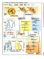
Figure1showsadiagramofoculariontophoresisofapositivelycharged
drug in a rabbit. The eyecup, with an internal diameter of $ 1 cm, is placed
over the cornea and filled with the drug solution. A metal electrode that is
connected to a direct current power supply is submerged in the solution in
the eyecup without making contact with the surface of the eye. The ground
electrode, connected to the other terminal of the power supply, is attached
to the ear of the rabbit via wet (0.9% NaCl) gauze to ensure a good con-
nection. With the hand-held applicator probe, the metal (platinum) elec-
trode extends into the eyecup that is filled with the drug solution. The
eyecup is placed against the eye and is held in place throughout the entire
iontophoresis procedure. Iontophoresis requires a complete electrical circuit
with direct current passing from the anode to the cathode and from the
cathode back to the anode. The two electrodes are placed as anatomically
close to each other as possible on the body, which is an excellent conductor
of electricity, to complete the circuit.
Ocular Iontophoresis 367
Copyright © 2003 Marcel Dekker, Inc.
II. MEDICAL APPLICATIONS OF IONTOPHORESIS
A. Archival Studies
The use of the shock of the torpedo, an electric fish, for the treatment of
gout was described by Aetius, a Greek physician, more than 1000 years ago
(14). In 1747, Veratti enunciated the concept of applying an electric current
to increase the penetration of drugs into surface tissues (15). In 1898,
Morton demonstrated that finely powdered graphite could be driven into
his arm under the positive electrode and produced small black spots that
persisted for weeks (16). In 1900, Leduc reported the first controlled studies
of iontophoresis as a therapeutic modality (17,18). Leduc showed that trans-
cutaneous iontophoretic delivery of strychnine and cyanide ions into rabbits
produced fatal tetanic seizures and cyanide poisoning.
The earliest description of ocular iontophoresis was published in 1908
by the German investigator Wirtz (19), who performed iontophoresis of zinc
salts for the treatment of corneal ulcers. In 1927, Morisot (20) enumerated
many successful ophthalmological applications of iontophoresis including
iontophoresis of magnesium for treatment of glaucoma, iontophoresis of
ammonium chloride for treatment of cataract, and iontophoresis of phos-
phoric acid for treatment of optic atrophy. Erlanger was one of the first
ophthalmologists to introduce iontophoresis to England and the United
States. In 1936, he delivered barium chloride iontophoretically into the
eyes of guinea pigs and observed cataract formation 48 hours later (21).
He went on to describe the usefulness of iontophoresis in the clinical treat-
ment of corneal ulcers, conjunctivitis, scleritis, glaucoma, and cataract
(22,23).
During the 1940s in the United States, Ludwig von Sallmann, a pro-
minent ophthalmologist, was one of the pioneers in the clinical use of ocular
iontophoresis. Von Sallmann showed that transcorneal iontophoresis of
penicillin was more effective than subconjunctival injection for the delivery
of penicillin into the aqueous humor (24,25) and demonstrated modest
success in the treatment of intraocular staphylococcal infection (26). In
1956, Witzel and his colleagues (27) published a report on the use of ocular
iontophoresis as a drug delivery system for a variety of antibiotics. They
found that iontophoresis was effective in the delivery of streptomycin, neo-
mycin, and penicillin.
Despite its widespread use and study during the first 60 years of the
twentieth century, iontophoresis was never fully adopted as a standard
procedure. The lack of carefully controlled trials and the paucity of toxicity
data were among the reasons that precluded its acceptance as a viable
alternative for drug delivery. However, over the past 30–40 years, ionto-
phoresis has been adapted for use in a variety of medical specialties, includ-
Ocular Iontophoresis 369
Copyright © 2003 Marcel Dekker, Inc.
inganesthesiology,dermatology,dentistry,andophthalmology.Table1
provides a list of selected reviews and reports describing the recent evolution
of this procedure and highlighting some of its medical applications.
B. Iontophoresis in General Medicine and Dentistry
1. Diagnosis of Cystic Fibrosis: Pilocarpine Iontophoresis
Iontophoresis of pilocarpine (28) to induce sweating to measure sweat
sodium and chloride concentration is the basis of the ‘‘sweat test’’ that is
used to diagnose cystic fibrosis (29–31). High concentrations of both sodium
and chloride ions in the sweat are considered as unequivocal evidence of the
disease. Although essential criteria have been established for a positive test
result in both children (30) and adults (31), pilocarpine iontophoresis should
be used in conjunction with other clinical features to make a definitive
diagnosis. This procedure is particularly useful in children younger than 1
year old because it is essentially painless and takes only 3–5 minutes to
complete.
2. Treatment of Hyperhidrosis: Tap-Water Iontophoresis
The first dermatological application of iontophoresis was to treat hyperhi-
drosis (32). Hyperhidrosis is a condition characterized by pathologically
excessive sweating due to abnormal secretion of the eccrine sweat glands
in various parts of the body [primarily the palms, soles and axillae (33–35)].
Iontophoresis of tap water has been very effective (!90% of patients) in
inhibiting palmar and plantar hyperhidrosis, but the results are less reward-
ing for axillary hyperhidrosis (33,35).
3. Treatment of Hypersensitive Teeth: NaF Iontophoresis
Iontophoresis of sodium fluoride to treat teeth that have become thermo-
sensitive is a very useful and successful therapy in dentistry (36,37). Teeth
previously shown to be hypersensitive to cold lose their sensitivity to heat
and cold immediately after iontophoresis of sodium fluoride, and the effects
are long lasting. The mechanisms by which teeth become hypersensitive and
by which iontophoresis alleviates this condition are not fully understood.
One theory is that exposed dentin allows fluid movement through micro-
tubules that stretch from the pulp. Occlusion of the patent dentinal tubules
would appear to be one mechanism by which sodium fluoride iontophoresis
could have a beneficial effect.
370 Myles et al.
Copyright © 2003 Marcel Dekker, Inc.
4.PediatricAnesthesia:LidocaineIontophoresis
Iontophoresisofanestheticsforinductionoflocalanesthesiainpediatric
officepatientshasbeenverysuccessful(38–40).Randomizedstudieshave
foundlidocaineiontophoresistobemoreeffectivethanEMLA(eutetic
mixtureoflocalanesthetics)creamortopicallidocaine(38,41),andits
effectivenesshasbeendocumentedforplacementofpediatricintravenous
catheters(39).Iontophoresisisadvantageousinpediatricpatientsbecauseit
alleviatesthepainandanxietyduetoneedleinjectionsandworksmore
rapidlythantopicallyappliedanesthetics,whichtakealongtimetobecome
effective(upto1h)andoftenprovideincompleteanesthesia.Squireetal.
(41)demonstratedthatthetimetoaccomplishtopicalanesthesiawasshorter
withiontophoresisof2%lidocainewithepinephrine1:100,000(13min)
comparedtoasurface-appliedEMLAcream[60min(41)].Iontophoresis
oflidocainewithepinephrineisasafe,rapid,andeffectivemethodoflocal
anesthesiadeliveryforpediatricprocedures.
C.IontophoreticTherapiesinOphthalmology
Table2providesalistofthedrugs,dyes,andotherchargedmolecules
summarizedbelow.Thecations(positiveions)areusedinanodal(positive
electrode)iontophoresis,whereastheanions(negativeions)areusedin
cathodal(negativeelectrode)iontophoresis.Iontophoresisofthevarious
classesofdrugs(antibiotics,antivirals,antifungal,antimetabolite,adrener-
gic,steroid,anesthetic,anddyes)canbedeliveredbytwoapproaches.
Transcornealiontophoresis(describedearlierandindiagrammaticformin
Fig.1)deliversahighconcentrationofdrugtotheanteriorsegmentofthe
eye(cornea,aqueoushumor,ciliarybody,andlens).Inphakicanimals,the
lens-irisdiaphragmlimitspenetrationofadrugtotheposteriortissuesof
theeyesuchasposteriorvitreousandretina.Thisbarriercanbeovercome
byapplyingthecurrentthroughtheparsplana(transscleraliontophoresis),
whichcanproducesignificantlyhighandsustaineddrugconcentrationin
thevitreousandretina.Fortransscleraliontophoresis,thedrugsolutionis
containedinanarrowtubewithinaneyecupheldtotheconjunctivaby
suction.Thetubeisplacedovertheparsplanatoavoidcurrentdamageto
theretina.Thistechniquecircumventsthelens-irisbarrieranddeliversdrugs
intothevitreousorretina.Figure2showsadiagramofatransscleral
iontophoresis device and setup.
Within the past 10 years, a number of excellent articles/chapters have
reviewed the application of iontophoresis in therapeutic approaches in
ophthalmology (11,12,42–44). Current research in ocular iontophoresis is
aimed at resolving the delivery problems associated with newly developed
Ocular Iontophoresis 371
Copyright © 2003 Marcel Dekker, Inc.
A review of the procedures and techniques used to study the flow of
aqueous humor in the eye was published by Brubaker in 1982 (48). He noted
that these methods were essentially equivalent to those described by Jones
and Maurice (45). After application of topical anesthesia, a gel 5 mm in
diameter containing 2% agar and 10% fluorescein was placed on the central
cornea. The power source was a 45 V battery. The agar gel constituted the
negative electrode, and to complete the circuit, the patient held the positive
electrode in his hand. The current (0.2 mA) was applied for 5–7 seconds. In
more than 10,000 iontophoretic applications of fluorescein, no obvious side
effects of the iontophoretic procedure were observed.
b. Dyes for Laser Sclerostomy Pulsed dye laser sclerostomy is an
adjunctive procedure used in the treatment of glaucoma (49). The pulsed
dye laser procedure uses a gonioscopic approach for the ab interno deliv-
ery of visible laser light. The procedure requires a full thickness penetra-
tion of a dye throughout a 1–2 mm
2
area of scleral tissue for adequate
absorption of the visible light energy. The light beam is transmitted
through the cornea, crosses the anterior chamber, and ablates stained lim-
bal scleral tissue with the formation of a fistula [a filtration channel for in-
traocular pressure (IOP) release (49)].
Methylene blue dye is water soluble and has a positive electrical charge
in solution. It has an absorption peak of 668 nm and is used to enhance the
optical absorption of scleral tissue (49). Latina et al. (50) iontophoresed 1%
methylene blue at the limbal region of 35 glaucoma patients. The current
applied was 5.0 mA for a duration of 4–8 minutes. To create the sclerostomy,
the laser energy (200–250 mJ), delivered by a slit-lamp, was focused onto the
dyed sclera, using a goniolens, so that only a light beam penetrated the eye.
The red wavelength of 660 nm generated by the laser was maximally
absorbed by the stained sclera and created a complete sclerostomy.
Successful sclerostomies were achieved in 21 of 35 patients ($60%) with a
reduction in IOP from a mean preoperative value of 35 mmHg to a mean
postoperative value (at 9 months) of 22 mmHg. Melamed (51) obtained
similar results with a 58% success rate for complete sclerostomies and a
reduction in IOP from a mean preoperative IOP of 36.6 mmHg to a mean
postoperative IOP of 23.7 mmHg. Grossman et al. (52) examined the sta-
bility of iontophoresed methylene blue in rabbit eyes. Decreased dye con-
centration of over 50% within 2 hours and a complete disappearance of dye
within 24 hours were demonstrated. They also noted that the stain tended to
bleach from the laser dye exposure. This prevented further absorption of the
laser energy, resulting in incomplete scleral ablation and fistula formation.
Melamed (51) reported blanching of the stained sclera after the first laser
shots, which adversely affected the efficacy of subsequent shots.
374 Myles et al.
Copyright © 2003 Marcel Dekker, Inc.
Reactive black-5 (RB5) is a water-soluble black dye that is negatively
charged at the physiological pH of the eye. RB5 has been proposed as an
alternative dye to stain the scleral tissue (53). RB5 stain was subjected to a
number of different conditions simulating laser treatment of the sclera. The
stain was stable over time (72 h) and stable when exposed to high tempera-
ture (1208C), to scleral breakdown products (collagen), to strong oxidants
(1.5% H
2
O
2
), and to laser light energy (53). Optimal parameters for ionto-
phoretic delivery of RB5 into limbal scleral tissue were also determined.
Ideal parameters for iontophoresis included a probe tip surface area
between 0.1 and 0.7 mm
2
, a current of 0.5 mA, and a duration of 5 minutes.
Using these parameters for iontophoresis, the maximum concentration of
RB5 achieved in sclera was 0.15% (53). This value is considerably greater
than the threshold for ablation of 0.001% RB5 using a laser energy of 250
mJ. Thus, iontophoresis can deliver an amount of RB5 stain to the sclera
that is more than sufficient for laser ablation. This approach obviates con-
junctival dissection and decreases the stimulus for episcleral scarring that
eventually could cause reelevation of intraocular pressure (49,52,53).
c. Adrenergic Agents for Treatment of Glaucoma 6-Hydroxydopa-
mine and a-methylparatyrosine are two pharmacological agents that block
the synthesis of norepinephrine. 6-Hydroxydopamine, a congener of nore-
pinephrine, causes the reversible destruction of nerve terminals in the
anterior segment. In the early to mid-1970s, a number of investigators
(54–58) used iontophoresis to deliver these substances to the eyes of rab-
bits, normal volunteers, and glaucoma patients with primary open-angle
glaucoma. The theory behind the treatment involved the depletion of ocu-
lar norepinephrine, which would result in an increased sensitivity to glau-
coma drugs such as epinephrine.
Kitazawa et al. (54,55) were the first to report the results of iontophor-
esis of 6-hydroxydopamine in rabbits and human eyes. A 1% solution of 6-
hydroxydopamine was iontophoresed at 0.75 mA for 3 minutes. High con-
centrations of 6-hydroxydopamine were achieved in ocular tissues in rab-
bits, and intraocular pressure was reduced in normal human eyes.
Subsequently, Kitazawa et al. (56) treated patients with primary open-
angle glaucoma with a combination therapy of iontophoresed 6-hydroxy-
dopamine and topical epinephrine. The results led them to conclude that
this combination therapy had clinical value in the management of open-
angle glaucoma.
Iontophoresis of 6-hydroxydopamine to treat primary open-angle glau-
coma was also examined by Watanabe et al. (57). For the patients for whom
they had sufficient data for analysis (49/100), this procedure was therapeuti-
cally effective in 41%, questionable in 31%, and ineffective in 28%.
Ocular Iontophoresis 375
Copyright © 2003 Marcel Dekker, Inc.
Colasanti and Trotter (58) performed ocular iontophoresis of a-
methylparatyrosine in rabbits. A 4.0% a-methylparatyrosine solution was
iontophoresed at a current of 3 mA for 5 minutes. This drug is similar to 6-
hydroxydopamine, and it also produced a significant decrease in the nore-
pinephrine concentration in rabbit ocular tissues. No clinical studies with a-
methylparatyrosine were done in normal human eyes or in patients with
primary open-angle glaucoma.
These results demonstrated that iontophoresis of 6-hydroxydopamine
was a viable method of sensitizing the eyes to glaucoma drugs and that this
procedure had some clinical value in the management of this disease.
However, with the advent of long-acting antiglaucoma drugs such as timo-
lol, levobunolol, and betaxolol, iontophoresis of 6-hydroxydopamine for the
therapy of glaucoma was discontinued.
d. 5-Fluorouracil for Control of Cellular Proliferation After Glaucoma
Surgery 5-Fluorouracil (5-FU) acts as an antiproliferative agent to pre-
vent cellular replication. The concentration of 5-FU required for 50% in-
hibition of rabbit conjunctival fibroblasts in culture is 0.2–0.5 mg/mL (59).
5-FU is a small, negatively charged molecule with a pKa of $ 8. Kondo
and Araie (60) were the first to report iontophoresis of 5-FU to the rabbit
eye. A 5% solution of 5-FU containing 8.47% tris(hydroxymethyl)amino-
methane was delivered transsclerally at 0.5 mA for 30 seconds. An elec-
trode 7 mm in diameter was placed on the bulbar conjunctiva 4 mm
posterior to the limbus in the superior temporal quadrant. Iontophoresis
of 30-second duration delivered enough 5-FU into ocular tissue such that
30 minutes later the drug concentration was 50 mg/g and 21 mg/g in con-
junctival and scleral tissue, respectively. Over the next 10 hours, the
amount of 5-FU decreased to 0.6 mg/g in the conjunctiva and 1.2 mg/g in
the sclera. These concentrations are still high enough to have a therapeu-
tic effect.
Transscleral iontophoresis of 5-FU would eliminate the need for sub-
conjunctival injection and its unwanted complications (risk of bleeding,
infections, scarring, and drug penetration into other ocular tissues).
Iontophoretically delivered 5-FU may improve the efficacy of antiglaucoma
surgery (e.g., sclerostomy) by interfering with healing and thereby maintain-
ing patency of the fistulas. To date, however, no studies have been done in
experimental models of disease or in human eyes.
2. Ocular Anesthesia
The deliver of local anesthetics by iontophoresis has been very successful
(61–63). Iontophoretically delivered anesthetics can provide topical anesthe-
sia within 5–15 minutes with limited systemic absorption (61). The anes-
376 Myles et al.
Copyright © 2003 Marcel Dekker, Inc.
thetic solutions used most often are a combination of lidocaine and epi-
nephrine.
Sisler (62) iontophoresed anesthetic solutions containing either 4%
lidocaine with 1:1000 epinephrine or 2% lidocaine with 1:2000 epinephrine
to patients with lesions of the tarsus and tarsal conjunctiva prior to surgi-
cal excision of conjunctival plaques. A current 0.5 mA was applied for 10
minutes. Twenty-seven patients were treated. None of the patients
reported any discomfort in the eyelids or the arm to which the negative
electrode was attached; only a mild sensation was described. Three
patients with lesions in the deeper portion of the tarsus reported pain
and were given an injection to achieve local anesthesia. This is the only
report describing iontophoretic delivery of an anesthetic agent to adnexal
areas for pain prevention.
Meyer et al. (63) iontophoresed a 4% lidocaine solution to eyelids of
patients for local anesthesia prior to blepharoplasty or ptosis repair. A
current of 2 mA was applied for 12 minutes. Ten normal volunteers were
used so as to compare pain sensations after anesthesia by iontophoresis or
topical application of the anesthetic. Both surgical patients and volunteers
reported significantly less pain after iontophoresis of the anesthetic. No side
effects of this iontophoresis procedure were observed.
3. Ocular Inflammation
Corticosteroids are the most common drugs used in treating ocular inflam-
matory disorders (64–67). Topical drop application is preferred to avoid the
serious systemic side effects of steroids (68). However, this mode of admin-
istration does not allow for sufficient drug delivery to the posterior segment
of the eye. Iontophoretic delivery of anti-inflammatory drugs into the eye
has been examined in human and various animal models and offers a viable
alternative to topical or systemic administration.
Lachaud (65) iontophoresed hydrocortisone acetate (0.1% solution)
into rabbit eyes with a current of 3 mA for 10 minutes. He demonstrated
that iontophoresis could deliver higher concentrations of steroid to rabbit
ocular tissue than either topical drops (0.5%) or subconjunctival injection
(0.1 mL, 2.5%). In human studies, Lachaud iontophoresed dexamethasone
acetate (7 mg%, 1–2 mA, 20 min) to treat a variety of clinical conditions,
including idiopathic uveitis. He reported that a significant proportion of the
patients with uveitis benefited in terms of more rapid recovery and/or
increased comfort. Lachaud (65) concluded that iontophoresis resulted in
therapeutic concentrations of the steroid(s) in ocular tissue. However, it
must be noted that this open clinical study did not involve comparisons
with eyes receiving other therapies or with untreated control eyes.
Ocular Iontophoresis 377
Copyright © 2003 Marcel Dekker, Inc.
Lam et al. (66) iontophoresed a 30% dexamethasone solution trans-
sclerally into rabbit eyes using 1.6 mA for 25 minutes. The diameter of the
cylinder holding the drug solution in contact with the sclera was 0.7 mm.
They compared peak steroid concentrations in the choroid-retinal tissue
following iontophoresis, subconjunctival injection (1 mg) or retrobulbar
injection (1 mg). The peak steroid concentration (mg/g tissue) for iontophor-
esis was 122, for subconjunctival injection 18.1, and for retrobulbar injec-
tion 6.6. In the vitreous humor the values were 140, 0.2, and 0.3 mg/mL,
respectively. Even at 24 hours after iontophoresis, significant therapeutic
levels of dexamethasone remained 3.3 mg/mL in the vitreous and 3.9 mg/g
in the choroid-retina.
Behar-Cohen et al. (67) investigated the efficacy of iontophoretic deliv-
ery of dexamethasone for the treatment of endotoxin-induced uveitis in the
rat. Dexamethasone was delivered by concurrent transcorneal-transscleral
iontophoresis (1% at 0.4 mA for 4 min) using a 1 mL reservoir electrode
that covered the cornea, the limbus, and the first millimeter of the sclera.
They showed that administration of dexamethasone by iontophoresis inhib-
ited anterior and posterior signs of intraocular inflammation (protein exu-
dation, cellular infiltration) as effectively as systemic administration.
Cytokine (TNF-a) (69) expression was inhibited in the anterior as well as
the posterior segment of the eye. No clinical or histological damage was
caused by iontophoresis. Thus, iontophoresis can deliver therapeutic doses
of this anti-inflammatory drug to the posterior as well as the anterior seg-
ment of the eye and may be a viable alternative to systemic administration
of corticoids in severe ocular inflammation.
4. Ocular Infection
Transcorneal iontophoresis of antibiotics is an effective means of treatment
for bacterial keratitis and other anterior segment infections. Transcorneal
iontophoresis delivers therapeutic concentrations of antibiotics to the cor-
nea and aqueous humor. In phakic animals, the lens-iris diaphragm limits
penetration of the drug into the posterior tissues of the eye such as posterior
vitreous and retina. Transscleral iontophoresis circumvents the lens-iris bar-
rier and delivers drugs into the vitreous or retina in amounts high enough to
be therapeutic in the treatment of posterior segment infections, such as
endophthalmitis. Numerous studies have documented successful ionto-
phoretic delivery of various antibiotics into ocular tissues of animal models.
Examples are summarized below.
a. Gentamicin: Transcorneal and/or Transscleral The aminoglyco-
side gentamicin has a molecular weight of approximately 430 daltons, is
lipid insoluble, and bears two positive charges at physiologic pH (70). It
378 Myles et al.
Copyright © 2003 Marcel Dekker, Inc.
possesses bactericidal properties against a wide variety of gram-negative
and gram-positive bacteria (71). Gentamicin concentrations of 5 mg/mL
inhibit over 90% of Pseudomonas (71), Proteus rettgeri, P. vulgaris, and
P. morganii strains, as well as 99% of Staphylococcus strains (70). These
molecular features, combined with the extreme sensitivity of the target or-
ganisms, make gentamicin an ideal drug for iontophoresis.
Hughes and Maurice (72) found that transcorneal iontophoresis of
gentamicin in the rabbit eye increased permeability to the antibiotic more
than 100-fold, compared with control eyes in which topical application was
performed under the same iontophoretic conditions but with no current
applied. Grossman et al. (70) iontophoresed 10% gentamicin in a 2%
agar solution into rabbit corneas. They demonstrated that significantly
higher and longer-lasting gentamicin concentrations were achieved in the
cornea with iontophoresis as compared to subconjunctival injection of a 20
mg dose. Frucht-Pery et al. (73) showed that higher current density did not
significantly enhance antibiotic penetration into rabbit cornea, but bacter-
icidal concentrations of gentamicin could be obtained. Frucht-Pery et al.
(74) also described the distribution of transcorneally iontophoresed genta-
micin in the rabbit cornea. They found that the highest concentrations of the
drug were in the central cornea, while the midperipheral cornea(s) had
higher levels than peripheral cornea(s).
Fishman et al. (71) described iontophoresis of gentamicin to unin-
fected aphakic rabbit eyes. Gentamicin iontophoresis (50 mg/mL at 0.75
mA for 10 min) yielded peak corneal (71 mg/g of tissue) and aqueous
humor (78 mg/mL) concentrations 30 minutes after treatment. The peak
vitreous concentration (10.4 mg/mL) was observed 16 hours after treatment.
Therapeutic concentrations of gentamicin were still present in the vitreous
24 hours after iontophoresis. This study suggests that even transcorneal
iontophoresis has the potential to deliver high concentrations of gentamicin
to the posterior segment in aphakic eyes. Since many patients with
endophthalmitis are aphakic, transcorneal iontophoresis could be a suitable
route of administration of antibiotics for therapeutic management of this
disease.
Barza et al. (75) modified the standard transcorneal procedures to
achieve direct delivery of high concentrations of gentamicin into the vitreous
by transscleral iontophoresis. First, a reservoir holding the drug was placed
over the pars plana, thereby bypassing the lens-iris barrier. Second, the
contact area of the fluid that delivered both the antibiotic and the current
was kept small (approximately 1 mm in diameter). They reported that trans-
scleral iontophoresis delivered therapeutic concentrations (94–207 mg/mL)
of gentamicin to the vitreous humor of uninfected rabbit eyes, thus obviat-
ing the need for intraocular injections.
Ocular Iontophoresis 379
Copyright © 2003 Marcel Dekker, Inc.
Another study from the same laboratory (76) demonstrated that trans-
scleral iontophoresis of gentamicin is a useful adjunct to intravitreal injec-
tions for the treatment of endophthalmitis caused by pseudomonas
aeruginosa. They showed that rabbits receiving both intravitreal injection
and transscleral iontophoresis of gentamicin had lower bacteria counts at
each treatment interval compared to rabbits that received gentamicin by a
single intravitreal injection of a 100 mg dose. These results support the use of
iontophoresis of gentamicin as a useful supplement to intravitreal injection
for the treatment of bacterial endophthalmitis.
Grossman et al. (70) reported that transscleral iontophoresis of genta-
micin produced results similar to their findings with transcorneal iontophor-
esis described above. Iontophoresis of 10% gentamicin in 2% agar solution
at 2 mA for 10 minutes with a contact area of 2 mm in diameter delivered
very high concentrations of gentamicin to the vitreous humor of rabbit eyes.
Vitreous concentrations peaked (53.4 mg/mL) 16 hours after iontophoresis
and remained at inhibitory levels even at 24 hours. As with transcorneal
iontophoresis (70), no ocular tissue toxicity or damage was observed with
transscleral iontophoresis.
Burstein et al. (77) reported that transscleral iontophoresis of genta-
micin into uninfected rabbit eyes resulted in antibiotic concentrations of 10–
20 mg/mL in the vitreous humor. These concentrations are significantly
lower than the concentration range (94–207 mg/mL) reported by Barza et
al. (75) in rabbit eyes. The authors found that the total surface area of the
electrode is inversely proportional to the amount of antibiotic delivered,
e.g., a 4.5 mm
2
applicator delivers approximately 20 times more drug to
the vitreous than a 28 mm
2
applicator. This result reinforced the conclusion
of Barza et al. (75,76) that a small area of contact in iontophoresis results in
a higher concentration of drug in the eye.
Barza’s research group (78) also reported the first use of a nonhuman
primate (the cynomolgus monkey) to study the pharmacokinetics of trans-
sclerally delivered gentamicin and/or potential histological damage of
transscleral iontophoresis. High and sustained concentrations of antibiotics
were achieved in the vitreous. Although small burns were observed in the
area of the pars plana where the electrode was applied, all electroretino-
grams were normal. The results suggest that transscleral iontophoresis is
well tolerated in the primate eye and that investigations with human eyes
may yield alternative treatment options. The absence of side effects with
the agar-based delivery system of Grossman et al. (70) is noteworthy. This
formulation may facilitate the use of transscleral iontophoresis as a treat-
ment of choice for patients with bacterial endophthalmitis or other clinical
conditions that require high concentrations of antibiotics in the posterior
segment.
380 Myles et al.
Copyright © 2003 Marcel Dekker, Inc.
b. Tobramycin: Transcorneal Transcorneal iontophoresis of the
aminoglycoside tobramycin has been examined by our group in studies
with the normal rabbit eye. Rootman et al. (79) were the first to demon-
strate the efficacy of iontophoresed tobramycin for the treatment of ex-
perimental Pseudomonas keratitis. Rabbit corneas were infected with 10
3
colony-forming units of P. aeruginosa. Transcorneal iontophoresis of a
2.5% tobramycin solution at 0.8 mA for 10 minutes was performed at 22,
27, and 32 hours after inoculation. On average, the treated corneas had a
6 log reduction in colony-forming units relative to untreated corneas. At
32 hours postinoculation, 67% of the corneas had no viable bacteria (i.e.,
were sterile). Topically applied or subconjunctival injection of tobramycin
did not yield corneas free of viable (infectious) Pseudomonas.
In other studies, Hill and associates examined the pharmacokinetics of
iontophoresed tobramycin and/or potential toxicity to the corneal epithe-
lium of transcorneal iontophoresis (80,81). In uninfected, mock-infected,
and P. aeruginosa–infected rabbit corneas, transcorneal iontophoresis pro-
duced high and sustained concentration of the antibiotic in the corneal
epithelium, corneal stroma, and aqueous humor (80,81). Iontophoresis
delivered five times more drug than bathing the cornea with a 2.5% tobra-
mycin solution and 20 times more the applying 1.36% tobramycin as for-
tified drops (80). No permanent abnormalities were observed by slit-lamp
biomicroscopy, scanning electron microscopy, or light microscopy (81).
After 10 minutes of iontophoresis, the epithelium showed focal edema
and disruption of all cell layers. Histological specimens obtained 8 and 16
hours after iontophoresis showed no defects in the corneal epithelium.
The efficacy of iontophoretically delivered tobramycin was examined
using a tobramycin-resistant strain of Pseudomonas (82). A strain of P.
aeruginosa with a minimum inhibitory concentration (MIC) for tobramycin
of 31 mg/mL was injected into the corneal stroma in rabbit eyes.
Transcorneal iontophoresis of 2.5% tobramycin resulted in a 3 log reduc-
tion in the number of bacteria. These results show that transcorneal ionto-
phoresis can deliver concentrations of tobramycin high enough to combat a
clinically tobramycin-resistant strain of Pseudomonas.
c. Ciprofloxacin: Transcorneal/Transscleral Ciprofloxacin, a very
potent fluoroquinolone antibiotic, is active against a broad spectrum of
gram-positive and gram-negative bacteria (83). Hobden et al. (84) used
transcorneal iontophoresis to deliver ciprofloxacin to rabbit corneas in-
fected with an aminoglycoside-resistant strain of Pseudomonas. Iontophor-
esis of 1% or 2.5% ciprofloxacin reduced the number of colony-forming
units by more than 5 log relative to untreated controls. This level of inhi-
bition was significantly greater than either topically applied drops (0.75%
Ocular Iontophoresis 381
Copyright © 2003 Marcel Dekker, Inc.