Textbook of Neuroanaesthesia and Critical Care - part 7 pptx
Bạn đang xem bản rút gọn của tài liệu. Xem và tải ngay bản đầy đủ của tài liệu tại đây (525.48 KB, 52 trang )
Pa
g
e 251
31. Bohlman HH. Acute fractures and dislocations of the cervical spine: an analysis of three hundred hospitalised patients and review
of the literature. J Bone Joint Surg 1979; 61: 1119
–
1142.
32. Aprahamian C, Thompson DM, Finger WA, Darin JC. Experimental cervical spine injury model: evaluation of airway
management techniques. Ann Emerg Med 1984; 13: 584
–
587.
33. Graham JJ. Complications of cervical spine surgery: a five year report on a survey of the membership of the Cervical Spine
Research Society by the Morbidity and Mortality Committee. Spine 1989; 14: 1046
–
1050.
34. Meschino A, Devitt JH, Koch JP, Szalai JP, Schwartz ML. The safety of awake intubation in cervical spine injury. Can J Anaesth
1992; 39: 114
–
117.
35. Sawin PD, Todd MM, Traynelis VC et al. Cervical spine motion with direct laryngoscopy and orotracheal intubation: an in vivo
cinefluoroscopic study of subjects without cervical abnormality. Anesthesiology 1996; 85: 26
–
36.
36. Horton WA, Fahy L, Charters P. Disposition of the cervical vertebrae, atlanto-axial joint, hyoid and mandible during X-ray
laryngoscopy. Br J Anaesth 1989; 63: 435
–
438.
37. Hauswald M, Sklar DP, Tandberg D, Garcia JF. Cervical spine movement during airway management: cinefluoroscopic appraisal
in human cadavers. Am J Emerg Med 1991; 9: 535
–
538.
38. Johnson RM, Hart, DL, Simmons EF. Cervical orthoses. J Bone Joint Surg 1977; 59A: 332
–
339.
39. Chandler DR, Nemejc C, Adkins RH, Waters RL. Emergency cervical spine immobilisation. Ann Emerg Med 1992; 21: 1185–
1188.
40. Podolsky S, Baraff LJ, Simon RR, Hoffman JR, Larmon B, Ablon W. Efficacy of cervical spine immobilisation methods. J
Trauma 1983; 23: 461
–
465.
41. Heath KJ. The effect on laryngosocoy of different cervical spine immobilisation techniques. Anaesthesia 1994; 49: 843
–
845.
42. Hastings RH, Wood PR. Head extension and laryngeal view during laryngoscopy with cervical spine stabilisation manoeuvres.
Anesthesiology 1994; 81: 1081
–
1082.
43. Majernick TG, Bieniek R, Houston JB, Hughes HG. Cervical spine movements during orotracheal intubation. Ann Emerg Med
1986; 15: 417
–
420.
44. Nolan JP, Wilson ME. Orotracheal intubation in patients with potential cervical spine injuries. An indication for the gum elastic
b
ougie. Anaesthesia 1993; 48: 630
–
633.
45. Bogdonoff DL, Stone DJ. Emergency management of the airway outside the operating room. Can J Anaesth 1992; 39: 1069–
1089.
46. Hastings RH, Marks JD. Airway management for trauma patients with potential cervical spine injuries Anesth Analg 1991; 73:
471
–
482.
47. Nolan JP. Resuscitation of the trauma patient. Care of the Critically Ill 1995; 11: 222
–
226.
48. Redan JA, Livingston DH, Tortella BJ, Rush BF. The value of intubating and paralysing patients with suspected head injury in
the emergency department. J Trauma 1991; 31: 371
–
375.
49. Talucci RC, Shaikh KA, Schwab CW. Rapid sequence induction with oral endotracheal intubation in the multiple injured patient.
Am Surg 1988; 54: 185
–
187.
50. Grande CM, Barton CR, Stene JK. Appropriate techniques for airway management of emergency patients with suspected spinal
cord injury. Anesth Analg 1988; 67: 714
–
715.
51. Lawes EG, Campbell I, Mercer D. Inflation pressure, gastric insufflation and rapid sequence induction. Br J Anaesth 1987; 59:
315
–
318.
52. Doolan LA, O'Brien JF. Safe intubation in cervical spine injury. Anaesth Intens Care 1985; 13: 319
–
324.
53. Narrod JA, Moore EE, Rosen P. Emergency cricothyrostomy – technique and anatomical considerations. J Emerg Med 1985; 2:
443
–
446.
54. DeLaurier GA, Hawkins ML, Treat RC, Mansberger AR Jr. Acute airway management: the role of cricothyroidotomy. Am Surg
1990; 56: 12
–
15.
55. Salvino CK, Dries D, Gamelli R, Murphy-Macabobby M, Marshall W. Emergency cricothyroidotomy in trauma victims. J
Trauma 1993; 34: 503
–
505.
56. McGill J, Clinton JE, Ruiz E. Cricothyrotomy in the emergency department. Ann Emerg Med 1982; 11: 361
–
364.
57. Esses B, Jafek BW. Cricothyroidotomy: a decade of experience in Denver. Ann Otol Rhinol Laryngol 1987; 96: 519
–
524.
58. Wood PR, Lawler PGP. Managing the airway in cervical spine injury: a review of the Advanced Trauma Life Support protocol.
Anaesthesia 1992; 47: 792
–
797.
59. McHale SP, Brydon CW, Wood MLB, Liban JB. A survey of nasotracheal intubating skills among Advanced Trauma Life
Support course graduates. Br J Anaesth 1994; 72: 195
–
197.
60. Logan AStC. Use of the laryngeal mask in a patient with an unstable fracture of the cervical spine. Anaesthesia 1991; 46: 987.
61. Calder I, Ordman AJ, Jackowski A, Crockard HA. The Brain laryngeal mask. An alternative to emergency tracheal intubation.
Anaesthesia 1990; 45: 137
–
139.
62. Benumof JL. Management of the difficult adult airway. Anesthesiology 1991; 75: 1087
–
1110.
63. Ovassapian A. Fiberoptic tracheal intubation. In: Ovassapian A (ed) Fiberoptic airway endoscopy in anesthesia and critical care.
Raven Press, NewYork, 1990.
64. Sidhu VS, Whitehead EM, Ainsworth QP, Smith M, Calder I. A technique of awake fiberoptic intubation. Anaesthesia 1993; 48:
910
–
913.
65. Ovassapian A, Krejcie TC, Yelich SJ, Dykes MHM. Awake fiberoptic intubation in the patient at high risk of aspiration. Br J
Anaesth 1989; 62: 13
–
16.
Pa
g
e 252
66. Ovassapian A, Yelich SJ, Dykes MHM, Brunner EE. Fiberoptic nasotracheal intubation – incidence and causes of failure. Anesth
Analg 1983; 62: 692
–
695.
67. Delaney KA, Hessler R. Emergency flexible fiberoptic nasotracheal intubation: a report of 60 cases. Ann Emerg Med 1988; 17:
919
–
926.
68. Mlineck EJ, Clinton JE, Plummer D, Ruiz E. Fiberoptic intubation in the emergency department. Ann Emerg Med 1990; 19:
359
–
362.
69. McNamara RM. Retrograde intubation of the trachea. Ann Emerg Med 1987; 16: 680
–
682.
70. Barriot P, Riou B. Retrograde technique for tracheal intubation in trauma patients. Crit Care Med 1988; 16: 712
–
713.
71. Fox DJ, Castro T, Rastrelli AJ. Comparison of intubation techniques in the awake patient: the Flexi-lum surgical light
(lightwand) versus blind nasal approach. Anesthesiology 1987; 66: 69
–
71.
72. Mallampati SR, Gatt SP, Gugino LD et al. A clinical sign to predict difficult tracheal intubation: a prospective study. Can
Anaesth Soc J 1985; 32: 429
–
434.
73. Wilson ME, Spiegelhalter D, Robertson JA, Lesser P. Predicting intubation. Br J Anaesth 1988; 61: 211
–
216.
74. Frerk CM. Predicting difficult intubation. Anaesthesia 1991; 46: 1005
–
1008.
75. Calder I. Predicting difficult intubation. Anaesthesia 1992; 47: 528
–
529.
76. Wilson ME, John R. Problems with the Mallampati sign. Anaesthesia 1990; 45: 486
–
487.
77. Oates JDL, Macleod AD, Oates PD, Pearsall FJ, Howie JC, Murray GD. Comparison of two methods for predicting difficult
intubation. Br J Anaesth 1991; 66: 305
–
310.
78. Laurent SC, De Melo AE, Alexander-Williams JM. The use of the McCoy laryngoscope in patients with simulated cervical spine
injuries. Anaesthesia 1996; 51: 74
–
75.
79. Hastings RH, Vigil AC, Hanna R, Yang BY, Sartoris DJ. Cervical spine movement during laryngoscopy with the Bullard,
Macintosh and Miller laryngoscopes. Anesthesiology 1995; 82: 859
–
869.
80. Marks RJ, Forrester PC, Calder I, Crockard HA. Anaesthesia for transoral craniocervical surgery. Anaesthesia 1986; 41: 1049–
1052.
81. Bland JH. Rheumatoid arthrits subluxation of the cervical spine (editorial). J Rheumatol 1990; 17: 134
–
137.
82. Macather A, Kleinman S. Rheumatoid cervical joint disease: a challenge to the anaesthetist. Can J Anaesth 1993; 40: 154
–
159.
83. Sherk H. Atlanto/axial instability and acquired basilar invagination in rheumatoid arthritis. Orthop Clin North Am 1978; 9: 1053–
1063.
84. Foley-Nolan D, Stack JP, Ryan M. Magnetic resonance imaging in the evaluation of patients with rheumatoid arthritis: a
comparison with plain film radiographs. Br J Rheumatol 1991; 30: 101
–
106.
85. Crockard HA, Calder I, Ransford AO. One-stage transoral decompression and posterior fixation in rheumatoid atlanto-axial
subluxation. J Bone Joint Surg (Br) 1990; 72: 682
–
685.
86. Calder I, Calder J, Crockard HA. Difficult direct laryngoscopy in patients with cervical spine disease. Anaesthesia 1995; 50:
756
–
763.
87. Cormack RS, Lehane J. Difficult tracheal intubation in obstetrics. Anaesthesia 1984; 39: 1105
–
1111.
88. Skues MA, Welchew EA. Anaesthesia and rheumatoid arthritis. Anaesthesia 1993; 48: 989
–
997.
89. Morton RE, Khan MA, Murray-Leslie C, Elliott S. Atlanto-axial instability in Down's syndrome: a five year follow up study.
Arch Dis Child 1995; 72: 115
–
119.
90. Powell JF, Woodcock T, Luscombe FE. Atlanto-axial subluxation in Down's syndrome. Anaesthesia 1990; 45: 1049
–
1051.
91. Kadis LB. Neurological disorders. In: Katz J, Berumof J, Kadis LB (eds). Anaesthesia and uncommon diseases: pathophysiologic
and clinical correlations, 2nd edn. WB Saunders, Philadelphia, 1981, pp 485
–
508.
92. Highland T, Salciccioli G, Wilson RF. Spinal cord injuries. In: Wilson RF, Walt AJ (eds) Management of trauma: pitfalls and
p
ractice, 2nd edn. Williams and Wilkins, Baltimore, 1996, pp 212
–
213.
93. Braken MB, Shepard MJ, Collins WF et al. A randomised, controlled trial of methylprednisolone or naloxone in the treatment of
acute spinal cord injury. Results of the second National Acute Spinal Cord Injury Study. N Engl J Med 1990; 322: 1405
–
1411.
94. Prendergast MR, Saxe JM, Ledgerwood AM, Lucas CE, Lucas WF. Massive steroids do not reduce the zone of injury after
p
enetrating spinal cord injury. J Trauma 1994; 37: 576
–
580.
95. Theodore J, Robin ED. Speculations on neurogenic pulmonary edema. Am Rev Respir Dis 1976; 113: 405
–
411.
96. John DA, Tobey RE, Homer L, Rice CL. Onset of succinylcholine-induced hyperkalaemia. Anesthesiology 1976; 45: 294
–
299.
97. Tobey RE. Paraplegia, succinylcholine and cardiac arrest. Anesthesiology 1970; 32: 359.
98. Stone WA, Beach TP, Hamilberg W. Succinylcholine
–
danger in the spinal-cord injured patient. Anesthesiology 1970; 32: 168.
99. Raeder JC, Gisvold SE. Perioperative autonomic hyperreflexia in high spinal cord lesion: a case report. Acta Anaesthesiol Scand
1986; 30: 672
–
673.
100. Erickson RP. Autonomic hyperreflexia; pathophysiology and medical management. Acta Physiol Med Rehab 1980; 61: 431–
440.
Pa
g
e 253
18—
Anaesthesia for Neurosur
g
er
y
Without Craniotom
y
Andrew C. Summors & Richard E. Erskine
Transsphenoidal Hypophysectomy 255
Surgery 259
Stereotactic Biopsy 262
Ventriculoperitoneal Shunts 263
References 265
Pa
g
e 256
eventually to the empty sella syndrome. The sphenoid bone surrounds it bilaterally and inferiorly.
The pituitary is close to several structures affected by its enlargement.
• The lateral walls of the sella are close to the cavernous sinus containing the internal carotid artery, oculomotor nerve, trochlear and
abducens nerve and the first two divisions of the trigeminal nerve.
• Tumour can spread into the sphenoid sinus lying anteriorly and inferiorly below the thin inferior sella.
• The optic chiasm lies directly above the sella diaphragma in front of the hypophyseal stalk and is easily compressed by suprasellar
extension.
• The hypothalamus and third ventricle of the brain lie above the roof of the sella. Compression by space-occupying lesions may give
rise to hypothalamic abnormalities.
The circulation of the pituitary gland is complex. Arterial supply arises from two paired arteries, the superior and inferior
hypophyseal arteries arising from the internal carotid arteries. A portal circulation provides 80–90% of the blood supply to the
anterior lobe from the infundibulum of the posterior lobe and pituitary stalk. This contains high concentrations of hypothalamic
neuroregulatory hormones controlling anterior hormone synthesis and release. The posterior pituitary receives blood from the inferior
hypophyseal artery and some branches of the superior hypophyseal artery and has a rich nerve supply of unmyelinated fibres from
the supraoptic and paraventricular nuclei and other areas of the hypothalamus. ADH and oxytocin are synthesized in the
hypothalamus and transported via neurones to the posterior pituitary. From here, they are released into the peripheral circulation.
1,2
A
crome
g
al
y
Acromegaly occurs as a result of an increase in growth hormone produced by a pituitary adenoma. Growth hormone (GH), a 191
amino acid polypeptide, is produced by the anterior pituitary in a pulsatile fashion. The temporal pattern of these pulses is determined
by a hypothalamic pulse-generating mechanism, influenced in turn by other areas of the brain such as the limbic system, the
amygdaloid nucleus and the brainstem. The pulsatile release of hormone into the blood results from the influence of GH-releasing
factor, which stimulates, and somatostatin, which inhibits release of the hormone from the anterior pituitary. The most important of
these influences is the sleep-stage cycle. Slow-wave sleep stimulates and REM sleep inhibits release.
Like ACTH and prolactin, GH is produced as part of the stress response. In addition, GH secretion is influenced by metabolism.
Hypoglycaemia and fasting stimulate its release while hyperglycaemia and food inhibit its release. This is the basis of the oral
glucose tolerance test (OGTT) utilized to test GH secretion. Blood is taken half-hourly for 2 hours after administration of 100 g
glucose. GH should remain below its nadir of 1 μg/l. In the liver and at the growth plate of longitudinal bones, activation of GH
receptors results in the production of insulin-like growth factor 1 (IGF-1). It is produced in the liver under the influence of GH but
also other influences and thus has to be considered a hormone in its own right. IGF-1 is transported in the blood bound to a protein,
insulin-like growth factor binding protein 3 (IGFBP-3). The concentration of this binding protein is regulated by GH. Unlike GH,
IGFBP-3 levels in the blood are fairly constant and can be used as an index of GH activity.
Ph
y
sical Findin
g
s
The head is elongated due to growth of the mandible with resultant malocclusion of the teeth. The tongue is enlarged, making
intubation difficult. In addition, there is an increase of the lymphoid tissue mass in the upper airway. These patients are prone to
develop a nocturnal oxygen deficit which may be compounded by a central sleep apneoa.
3
Kyphoscoliosis may be present, leading to
restrictive lung disease.
Hypertension is a feature and may be the result of the direct antinaturetic effect of GH, leading to the activation of the renin-
angiotensin system and an increase in blood volume.
4,5
Patients with long-standing acromegaly develop cardiomegaly. Thickening of
the left ventricle is the most consistent finding.
6
This hypertrophy is reversible when the GH returns to normal.
7
An excess of GH prior to closure of the epiphyseal plates leads to gigantism. After the plates have fused, the bony growth is by
apposition, i.e. thickening of the cortices. The muscles are paradoxically weak as a result of both a specific acromegalic myopathy
and a peripheral neuropathy resulting from endo- and perineural connective tissue thickening.
GH plays a pivotal role in intermediate metabolism. It acts with insulin to encourage protein production and, in insulin-depleted
states, as a fat-mobilizing agent. It increases amino acid uptake into muscle. In excess, it causes glucose intolerance and a third of
acromegalics present with diabetes mellitus which reverts to normal after the acromegaly is successfully treated. Lean body mass is
increased as protein is laid down at the expense
Pa
g
e 257
of fat. This is accompanied by an increase in total body water, possibly resulting from the antinaturetic effect of GH. Acromegaly is
diagnosed by the demonstration of autonomous GH production which is not inhibited by oral glucose. However, some response to
oral glucose load may persist. Because IGF-1 has a longer half-life it does not exhibit the same pulsatility in the blood and hence its
blood level is used as a diagnostic test. It is also used to measure the response of acromegalic patients to treatment. PRL is raised in a
third of acromegalic patients. In some cases this is because both hormones are produced in the same cell (mammosomatotroph cell)
or because a GH-producing macroadenoma has expanded to compress the pituitary stalk and reduce the hypothalamic dopaminergic
inhibition of PRL production. As with other pituitary adenomas, the expanding GH-secreting tumour may compress the normal areas
of the gland and inhibit the production of, in ascending order of occurrence: gonadotrophins, prolactin, ACTH and, rarely, TSH. This
results in secondary inhibition of the peripheral glands. After treatment, either medical or by surgery or irradiation, the response is
measured by oral glucose tolerance or by estimation of IGF-1 in the blood.
Anaesthetic Considerations
The patient with acromegaly, or gigantism if excess growth hormone is secreted prior to pubertal closure of bony epiphyses, presents
a variety of challenges to the anaesthetist. Acromegalic symptoms are present before surgery on average for 6–7 years. This means
most patients have anatomical and physiological changes when they present for anaesthesia. Anatomical distortions of the face,
tongue, vocal cords and pharyngeal and glottic structures, cardiovascular disease including hypertension and idiopathic
cardiomyopathy, pulmonary disease and endocrine dysfunction such as diabetes mellitus are not uncommon.
8
The airway probably
presents the most significant problem. Difficulty may be experienced with intubation due to both the longjaw and the connective
tissue disturbances of the vocal cords. A fibreoptic bronchoscope may be needed to facilitate intubation. Problems with the upper
airway may be suspected with exertional dyspnoea, hoarseness, stridor, macroglossia and decreased mobility of the neck and
temporomandibular joints. Hypertrophy of pharyngeal and laryngeal soft tissue obscures the glottis. Thyroid enlargement may distort
the airway and cause glottic stenosis. Elective tracheostomy has been suggested for the severe grades of airway involvement but
fibreoptic-guided intubation has also been used.
9
Indirect laryngoscopy, soft tissue X-ray assessment of the neck and inspiratory or
expiratory flow volume studies may also be helpful.
10
The heart needs to be carefully assessed for involvement as described above. The commonly present diabetic state needs to be
recognized and treated. This usually reverts back to normal when GH levels return to more physiological levels in the postoperative
p
eriod.
Muscle relaxants should be used with caution and each increment monitored with a peripheral nerve stimulator. As for any
transsphenoidal pituitary operation, the patient needs to be observed carefully for any signs of generalized hypopituitarism and
specifically for the development of diabetes insipidus (DI).
Close attention to the use of narcotic analgesics during anaesthesia and supervision in the postoperative period is required.
Postoperatively, these patients are prone to airway obstruction due to both their preoperative anatomy and nasal packing and a sleep
apnoea caused by the central disturbance of ventilation that occurs with elevated growth hormone. Nasal airway catheters through the
nasal packing have been used to overcome this and provide a means of positive pressure ventilation if required.
8
Their use has been
well described and advantages include: suctioning and airway toilet; bypassing the tongue with a high FiO
2
; avoiding unnecessary
and complex tracheostomies; and avoiding the problems of mechanical ventilation.
11,12
Large respiratory volumes may be necessary to maintain PaCO
2
, especially in the patient with gigantism. These patients pose special
problems with moving and positioning.
10
Raised intracranial pressure may be a problem if extrasellar extension is present but it is
likely a transcranial route would be used for large extrasellar extensions.
Sur
g
ical Treatment of Acrome
g
al
y
Acromegaly almost always results from an adenoma of the pituitary. The treatment is surgical in the first instance. Surgery is
additionally indicated for those patients in whom upward pressure on the optic chiasm above has resulted in visual field defects. Fifty
percent of patients will be cured by surgery alone, as tested by a GH level persistently below 2 μg/l on OGTT. A further half of the
remainder will respond satisfactorily to additional measures. These include medical treatment with the dopaminergic agent
bromocriptine or the somatostatin analogue octreotide, or radiotherapy applied to the gland. Factors which predispose to a less
favourable surgical result include tumour involvement of the dura lining the pituitary cavity, extention out of the sella and younger
p
atients in whom gigantism may be a factor.
Pa
g
e 258
Cushin
g
's Disease
Cushing's disease, first described by Harvey Cushing in 1912, is an adenoma of corticotrophic cells of the anterior pituitary.
Physiologically, corticotrophin-releasing hormone (CRH) from the hypothalamus stimulates the synthesis and release of ACTH and
other pro-opiomelanocortin (POMC)-derived peptides in the pituitary. ACTH in turn induces secretion of cortisol and adrenal
androgens by the adrenal gland with complex feedback mechanisms regulating plasma cortisol. Normal levels of urine free cortisol
are under 250 nmol/day
13
and this can be used as a basis for screening. Total plasma cortisol levels vary widely as most is protein
b
ound to cortisol-
b
inding globulin (80%) or albumin (10%) and protein levels change with various disease states.
Most ACTH-secreting tumours are microadenomas, less than 1 cm diameter, lying centrally in the anterior pituitary;
14
10% are large
enough to produce changes in the sella turcica. Most tumours are basophilic. Immunochemical stains detect ACTH and POMC-
related peptides. A minority are chromophobic or mixed basophilic/chromophobic with minimal ACTH. These are often larger, faster
growing and less hormonally active.
15
Diagnosis of adenoma can be confirmed with a low-dose overnight dexamethasone
suppression test causing feedback decrease in ACTH secretion and subsequent cortisol release. A plasma cortisol level <140 nmol/l
is normal after 0.5 mg dexamethasone every 6 h for two days or 1mg dexamethasone the night before and blood sampled at 8.00 am
the following morning. High-dose dexamethasone can still partly suppress ACTH from adenomas and is used to distinguish pituitary
adenomas from ectopic ACTH sources.
15,16
Ph
y
sical Findin
g
s
Symptoms and signs depend on the degree and duration of disease and are caused by:
• endocrine effects of ACTH on cortisol, adrenal androgens and aldosterone, to a lesser degree;
• mass effects.
Patient appearance is one of generalized obesity of face, neck, trunk and abdomen with atrophic limb muscles. The neck appears
short and thick from the dorsocervical fat pad that may cause difficulty with intubation. Changes in cellular glucose transport may
result in steroid-induced diabetes mellitus.
17
Mild hypertension is common and hypertension is severe in 10% with diastolic BP >130
mmHg due to both low renin and an elevated response to vasoactive substances.
18
Congestive cardiac failure is common. The skin is atrophic, thin and very fragile with loss of connective tissue. Osteoporosis and
bone fractures are common and care is needed when gaining vascular access and positioning the patient. Wound healing is slow and
the immune response is suppressed by glucocorticoids. Psychiatric symptoms occur in over half of patients.
Hypokalaemia is rare in contrast to other causes of hypercortisolism. Peptic ulcers usually occur if NSAIDS have been given. Mass
effects usually produce headaches and visual field defects.
15
Treatment and Outcomes
The preferred treatment is surgical in most instances. Remission rates of 85–95% can be expected following surgery in experienced
hands. Determination of ACTH levels in the inferior petrosal veins has helped to identify the position of the microadenoma in the
p
ituitary gland.
P
rolactinoma
Prolactinoma is the most common pituitary disorder. Symptoms relate to central decrease in gonadotrophin secretion, giving
menstrual disturbances in females and loss of libido in men, and to stimulation of the mammary gland, giving galactorrhoea (Table
18.2). Over half are microadenomas, sometimes occurring as part of the multiple endocrine neoplasia syndrome type 1. Their small
size means adequate functioning pituitary gland often remains after surgery.
19
Table 18.2 Symptoms and signs of prolactinoma
(reproduced in part from reference
21
)
Space occupation
Visual field defects
Hydrocephalus (blockage of foramen of Monro)
Anterior pituitary insufficiency
Ophthalmoplegia
Endocrine disturbance
Males Females
Decreased libido
Hypogonadism
(
decreased andro
g
en-
Amenorrhoea
Oligomenorrhoea
Anovulatory cycles
Galactorrhoea
dependent hair growth,
testicular atrophy)
Galactorrhoea
G
y
naecomastia
Virilization
Pa
g
e 259
PRL is the only anterior pituitary hormone under the dominant tonic inhibitory control of dopamine from the hypothalamus. Stress or
suckling has a stimulatory effect on PRL secretion via hypothalamic TSH release. Prolactin levels correlate well with adenoma size,
in contrast to other pituitary adenomas. Normal levels in males are <
15 μg/l and in non-
p
regnant females 5
–
20 μg/l.
20,21
Treatment and Outcomes
Prolactinomas tend to be very slow growing and remain stable in size and the amount they secrete over many years. Most
prolactinomas are treated with the dopamine agonist bromocriptine which is successful in reducing PRL levels to normal in 60–90%
of patients. Treatment needs to be continued for a very long time, often for life. Surgery is successful mainly when the tumour is
small and is thus considered when patients do not tolerate medical treatment or for a small tumour that does not respond adequately
to bromocriptine.
E
ndocrine Inactive Adenomas
Up to 30% of patients with pituitary adenomas have endocrinologically silent tumours and present with symptoms secondary to mass
effects (headache, visual field disturbance) or pituitary hypofunction secondary to destruction of normal gland. Compression of the
optic chiasm often results in visual field defects and extension into the cavernous sinus may cause cranial nerve palsies. Headache is
the most frequent presenting symptom of mass effect (Table 18.2).
The absence of specific hormone markers makes it difficult to distinguish these tumours from other intrasellar and suprasellar lesions
such as meningiomas, craniopharyngiomas or metastases.
22
P
ituitar
y
A
p
o
p
lex
y
Pituitary apoplexy is a clinical syndrome characterized by a sudden onset of headache, accompanied by a loss of vision and
impairment of ocular mobility due to the rapid enlargement of a pituitary adenoma subsequent to a vascular event. The rise in
pressure within the pituitary fossa constitutes a neurosurgical emergency to reduce this pressure, usually by operation via the
transnasal route. Enlargement upwards compresses the optic pathways and may produce effects on the function of the midbrain and
hypothalamus. This may result in altered levels of autonomic function and a deterioration in conscious level. The
hypothalamicpituitary axis (HPA) is nearly always compromised and systemic steroid replacement should be immediately
commenced in the diagnosis of pituitary apoplexy.
23
Other Sella Masses
Cranio
p
har
y
n
g
iomas
These histologically benign tumours are formed of epithelial remnants in the region of the pituitary stalk and may represent a
persistent form of Rathke's pouch. They are mostly suprasellar but present for surgery via the transseptal route if intrasellar and
require biopsy or cyst decompression. The anaesthetist should be aware that they are usually adherent to critical vascular, endocrine
and cerebral structures, making surgery difficult. Stereotactic irradiation under local anaesthesia may be an option for some.
19,24
Em
p
t
y
Sella S
y
ndrome
This is a dynamic process due to prolapse of arachnoid through an incomplete diaphragmatic sella or from previous pituitary surgery
or irradiation. The sella is enlarged by CSF and pituitary tissue is compressed against the posterior floor of the sella.
19
Sur
g
er
y
Transseptal pitiuitary surgery is a safe and effective method of management of pitiutary adenomas and related parasellar anomalies.
The extracranial approach to the hypophysis was first described by Schloffer in 1906, who reported the removal of a pituitary tumour
through the nose.
19
Four transseptal approaches to the pitiuitary fossa are described (direct transnasal transsphenoidal, transethmoidal, sublabial
transseptal and transantral), the most common approach today being the direct transnasal transsphenoidal route.
25
Since the 1970s the
transsphenoidal has been the preferred route for removal of pituitary tumours, even those with suprasellar extensions.
26
In
comparison with a transcranial approach, a transsphenoidal approach is faster and less traumatic. There is a better cosmetic outcome
afterward and less frequent panhypopituitarism. The posterior pituitary often remains intact, decreasing the incidence of DI. There is
a faster recovery and shorter hospitalization and no external scarring. Transfusion requirements are also less. A more selective
resection of tumour avoids injury to the frontal lobes, olfactory tracts, pituitary stalk and optic chiasm.
1,26
However, all the
transsphenoidal operations are liable to have complications relating to damage within and adjacent to the pituitary fossa, e.g. CSF
leak, meningitis, diabetes insipidis, carotid and cavernous sinus injury and optic chiasm injury. It is not the best route to use if spread
is extensive. There are also complications due to the route of access to the fossa, e.g. hypertensive responses and dental or septal
complications.
1,25,27
Pa
g
e 260
A
naesthetic Considerations
Position
The patient is intubated via the the mouth and the throat is packed loosely with moistened gauze. The eyes are closed and covered to
prevent corneal abrasions. The patient is placed with the head on a headrest. The anaesthesia machine is placed on the patient's left
and the breathing circuit is secured to prevent drag on the endotracheal tube.The surgeon may operate from the patient's right side or
from above the head, so the patient will be semirecumbent or supine. The image intensifier is placed for lateral views of the fossa
with the C-arm below the head. Surgical access is via the nose to approach the pituitary fossa, using the image intensifier to check
p
osition. The patient's thigh is exposed for autografts to help seal the sella at the conclusion of surgery.
Anaesthetic Mana
g
ement
Anaesthetic management of patients for transsphenoidal surgery is similar to that for transcranial surgery. The patient should undergo
appropriate preoperative investigation and preparation, including baseline endocrine function tests and investigation of anatomical
manifestations (e.g. changes in airway). The patient should receive appropriate premedication and explanation, a smooth induction,
stable maintenance and recovery with appropriate monitoring and fluid management with control of intracranial pressure.
Preo
p
erative Assessment
Patients routinely have a clinical examination for neurological deficit including ophthalmoscopy, examination of visual fields and
visual acuity, an ENT examination, full blood count, nose swabs for microbial growth and antibiotic sensitivity, a detailed
neuroradiological investigation for size and location of pathology including high-resolution unenhanced and intravenous contrast-
enhanced CT scan of the pituitary fossa and surrounding structures. Patients may also undergo MRI scan or, less frequently, sellar
tomograms for bone thickness and sinus symmetry and carotid angiography.
19
A number of investigations may be required to assess endocrine dysfunction, including:
•
b
aseline glucose, cortisol, ACTH, GH, FSH, LH, TSH, PRL and testosterone in males, with repeat levels after stimulation;
• thyroid function tests;
• water deprivation tests for DI (measurement of serum electrolytes and plasma and urine osmolalities).
Coagulopathy is a rare problem with primary brain tumours but has been reported with chronic activation of the coagulation system
during episodes of chronic DIC. The risk of DIC often worsens with surgery with the risk extending to several days
postoperatively.
28
An explanation of the operative risk should be given. A 1% mortality has been reported.
19
The patient should also
be given an explanation of packing of the nose and nasopharyngeal space that will require mouth breathing postoperatively.
Admission to the intensive care unit may also be required after surgery.
Evidence suggests that the current amount of peroperative glucocorticoid coverage is excessive and based on anecdotal information.
N
ew recommendations based on preoperative glucocorticoid dose, preoperative glucocorticoid duration and the nature and duration
of surgery have been proposed. Transsphenoidal hypophysectomy may equate to minor or moderate surgical stress with targets of
25–50 mg hydrocortisone equivalents per day for 1–2 days based on daily cortisol secretion rates and static plasma cortisol
measurements. There is no information suggesting that these new recommended equivalent doses need to be exceeded, so a patient
receiving maintenance glucocorticoid therapy exceeding the estimated stress requirement will not need more steroid cover during the
stress period. After uncomplicated surgery, circulating cortisol concentration returns to normal by 24
–
48 h in most patients.
29
Premedication
Our practice is to administer a small dose of benzodiazepine orally to reduce anxiety. Assessment of the HPA axis will establish the
requirement for glucocorticoid replacement. Thyroid function should be controlled and diabetes stabilized. Antibiotic prophylaxis is
commonly given.
Explanations are given for mouth breathing postoperatively, intensive care admission or awake fibreoptic intubation if required.
Maintenance of Anaesthesia
Vascular access is established with a 16G peripheral intravenous cannula. Invasive arterial monitoring can be achieved with
cannulation of a radial artery or dorsalis pedis if collateral flow is inadequate.
30
Capnography is used to ensure adequate ventilation and a moderate reduction in PaCO
2
to 4–4.5 kPa.
Pa
g
e 261
Hypocapnoea is induced before adding a volatile anaesthetic agent but it is rare to have raised intracranial pressure without
suprasellar extension. Surgical access may be aided by normalization of PaCO
2
to induce descent of the gland as CO
2
rises to normal.
The surgeon may request a Valsalva manoeuvre at the end of surgery to check for CSF leaks.
Anaesthesia and Choice of Dru
g
s
Techniques and agents are chosen based on their effects on CMR, CBF and intracranial blood volume. Thus, thiopentone, propofol
and fentanyl are used frequently. Volatile agents can be used after modest hyperventilation, with isoflurane being safer than enflurane
or halothane, but care should still be exercised with midline shift on CT scan. Sevoflurane also has advantages in maintaining
cerebral autoregulation.
31–33
A TIVA technique with propofol is favoured by some neuroanaesthetists.
Generally, CPP is controlled and cerebral vasodilatation avoided with hypocapnia. One study with a large number of patients showed
no major untoward effect of nitrous oxide in transsphenoidal hypophysectomy but isoflurane requirements decreased. Nitrous oxide
increases cerebral blood flow and intracranial pressure and, unlike other inhaled agents, gives no brain protection by decreasing
cerebral metabolic rate. A trend toward a higher incidence of early postoperative nausea and vomiting may be seen with nitrous oxide
after transsphenoidal hypophysectomy.
34
The same authors found an association with an increased incidence of atrioventricular
dissociation and a trend toward more premature ventricular contractions in these same patients receiving infiltrations of lignocaine
with adrenaline for haemostasis.
35
Blood pressure control is important for haemostasis, enabling the surgeon to perform an optimal resection. Blood pressure should be
maintained at least at preoperative levels. Bleeding is more likely in Cushing's disease and acromegaly, with hypertension and fragile
blood vessels, and in metastatic cancer, with thrombocytopaenia due to bone marrow invasion. These problems can be exacerbated
with the severe cardiovascular reactions that have been reported with transsphenoidal surgery. Hypertension can be severe and
uncontrollable, obscuring the operative field. Use of at least 1% lignocaine with the adrenaline has been shown to minimize
hypertensive responses.
27
Cocaine and adrenaline combinations may sometimes be used to help achieve haemostasis. Cocaine provides topical anaesthesia and
vasoconstriction and adrenaline increases vasoconstriction further and decreases cocaine absorption. However, life-threatening
complications with arrhythmias and hypertension have occurred when maximum recommended doses are exceeded, especially with
the high-concentration solutions and pastes used to shorten the onset time and improve duration. The value of adding cocaine to
adrenaline has recently been called into question.
36
Moderate hypotension has been used to improve the surgical view by reducing bleeding. However, hypotension may be best avoided,
as one study of intrasellar pressures suggests that the blood supply to the pituitary is mixed arterial and venous with the arterial
supply pressure lower than normal arterial pressure. Hence a small decrease in mean blood pressure or an increase in intrasellar
p
ressure may give rise to pituitary apoplexy.
37
Small amounts of vasopressors may be required to support the circulation but hypertension is frequently seen initially.
A mean blood pressure between 60 and 80 mmHg at the highest point of the skull maintains cerebral perfusion pressure and
minimizes blood in the surgical field. At the conclusion of the operation, a piece of muscle or fascia taken from the the thigh may be
used to seal the operation site and reduce the risk of CSF leak. The muscle relaxant is reversed, the pack removed and the oropharynx
suctioned under direct vision. The patient is extubated when responsive to command with adequate ventilation, given humidified
oxygen and reminded to mouth breathe.
38
The patient is positioned 30° head up to minimize CSF leakage and a mini neuroexam may
b
e performed.
Intravenous Fluids and Blood Loss
Blood loss is usually less than 100 ml and a normal perioperative fluid regimen includes 11 of Hartmann's solution intraoperatively
and for the first 8 h thereafter followed by a further 1 l over the next 16 h. Most patients have resumed normal oral intake by then.
Blood transfusion is rare and occurs in 1
–
2%.
38
P
osto
p
erative Course and Com
p
lications
As the transcranial route is avoided, neurological complications are rare and the postoperative course is generally smooth. The patient
is closely monitored in the recovery area for 1–2 h for assessment of vital signs, neurologic examination and fluid monitoring. Most
patients are ambulatory and resuming normal oral intake in 48 h. Hypertension lasting more than 12 h is common, especially in
Cushing's syndrome, and may
Pa
g
e 262
only resolve after nasal packs are removed on the fifth or sixth postoperative day.
Particular care with ventilation may be required depending on the level of consciousness. Problems with acromegaly and gigantism
have been detailed above. Ventilation may also be required postoperatively in the patient with advanced Cushing's disease who has a
reduced chest wall compliance.
Antibiotic prophylaxis is continued for at-risk patients including those with nasal or sinus infections, previous pituitary surgery and
Cushing's syndrome. Corticosteroid substitution is continued for 24 h (e.g. 25–50 mg hydrocortisone/day) and decreased to a
maintenance dose depending on preoperative adrenal function and the amount of pituitary tissue remaining.
Com
p
lications of Sellar Re
g
ion Sur
g
er
y
Cranial DI is a well-recognized cause of fluid balance disturbance after pituitary surgery and can occur in up to 20% of patients. A
greater than 90% destruction of the paraventricular and supraoptic nuclei is usually associated with permanent DI. Most patients tend
to retain fluids on the day of surgery followed by a slight diuresis therafter but without the signs of DI. Transient DI from
manipulation of the posterior lobe or irritation of the pituitary stalk resolves after several days and a urine output of up to 2.5 l/day
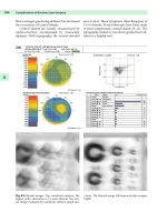
may be normal. DI is suspected when dilute urine is excreted in volumes greater than the fluid intake. Most cases have polyuria (UO
>
30 ml/kg/h), hypernatraemia and elevated plasma osmolality. Patients with a complete DI have urine volumes in excess of 10 l/day
with urine osmolality 50–200 mosmol/kg and specific gravity of 1.001–1.005. They are sensitive to exogenous ADH. Partial DI
patients have smaller urine volumes and urine osmolalities between 290 and 600 mosmol/kg. The diagnosis can be confirmed with
the ADH synthetic analogue desmopressin (DDAVP) which causes the urine output to fall and urine osmolality to increase. Other
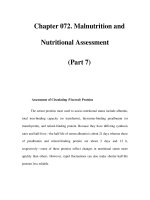
causes of polyuria include osmotic diuretics (e.g. mannitol, radiocontrast agents), severe hyperglycaemia and fluid overload.
Comparison of plasma and urine osmolalities may help (Figure 18.1).
Treatment consists of adequate fluid resuscitation and correction of electrolytes with regular monitoring of intake and output. Severe
polyuria can be treated with IV desmopressin 1–4 μg/day. The duration of DDAVP is dose dependent and ranges from 8 to 12 h.
SIADH due to inappropriate release of ADH from damaged posterior pituiary cells is much more rare but has also been reported,
usually in the late postoperative period after discharge.
39
Figure 18.1
Plasma and urine osmolalities (redrawn
from Moses AM, Blumenthal SA, Streeten DH.
Acid–base and electrolyte disorders associated
with endocrine disease: pituitary and thyroid.
In: Arief AI, Defronzo RA (eds) Fluid, electrolyte
and acid–base disorders. Churchill Livingstone,
New York, 1985,
pp
851
–
892
)
.
CSF rhinorrhoea associated with diaphragmatic lesions may occur and be complicated by bacterial meningitis from nasal flora. Some
centres routinely continue antibiotic prophylaxis.
Vision changes are rare and cranial nerve palsies affecting the third, fourth and sixth cranial nerves are usually transient if they occur.
Anterior pituitary function after surgery depends on the size of the tumour.
40
Lesions to adjacent anatomical structures occur, including bleeding from the carotid artery and cavernous sinus or hypothalamic
lesions leading to hyperthermia, electrolyte disturbance and cerebral coma.
41
Venous air embolism is rare with the supine patient.
Sinusitis, fractures from surgical instruments, deviated nasal septum with obstruction of nasal ventilation and dental problems have
all been described.
Stereotactic Bio
p
s
y
Image-guided stereotactic surgery allows the biopsy or destruction of intracranial lesions without the need for craniotomy. It gives
greater accuracy and diagnostic yield in tissue sampling and may be of great benefit in deciding the question of histology prior to a
major surgical undertaking.
42
It is most commonly used for biopsy of lesions seen on CT and to locate small lesions difficult to find
visually, especially lesions deep to the brain's surface. It also enables the surgeon to
Pa
g
e 263
b
iopsy lesions adjacent to vital centres more safely, thereby avoiding the risks of freehand techniques.
Stereotaxis is also used for radiotherapy with radioactive wires placed through plastic cannulae or sheaths.
M
ethod
A mechanical frame is attached to the patient's head and a scan obtained as usual. The scanner's computer calculates the 3D
coordinates of points in the brain and relates them to the stereotactic space outlined by the frame. Simple calculations then convert
these radiographic coordinates to the coordinate system used by the frame. These coordinates are set on the micrometers on the frame
through which biopsy instruments can be directed with an accuracy of 1 mm.
43
Many of these procedures can be carried out under local anaesthesia (together with supplementation by IV sedation). This has the
advantage of continuous patient evaluation during biopsy. General anaesthesia is used for patient comfort in prolonged procedures
and particularly in children.
A potential complication of the technique is intracerebral haemorrhage from accidental puncture of major arteries, sinuses and
b
ridging veins. This may require urgent transfer of the patient for CT or even urgent craniotomy.
43
A
naesthesia
Anaesthetic technique aimed at cardiovascular and respiratory stability throughout will maintain a stable CBF and thus ICP and CPP.
Localized or general oedema is thus minimized and therefore lesion movement with respect to the skull. Bone and Bristow
44
describe
results from a year's experience with total intravenous anaesthesia for stereotactic surgery. The technique consists of a combination
of propofol, fentanyl, vecuronium and O
2
in air. It provides good cardiovascular stability and adequate depth of anaesthesia for all
stages in stereotaxis and a reliable rapid recovery. Furthermore, it can also be useful for transferring head-injured patients from the
ICU to the CT scanner. Propofol is also believed to be the ideal agent for easing the discomfort of siting the stereotactic frame if the
p
rocedure to follow requires the patient to be awake, e.g. thalamotomy.
45
After careful preoperative assessment, a short-acting benzodiazepine premed is given and large-
b
ore IV access is established. A level
of monitoring is chosen that maintains patient care in transit, commonly NIBP, SpO
2
, EGG, EtCO
2
. All monitors, pumps, O
2
cylinder
and ventilator are fixed to the trolley securely if transported and arranged for easy observation from outside the CT scanner.
Bone and Bristow
44
used IV lignocaine 1 mg/kg, fentanyl 2 μg/kg, propofol 1–2 mg/kg over 30 s, vecuronium 0.1 mg/kg followed by
intubation and positive pressure ventilation with a portable ventilator with fresh gas flow titrated against EtCO
2
. Anaesthesia was
then maintained by a propofol infusion according to Roberts et al,
46
i.e. 10 mg/kg/h for the first 10 min, followed by 8 mg/kg/h for
the next 10 min and 6 mg/kg/h thereafter and adjusted according to depth of anaesthesia. A volatile agent is not required. This has the
advantage of avoiding the need for an anaesthetic machine during transfer and avoiding the adverse effects of a volatile agent on the
cerebral vasculature. The problem of pollution and scavenging requirements is also overcome. Muscle relaxant was provided with
b
oluses or infusion of vecuronium.
After skin preparation, a minimum of four cranial fixation pins are applied to the skull. This stimulus requires careful attention to
anaesthetic technique to avoid increases in ICP (a bolus of propofol or opiate is commonly used at this point to deepen anaesthesia
quickly and overcome any sympathetic response). The propofol infusion is stopped just prior to reversal with neostigmine and
glycopyrrolate. The patient usually remains supine throughout.
The advantages of this total intravenous technique are the haemodynamic and respiratory stability during the stereotactic procedures,
patient transfer between CT and operating theatre and after use of radiocontrast in CT. It is a simple technique and the same
anaesthetic technique can be continued in the operating room.
Postoperative patient care includes frequent neurological monitoring. There is a potential risk of air embolism through transgressed
venous channels in bone.
Overall, there is a low incidence of nausea and vomiting and morbidity and mortality are very low.
44
Ventriculo
p
eritoneal Shunts
Hydrocephalus is usually due to obstruction of CSF flow giving rise to increases in volume and thus intracranial pressure. Surgical
treatment aims are to provide a shunt through which excess fluid can drain to a site to be absorbed. The most common drains are to
the peritoneal cavity but the right atrium, cisterna magna and pleural cavity are also sometimes used.
Page 264
VP shunts drain CSF from the fourth or lateral ventricles to the peritoneal cavity. Systems usually involve three components: a
ventricular catheter, a one-way valve and a distal catheter. Other devices may also be used (on–off valves, siphon control devices and
chambers for flushing the system). The ventricular catheter is inserted through a burr hole (usually on the right), connected to the
one-way valve that determines the draining pressure from the ventricle and then connected to the peritoneal catheter, passed over the
chest wall and inserted into the peritoneal cavity through a small incision.
P
osition
Children
For VP shunts the child is placed supine with the head turned to the opposite side for the occipital insertion of the ventricular
catheter. A towel is placed under the nape of the neck so the head, neck and abdomen are in one plane for the passage of the
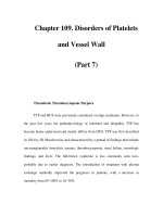
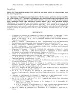
subcutaneous tunneller for shunt placement (Fig. 18.2). Children are usually positioned with towels, rolls and pillows rather than
table adjustment. The tunneller theoretically reduces chest wall compliance and may cause underventilation or dangerous increases in
airway pressures.
Adults
Adults are placed supine with the head turned to the opposite side and a roll or pad under the ipsilateral shoulder and neck giving a
straight path for the tunnelling device.
The shunt tubing is passed subcutaneously from the scalp incision over the occipitoparietal region to the abdominal incision made
either in the midline above the umbilicus or just lateral to and above the umbilicus. Local anaesthetic with adrenaline may be injected
along the incision lines. Intravenous antibiotics are given perioperatively.
47
Figure 18.2
The shunt is passed subcutaneously from the
cranial end to the peritoneal incisions through a
shunt-passing device (redrawn from reference
47
).
A
naesthetic Managemen
t
The child presenting for insertion of VP shunt or shunt revision must be considered to have overt or potentially raised intracranial
p
ressure. Anaesthetic technique should be tailored to minimize sympathetic responses and steps taken to reduce intracranial pressure.
Preoperative respiratory depressant drugs are avoided in the presence of a raised ICP. A smooth induction is desirable as crying will
further increase the ICP. An inhalation induction is acceptable if the fontanelle and sutures are opened, otherwise an intravenous
induction is preferred. EMLA cream is used over the venepuncture site. For induction, thiopentone and propofol have the advantage
of decreasing cerebral metabolic rate and cerebral blood flow.
Vecuronium has the fewest cardiovascular effects during induction but the other muscle relaxants have also been used satisfactorily.
Other measures may be used to limit the pressor responses to laryngoscopy and intubation, such as the use of a bolus of lignocaine or
fentanyl. This, however, seems less of a problem in children with open sutures than in adults. A RAE tube or armoured tube is used
either orally or nasally and fixed well to prevent movement. The chest is auscultated for breath sounds bilaterally after the patient is
p
laced in their final position. The breathing circuit is secured firmly to prevent dragging on the endotracheal tube. The eyes and limbs
are protected from injury.
Anaesthesia is maintained with a total intravenous technique using propofol or with volatile agents and IV analgesics such as
fentanyl or alfentanil or with combinations of these.
TIVA with propofol decreases CMRO
2
, may give neuroprotection, reduces relaxant needs and gives rapid wakening postoperatively.
Isoflurane also decreases CMRO
2
and relaxant requirements and gives easy control of blood pressure if required. Opioids are
adjusted to avoid postoperative respiratory depression. Their use increases the incidence of postoperative nausea and vomiting.
Moderate hypocapnia using capnography to monitor ventilation reduces brain mass by cerebral vasoconstriction.
Surgical infiltration with adrenaline reduces bleeding but the addition of at least 1% lignocaine provides scalp analgesia and helps
reduces any hypertensive response.
27
Pa
g
e 265
Intravenous fluids without glucose are used as elevated levels of glucose worsen outcome.
At the conclusion of surgery, the pharynx is cleared of secretions under direct vision, muscle relaxants reversed and the patient
extubated, avoiding coughing if possible.
References
1. Messick JM, Laws ER, Abboud CF. Anesthesia for transsphenoidal surgery of the hypophyseal region. Anesth Analg 1978; 57:
206
–
215.
2. Riskind PN, Martin JB. Functional anatomy of the hypothalamic-anterior pituitary complex. In: DeGroot LJ (ed) Endocrinology,
3rd edn. WB Saunders, Philadelphia, 1995, pp 151
–
159.
3. Ho KY, Sullivan CE. Sleep apnea in acromegaly. Ann Intern Med 1991; 115: 527
–
532.
4. Ho HI, Weissberger JA. The antinaturetic effect of biosynthetic human growth hormone in man invoves activation of the renin-
angiotensin mechanism. Metabolism 1990; 39: 133
–
137.
5. Hirsch EZ, Sloman JG, Martin FIR. Cardiac function in acromegaly. Am J Med Sci 1969; 257: 1
–
8.
6. Savage DD, Henry WL, Eastman RC. Echocardiographic assessment of cardiac anatomy and function in acromegalic patients. Am
J Med 1979; 67: 823
–
829.
7. Lim MJ, Barkan AL, Buda AJ. Rapid reduction of left ventricular hypertrophy in acromegaly after suppression of growth hormone
hypersecretion. Ann Intern Med 1992; 117: 719
–
726.
8. Young ML, Hanson CW. An alternative to tracheostomy following transsphenoidal hypophysectomy in a patient with acromegaly
and sleep apnoea. Anesth Analg 1993; 76: 446
–
449.
9. Southwick JP, Katz J. Unusual airway difficulty in the acromegalic patient – indications for tracheostomy. Anesthesiology 1979;
51: 72
–
73.
10. Chan VWS, Tindall S. Anaesthesia for transsphenoidal surgery in a patient with extreme gigantism. Br J Anaesth 1988; 60: 464–
468.
11. Singelyn FJ, Scholtes JL. Airway obstruction in acromegaly: a method of prevention. Anaesth Inten Care 1988; 16: 491
–
492.
12. Perks WH, Horricks PM, Cooper RA et al. Sleep apnoea in acromegaly. BMJ 1980; 280: 894
–
897.
13. Burke CW, Beardwell CG. Cushing's syndrome: an elevation of the clinical usefulness of urinary free cortisol and other urinary
steroid measurements in diagnosis. Quart J Med 1973; 42: 175
–
204.
14. Hardy J. Cushing's disease: 50 years later. Can J Neurol Sci 1982; 9: 375
–
380.
15. Schulte HM, Petersenn S. Cushing's disease – clinical findings and endocrinology. In: Landolt AM, Vance ML, Reilly PL (eds)
Pituitary adenomas. Churchill Livingstone, NewYork, 1996, pp 101
–
110.
16. Liddle GW. Test of pituitary-adrenal suppressibility in the diagnosis of Cushing's syndrome. J Clin Endocrinol Metab 1960; 20:
1539
–
1560.
17. Murray DK, Hill ME, Nelson DH. Inhibitory action of sphigosine, sphinganine and dexamethasone on glucose uptake: studies
with hydrogen peroxidase and phorbol esters. Life Sci 1990; 46: 1843
–
1849.
18. Ritchie CM, Sheridan B, Fraser R et al. Studies on the pathogenesis of hypertension in Cushing's disease and acromegaly. Quart J
Med 1990; 76: 855
–
867.
19. Kern EB, Pearson BW, McDonald TJ, Laws ER. The transseptal approach to lesions of the pituitary and parasellar regions.
Laryngoscope 1979; 89 (5 pt 2 suppl 15): 1
–
34.
20. Vance ML, Thorner OM. Prolactin and hyperprolactinaemic syndromes and management. In: DeGroot L (ed) Endocrinology.
WB Saunders, Philadelphia, 1989, pp 408.
21. Von Werder K. Prolactinoma: clinical findings and endocrinology. In: Landolt AM, Vance ML, Reilly PL (eds) Pituitary
adenomas. Churchill Livingstone, New York, 1996, pp 111
–
126.
22. Katznelson L, Klibanski A. Endocrine-inactive, FSH, LH and -subunit adenomas: clinical findings and endocrinology. In:
Landolt AM, Vance ML, Reilly PL (eds) Pituitary adenomas. Churchill Livingstone, New York, 1996, pp 127
–
131.
23. McFadzean RM, Teasdale GM. Pituitary apoplexy. In: Landolt AM, Vance ML, Reilly PL (eds) Pituitary adenomas. Churchill
Livingstone, NewYork, 1996, p 485.
24. Pollack IF, Lunsford LD, Slamovits TL, Gunerman LW, Levine G, Robinson AG. Stereotaxic intracavitary irradiation for cystic
craniopharyngiomas. J Neurosurg 1988; 68: 227
–
233.
25. Cooke RS, Jones RAC. Experience with the direct transnasal transsphenoidal approach to the pituitary fossa. Br J Neurosurg
1994; 8: 193
–
196.
26. Peter M, DeTribolet N. Visual outcome after transsphenoidal surgery for pituitary adenomas. Br J Neurosurg 1995; 9: 151
–
157.
27. Abou-Madi MN, Trop D, Barnes J. Aetiology and control of cardiovascular reactions during transsphenoidal resection of
p
ituitary microadenomas. Can Anaesth Soc J 1980; 27: 491
–
495.
28. Weinberg S, Phillips L, Twersky R, Cottrell JE, Braunstein KM. Hypercoagulability in a patient with a brain tumor.
Anesthesiology 1984; 61: 200
–
202.
29. Salem M, Tainsh RE Jr, Bromberg J, Loriaux DL, Chernow B. Perioperative glucocorticoid coverage. A reassessment 42 years
after emergence of a problem. Ann Surg 1994; 219: 416
–
425.
30. Campkin TV: Radial artery cannulation. Potential hazard in patients with acromegaly. Anaesthesia 1980; 35: 1008
–
1009.
31. Grosslight K, Foster R, Colohan AR, Bedford RF. Isoflurane for neuroanesthesia: risk factors for increases in intracranial
p
ressure. Anesthesiology 1985; 63: 533
–
536.
Pa
g
e 266
32. Messick JM Jr, Newberg LA, Nugent M, Faust RJ. Principles of neuroanesthesia for the neurosurgical patient with CNS
p
athophysiology. Anesth Analg 1985; 64: 143
–
174.
33. Gupta S, Heath K, Matta BF. The effect of incremental doses of sevoflurane on cerebral pressure autoregulation in humans: a
transcranial doppler study. Br J Anaesth 1997; 79: 469
–
472.
34. Eger EI II, Lampe GH, Wauk LZ, Whitendale P, Calahan MK, Donegan JH. Clinical pharmacology of nitrous oxide: an
argument for its continued use. Anesth Analg 1990; 71: 575
–
585.
35. Lampe GH, Donegan JH, Rupp SM et al. Nitrous oxide and epinephrine induced arrythmias. Anesth Analg 1990; 71: 602
–
605.
36. Nicholson KEA, Rogers JEG. Cocaine and adrenaline paste: a fatal complication. BMJ 1995; 311: 250
–
251.
37. Kruse A, Astrup J, Cold GE, Hansen HH. Pressure and blood flow in pituitary adenomas measured during transsphenoidal
surgery. Br J Neurosurg 1992; 6: 333
–
342.
38. Landolt AM, Schiller Z. Surgical technique – transsphenoidal approach. In: Landolt AM, Vance ML, Reilly PL (eds) Pituitary
adenomas. Churchill Livingstone, New York, 1996, pp 315
–
331.
39. Tymms J, Griffith HB, Hartog M, Clark JDA, Reckless JPD. Pituitary surgery and inappropriate antidiuretic hormone secretion. J
Roy Soc Med 1992; 85: 302.
40. Maclanahan CS, Christy JH, Tindall GT. Anterior pituitary function before and after transsphenoidal microsurgical resection of
p
ituitary tumors. Neurosurgery 1978; 3: 142
–
144.
41. Fahlbusch R, Buchfelder M. Surgical complications. In: Landolt AM, Vance ML, Reilly PL (eds) Pituitary adenomas. Churchill
Livingstone, NewYork, 1996, pp 394
–
408.
42. Apuzzo MJL, Chandrasoma PT, Cohen D, Zee CJ, Zelman V. Computed imaging stereotaxy: experience and perspective related
to 500 procedures applied to brain masses. Neurosurgery 1987; 20: 930
–
937.
43. Salcman M. The surgical management of gliomas. In: Tindall GT, Cooper PR, Barrow PL (eds) The practice of neurosurgery.
Williams & Wilkins, Baltimore, 1996, pp 649
–
670.
44. Bone ME, Bristow A. Total intravenous anaesthesia in stereotactic surgery – one years clinical experience. Eur J Anaesthesiol
1991; 8: 47
–
54.
45. Anderson BJ, Marks PV, Futter ME. Propofol
–
contrasting effects in movement disorders. Br J Neurosurg 1994; 8: 387
–
388.
46. Roberts FL, Dixon J, Lewis GTR, Tackley RM, PrysRoberts C. Induction and maintenance of propofol anaesthesia. Anaesthesia
1988; 43(suppl): 14
–
17.
47. Roth PA, Cohen AR. Management of hydrocephalus in children. In: Tindall GT, Cooper PR, Barrow PL (eds) The practice of
neurosurgery. Williams & Wilkins, Baltimore, 1996, pp 2707
–
2728.
48. Trouillas J, Girod C. Pathology of pituitary adenomas. In: Landolt AM, Vance ML, Reilly PL (eds) Pituitary adenomas.
Churchill Livingstone, New York, 1996, pp 27
–
46.
Pa
g
e 267
19—
Anaesthesia for Posterior Fossa Sur
g
er
y
Catherine Duffy
Anatomy 269
Posterior Fossa Lesions 269
Patient Positioning 272
Complications of the Sitting Position 273
Central Nervous System Monitoring 276
Anaesthetic Managemen
t
277
Conclusion 278
References 278
Pa
g
e 269
The posterior fossa is a small, rigid compartment which houses the cerebellum, pons, medulla oblongata and fourth ventricle. Life-
threatening symptoms can result from compression of these vital structures. Because of the narrow outflow of cerebrospinal fluid
(CSF), intracranial hypertension can develop suddenly. The common goal of the anaesthetist and surgeon is an operation in which the
need for retraction, with consequent tissue damage, is minimized. This is achieved by optimizing the surgical approach and patient
p
osition and by minimizing congestion of cerebral vasculature.
Anatom
y
The anterior portion of the posterior fossa is the dorsum sellae and basilar portion, or clivus, of the occipital bone. Laterally are the
petrous portions of the temporal bones. The roof is the tentorium cerebelli and the floor contains the foramen magnum. Cranial nerve
V arises from the pons and cranial nerves VI-XII from the medulla. The posterior fossa is traversed by the transverse, superior
p
etrosal, occipital and sigmoid sinuses.
1
Posterior Fossa Lesions
Tumours
Intraaxial lesions tend to be malignant whereas extraaxial lesions are usually benign. Posterior fossa tumours are more common in
children than in adults, with 55% occurring in children and 45% in adults. 54–70% of childhood brain tumours originate in the
posterior fossa. Primitive neuroectodermal tumours (PNET)/medulloblastomas, ependymomas and astrocytomas all occur in infants
and children. Mixed gliomas are unique to childhood. In the adult population, 15–20% of brain tumours occur in the posterior fossa.
Metastases, haemangioblastomas and lymphoma are all more common in adults than in children.
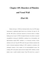
Figure 19.1
Acute h
y
droce
p
halus in a child with medulloblastoma.
Si
g
ns and S
y
m
p
toms
Headache and vomiting are the most common symptoms in patients with posterior fossa tumours. Hemicerebellar lesions are
characterized by unilateral ataxia, dysmetria, intention tremor and hypotonia. Vermian lesions tend to present with a wide-based gait,
truncal ataxia and abnormal extraocular movements. Brainstem tumours present with bulbar palsies, secondary to cranial nerve
dysfunction, and contralateral motor and sensory deficits. Depending on the level of the lesion, respiratory changes may occur.
When a posterior fossa lesion is complicated by obstructive hydrocephalus, headache and vomiting are exacerbated. The patient
develops an unsteady gait, mental state changes, diplopia, incontinence and signs of meningism.
Intraaxial Tumours
P
NET/Medulloblastoma
These tumours represent 25% of childhood intracranial tumours. They occur mainly in the 5–9-year-old age group with equal sex
distribution. Because of their midline position, they tend to present early with intracranial hypertension due to obstructive
hydrocephalus (Figure 19.1).
Cerebellar Astroc
y
toma
These account for 12–28% of childhood brain tumours. They are relatively benign lesions with very good long-term survival
following surgical resection.
B
rainstem Glioma
These represent 25–30% of posterior fossa tumours in childhood. Most are low-grade astrocytomas. They tend to be slow growing
with insidious onset of symptoms. The exception is pontine gliomas which are malignant and present with a short and aggressive
history.
Pa
g
e 270
Ep
end
y
moma
These lesions tend to occur in childhood and adolescence, with 50% occurring in children less than three years old. They arise from
the floor of the fourth ventricle. Treatment involves surgical removal and adjuvant irradiation. Complete removal may not always be
p
ossible if there is brainstem involvement.
Choroid Plexus Papilloma/Carcinoma
These are rare lesions arising from the fourth ventricle. They account for only 3% of paediatric intracranial tumours. They
hypersecrete CSF and are associated with communicating hydrocephalus.
D
ermoid Tumour
These are childhood tumours involving the cerebellar midline. They often present with recurrent episodes of bacterial or aseptic
meningitis due to rupture. Eventually arachnoid scarring may result in hydrocephalus. The surgical goal is total excision.
H
aeman
g
ioblastoma
These are histologically benign tumours which can occur throughout the central nervous system but are most commonly seen in the
posterior fossa. They account for 7–12% of adult posterior fossa tumours. They may occur as part of the Von Hippel-Lindau
syndrome, an inherited disorder characterized by visceral cysts and tumours, retinal haemangiomas and polycythaemia. Due to their
vascular nature, preoperative embolization is desirable. Complete surgical resection is usually curative.
M
etastatic Tumour
These are the most common posterior fossa tumours in adults. The usual primary tumour sites are lung, breast, skin and kidney.
Surgical removal is indicated when the lesions cause a mass effect. In the case of a solitary lesion, excision may improve survival.
Extraaxial Tumours
S
chwannoma
An acoustic neuroma is a schwannoma arising from the vestibular portion of cranial nerve VIII. The usual presenting symptom is
hearing loss. Schwannomas do not invade tissues but tend to displace them. They may cause compression of cranial nerves V and
VII, manifesting as tic douloureux and hemifacial spasm respectively. If large, they may be associated with hydrocephalus. Acoustic
neuromas occur in the fifth and sixth decades with equal sex distribution. Total resection is usually feasible. Acoustic neuromas may
occur as part of a type II neurofibromatosis syndrome in which case they are multiple and behave more aggressively than single
lesions.
Schwannomas may also affect cranial nerves V, VII, IX, X and XII.
M
eningioma
Ten percent of all meningiomas are in the posterior fossa. They may arise from the petrous ridge of the temporal bone and extend
into the cerebellopontine angle. Other possible sites are the clivus, tentorium cerebelli, fourth ventricle and, rarely, foramen magnum.
Because they are vascular lesions, preoperative embolization is usually attempted.
Clival Tumour
These are rare tumours and comprise chordomas, chondrosarcomas and carcinomas. Surgery is technically difficult and local
recurrence rates are high.
A
rachnoid Cys
t
These are common congenital lesions affecting sites throughout the central nervous system. In the posterior fossa, they are found
around the foramen magnum, the cerebellopontine angle and in the pineal region. They are often asymptomatic. Should a mass effect
occur, surgical treatment involves marsipualization of the cyst.
Glomus
J
u
g
ulare Tumour
These are tumours of the extraadrenal paraganglion system. They arise in the region of the middle ear and may extend through the
j
ugular foramen or through the petrous bone to the cerebellopontine angle.
Ep
idermoid C
y
s
t
These are benign tumours arising from epithelial cells. They are usually found in the cerebellopontine angle where they may enlarge
to cause cranial nerve or brainstem dysfunction.