Cataract and Refractive Surgery - part 7 pptx
Bạn đang xem bản rút gọn của tài liệu. Xem và tải ngay bản đầy đủ của tài liệu tại đây (527.65 KB, 18 trang )
8
104 Complications of Excimer Laser Surgery
blows nitrogen gas during ablation has decreased
the occurrence of central islands.
Central islands are usually characterized by
undercorrection accompanied by monocular
diplopia. With topography, the central elevated
area is clear. ese symptoms oen disappear in
3 to 6 months. Several attempts have been made
to treat symptomatic central islands [9, 21]. e
topography-linked or wavefront-guided laser ab-
lation is a helpful tool.
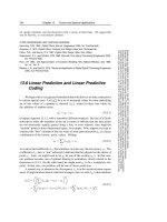
Fig. 8.2 Retinal images. Top: wavefront analysis. e
higher order aberration is 1.14 μm.
Bottom: the reti-
nal image evaluated by wavefront analysis (pupil size,
3 mm). e blurred image le improved aer surgery
(right)
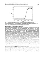
Fig. 8.3 Orbscan (irregular astigmatism). Top : kera-
tometric map shows irregular astigmatism. e visual
acuity is 0.7 (1.5 × sphere: –0.5 D; cylinder: –2.0 D;
axis: 153°). e patient complained of monocular dip-
lopia during the day and at night. Bottom: aer wave-
front-guided laser ablation, the uncorrected visual
acuity improved to 1.5
8.3 Intraoperative Complications 105
8
106 Complications of Excimer Laser Surgery
8.3.4 Undercorrection
Undercorrection is the result of incorrect preop-
erative evaluation of refraction, excessive mois-
ture in the stromal bed, decentration, and prob-
lems with laser calibration. If the preoperative
refraction is unstable, the refraction should be
Fig. 8.4 Retinal images. Top: wavefront analysis. e
higher order aberration is 0.66 μm.
Bottom: the reti-
nal image evaluated by wavefront analysis (pupil size,
3 mm). e blurred image (le) improved aer surgery
(right)
repeated aer an interval and the ablation post-
poned. If the patient uses hard contact lenses, the
refraction should be done at least 2 weeks aer
the patient stops wearing them. Because the re-
fraction is the key to achieving successful results,
the examination should be repeated until the sur-
geon is comfortable with the status of the refrac-
tion. Some patients require more than 2 months
to obtain a stable refraction aer wearing contact
lenses. If the cycloplegic refraction diers sub-
stantially from the non-cycloplegic refraction,
the refraction should be repeated using both val-
ues at another visit. Using uncertain refractive
results will cause unnecessary complications.
Excessive moisture in the stromal bed can re-
sult in undercorrection. is oen happens when
the surgeon is inexperienced. Undercorrection
also can result if the patient’s xation is poor and
the laser is not ideally applied. e laser calibra-
tion is also important. e laser operator should
be aware of the condition of the laser. e laser
should be recalibrated with each use.
Enhancement usually results in good refrac-
tive outcomes. e timing of the enhancement
surgery depends on the cause of the undercor-
rection. If the refraction is stable, retreatment
can be done at any time. To avoid repeating the
problems that arose in the initial surgery, the
cause should be well considered. In addition,
the postoperative corneal thickness should be
conrmed. If the total corneal thickness is less
than 400 μm, further laser ablation should be
avoided to prevent corneal ectasia. e patient’s
age also should be considered. If the patient is
over 40 years, monovision may be an option.
A surgeon can perform unilateral enhancement
and see both far and near visual function. Some
patients enjoy unplanned monovision.
8.3.5 Overcorrection
e causes of overcorrection are similar to those
of undercorrection: the accuracy of the refrac-
tion, the condition of the stromal bed, and the
laser calibration. Dryness of the stromal bed usu-
ally causes overcorrection. If the surgeon delays
starting the laser ablation, the cornea becomes
dry and the eect of the laser is intensied. Tran-
sient overcorrection aer PRK is a well-known
phenomenon. Although this problem has de-
creased with the latest generations of excimer
lasers, the changes in the refraction should be
observed over time.
Patient age also plays an important role.
Younger patients can tolerate overcorrection;
however, older patients are very sensitive to over-
correction. Unfortunately, the risk factors for
overcorrection are age and attempted correction.
e higher these factors are, the more frequently
patients encounter this complication.
If overcorrection occurs aer PRK, the admin-
istration of steroid eye drops should be stopped.
Some physicians also recommend stopping the
use of articial tears. Regarding hyperopic cor-
rection, transient overcorrection is the goal be-
cause subsequent regression is common. Patients
should be well informed about this before sur-
gery.
e treatment of overcorrection aer the
treatment of myopia is mandatory. Recently,
the use of diclophenac eye drops with contact
lenses produced good results. If the correction
meets the desired target, the eye drops should be
stopped immediately. If this does not change the
results, excimer laser with hyperopic correction
or holmium laser thermoplasty is frequently per-
formed.
Summary for the Clinician
■
Preoperative examination of the refrac-
tion and calibration of the laser are fun-
damental to achieving the best visual
outcomes.
■
Corneal topography should be per-
formed even if the patient achieved good
visual outcome to conrm the ideal laser
ablation.
■
Wavefront-guided ablation is a helpful
tool in patients with decentered ablation
or irregular astigmatism.
8.4 Postoperative Complications
Even though patients achieve good outcomes,
some complications can develop later. Since pa-
8.4 Postoperative Complications 107
8
108 Complications of Excimer Laser Surgery
tients enjoy their improved uncorrected visual
acuity, even a slight decrease in visual acuity is
unacceptable. However, the cause of decreased
uncorrected visual acuity should be well evalu-
ated and the treatment planned.
8.4.1 Regression
Regression is a common problem for any laser
surgery including LASIK. If regression occurs,
retreatment is considered. Regression accompa-
nied by corneal haze requires a dierent treat-
ment approach, such as the application of steroid
eye drops, PRK, or phototherapeutic keratec-
tomy (PTK). Recently, the use of beta-blocker
eye drops to decrease the intraocular pressure
has achieved good results. e eect of improv-
ing the vision in these cases is still under dis-
cussion. Why beta-blocker eye drops work and
Latanoprost eye drops do not is a question for
future research. is approach does not work
in every case; however, it is worth trying beta-
blocker eye drops. e interval since the time of
laser surgery, patient gender and age, and the pre-
operative refraction are not correlated with the
amount of improvement. If this does not produce
a satisfactory result, retreatment is planned aer
conrming that the refraction is stable. Gener-
ally, enhancements should be planned at least
3 months aer the previous surgery. If the cor
-
rection exceeds 6 D, waiting more than 6 months
may be necessary to achieve a stable refraction.
8.4.2 Corneal Haze
Corneal haze is a well-known complication aer
PRK. Histopathologic and confocal microscopic
studies revealed that haze is induced by activa-
tion and proliferation of corneal keratocytes [3].
e haze usually appears 1–3 months aer sur
-
gery and gradually resolves within 1 year. With
slit-lamp microscopy, subepithelial haze can be
observed in the central area and classied from
grades 0 to 4 [16]. Recently, objective scoring
was introduced using digital images and con-
focal microscopy [2, 6, 10]. e incidence of
haze was higher with previous laser treatment
[30]; however, the incidence decreased with re-
cent technological advances that produced a
smoother ablation. e risk factors are greater
tissue ablation such as in the treatment of high
myopia, ultraviolet exposure, atopic dermatitis,
and autoimmune conditions [8, 12, 24]. Despite
the appearance of haze, most cases achieve good
visual acuity. If the haze becomes substantial, the
best-corrected visual acuity decreases with some
regression (Fig. 8.5). Problems may develop with
night vision and decreased contrast sensitivity
[13, 18]. Most surgeons use steroid eye drops
immediately or shortly aer laser treatment and
gradually taper the drops. Although the eects of
corticosteroid eye drops used clinically have been
positively or negatively reported, theoretical ben-
ets have been described in experimental stud-
ies. Special attention should be paid to the side
eects of corticosteroids, especially increased
intraocular pressure.
Recently, the eects of chilled irrigation solu-
tion and mitomycin C were reported. Mitomy-
cin C is used for glaucoma ltering surgery and
pterygium surgery and has been introduced into
laser surgery [17, 28, 34]. e concentration of
mitomycin C and the duration of its application
have been discussed; 0.01 mg/ml is the mini
-
mum concentration reported to be eective and
0.4 mg/ml is the maximum to avoid complica
-
tions [4]. e 0.02% concentration is widely
used. Aer laser application, a 6-mm diameter
Fig. 8.5 Corneal haze. e best corrected visual acuity
decreases
sponge is soaked in 0.02% mitomycin C diluted
with balanced saline solution (BSS) and applied
over the ablated cornea for 2–3 min. e eye
then is washed with BSS. Complications such as
thinning of the scleral tissue and delayed epithe-
lialization were reported in cases of glaucoma-l-
trating surgery and pterygium. An experimental
study showed dose-dependent corneal edema
and endothelial apoptosis. However, the prophy-
lactic use of 0.02% mitomycin C in laser surgery
seems to be safe and eective at preventing haze
[33] and achieved better visual acuity [7]. Mito-
mycin C is also used to treat haze [20, 29] in the
same technique used during PRK, or the drug
can be administered as an eye drop. Use of vita-
min C and amniotic membranes also have been
reported; however, the eects need to be studied
further [33, 36].
8.4.3 Delayed Epithelialization
Following PRK, LASEK, and Epi-LASIK, ban-
dage contact lenses are applied. Aer 3 days,
most eyes achieve re-epithelialization and the
contact lenses can be removed. e preservative
in the eye drops sometimes delays the recovery of
the epithelium. e use of eye drops without pre-
servatives is preferable. If the eye developed epi-
thelial problems due to the toxicity of eye drops,
the drops should be discontinued.
8.4.4 Infections
Infections aer refractive surgery are rare, but
can be the most severe complications aer any
ophthalmic surgery. Regarding laser surgery,
corneal opacity remains even though the infec-
tion was treated with antibiotics. e common
cause is staphylococcus and mycobacteria; the
prophylactic application of antibiotics is recom-
mended [15].
An epithelial defect is the optimal site for
the development of a bacterial infection. If the
process of re-epithelialization is prolonged, spe-
cial steps should be taken to avoid infections. In
LASIK cases, the focus of the infection is under
the ap and the risk of perforation increases.
Cultures should be performed to conrm the
bacteria in severe cases; however, topical antibi-
otics should be started immediately. Liing the
ap and irrigation are necessary in certain cases.
Aer treatment, PTK can be performed if the
opacity remains on the corneal surface. Penetrat-
ing or lamellar keratoplasty is needed in patients
with poor visual acuity. Infection usually results
in poor corrected visual acuity.
8.4.5 Adverse Eects
on the Corneal Endothelium
Experimental and clinical studies have shown
no side eects from refractive procedures on the
corneal endothelium [3, 11]. One study reported
that the number of endothelial cells decreased af-
ter a tranquilizer was administered to the patient
before PRK [25].
8.4.6 Corneal Ectasia
Aer LASIK was introduced, a new complica-
tion, keratectasia, was reported [5, 26, 31] in
which continuous regression with irregular
astigmatism develops. e risk factors are form
fruste keratoconus, thin cornea with high myo-
pia, and pellucid marginal degeneration. Preop-
erative evaluation with corneal topography and
pachymetry are necessary. e postoperative
corneal condition should be assessed to maintain
a corneal thickness greater than 400 μm or a re
-
sidual stromal bed greater than 250–300 μm. En
-
hancements performed without measuring the
corneal thickness can cause ectasia.
Orbscan can be performed to diagnose kera-
tectasia. e posterior oat map shows obvious
thinning. If this complication occurs, hard con-
tact lenses are tted. If the vision cannot be cor-
rected with contact lenses, ICR or cross-linking
may be performed, followed if not successful by
corneal transplantation. If the surgeon does not
recognize the corneal thinning and continues to
treat with the excimer laser to improve the vi-
sion, the cornea may be perforated.
8.4 Postoperative Complications 109
8
110 Complications of Excimer Laser Surgery
Summary for the Clinician
■
Some postoperative complications are
well treated with eye drops.
■
Regarding postoperative complications
concerning the refractive error, en-
hancement should be considered when
the refraction is conrmed to be stable.
■
Before enhancement, the corneal thick-
ness and shape should be considered.
■
Some postoperative complications are
related to the failure of the indication.
References
1. Alkara N, Genth U, Seiler T. Diametral abla-
tion—a technique to manage decentered photore-
fractive keratectomy for myopia. J Refract Surg
1999;15:436–440.
2. Allerman N, Charmon W, Silverman RG, et al.
High-frequency ultrasound quantitative analysis
of corneal scarring following excimer laser kera-
tectomy. Arch Ophthalmol 1993;111:968–973.
3. Amm M, Wertzel W, Winter M, et al. Histopatho-
logical comparison of photorefractive keratec-
tomy and laser in situ keratomileusis in rabbits. J
Refract Surg 1996;12:758–766.
4. Ando H, Ido T, Kawai Y, et al. Inhibition
of corneal wound healing. Ophthalmology
1992;99:1809–1814.
5. Argento C, Cosentino MJ, Tytium A, et al. Cor-
neal ectasia aer laser in-situ keratomileusis. J
Cataract Refract Surg 2001;27:1440–1448.
6. Braustein RE, Jain S, McCally RL, et al. Objec-
tive measurement of corneal light scattering af-
ter excimer laser keratectomy. Ophthalmology
1996;103:439–443.
7. Carons F, Vigo L, Scadola E, Vacchini L. Evaluation
of the prophylactic use of mitomycin C to inhibit
haze formation aer photorefractive keratectomy.
J Cataract Refract Surg 2002;28:2088–2095.
8. Carson CA, Taylor HR Excimer laser treatment
for high and extreme myopia. Arch Ophthalmol
1995; 113:431–436.
9. Castill A, Romero F, Martin-Valverde JA, et al.
Management and treatment of steep islands aer
excimer laser photorefractive keratectomy. J Re-
fract Surg 1996;12:15–20.
10. Chew SJ, Beuerman RW, Kaufman HE, et al. In
vivo confocal microscopy of corneal wound heal-
ing aer excimer laser photorefractive keratec-
tomy. CLAO J 1995;25:273–280.
11. Colin J, Cochener B, Le Floch G. Corneal en-
dothelium aer PRK and LASIK. J Refract Surg
1996;12:674.
12. Corbett MC, O’Brart DPS, Warburton FG, Mar-
shall J. Biological and environmental risk factors
for regression aer photorefractive keratectomy.
Ophthalmology 1996;103:1381–1391.
13. Corbett MC, Prydol JI, Verma S, et al. An in vivo
investigation of the structures responsible for cor-
neal haze aer photorefractive keratectomy and
their eect on visual function. Ophthalmology
1996;103:1366–1380.
14. Doanne JG, Cavanaugh TB, Durrie DS, Hassa-
nein KH. Relation of visual symptoms to topo-
graphic ablation zone decentration aer excimer
laser photorefractive keratectomy. Ophthalmol-
ogy 1995;102:42–47.
15. Donnefeld ED, O’Brien TP, Solomon R, et al.
Infectious keratitis aer photorefractive keratec-
tomy. Ophthalmology 2003;110:740–747.
16. Fantes FE, Hanna KD, Waring GO III, et al.
Wound healing aer excimer laser keratomileusis
(photorefractive keratectomy) in monkeys. Arch
Ophthalmol 1990;108:665–675.
17. Gambato C, Ghirlando A, Moretto E, et al. Mito-
mycin C modulation of corneal wound healing af-
ter photorefractive keratectomy in highly myopic
eyes. Ophthalmology 2005;112:208–218.
18. Hersh PS, Stulting RD, Steinert RF, et al. e
Summit PRK Study Group. Results of phase III
excimer laser photorefractive keratectomy for
myopia. Ophthalmology 1997;104:1535–1553.
19. Krueger R, Saedy NF, McDonnell PJ. Clinical
analysis of steep central islands aer excimer laser
photorefractive keratectomy. Arch Ophthalmol
1996;114:377–381.
20. Majmudar PA, Forstot SL, Dennis RF, et al. Topi-
cal mitomycin C for subepithelial brosis af-
ter refractive corneal surgery. Ophthalmology
2000;107:89–94.
21. Manch EE, Maloney RK, Smith RJ. Treatment of
topographic central islands following refractive
surgery. J Cataract Refract Surg 1998;24:464–470.
22. Mulhern MC, Foley-Nolan A, O’Keefe M, et al.
Topographical analysis of ablation centration
aer excimer laser photorefractive keratectomy
and laser in situ keratomileusis for high myopia. J
Cataract Refract Surg 1997;23:488–494.
23. Nagy ZZ, Hiscott P, Seitz B, et al. Ultraviolet-
B enhances corneal stromal response to 193-
nm excimer laser treatment. Ophthalmology
1997;104:375–380.
24. Nakaya-Onishi M, Kiritoshi A, Hasegawa T, et al.
Corneal endothelial cell loss aer excimer laser
keratectomy, associated with tranquillizers. Arch
Ophthalmol 1996;114:1282–1283.
25. Pallikaris IG, Kymionis GD, Astyrakakis NR. Cor-
neal ectasia induced by laser in-situ keratomileu-
sis. J Cataract Refract Surg 2001;27:1796–1802.
26. Pande M, Hillman JS. Optical zone centration
in keratorefractive surgery; entrance pupil cen-
ter, visual axis, coaxially sighted corneal reex.
Or geometric corneal center? Ophthalmology
1993;100:1230–1237.
27. Porges Y, Ben-Haim O, Hirsh A, et al. Photothera-
peutic keratectomy with mitomycin C for corneal
haze following photorefractive keratectomy for
myopia. J Refract Surg 2003;19:40–43.
28. Raviv T, Majmudar PA, Dennis RF, et al. Mitomy-
cin-C for post-PRK corneal haze. J Cataract Re-
fract Surg 2000;26:1105–1106.
29. Seiler T, Holschbach A, Derse M, et al. Com
-
plications of myopic photorefractive keratec-
tomy with the excimer laser. Ophthalmology
1994;101:153–160.
30. Seiler T, Koufala K, Richter G. Iatrogenic keratec-
tasia aer laser in-situ keratomileusis. J Refract
Surg 1998;14:312–317.
31. Stojanovic A, Ringvoid A, Nitter T. Ascorbate
prophylaxis for corneal haze aer photorefractive
keratectomy. J Refract Surg 2003;19:338–343.
32. Suzuki T, Bissen-Miyajima H, Nakamura T, et al.
Use of mitomycin C for enhancement following
photorefractive keratectomy (in Japanese). Jpn J
Clin Ophthalmol 2004;58:461–464.
33. Talamo JH, Collamudi S, Green WR, et al. Modu-
lation of corneal wound healing aer excimer la-
ser keratomileusis using topical mitomycin C and
steroids. Arch Ophthalmol 1991;109:1141–146.
34. Tamayo GE, Serrano MG. Computerized topo-
graphic ablation using the VisX CAP method.
In: MacRae SM, Krueger RR, Applegate RA,
eds. Customized corneal ablation. orofare, NJ:
SLACK, 2001.
35. Tsai Y, Lin JM. Ablation centration aer active
eye-tracker-assisted photorefractive keratectomy
and laser in situ keratomileusis. J Cataract Refract
Surg 2000;26:28–34.
36. Wang MX, Gray TB, Park WC, et al. Reduction
in corneal haze and apoptosis by amniotic mem-
brane matrix in excimer laser photoablation in
rabbits. J Cataract Refract Surg 2001;27:310–319.
References 111
Core Messages
■
Refractive lens exchange in myopic eyes
carries a signicant risk of postoperative
retinal detachment.
■
Particular risk factors are:
■
Higher myopia;
■
Younger age, (less then 50 years);
■
Surgical complications (capsule rup-
ture and vitreous loss);
■
Neodymium:YAG capsulotomy re-
lation to rhegmatogenous retinal
detachment aer refractive lens ex-
change is controversial and indeter-
minate
9.1 Introduction
About 60 years ago the concept of intraocular lens
implantation was pioneered. About 30 years ago,
small incision lens extraction by phacoemulsi-
cation was realized. Both pioneering eorts have
subsequently led to perfecting the respective pro-
cesses. us, refractive surgery, the correction
of ametropia through lens-based surgery was
initiated. e era of corneal laser surgery com-
mencing about 25 years ago focused public and
professional attention on the wider opportunity
for permanent refractive correction and thereby
created in practice the sub-specialty of refractive
surgery. Lens-based refractive surgery was side-
tracked for a time as the surgical process matured
until it was able to oer ophthalmic surgeons
with that interest more security and scope for in-
tervention. Initially, surgical techniques evolved
more rapidly than lens implant technology. e
crystalline lens, whether cataractous or ‘clear,’
could be removed by sub-2.5-mm incisions.
However, intraocular lens implants (IOLs), made
of PMMA before the foldable materials were ap-
proved, required a 5- to 6-mm incision, not the
ideal basis for a refractive surgical procedure.
Gradually, though, lens implants became more
rened and eventually developed spectacularly
in form, eect, and enhanced small incision ca-
pability, an essential component of the refractive
surgical process. Today, modern lens extraction
and implant replacement is a safe, predictable,
and stable process in general; however, nothing is
absolute in this sense. All surgeons are aware that
no surgical intervention is absolutely risk-free.
As we age, the crystalline lens is the ever-chang-
ing element in the eye. Its replacement (the lens
implant) provides a permanent result in the op-
tical sense, leaving the cornea for enhancement
of eect if necessary. As with all surgical proce-
dures there are risk factors to be weighed against
the benets. Refractive surgery in general is
about risk management. One issue that requires
in-depth exploration is retinal complications of
refractive surgery. is applies in particular to
refractive lens exchange (RLE) and especially its
application in myopic eyes, which are more vul-
nerable in the retinal sense than hyperopic eyes.
9.2 RLE: Need to Know
A refractive surgeon needs to know the risks
inherent in an RLE procedure, risks for hyper-
metropic eyes, and those for myopic eyes. e
surgeon needs to know the risk odds so that the
patient can be reliably informed what they are
getting into. In the case of myopic eyes, evidence
suggests that the degree of myopia or size of the
globe is one type of risk that could be graded. Age
is another as is surgical complications. A study of
the literature enables the risks to be quantied,
Refractive Lens Exchange:
Risk Management
Emanuel Rosen
Chapter 9
9
9
114 Refractive Lens Exchange: Risk Management
despite the signicant variations in study proles
(Tables 9.1, 9.2).
e literature is an aid to learning about the
risk of RLE as well as the outcomes from cata-
ract and lens implant surgery in myopic eyes. It
is necessary to dene risk factors as well as the
outcome for myopic and hyperopic eyes that
have suered pseudophakic retinal detachments.
Surgical complications are fortunately very rare
in eyes undergoing RLE in experienced surgical
hands. However, what are the risks and potential
outcomes if complications do occur?
9.3 Cystoid Macular Edema
ere are other retinal risks of RLE apart from
rhegmatogenous retinal detachment (RRD), but
they are of less importance in incidence and ef-
fect. Cystoid macular edema, which if unre-
solved will lead to permanent visual impairment
through cystoid macular changes, is fortunately
rare following uncomplicated surgery. It tends
to be transient, causing short-term visual distur-
bances. Invariably, it will resolve with appropri-
ate anti-inammatory medication for it is medi-
ated by the post-surgical inammatory cascade
and temporary loss of the blood–retinal barrier.
e incidence and causes of clinical and angio-
graphic cystoid macular edema (CME) aer un-
complicated phacoemulsication and intraocu-
lar lens implantation in otherwise normal eyes
were investigated by Mentes et al. [31]. Clinical
and uorescein angiographic macular edema
was evaluated 45 days postoperatively in a study
comprising 252 eyes following uncomplicated
phacoemulsication with in-the-bag acrylic IOL
implantation. Clinical CME was not detected
in any eye at any postoperative visit, but angio-
graphic macular edema was detected in 9.1% of
eyes. e visual outcome did not dier between
eyes with no clinical edema and those with fun-
dus uorescein angiography-detected edema.
Treatment of clinically evident and visually dis-
abling CME aer RLE is by topical application
of steroidal and non-steroidal anti-inammatory
agents coupled with low-dose acetazolamide.
Only in circumstances where there is a poor re-
sponse to topical therapy should systemic high-
dose, short course steroid therapy be contem-
plated. Other aids include sub-Tenon’s steroid or
as a last resort intraocular steroids though this is
a remote requirement.
9.4 Risk Management
and Rhegmatogenous
Retinal Detachment
A meta-analysis of papers concerning the inci-
dence of retinal detachment aer lens extraction
and IOL implantation for 12 years between 1994
and 2005 reveals that these studies are not uni-
form in their protocols (Table 9.1) and most were
retrospective reviews. ere were many variables
that have to be evaluated in an attempt to isolate
the identiable risk factors for RRD [1–3, 5–7,
9–12, 14, 16, 18–20, 22, 23, 25, 26, 28–30, 32, 36,
39, 40, 43–45, 48, 50, 52–54].
Factors not apparent from this study, but
hinted at in some papers, are the consistently
inuential factor of age of the patient. Younger
patients, i.e., less than 50 years old, have a dis
-
proportionately higher risk of RRD according to
the general cataract studies of Polkingshorne and
Craig [38], e.g. less than 50 years related to an in
-
cidence of 5.1% RRD (which is a more relevant
rate for RLE comparisons) whereas over 70 years
the rate was less than 0.7% [8]. One hundred and
forty-one patients presented between May 1997
and April 1998 with an RRD, i.e., an annual inci-
dence of 1.18 cases per 10,000 people (0.0118%),
5 of whom presented with bilateral RRD and the
mean age at presentation was 53.9 years. RRD
was more common in males than in females with
a ratio of 1.3:1. Ocular trauma, high myopia, and
Table . Meta analysis publications on refractive lens
exchange (RLE) and myopic cataract surgery (1994–
2005): variables
Variables include:
Eye axial length
Number of eyes studies
Follow up duration and range
Neodymium:YAG capsulotomy rates
Pre-operative retinal prophylaxis
Patient age range
Operative complications
Table . Order of frequency of retinal detachment (RD) aer refractive lens exchange (RLE) and cataract and
IOL surgery in myopic eyes (1994–2005). ECCE extracapsular cataract extraction, AC anterior chamber, RRD
rhegmatogenous retinal detachment
Reference Year Eyes in study RD rate (%) No. of eyes Comment
[51] 2005 14 0 0
[53] 2001 26 0 0
[2] 1998 40 0 0
[23] 1996 24 0 0
[54] 1998 120 0 0
[20] 1998 26 0 0
[1] 1996 80 0 0
[14] 2003 44 0 0
[19] 2003 526 0 0
[43] 2003 358 0.26 1
[11] 1998 581 0.3 1
Rosen 2005 583 0.3 2 Unpublished data
[52] 1999 38 0.7 1
[12] 2002 72 0.7 1
[39] 1995 430 0.8 4
[18] 1997 386 0.8 1 Myopic cataract
[28] 1996 109 0.9 2
[25] 1997 90 1.1 1 ECCE
[50] 2003 73 1.3 1 Aphakia
[32] 1998 245 1.4 4 ECCE
[22] 2002 125 1.7 2
[9] 1999 118 1.7 2
[10] 2004 190 2.1 4
[30] 2005 194 2.1 4 Phakic IOL
[36] 2002 151 3.0 4
[16] 2005 37 3.2 2
[26] 1994 136 3.6 4 ECCE
[40] 2001 25 4.0 1
[45] 1999 166 4.8 8 AC phakic IOL
[3] 1998 33 6.1 2
[44] 2003 930 8.0 72
[44] 2003 1020 1.2 10 Control group
[5] 1994 52 0 0 Same cohort
[6] 1997 49 1.9 2 Same cohort
[7] 1999 52 8.1 4 Same cohort
9.4 Risk Management and Rhegmatogenous Retinal Detachment 115
9
116 Refractive Lens Exchange: Risk Management
cataract extraction were found to be signicant
risk factors in the development of RRD.
Lois and Wong [27] quoted an incidence of
RRD aer phacoemulsication cataract surgery
ranging from 0 to 3.6% and averaging 0.7% in
the general population. ey calculated that the
excess risk of developing a retinal detachment af-
ter cataract surgery in the rst 10 years over eyes
without surgery was 5.5. Desai [8] estimated that
94% of retinal detachments occurring in the rst
year aer surgery were the result of the surgery.
Ivanesivic and colleagues [17] studied the
epidemiological characteristics of non-traumatic
phakic RRD in a dened population of a county
in Croatia. Of 278 eyes (272 patients) developed
RRD during an 11-year period, 1988–1998, with
a population of 465,947. e annual incidence
was 0.54 per 10,000 of the population (0.005%).
e mean age of patients was 58.3 years, and the
sex distribution corresponded with that in the
general population. Bilaterality was observed
in 2.2%. e presence of myopia was diagnosed
in 46.9% eyes, although the range was not dis-
closed
Li, in China [24], estimated the incidence and
epidemiologic characteristics of RRD in Beijing,
in a prospective population-based incidence
study with 6.5 million subjects. A total of 526 pa-
tients with RRD were newly diagnosed between
October 1999 and September 2000. ere was an
annual incidence of 0.8/10,000 people (95% con-
dence interval = 7.30–8.67; 0.008%). e 60–69
age group had the highest incidence (2.22/10,000
(0.022%). ree subtypes of RRD were iden-
tied; 0.0093% were related to blunt trauma,
0.0080% were either aphakic or pseudophakic,
and 0.006.25% for non-traumatic phakic retinal
detachment. High myopia greater than 6D was
more prevalent in bilateral RRD (57.1%) than in
the unilaterally aected patients (32.4%).
However, in considering refractive lens ex-
change as opposed to cataract extraction, inevi-
tably the age range will be much lower and as
noted above a signicant risk factor for RRD af-
ter lens extraction is being under 50 years of age.
e cause presumably relates to the vitreo-retinal
interface and the promotion of posterior vitreous
detachment by the volumetric change in the eye
aer removal of the crystalline lens even if it is
replaced by a lens implant.
9.5 Complicated Lens Surgery
e eects of complicated or traumatic surgery
were reported by Onal et al. [34] in another gen-
eral cataract study that indicates that the rate for
RRD was signicantly magnied by that event.
Bearing in mind that RLE applies to presbyopic
patients in general and younger patients rather
than older, it seems reasonable to presume that
the more vulnerable myopic eye entertains an ad-
ditional risk factor above and beyond the general
risk because of its inherent retinal instability as
dened by the above statistics.
Ripandelli et al. [44] discussed cataract sur-
gery as a risk factor for retinal detachment in
very highly myopic eyes. Studying 930 in a retro-
spective, paired-eye, case-control trial in which
axial length ranged from 29.7 to 35.5 mm with
a follow-up of 36 months and an neodymium:
YAG rate of 34% utilizing IOLs made of PMMA,
they noted a RRD rate of 8%, whereas in their
control group it was only 1.2%.
Uhlman et al. [51] combined RLE with si-
multaneous pars plana vitrectomy (PPV) in the
Table . (continued)
Reference Year Eyes in study RD rate (%) No. of eyes Comment
Total eyes 6,042 2.2 (133) Mean RRD rate
Hyperopia RLE
[48] 1998 35 0 0 Hyperopia
Rosen 2005 433 0.25 1 Unpublished data
[29] 1997 20 0 0 Follow-up 3–60 months
management of severe myopia. Retrospectively,
they reviewed 14 eyes of 8 patients who had RLE
to treat myopia of –19.0±5.4 D in whom phaco
-
emulsication posterior chamber (PC) IOL im-
plantation, and standard three-port vitrectomy
were performed. With a mean postoperative fol-
low-up time of 2.5 years (range 1–4 years), 21.4%
required Nd:YAG capsulotomy for posterior
capsule opacication. No retinal detachments or
cases of CME were observed during the follow-
up. e authors considered that simultaneously
performed PPV may reduce the risk of postop-
erative retinal detachment, but they advised that
a denitive conclusion would have to be based on
a prospective study.
In our own clinic we studied 583 eyes (se-
lected aer V-R review and prophylactic treat-
ment if indicated) in which the mean axial length
was 27.1 mm (range 24–31 mm) with a follow-
up range of 3–96 months. e Nd:YAG rate was
35% utilizing both silicone and acrylic IOLs. e
RRD was 0.3%, i.e., 2 eyes aected, both of which
resumed near normal vision aer retinal surgery.
One patient, who had RRD 18 months aer sur
-
gery with a preoperative best corrected visual
acuity (BCVA) of 20/40 (amblyopic), achieved
a postoperative BCVA of 20/40 18 months aer
RRD surgery. Another patient, who had RRD
20 months aer RLE with a BCVA of 20/15,
achieved a postoperative retinal repair BCVA of
20/30 within 3 months.
Martinez-Castillo et al. [30] and Ruiz-Moreno
and Alio [45] investigated RRD following phakic
IOL implantation, which has some parallels with
RLE in myopic eyes. eir studies provide im-
portant information, not only on the incidence
of RRD, but the mechanisms, treatment, and out-
come investigated (see prognosis for RRD).
9.6 Age and Pseudophakia
in Myopic Eyes
Younger patients are more vulnerable to RD in
pseudophakia so particular care in case selection,
vitreo-retinal expert preoperative advice, and pa-
tient informed consent are essential.
An epidemiological study of RRD in a gen-
eral population by Polkinghome and Craig [38]
is provided for comparative purposes. Of 141
patients presenting between May 1997 and April
1998 with a RRD:
• Five presented with bilateral RRD;
• Mean age at presentation was 53.9 years;
• Annual incidence of RRD was 11.8 cases per
100,000 people;
• RRD was more common in males than in fe
-
males (1.3:1);
• Ocular trauma,
high myopia, and cataract ex-
traction were found to be signicant risk fac-
tors in the development of RRD.
9.7 Odds of RRD Occurrence
Because of the temporal sequence of events, RRD
following RLE/cataract surgery is usually as-
sumed to be causally related to the lens surgery.
e evidence for this relation has been based on
the observed frequency of such events follow-
ing cataract surgery, particularly the excess fre-
quency previously observed aer intracapsular
cataract extraction. Such studies were character-
ized by the lack of a control group of patients who
did not have lens surgery and their experience of
retinal detachment for comparison. Measures of
eect, such as relative risk, provide some assess-
ment of the magnitude of an association between
myopic RLE/cataract surgery and RRD, indicat-
ing the likelihood of developing the condition in
the exposed group relative to those who are not
exposed. e identication of a control group
(nonmyopic eyes) by Ripandelli and colleagues
[44] permits this kind of assessment of the risk
of RRD associated with RLE/cataract surgery
in which they found a factor of 4 applied to the
myopic group with very long eyes.
Norregaard et al. [33] suggested that about
60% of detachments following extracapsular
cataract extraction (ECCE) and IOL occurred
within 1 year, with about a quarter occurring
aer 3 or more years, which is consistent with
previous reports that have indicated that up to
75% of detachments may occur within 1 year of
surgery.
Polkinghome and Craig [38] demonstrated
that for the general population undergoing Kel-
man’s phacoemulsication (KPE) for cataracts the
RRD rate was 1.17% per year, which is 100 times
the rate for the unoperated eyes. Using their data,
9.7 Odds of RRD Occurrence 117
9
118 Refractive Lens Exchange: Risk Management
in other words, removal of the crystalline lens
and replacing it with a lens implant, dramatically
increases the risk of RRD even if the actual rate is
low, but nevertheless signicant. e RRD rate in
myopic eyes of axial length greater than 25 mm
aer KPE embracing both RLE and cataractous
eyes was about 2.2% (see average of eyes aected
by RRD in Table 9.2). is rate of occurrence of
220 eyes per 10,000 is double that of the general
population rate or approximately 200 times the
natural rate (Table 9.3). However, the rate of
spontaneous retinal detachment in a population
of myopic eyes with more than –10 D is quoted as
0.68% [35], which is equivalent to an axial length
of more than 26 mm. us, comparing like with
like as far as can be achieved, 0.68% increases to
at least 2.2%, i.e., by a factor of 3 as a result of lens
exchange.
Summary for Clinicians
■
e overall rate of RRD in myopic eyes
aer RLE is a mean of 2.2% (range
0–8%).
■
e mean time for occurrence of RRD
aer RLE is 39 months.
■
PVD is an initiating factor.
9.8 Why Should Myopic Eyes
Be Vulnerable to RRD?
Ramos and Kruger [41] articulate the widely held
belief that volumetric changes in eyes undergo-
ing removal of the crystalline lens induce the cir-
cumstances of exciting vitreo-retinal pathology.
Table . Annual incidence of RRD. KPE Kelman’s phacoemulsication, ICCE intracapsular cataract extraction
Reference Eyes in study Incidence
[37] General population 0.012% = 1.2:10,000
[24] General population 0.008% = 0.8:10,000
60–69 years
0.022% = 2.2:10,000
Phakic blunt trauma 0.009% = 0.9:10,000
Nontraumatic 0.006% = 0.6:10,000
[17] 0.005% = 0.5:10,000
[47] General population 0.2% = 20:10,000
[35] More than -10 D 0.68% = 68:10,000
[38] Aer KPE 1.17% = 117:10,000
<50 years 5.1% = 510:10,000
>70 years 0.7% = 70:10,000
[24] Pseudophakia and aphakia 0.008% = 0.8:10,000
Overall incidence of RRD
following RLE for myopia
2.2% = 220:10,000
Risk of RRD follow-
ing RLE for myopia
1 in 45 eyes
[41] RRD rate aer ICCE = 0.40–3.6%
ECCE = 0.55–1.65%
Phaco = 0.75–1.65%.
e volume of the eye obviously varies accord-
ing to its diameter. Myopic eyes are large and as
is well accepted the retina does not expand but
stretches. If the crystalline lens is removed the
vitreous degenerates more so the larger the eye
will expand to ll the void. erein lies the prob-
lem, for if the vitreous is attached prior to lens
exchange, the extra volume at its disposal sharply
increases the risk of a posterior vitreous detach-
ment. Because of the intrinsic vitreo-retinal pa-
thology in large eyes, anomalous vitreo-retinal
attachments are more likely than in emmetropic
eyes or hyperopic eyes of smaller dimensions.
As vitreous detaches it may tear the retina at the
point of attachment and thereby create the con-
ditions for the retina to detach (Table 9.4).
9.9 Prophylaxis
erefore, retinal prophylaxis should have a mar-
ginal eect on the incidence of RD. e litera-
ture supports this view in terms of pre-existing
identiable retinal pathology (1999 data). Colin
and colleagues’ three papers [5–7] on retinal de-
tachment in myopic eyes were based on a very
small sample, of which 3 patients had 4 retinal
detachments occurring some years aer cataract
extraction using methods not comparable to
today’s surgical procedure. He did demonstrate
that prophylactic treatment seemingly had little
value in preventing detachment. Particular risk
factors he illustrated were higher myopia (>10 D)
and the passage of time in a pseudophakic myo-
pic eye, despite prophylactic retinal treatment.
If a conclusion were to be reached on the basis
of his ndings it would have to be that regular
sequential monitoring of myopic pseudophakic
eyes is required to assess retinal pathology and
then apply prophylaxis if clinical signs most likely
to appear with or without symptoms warrant that
degree of follow-up observation.
On the other hand Sharma et al. [46] studied
64 patients with an RRD in one eye, but who
were phakic in the fellow eye. During an average
follow-up of 57.4 months, 5 (7.8%) fellow eyes
developed retinal detachment while still phakic.
In addition to the 5 eyes with a phakic RD, 10
originally phakic fellow eyes underwent cataract
surgery. Of these, 1 (10%) suered an RRD. us,
they concluded that the fellow eyes of patients
with an RRD are at signicant risk of RD even
if they do not undergo cataract surgery. How-
ever, this does not mean that signs of impending
RRD would be discernable or that prophylactic
therapy was admissible. In terms of myopic eyes
the need to carefully evaluate vitreo-retinal signs
is thus demonstrated.
More circumstantial evidence of the eect of
lens extraction on the eye’s internal structures is
oered by Grand [13], who studied the risk of
a new retinal break or detachment following cat-
aract surgery in eyes that had undergone success-
ful repair of phakic break or detachment. In a 10-
year study of patients who had undergone prior
repair of retinal breaks or detachment, cataract
surgery was associated with a 4.6% incidence of
new breaks or detachment. Cataract surgery, i.e.,
lens extraction, appears to be an independent
risk factor for retinal tears or detachments. It fol-
lows that a dilated retinal examination following
cataract surgery is advisable in patients who have
previously undergone repair of a phakic retinal
tear or detachment, and even more so in myo-
Table . Axial length
Axial length Approx. eye volume . Approx. lens volume Approx. IOL volume
26 mm 9 ml 0.5 ml 0.05 ml
28 mm 12 ml 0.5 ml 0.05 ml
30 mm 14 ml 0.5 ml 0.05 ml
32 mm 17 ml 0.5 ml 0.05 ml
34 mm 20.5 ml 0.5 ml 0.05 ml
36 mm 24 ml 0.5 ml 0.05 ml
9.9 Prophylaxis 119
9
120 Refractive Lens Exchange: Risk Management
pic eyes that become pseudophakic even without
prior detachment or retinal tear, for this study
seems to conrm the theory that expanding the
internal volume of the eye by lens extraction and
the internal dynamic changes that take place dur-
ing the extraction process may be the precursor
of retinal breaks and subsequent RRD.
Summary for Clinicians
■
75% of RRD aer RLE occur within
12 months of RLE.
■
91% of RRD result in retinal attach-
ment.
■
e mean visual acuity loss is 2 Snellen
lines.
■
e corollary is that 9% do not repair,
resulting in serious visual loss.
9.10 Nd:YAG Laser
Posterior Capsulotomy
and Retinal Detachment
Tielsch et al. [49] addressed the odds ratio for
RRD aer cataract surgery in general. “Condi-
tional logistic regression models showed that
a number of factors were associated indepen-
dently with an excess risk of retinal detachment
aer cataract surgery. ese included Nd:YAG
laser capsulotomy (odds ratio [OR] = 3.8; 95%
condence interval [CI], 2.4–5.9), a history of
retinal detachment (OR = 2.7; 95% CI, 1.2–6.1),
a history of lattice degeneration (OR = 6.6; 95%
CI, 1.6–27.1), axial length (OR = 1.21 mm; 95%
CI, 1.03–1.43), refractive error (OR = 0.92/diop-
ter; 95% CI, 0.88–0.95), and a history of ocular
trauma aer cataract surgery (OR = 6.1; 95% CI,
4.3–28.2).”
Other authors [19, 26, 39] are more reticent
regarding the eect of Nd:YAG capsulotomy on
RRD rates. Koch et al. [21] conducted a retro-
spective analysis of Q-switched Nd-YAG laser
capsulotomies performed in 122 eyes between
April 1984 and June 1987. Retinal complications
occurred in 3 (2.5%) out of 121 eyes followed up
for 1 year and in 2 (3.6%) out of 55 eyes followed
up for 2 years. Four eyes developed RRD and 1
developed an acute symptomatic retinal tear that
correlated with axial myopia, pre-existing vitreo-
retinal disease, male gender, younger age, vitreous
prolapse into the anterior chamber, and sponta-
neous extension of the capsulotomy. However, if
there is an increased risk of retinal detachment
occurring in myopic pseudophakic eyes aer Nd:
YAG capsulotomy, the literature shows a signi-
cant variation of RRD rate and time aer capsu-
lotomy [1–3, 5, 6, 9, 10–12, 14, 16, 18–20, 22, 23,
25, 26, 28, 30, 32, 36, 39, 40, 43–45, 50, 52–54].
e methodology of Nd:YAG capsulotomy may
be an explanatory factor causing the variance.
e energy used during treatment, the diameter
of the capsulotomy, and previous preoperative
and postoperative retinal scrutiny may all play
a part; however, this degree of detail can simply
not be extracted from the literature [47, 49].
9.11 Relationship of RRD
Occurrence to Surgical
Complications
of Lens Extraction
Several papers conrm the increased risk of reti-
nal detachment if a capsular tear occurs, if an
anterior vitrectomy is performed, or if vitreous
loss is recorded [16]. Onal et al. [34] suggest that
the odds for a complicated outcome of a capsular
tear during phacoemulsication can be calcu-
lated. ey suggest, for example, that retinal
complications have the following ratios: 12:1
for RD and 26:1 for CME, which compare with
15:1 for raised IOP and 33:1 for IOL decentration
(Tables 9.5–9.7).
9.12 Risk of RRD After RLE
in Hyperopic Eyes
Hyperopic eyes do not have the intrinsic retinal
pathology associated with myopia and increased
axial length. In only one paper in the literature
for RLE in hyperopia did the authors indicate
that no retinal detachments occurred in that se-
ries [29]. is is mirrored in our (Rosen Eye As-
sociates Clinic) results in a signicant but unpub-
lished series of 421 eyes studied with a minimum
follow-up of 1 year and a maximum of 5 years in
which 1 RRD occurred.
9.13 Prognosis of RRD
Following RLE:
Outcome of Pseudophakic
Retinal Detachment
If retinal detachment does occur in pseudopha-
kia, does it spell doom or can the retina be suc-
cessfully reattached with a good visual outcome,
i.e., what is the probable functional and anatomic
outcome of RD in pseudophakic eyes?
In considering these issues in myopic eyes in
particular, Ranta et al. [42] reported the outcome
of 138 eyes treated by uncomplicated ECCE, but
followed by RRD. ere was a 35% Nd:YAG cap-
sulotomy rate. Seventy-four percent achieved
a successful retinal repair following one proce-
dure. Overall, 91% achieved long-term retinal
attachment, i.e., there was a 9% failure rate or 1
in 10 eyes. Many had some reduction of BCVA.
Because of life-long risks of RRD in myopic eyes,
those that undergo RLE or cataract extraction
should have a large diameter IOL and wide CCC
to facilitate postoperative retinal scrutiny. Sili-
cone IOLs should be avoided to limit PCO and
emulsication of silicone oil if it is required. It
should also be noted that the mean time for an
RRD to occur postoperatively is 39 months.
In a study of 114 cases of RRD aer phaco-
emulsication, Haddad et al. [15] indicated that
once RRD occurred, there was no statistically
signicant correlation between the nal visual
outcome and KPE intraoperative complications
including: posterior capsular rupture, vitreous
loss, and posteriorly dislocated lens fragments.
Christensen et al. [4] compared pre- and post-
operative ndings in 120 pseudophakic patients
and 280 phakic patients who had RRD surgery
over a 4-year period. An identical scleral buck-
ling procedure was used for primary surgery
in both groups. Cataract surgery had been per-
formed using ECCE in most eyes; phacoemul-
sication was used in 67.5% of the pseudopha-
kic eyes. e mean follow-up was 13.5 months.
Pseudophakic patients with RRD presented with
signicantly worse preoperative visual acuity
than phakic patients due to a higher frequency
of total RRD and macula-o RRD. Retinal breaks
were found signicantly less frequently and reop-
erations were performed with a higher frequency
in pseudophakic patients than in phakic patients.
Table . Onal [34]: following capsule rupture during
lens exchange. CME cystoid macular edema
• RD rate 8% = 1:12
• CME rate 4% = 1:26
• IOP rise 7% = 1:15
• Dislocated IOL 3% = 1:33
Table .
Annual incidence of RRD and risk factors
In general
population
= 0.018% [37]
Aer KPE in gen-
eral population
= 1.17% (100x) range [8]
Aer KPE in
myopic eyes
= 2.2% (range 0–8.1%)
(see Table 9.2)
Aer KPE with
capsular tear, etc.
= 8.0% [49]
Table .
Annual incidence of RRD and risk factors
expressed as :1,000 per annum
• RRD in general
population [37]
= 0.18:1,000
• RRD aer KPE gen
-
eral population
= 11.7:1,000
• RRD aer KPE myo
-
pic population
= 22:1,000
• RRD aer KPE male
myopic population
= 28:1,000
• RRD aer KPE myopic
population <50 years
= 55:1,000
• RRD aer KPE myopic pop
-
ulation with capsular tear
= 99:1,000
Retinal reattachment
rates aer RRD
= 90%+ [42]
Mean visual decit = 2 lines [42]
Retinal reattachment failure = 1 in 10
eyes [42]
9.13 Prognosis of RRD Following RLE: Outcome of Pseudophakic Retinal Detachment 121
9
122 Refractive Lens Exchange: Risk Management
e overall anatomic reattachment rate was 94%
and 96% in the two groups respectively, and the
visual outcome was also similar, with a visual
acuity better than 0.4 in about 60% of patients.
e authors concluded that the anatomic and vi-
sual prognosis of pseudophakic detachments was
identical to that of phakic detachments.
In a recent study, Martinez-Castillo et al. [30]
provide the most detailed information, albeit in
relation to phakic IOL implantation in myopic
eyes. Although the surgical process is dierent,
the phakic IOL is an additive process, whereas
RLE removes the crystalline lens. Nevertheless,
it is reasonable to suppose that in many if not all
operated eyes the interior milieu of the eye uc-
tuates with consequentially adverse eects on the
vitreous body. erefore, it is not unreasonable to
infer that RRD data may be relevant to the con-
sideration of RRD following RLE. e authors’
data are summarized in Table 9.8. For a wider
summary of odds for RRD see Table 9.9.
9.14 Ethical and Medico-Legal
Considerations
While the potential benets of RLE can be
successfully argued, the Ophthalmic Mutual
Insurance Company of USA (OMICS), which
insures more than 3,500 policyholders (35%
of whom perform refractive surgery), takes a
conservative approach. According to OMICS
data, the company has oered coverage for RLE
since 1999 and revisited its guidelines when the
Crystalens was approved by the FDA for use in
cataract surgery.
Table . Data aer phakic IOL implantation with re-
gard to RRD [30]
• e incidence of RRD aer PCP
IOL implantation was 2.07%
• Mean patient age was 32.9 years (range, 23–46)
• Nine patients underwent bilateral
PCP IOL implantation (60%)
• Primary RRD developed in 16 eyes of 15 patients
• Prophylactic laser photocoagulation was per
-
formed in 3 eyes in 3 patients (18.75%)
• Mean preoperative spherical equivalent (SE)
was –17.3±2.47 D (range, –13.75 D to –22 D)
• Rhegmatogenous retinal detachment oc
-
curred between 1 and 70 months aer PCP
IOL implantation (mean, 29.12 months)
• Each of 11 RRDs (68.75%) had one causative break
• Fourteen breaks (60.86%) were horseshoe
tears and 9 (39.14%) were atrophic holes
• Scleral buckling was performed in 10 eyes (62.5%)
• Pars plana vitrectomy alone was performed
in 5 cases (31.25%) with posterior breaks.
Initial reattachment rate was 90.9%
• Final retinal reattachment was 100%. Mean
postoperative BCVA was 20/28 (0.72±0.25)
• Mean follow-up aer retinal detachment surgery
was 35.25±17.29 months (range, 12–67 months)
Table . Odds for RRD
Perkins [35] more than -10 D 1 in 140 un-
operated eyes will suer RRD
Polkinghome and Craig [37] suggest that 1 in 8,333
(all) eyes will suer RRD on an annual basis
Polkinghome and Craig [38] suggest that
1 in 85 (all) eyes will suer RRD on an an-
nual basis aer uneventful KPE
Table 9.2 suggests mean gure of 2.2% for RRD,
i.e., for every 1,000 myopic eyes 22 will suer
RRD at some time aer lens surgery = 1:48
If a peak gure of 8% is accepted (Ripandelli et
al. [44] and Colin [7]) then 80 myopic eyes will
suer RRD at some time aer lens surgery = 1:12
If a capsule rupture were to occur dur-
ing lens surgery the rate increases to
1 in 12 (irrespective of myopia)
If the patient is less than 50 years, rates may increase
by a factor of 5 (Polkinghome and Craig [38])
If the patient is male rates may increase by fac-
tor of 1.25 (Polkinghome and Craig [38])