Tropical Neurology - part 6 pptx
Bạn đang xem bản rút gọn của tài liệu. Xem và tải ngay bản đầy đủ của tài liệu tại đây (8.08 MB, 55 trang )
264
Tropical Neurology
15
Laboratory Findings
Hematological Abnormalities
Mild eosinophilia, usually below 10%, is the most common hematological ab-
normality in patients with NCC and may be found in 6-37% of cases.
10
Some pa-
tients also have moderate leukocytosis (up to 17,000 per mm
3
) as well as an erythrocyte
sedimentation rate above 30 mm. These findings appear to be more common in
patients with massive cysticercal infestation.
Stool Examination for Taenia solium Eggs
Eggs of T. solium are detected in the stool of 3-27% patients with NCC. It is
more important to search for T. solium carriers in the patients’ close environment
than to determine the prevalence of taeniasis.
2
Recognition of Taenia eggs is not easy
and many patients may remain undiagnosed if a single sample study is performed.
Serial stool specimens, therefore, must be examined before the patient is considered
negative. Two recent advances have been made for the diagnosis of human taeniasis:
ELISA for coproantigen detection and DNA hybridization for egg identification.
These new tests will greatly improve the screening for T. solium carriers among healthy
individuals in the endemic areas.
Cytochemical Analysis of CSF
CSF abnormalities have been reported in about 80% of patients with NCC
(Table 15.2). These abnormalities correlate with the activity of the disease and sub-
arachnoid location of the parasite.
9,10
A normal CSF examination, therefore, does
not rule out the diagnosis of NCC. The most consistent CSF finding is a moderate
mononuclear pleocytosis, with an increase in the number of eosinophils. The cell
count rarely exceeds 300 per mm
3
; however, in severe cysticercus meningitis CSF
cell count may rise up to 5000/mm
3
. CSF glucose levels are usually within the nor-
mal range despite active meningeal disease. Hypoglycorrhachia (<40 mg/dl) has been
associated with poor prognosis. Elevated CSF protein is common in patients with
pleocytosis. Protein usually ranges from 50 to 300 mg/dl, although it may be as high
as 1,600 mg/dl.
Immunologic Diagnosis
Immunologic diagnostic tests have been used to assess the prevalence of cysticer-
cosis in a population and to exclude or confirm the diagnosis of NCC.
2
As the
accuracy of these tests depends on the complex humoral immune response of the
host against cysticerci, these tests have limitation of suboptimal sensitivity and speci-
ficity. False-negative results are related to local production of antibodies within the
CNS without a parallel increase of antibodies in peripheral blood, or to immune
tolerance to the parasite without production of anticysticercal antibodies. The
false-positive results are due to previous contact with adult T. solium or to
cross-reactivity with other helminths. We are still far from a reliable test that serves
as a “gold standard” for the diagnosis of human cysticercosis.
Complement Fixation Test
Complement fixation test has been used for the diagnosis of cysticercosis for
more than 80 years. Initially this test was developed to determine the presence of
anticysticercal antibodies in serum but later it has been found to be more useful
265
Neurocysticerosis
15
when used in CSF. The complement fixation test is positive in 83% of patients with
NCC who also had inflammatory changes in the CSF but only in 22% of patients
with normal CSF. The complement fixation test is less sensitive in ventricular than
subarachnoid CSF; therefore, a negative result in a CSF sample obtained at the time
of shunt surgery does not exclude the diagnosis of NCC.
Enzyme-Linked Immunosorbent Assay (ELISA)
ELISA has been one of the most widely evaluated immunologic diagnostic tests
for human cysticercosis. Preliminary experience with the ELISA postulated that a
positive result “strongly suggests” cysticercosis and a negative result indicates that
the diagnosis is “highly unlikely”. Subsequent studies, however, showed that up to
30% of patients with NCC may have a false-negative ELISA, particularly if the test
is performed in serum. A similar percentage of individuals may have a false-positive
result due to cross-reactivity with other infectious diseases. Detection of anticysticercal
antibodies in CSF by ELISA is more accurate than in serum. Some authors have
found 87% sensitivity and 95% specificity of ELISA in CSF. These results, however,
depend on the presence of active disease, since many patients with parenchymal
brain calcifications or granulomas have a negative ELISA, even if the test is per-
formed in CSF.
12
Enzyme-Linked Immunoelectrotransfer Blot (EITB) Assay
The demonstration that antibodies to species-specific antigens of T. solium can
be detected by EITB stimulated investigators to develop highly purified
antigens of cysticercus to be used in a new immunologic diagnostic test for cysticer-
cosis. Seven antigenic bands are usually recognized by antibodies of patients with
cysticercosis. Among these GP13, GP14, GP24 and GP39-42 are the most fre-
quently recognized antigens. The EITB has been extensively evaluated. Some re-
ports suggest that serum EITB is 94-98% sensitive and 100% specific for the diagnosis
of human cysticercosis, while results from other studies have been disappointing. In
patients with a single cyst, a high frequency of false negative results has been
Table 15.2. Cerebrospinal fluid findings in neurocysticercosis
Finding Range of Abnormality Prevalence
Pleocytosis 10-300 x mm
3 π
50-70%+
Eosinophils in sediment —— 4-60%
Increased proteins 100-300 mg/dl
β
50-70%+
Hypoglycorrhachia <40 mg/dl
ξ
12-18%
Increased IgG >15% of protein count ——————
Oligoclonal bands ——- Unknown
Increased neopterin ——- Unknown
Increased alcohol ——- Unknown
π Pleocytosis may be up to 5,000 per mm
3
.
β Protein count may be as high as 6,000 mg/dl.
ξ Glucose levels<10 mg/dl carry a poor prognosis.
+ The percentage is higher when only patients with arachnoiditis are considered.
266
Tropical Neurology
15
reported, reducing the sensitivity of EITB to 30%. Patients with calcified lesions
are less likely to have positive EITB compared to those with active disease. Another
limitation is that EITB assay may be positive in patients with taeniasis, which is
especially important in endemic areas since many patients who had been exposed to
the adult parasite without developing cysticercosis may test positive. The EITB re-
sults, therefore, must be interpreted with caution in light of the clinical manifesta-
tions, neuroimaging findings and, more importantly, the habitat of the patient. A
positive EITB in serum is of lesser value in patients coming from areas where cys-
ticercosis is endemic than in those living in areas where it is rare. A negative EITB
does not exclude NCC in patients with a single cerebral lesion or in those with
parenchymal brain calcifications.
13
Computed Tomography
With computed tomography (CT) cysticerci can be easily visualized. Computed
tomography also allows assessment of the topography, number and stage of cys-
ticerci. In addition, CT studies are important for determining the rational thera-
peutic approach.
Parenchymal Neurocysticercosis
CT findings in parenchymal NCC depend on the stage of development of the
parasites (Fig. 15.5). Vesicular cysticerci appear as small and rounded low-density
areas that are well demarcated from the surrounding brain parenchyma. These cysts
lack perilesional edema and do not enhance after contrast administration. Most of
these lesions contain an eccentric hyperdense nodule representing the scolex. Some-
times cysts are so numerous that the brain resembles “Swiss cheese”.
11
Colloidal
cysticerci appear on CT scan as ill-defined hypodense or isodense lesions surrounded
by edema. Most of these show a ring pattern of enhancement after contrast admin-
istration. Colloidal cysticerci represent the so-called “acute encephalitic phase” of
NCC in which the host’s immune system is actively reacting against the parasite.
Parenchymal brain cysticerci may also appear on CT scan as hyperdense lesions
surrounded by edema with nodular enhancement after contrast administration. This
CT scan picture corresponds to the granular stage of cysticerci and is commonly
referred as to “cysticercus granuloma.” Finally, calcified cysticerci appear as small
hyperdense nodules without perilesional edema or enhancement after contrast ad-
ministration. Another CT scan pattern of parenchymal NCC is observed in patients
with cysticercous encephalitis. In these patients, CT shows diffuse brain edema and
sinking of the ventricular system without midline shift. Following contrast adminis-
tration, multiple small ring-like or nodular lesions appear disseminated within the
brain parenchyma (Fig. 15.6).
Most of the described CT patterns are characteristic of parenchymal NCC. The
differential diagnosis, however, with other infectious or neoplastic diseases of the
CNS may be difficult in some cases. The main problem arises with single or multiple
ring enhancing lesions, since pyogenic brain abscess, toxoplasma, tuberculoma,
mycotic granuloma and primary or metastatic brain tumors may present with simi-
lar findings on CT scan. In such situations, the presence of different stages of the
cyst on CT, clinical findings, immunodiagnostic tests and epidemiological data, as
well as the empirical administration of anticysticercal drugs, help in the diagnosis.
13
267
Neurocysticerosis
15
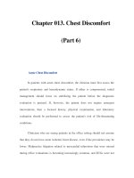
Fig. 15.5a. Computed tomographic appearance of parenchymal brain cysticercus: cys-
tic vesicular lesion showing scolex.
Subarachnoid Neurocysticercosis
Hydrocephalus, caused by inflammatory occlusion of the foramina of Luschka
and Magendie, is the most common CT finding in patients with subarachnoid NCC.
The fibrous arachnoiditis responsible for the development of hydrocephalus is seen
on CT scan as areas of abnormal leptomeningeal enhancement at the base of the
brain after contrast administration. Some patients with hydrocephalus due to
268
Tropical Neurology
15
cysticercus arachnoiditis also have single or multiple subarachnoid and parenchy-
mal brain cysts or calcifications, a finding that facilitates the diagnosis of NCC
(Fig. 15.7). Cystic subarachnoid lesion may be small when located within the corti-
cal sulci or may reach a large size if these are located in the Sylvian fissure or within
the basal CSF cisterns. The latter usually have a multilobulated appearance, displace
neighboring structures and behave like space occupying lesions.
Fig. 15.5b. Computed tomographic appearance of parenchymal brain cysticercus: col-
loidal cyst appearing as ring-enhancing lesion surrounded by edema.
269
Neurocysticerosis
15
Ischemic stroke in subarachnoid NCC can be seen on CT scan. These findings
are nonspecific since the CT appearance of cysticercus-related cerebral infarctions
are similar to cerebral infarctions from other causes. In some patients, the association
of subarachnoid cysts or the presence of abnormal enhancement of basal leptom-
eninges in the opticochiasmatic region suggests the correct diagnosis. The differen-
tial diagnosis should include fungal, tuberculous and carcinomatous meningitis since
these conditions are also associated with cerebral infarctions and abnormal enhance-
ment of the leptomeninges.
Fig. 15.5c. Computed tomographic appearance of parenchymal brain cysticercus: granular
cysticercus appearing as nodular lesion.
270
Tropical Neurology
15
Ventricular Neurocysticercosis
Ventricular cysticerci appear on CT scan as hypodense lesions that cause asym-
metric or obstructive hydrocephalus. Ventricular cysts are usually isodense with the
CSF; therefore, these can only be inferred on the basis of distortion of the ventricu-
lar system (Fig. 15.8). The administration of positive intraventricular contrast al-
lows precise localization of intraventricular cysticerci. This is usually performed by
Fig. 15.5d. Computed tomographic appearance of parenchymal brain cysticercus: cal-
cified cysticerci.
271
Neurocysticerosis
15
Fig.15.6. Computed tomography of a patient with cysticercus encephalitis. There is dif-
fuse brain edema, collapse of the ventricular system and multiple small areas of nodular
enhancement disseminated throughout the brain parenchyma (with permission from Del
Brutto OH, Sotelo J, Roman GC. Neurocysticercosis: A Clinical Handbook. © Lisse:
Swets & Zeitlinger).
transcutaneous puncture of the antechamber of a ventricular shunt previously placed
for the hydrocephalus or through a ventriculostomy tube. The contrast may also be
administered through a lumbar puncture; however, this procedure should be con-
ducted cautiously since this may result in brain herniation in patients with hydro-
cephalus or intraventricular cysts.
Magnetic Resonance Imaging
Magnetic resonance imaging (MRI) has the advantage of multiplanar (axial, coro-
nal and sagittal) reconstruction of images and the capacity to visualize the posterior
fossa without bone artifacts and high contrast resolution. By MRI it is possible to
recognize forms of cysticerci that were not seen on CT scan. The main shortcoming
of MRI, however, is failure to detect small calcifications. Parenchymal brain calcifi-
cations are the most common CT finding in patients with NCC and, in many
patients, these may be the only radiological evidence. Because of this limitation of
MRI, CT still remains the best screening neuroimaging procedure for patients with
suspected NCC.
1
Parenchymal Neurocysticercosis
MRI appearance of parenchymal brain cysticerci depends on their stage of devel-
opment. Vesicular cysts appear as rounded lesions with signal properties similar to
that of CSF in both T1- and T2- weighted images. The scolex is usually seen within
the cyst as a high intensity nodule giving the lesion a “hole-with-dot” image which
is pathognomonic of vesicular cysts (Fig. 15.9). The appearance of colloidal cysts is
272
Tropical Neurology
15
quite different because as the parasite begins to degenerate, proteins from the scolex
combine with the vesicular fluid, and the whole cysticercus becomes discretely
hypointense with the surrounding brain parenchyma. The wall of the cyst becomes
thick and hypointense and there is marked perilesional edema; these findings are
better visualized on T2-weighted images. Administration of gadolinium results in a
ring-like pattern of enhancement (Fig. 15.10). Granular cysticerci are visualized as
areas of signal void on both T1- and T2-weighted images surrounded by edema or
gliosis with hyperintense rims around the area of signal void. MRI findings in
cysticercus encephalitis consist of multiple small rounded areas of decreased signal
intensity on a T1-weighted sequence; these lesions become hyperintense on T2 and
Fig. 15.7. Computed tomography showing hydrocephalus, subarachnoid cysticercus in
Sylvian fissure and lacunar infarction in genu of internal capsule.
273
Neurocysticerosis
15
most of these are surrounded by edema. Due to the small size of the cysts and their
stage of development, some cysts are only discernible on proton-density and
T2-weighted sequences.
Fig.15.8. Computed tomography showing distortion of ventricular system caused by
ventricular cysticercus.
274
Tropical Neurology
15
Subarachnoid Neurocysticercosis
MRI provides an excellent opportunity to visualize subarachnoid cysticerci lo-
cated over the convexity of cerebral hemispheres (Fig. 15.11). Likewise, large cys-
ticerci within the Sylvian fissure or the basal CSF cisterns are seen on MRI as
multilobulated hypointense lesions that may not show gadolinium enhancement.
Sometimes the cysts have the same signal properties as CSF and may only be in-
ferred by the distortion of CSF cisterns. The diagnosis is difficult because cysts
usually lack a scolex as these are of racemose type. In cysticerus arachnoiditis, MRI
Fig. 15.9. T1-weighted magnetic resonance imaging showing characteristic
“hole-with-dot” imaging of vesicular cysticercus in cerebellum in axial section.
275
Neurocysticerosis
15
shows hydrocephalus and signal changes in the basal leptomeninges, with entrap-
ment of cranial nerves and blood vessels at the circle of Willis. Gadolinium en-
hancement usually increases the sensitivity for detection of areas of meningeal
inflammation.
Fig. 15.10. T2-weighted magnetic resonance imaging of colloidal cysticercus. Cystic
fluid appears hyperintense and cyst’s capsule is visualized as a hypointense rim. Marked
edema is seen surrounding the lesion.
276
Tropical Neurology
15
Ventricular Neurocysticercosis
Noninvasive diagnosis of intraventricular cysticerci represents one of the greater
advantages of MRI. Most ventricular cysts are readily seen on MRI because the
signal properties of the cystic fluid or the scolex differ from those of the CSF (Fig.
15.12). Cyst mobility within the ventricular cavities in response to movements of
Fig. 15.11. Gadolinium-enhanced T1-weighted magnetic resonance imaging showing
multiple cysticerci located over a convexity of cerebral hemispheres and Sylvian fis-
sures.
277
Neurocysticerosis
15
the patient’s head, the so-called “ventricular migration sign,” is better observed on
MRI than CT scan.
Spinal Cysticercosis
On MRI, intramedullary cysticerci appear as rounded or septated lesions. There
may be an eccentric hyperintense nodule representing the scolex. The periphery of
the cyst usually enhances due to a breakdown of the blood-spinal cord barrier sur-
rounding the cyst. The spinal cord is usually enlarged and if the scolex is not identi-
fied it becomes difficult to differentiate cysticercosis from ependymoma or
astrocytoma. Visualization of a spinal leptomeningeal cyst may be difficult if
perilesional inflammatory changes are absent.
Other Diagnostic Imaging Tests
Myelography
In patients with spinal leptomeningeal cysticercosis, multiple filling defects are
seen in the contrast column on myelography (Fig. 15.13). Leptomeningeal cysts
may be freely mobile within the spinal subarachnoid space and may change their
position during myelography. On the other hand, myelography is not specific in
patients with intramedullary cysts since it produces partial or complete block at the
level of the lesion. This finding may also be observed in other intramedullary spinal
cord lesions.
Cerebral Angiography
During the past decade, a number of reports dealing with the cerebrovascular
complications of NCC have described in detail the full spectrum of angiographic
changes. Angiographic findings in NCC include segmental narrowing of anterior or
middle cerebral arteries in patients with lacunar infarctions, occlusion of the ante-
rior or middle cerebral arteries or even the internal carotid artery in patients with
large cerebral infarctions and mycotic aneurysms in patients with subarachnoid hem-
orrhages. The prevalence of angiographic abnormalities in patients with NCC is
unknown; however, some studies suggest that angiographically documented arteri-
tis is a common finding in patients with subarachnoid NCC, even in patients lack-
ing clinical or neuroimaging evidence of a cerebral infarction.
Treatment
Neurocysticercosis is a pleomorphic disease that causes several neurological syn-
dromes and patholgical lesions; therefore, uniform therapeutic scheme is not practi-
cal in every patient. Characterization of the disease in terms of cysts’ viability, degree
of the host’s immune response to the parasites and location of the lesions are of
importance for rational management. Therapy of NCC includes a combination of
symptomatic drugs, specific cysticidal drugs, surgical resection of the lesion and
placement of ventricular shunts.
14
Cysticidal Therapy
Praziquantel is an isoquinoline with proven cysticidal properties that have been
used to treat human NCC since 1979. Subsequent studies showed that praziquantel
destroys up to 70% of parenchymal brain cysticerci after a 15 day treatment with
daily doses of 50 mg/kg. Albendazole is an imidazole derivative that also has cysticidal
properties. This drug was initially administered at a dose of 15 mg/kg per day for
278
Tropical Neurology
15
one month. Further studies, however, have shown that at similar doses, the duration
of therapy could be shortened to one week without compromising the results.
Albendazole destroys 75-90% of parenchymal brain cysts and has been considered
superior to praziquantel in several trials comparing the efficacy of these drugs (Table
15.3). Another advantage of albendazole over praziquantel is that the former also
destroys subarachnoid and ventricular cysts due to its better CSF penetration. The
initial regimen of praziquantel at a daily dose of 50 mg/kg for 15 days was arbitrarily
Fig. 15.12. T1-weighted magnetic resonance imaging showing cysticercus in the lateral
ventricle. The capsule and scolex of the cyst are clearly differentiated from cerebrospi-
nal fluid.
279
Neurocysticerosis
15
Fig. 15.13. Myelogram showing multiple filling defects in contrast column correspond-
ing to small spinal leptomeningeal cysticerci (With permission from: Del brutto OH,
Sotelo J, Roman GC. Neurocysticercosis: A Clinical Handbook. © Lisse: Swets &
Zeitlinger).
280
Tropical Neurology
15
chosen. Since then, recommended doses of praziquantel have ranged from 10-100
mg/kg for 3-21 days. In these studies, praziquantel has been administered eight
times hourly. Since plasma levels of praziquantel decline within three hours of ad-
ministration, it seems that the cysticidal effect has been reached by exposing the
parasites to several intermittent peaks of the drug. On this basis, it was suggested
that if parenchymal brain cysticerci are exposed to high concentrations of praziquantel
which are maintained for six hours by giving three individual doses of 25 mg/kg at
two-hour intervals, it might be sufficient to destroy the parasites. Preliminary results
were encouraging since the percentage of cyst disappearance on neuroimaging stud-
ies was similar to that observed in patients receiving larger doses.
The introduction of praziquantel and albendazole has radically changed the prog-
nosis of most patients with NCC; however, the anecdotal nature of initial studies on
these drugs generated doubts about their usefulness. Some authors claim that cysticidal
drugs only improved the CT scans without improving the patients. Nevertheless,
more recent studies have focused on the clinical outcome of the patients following
cysticidal therapy and have shown that such therapy also produces significant clini-
cal improvement. The control of seizures in patients with NCC is better after treat-
ment with anticysticidal therapy compared to those who don’t receive anticysticidal
treatment.
15
Moreover, some studies have shown that patients with focal neurologi-
cal deficits also have recovered after a trial of cysticidal drugs because the pressure
effects exerted by parasites regress following cysticidal therapy. Cysticidal drugs have
a diagnostic role in epileptic patients with single enhancing lesions on CT or MRI.
11
While most of these lesions are dying cysticerci in the acute encephalitic phase,
some single enhancing lesions are, however, due to tuberculoma, mycotic granu-
loma or low-grade gliomas and may require a follow-up study. Therapeutic trial
with cysticidal drugs, if it results in prompt resolution of an intracranial lesion, may
suggest the diagnosis of cysticercosis and obviate expensive and hazardous investiga-
tion and treatment.
Patients with cysticercus encephalitis should not receive cysticidal drugs because
these may aggravate intracranial hypertension. In patients with both hydrocephalus
and parenchymal brain cysts, cysticidal drugs may be used only after a ventricular
shunt has been placed to avoid further increases in the intracranial pressure follow-
ing cysticidal therapy. Cysticidal drugs must be used with caution in patients with
giant subarachnoid cysticerci because the inflammatory reaction developed by the
host in response to the acute destruction of the parasite within the subarachnoid
space may occlude small leptomeningeal vessels surrounding the cyst. In such pa-
tients, concomitant steroid administration is mandatory to avoid cerebral infarc-
tion. In patients with ventricular cysts, the therapeutic approach with cysticidal
drugs should be individualized. Albendazole therapy successfully destroys many ven-
tricular cysts; however, the inflammatory reaction surrounding those cysts may cause
acute hydrocephalus if these are located within the fourth ventricle or near the
foraminae of Monro. Patients with calcifications alone should not receive cysticidal
drugs since these lesions represent already dead parasites.
10
Symptomatic Therapy
In patients with seizures due to calcified cysticerci, the administration of stan-
dard doses of a single, first-line antiepileptic drug usually results in adequate control
of seizures. In contrast, patients with parenchymal brain cysts must first be treated
with cysticidal drugs to achieve adequate control of seizures with antiepileptic drugs.
281
Neurocysticerosis
15
In two large series of patients with epilepsy due to NCC, a statistically significant
correlation between the use of praziquantel or albendazole and the rate of seizure
control was demonstrated. The optimal duration of antiepileptic drug therapy in
patients with NCC has not been decided. A recent prospective study showed that
up to 50% of these patients had relapses after withdrawal of antiepileptic drugs.
Such patients had been free of seizures for two years, and their parenchymal brain
cysts had been successfully destroyed with albendazole. Prognostic factors associated
with seizure recurrence included the development of parenchymal brain calcifica-
tions as the result of albendazole therapy and presence of recurrent seizures and
multiple brain cysts before the institution of cysticidal therapy.
Corticosteroids are frequently used in patients with NCC and are the primary
form of therapy for cysticercus encephalitis, and arachnoiditis causing hydroceph-
alus and progressive entrapment of cranial nerves. According to the severity of the
disease, up to 32 mg per day of dexamethasone may be needed for the control of
symptoms. In patients with cysticercus encephalitis, corticosteroids may be used
alone or in combination with mannitol 2 mg/kg per day. The initial trial with high
doses of intravenous dexamethasone may be followed by oral therapy with pred-
nisone (50 mg/day) or dexamethasone (10 mg/day). The simultaneous administra-
tion of corticosteroids and cysticidal drugs is controversial. This combination has
been recommended to ameliorate the features of raised intracranial tension that may
occur during praziquantel or albendazole therapy. Such manifestations are not re-
lated to toxic effects of cysticidal therapy but to the acute destruction of parasites
within the brain and are reliable indicators of drug efficacy. Common analgesics and
antiemetics may be used to ameliorate such complaints, avoiding the routine use of
corticosteroids in every patient. Absolute indications for corticosteroid administra-
Table 15.3. Results of controlled clinical trials comparing efficacy of
praziquantel and albendazole in treatment of parenchymal brain
cysts
Reference Drug Regimen No. % of Cyst
Pts Reduction on CT
Sotelo, Escobedo et al, PZQ 50 mg/kg/d x 15 d 10 73%
1988 ALB 15 mg/kg/d x 30 d 10 76%
None ——- 5 0
Sotelo et al, 1990 PZQ 50 mg/kg/d x 15 d 52 60%
PZQ 50 mg/kg/d x 8 d 13 40%
ALB 15 mg/kg/d x 30 d 25 85%
ALB 15 mg/kg/d x 8 d 24 85%
None ——- 33 0
Takayanagui & Jardim PZQ 50 mg/kg/d x 21 d 20 50%
1992 ALB 20 md/kg/d x 21 d 20 88%
None — 16 7%
PZQ-praziquantal, ALB-albendazole, Pts –Patients.
Used with permission from Del Brutto OH, Sotelo J, Roman GC.
Neurocysticercosis: A Clinical Handbook. Lisse: Swets & Zeitlinger Publishers.
282
Tropical Neurology
15
tion during cysticidal drug therapy include patients with giant subarachnoid cys-
ticerci, ventricular cysts, spinal cysts, and multiple parenchymal brain cysts. In such
patients, corticosteroids must be administered before, during and even a few days
after the course of cysticidal drugs to avoid the risk of cerebral infarctions, acute
hydrocephalus, spinal cord swelling and massive brain edema.
Surgery
Patients with hydrocephalus secondary to cysticercus arachnoiditis require ven-
tricular shunt surgery. The main problem in these patients is the high prevalence of
shunt dysfunction. The protracted course of these patients and their high mortality
rates (up to 50% in two years) is directly related to the number of surgical interven-
tions for shunt revision. A new shunt device functioning at a constant flow without
a valve mechanism has been recently developed. This shunt does not allow the en-
trance of spinal CSF into the ventricular system. In patients with NCC, this inver-
sion of CSF transit is the most common cause of shunt dysfunction as it allows the
entrance of subarachnoid inflammatory cells and parasitic debris into ventricular
cavities.
Ventricular cysts may be removed by surgical excision or by endoscopic aspira-
tion. However, the possibility of cyst migration between the time of diagnosis and
the surgical procedure, must be ruled out by a control CT or MRI immediately
before surgery to avoid unnecessary craniotomies. Permanent shunt placement is
usually not necessary after removal of a ventricular cyst in the absence of ependymi-
tis. In contrast, shunt placement should follow or even precede the excision of a
ventricular cysts associated with ependymitis. For a rare patient with double com-
partment hydrocephalus related to both granular ependymitis of the cerebral aque-
duct and occlusion of the foraminae of Luschka and Magendie, two independent
shunt devices may be needed; one draining the supratentorial ventricular system
and the other draining the isolated fourth ventricle.
Summary
Cysticercosis is the most common parasitic disease of the nervous system. The
disease occurs when humans become the intermediate host in the life cycle of Ta e nia
solium by ingesting its eggs from contaminated food. Cysticercosis is endemic in
developing countries of Latin America, Asia and Africa. Massive immigration of
people to industrialized nations has caused a recent increase in the number of pa-
tients with cysticercosis in the United States and in some European countries as
well. Neurocysticercosis is a pleomorphic disease due to individual differences in the
number, size and location of the parasites within the nervous system, as well as to
differences in the severity of the host’s immune reaction. Epilepsy, focal neurological
signs and intracranial hypertension are the most common clinical manifestations of
NCC. Since the diagnosis is not possible on clinical grounds, it is necessary to un-
dertake radiological and immunological investigations. Neuroimaging studies (CT
or MRI) usually permit the diagnosis as these show objective evidences of the para-
sites and the inflammatory changes induced in the surrounding nervous tissue. Im-
munological tests developed to detect anticysticercal antibodies in serum or CSF
have many problems due to inherent lack of specificity or sensibility. The immuno-
logical tests, therefore, should not be the sole basis for confirming or excluding the
diagnosis of NCC. The development of potent cestocidal drugs, albendazole and
praziquantel, have greatly improved the prognosis of NCC. Nevertheless, some pa-
283
Neurocysticerosis
15
tients still have torpid clinical courses despite proper therapy. Surgery has a role in
the management of selected patients, particularly in patients with hydrocephalus
and intraventricular cysts.
References
1. Del Brutto OH, Sotelo J. Neurocysticercosis: An update. Rev Infect Dis 1988;
10:1075-1087.
2. Garcia HH, Gilman R, Tovar MA et al. Factors associated with Taenia solium cys-
ticercosis: Analysis of nine hundred forty-six Peruvian neurologic patients. Am J
Tr op Med Hyg 1995; 52:145-148.
3. Flisser A. Taeniasis and cysticercois due to Taenia solium. In: Sun T, ed. Progress in
Clinical Parasitology. Boca Raton: CRC Press, 1994:77-116.
4. Trelles JO, Trelles L. Cysticercosis of the nervous system. In: Vinken PJ, Bruyn
GW, eds. Handbook of Clinical Neurology. Vol. 35. Amsterdam: North Holland,
1978:2910-320.
5. Escobar A. The pathology of neurocysticercosis. In: Palacios E, Rodriguez-Carbajal
J, Taveras JM, eds. Cysticercosis of the Central Nervous System. Springfield: Charles
C. Thomas Publisher, 1983:27-54.
6. Pittella JEH. Neurocysticercosis. Brain Pathol 1997; 7:681-693.
7. Sotelo J, Del Brutto OH, Roman GC. Cysticercosis. In: Remington JS, Swartz
MN, eds. Current Clinical Topics in Infectious Diseases, 16. Cambridge: Blackwell
Science, 1996:240-259.
8. White Jr. AC, Tato P, Molinari JL. Host-parasite interactions in Taenia solium cys-
ticercosis. Infect Ag Dis 1992; 1:185-193.
9. Sotelo J, Guerrero V, Rubio F. Neurocysticercosis: A new classification based on
active and inactive forms. Arch Intern Med 1985; 145:442-445.
10. Dixon HBF, Lipscomb FM. Cysticercosis: An analysis and follow-up of 450 cases.
Medical Research Council Special Report Series No 299. London: Her Majesty’s
Stationary Office, 1961:1-58.
11. Wadia NH. Neurocysticercosis. In: Shakir RA, Newman PK, Poser CM, eds. Tropi-
cal Neurology. London: W.B. Saunders Co., 1996:247-274.
12. Del Brutto OH, Sotelo J, Roman G. Neurocysticercosis: A clinical handbook.
Lisse: Swets & Zeitlinger Publishers, 1998.
13. Del Brutto OH, Wadia NH, Dumas M, Tsang VCW, Schantz PM. Proposal of
diagnostic criteria of human cysticercosis and neurocysticercosis. J Neurol Sci 1996;
142:1-6.
14. Del Brutto OH, Sotelo J, Roman GC. Therapy for neurocysticercosis: A reap-
praisal. Clin Infect Dis 1993; 17:730-735.
15. Vazquex V, Sotelo J. The course of seizures after treatment for cerebral cysticerco-
sis. N Engl J Med 1992; 327:696-701.
CHAPTER 16
Tropical Neurology, edited by U. K. Misra, J. Kalita and R. A. Shakir.
©2003 Landes Bioscience.
Cerebral Malaria
J. E. Touze, L. Fourcade, P. Heno, P. Riviere and P. Paule
Cerebral malaria, mostly caused by Plasmodium falciparum, remains a major health
problem in the endemic areas. The last report of the World Health Organization
indicates an increasing incidence of malaria. Over three billion people are poten-
tially exposed to malaria each year with an estimated death rate of two million every
year, mainly among African children and nonimmune adults, such as travellers.
Failure to provide appropriate chemoprophylaxis, lack of convenient methods for
screening high-risk populations, delay in the diagnosis, use of inappropriate therapy
and widespread falciparum resistance to chloroquine have contributed to high mor-
bidity and mortality. Most patients with falciparum malaria do not have CNS mani-
festations. Cerebral malaria, however, is one of the manifestations of severe falciparum
infection and should be considered a medical emergency as it may rapidly progress
to a fatal multisystem disease. Cerebral malaria is a major cause of death, accounting
for more than 80% of patients dying of falciparum infection. The last two decades
have witnessed better understanding in the pathophysiology of cerebral malaria.
Sequestration of parasitized erythrocytes in deep capillaries and host response are
better approached with new mechanisms such as rosetting, cytoadherence and
cytokine secretion. The clinical presentation of cerebral malaria is now better de-
fined. It may be associated with other complications such as pulmonary edema,
hemodynamic failure, renal insufficiency, severe anemia and hypoglycaemia.
1
At the
same time of widespread resistance to chloroquine, new antimalarial drugs, e.g.,
artemisinine derivatives, have been introduced.
Pathophysiology of Cerebral Malaria
Two main pathophysiological mechanisms of cerebral malaria, namely sludging
of red blood cells (RBC) and change in capillary permeability, are thought to be
responsible for cerebral malaria.
The Sludging Theory
Sequestration of the late stages of intra-erythrocytic cycle is believed to cause
cerebral malaria. Pathological observations in patients with fatal cerebral malaria
showed a high concentration of parasitized erythrocytes. Sequestration of parasit-
ized erythrocytes in the relatively hypoxic venous beds allows optimal parasite growth
and prevents parasitized RBCs from being destroyed by the spleen. Several decades
ago, Gaskell and Millar postulated that parasitized RBCs reduce blood flow and
induce sequestration. This hypothesis was criticized for three reasons: 1) sequestra-
tion was observed elsewhere in arterial and capillary venules; 2) anoxic changes were
inconstantly observed in histological samples; and 3) sequestration was never ob-
served with P. v i v a x which has large trophozoites and schizonts.
Several authors have noted the discrepancy between autopsy findings and clini-
cal features of cerebral malaria although a study has shown correlation between the
degree of parasitized red blood cell sequestration and depth of coma.
285
Cerebral Malaria
16
The Permeability Hypothesis
The permeability hypothesis of cerebral malaria was based on an experiment by
Maigraith and Fletcher. The essential observation was an increase in blood-brain
barrier permeability to I-labelled albumin in rhesus monkeys infected with P. knowlesi.
This increase in capillary permeability was rapidly reversed by hydrocortisone. In-
creased capillary permeability results in leakage of plasma and cerebral edema. Based
on this theory corticosteroids were widely used in cerebral malaria. The permeabil-
ity hypothesis of cerebral malaria has been criticized for following reasons: 1) Dex-
amethasone has been found to be deleterious in severe falciparum malaria.
2
In a
double blind placebo controlled trial, coma was significantly prolonged in the corti-
costeroid group; 2) many studies carried out in Africa have shown that cerebrospi-
nal fluid pressure was not significantly increased in cerebral malaria; and 3)
computerized tomography studies have never demonstrated features of brain edema
in cerebral malaria.
Cerebral malaria is presently considered a result of a cascade of events including
parasite cytoadherence, enhanced cytokine secretion in the background of a high
parasite load and specific host factors.
3
Cytoadherence
Parasitized RBCs appear to stick to vascular endothelium through a specific in-
teraction between plasmodium-derived protein on the surface of parasitized RBCs
and ligands on the endothelial cells. Cytoadherence is the result of rosetting, “knob”
formation and attachment of infected erythrocytes to specific endothelial receptors.
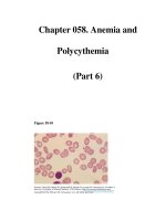
Rosetting is a phenomenon where parasitized red cells agglutinate around normal
red cells (Fig. 16.1). This complex of red cells induces sequestration in deep capilla-
ries. The second component in cytoadherence is the presence of protrusions, or
“knobs” on red cells. These knobs contain specific falciparum antigens such as histi-
dine rich protein and RESA (ring erythrocyte surface antigen) protein and plasmo-
dium falciparum erythrocyte membrane protein-1(PfEMP-1). These proteins show
clonal variation allowing them to evade host immune responses. Knobs are essential
for cytoadherence and facilitate the attachment of the red cells to the vascular en-
dothelial cell. The third component implicated in cytoadherence is a number of
specific endothelial receptors: ICAM-1, CD-36 protein, VCAM-1, E-selectin,
thrombospondin and chondroitin-sulfate-A (CSA). Infected red cells are attached
to these receptors. Some of these, such as thrombospondin and CD36, are expressed
in a wide range of vascular beds and are not related to the severity of the disease. On
the other hand, other receptors such as ICAM-1 and E-selectin are expressed in the
cerebral capillaries of patients with cerebral malaria highlighting the role of
cytoadherence. This phenomenon appears to play an important role in parasite sur-
vival. It is generally believed that sequestration initiates many pathogenic processes
associated with cerebral malaria and malaria during pregnancy. The role of CSA has
been recently demonstrated in pregnant women, especially during first pregnancy.
In the primigravida, large number of parasitized red blood cells (PRBCs) sequester
in the maternal compartment of the placenta, binding to CSA. Preliminary studies
have shown that antibodies developed after multiple pregnancies are associated with
reduced number of parasitized RBCs in the placenta and block CSA-binding of
parasitized RBCs, suggesting that protection is conferred by a conserved Pf-antigen.
4
286
Tropical Neurology
16
Cytokines
Plasma concentration of cytokines are elevated in adults and children with severe
malaria. Cytokines have an important role in the pathogenesis of cerebral malaria.
Injection of TNFα into mice resulted in a disorder similar to cerebral malaria. The
role of TNFα was suggested in the pathogenesis of fever, hypoglycemia and pulmo-
nary edema associated with cerebral malaria. Kwiatowski has shown that the severity
of falciparum malaria strongly correlated with TNFα levels.
5
However, its mecha-
nism of action remains unclear. Currently the best hypothesis about the pathogen-
esis of cerebral malaria would be the recruitment and modulation of endothelial
receptors which could also enhance nitric oxide and lactic acid production which
may interfere with synaptic transmission, causing coma.
TNF release has been correlated with severity of malaria but there are still a
number of inconsistencies. Increased TNF secretion was noted in different patient
groups with malaria, especially in patients infected by P. v i v a x . Secondly, although
cytokine release in rodent models has been demonstrated, their implications in hu-
man malaria remain doubtful. Thirdly, TNF was not the only cytokine involved in
the pathophysiology of cerebral malaria. Gamma interferon, IL-1, GMCSF, IL-2
and IL-10 are also implicated in macrophage activation and TNF secretion.
Host Factors
Genetic factors, immunity and nutritional status may play a part in the patho-
genesis of cerebral malaria and may account for why some patients develop cerebral
malaria and others do not. Among the genetic factors, sickle cell anemia and thalas-
semia may protect against cerebral malaria. Studies in Africa have demonstrated
that heterozygous children with sickle cell traits (AS) in malaria endemic areas have
fewer parasites and a better outcome than children with normal hemoglobin (AA).
Moreover, polymorphic genes could explain the resistance of some individuals against
Fig. 16.1. Rosetting phenomenon. Agglutination of infected red blood cells around a
normal erythrocytes.
287
Cerebral Malaria
16
malaria infection. Some genes are linked to the HLA system (BW3 alleles) and
control liver stages of Plasmodium falciparum. Others (DRW-3) play a role in pro-
tection against severe anemia. These genes could be implicated in semi-immune
patients but are inefficient against severe falciparum malaria with high parasitemia.
Nutritional Status
Nutritional status could also have a role in the pathophysiology of severe
falciparum malaria. Convulsions in cerebral malaria seem to occur more frequently
in well-nourished children than in those with marasmus or kwashiorkor. Neverthe-
less, mortality is higher when severe falciparum malaria occurs in a malnourished
child. The individual response against P. f a lciparum malaria is linked to host immu-
nity. Two types of responses have been observed: 1) either an increase in T4 lym-
phocyte production with a high TNF secretion; or 2) a low response of T4
lymphocytes, decreased TNF secretion and blockage of specific inhibitors. In this
latter situation the host is also protected by genetic factors and immunity against
cerebral malaria.
6
Parasite Factors
Differences in virulence between plasmodium strains were noted in infected aotus
monkeys but were never demonstrated in P. f a lciparum-infected humans. Moreover,
there is no strong evidence to correlate the severe falciparum malaria to the inocu-
lum or the level of chemoresistance of the strain. The main parasite factor impli-
cated in severe falciparum malaria remains within the susceptibility level to
antimalarial drugs. Despite these new advances, the cause of coma in cerebral ma-
laria remains unclear. Many hypotheses have been proposed, such as changes in
neurotransmitter synthesis, release of nitric oxide and raised intracranial pressure,
but none of these are proven.
3
Pulmonary Edema
The pathophysiology of pulmonary edema in cerebral malaria remains unclear.
In severe falciparum malaria, increased capillary permeability, hypoalbuminemia
and overhydration are the main causes of acute pulmonary edema. Pregnant women
and children are at increased risk of developing pulmonary edema which can de-
velop even after several days of appropriate chemotherapy. In those patients one
should be cautious in maintaining fluid balance, and hypertonic solutions such as
bicarbonates, hypertonic saline and mannitol should be avoided. In some patients,
the course of pulmonary edema is similar to adult respiratory distress syndrome.
Ultrastructural studies of the lung show neutrophil deposits, formation of hyaline
membranes and septal thickening of lung alveolus which are attributed to
cytoadherence and cytokines.
Hypoglycemia
This is an important complication of cerebral malaria. It is attributed to in-
creased glucose consumption by the parasite, impaired glycogenosis and to the use
of quinine. Quinine is a potent stimulus for insulin release, and this effect is in-
creased in pregnant women and children.
Renal Impairment
Renal impairment is usually observed among adults and is attributed to fluid
imbalance and hemodynamic failure. In severely affected patients acute tubular ne-
crosis may be observed. Its pathophysiology remains unclear, although a decreased
288
Tropical Neurology
16
renal cortical blood flow has been noted in earlier studies using
133
Xe clearance method
and contrast urography. In most clinical studies, renal dysfunction is strongly asso-
ciated with high parasitemia, jaundice, hemoglobinuria and pulmonary edema. These
changes could be due to cytoadherence, increased TNF secretion and immune com-
plex deposition in glomeruli. In view of this, hemoglobinuria in cerebral malaria
should be distinguished from black water fever and hemolysis induced by
glucose-6-phosphate dehydrogenase (G-6-PD) deficiency. In the former, sensitiza-
tion of red cells has been observed after intermittent use of aminoalcohol drugs such
as quinine, quinidine, halofantrine and mefloquine. In the latter group hemoglobi-
nuria occurs in patients with G-6-PD deficiency who take oxidant drugs such as
primaquine or certain foodstuffs.
Hematological Disorders
Anemia is an unavoidable complication of P. falciparum malaria. In cerebral
malaria, erythrocyte destruction is often underestimated because of red cells seques-
tration in deep capillaries. The mechanisms of anemia in cerebral malaria are multi-
factorial, involving dyserythropoiesis, iron deficiency, RBC sequestration in the spleen
and hemolysis. In some patients, immune-mediated hemolysis may be the mecha-
nism of anemia in endemic areas. Coombs’ test may be positive in those patients.
Thrombocytopenia
Thrombocytopenia is commonly observed in malaria. In cerebral malaria the
role of disseminated intravascular coagulation has been probably over-emphasized
for thrombocytopenia; this, however, may be observed in severe disease with acute
renal failure and pulmonary edema. Thrombocytopenia is attributed to enhanced
spleen sequestration or peripheral destruction by an immune mechanism involving
CD4
+
lymphocytes and antiplatelet IgG antibodies. Other hemostatic abnormali-
ties include failure to synthesize clotting factors such as prothrombin complex, ac-
celerated fibrinogen metabolism and changes in platelet function.
Other Infections
Gram negative septicemia, bronchopneumonia, urinary tract infections and men-
ingitis are frequently encountered in patients with cerebral malaria, particularly in
an intensive care unit. Salmonella septicemia is also commonly associated with ma-
laria in endemic areas. These associated infections must always be considered in the
management of cerebral malaria. These can induce hypotension, “algid” shock and
lactic acidosis.
Clinical Features
In the past, many patients with impaired consciousness, neck stiffness, febrile
convulsions or focal neurological signs were considered to have cerebral malaria.
Mostly, those manifestations resulted from other diseases such as meningitis, men-
ingoencephalitis and vascular diseases. Moreover, impairment of consciousness may
be observed in patients with high fever or in children after febrile seizures. For these
reasons, Warrell et al proposed a strict definition of cerebral malaria. This definition
requires an unarousable coma in P. falciparum malaria defined in adults by a Glasgow
scale lower than 10 and in children by a Blantyre scale lower than 3 (Table 16.1), in
the absence of another cause of altered sensorium.
7
In adults, cerebral malaria commonly occurs after several days of fever or non-
specific symptoms. Convulsions are usually explained by hypoglycemia, particularly
in pregnant women with or without quinine treatment. Neck rigidity is uncom-