Antibody Phage Display Methods and Protocols - part 6 docx
Bạn đang xem bản rút gọn của tài liệu. Xem và tải ngay bản đầy đủ của tài liệu tại đây (457.31 KB, 39 trang )
3. Transfer an aliquot of the eluted Ab-phage clone to 2YT medium containing
the appropriate antibiotics and amplify according to standard protocols. Isolate
the amplifi ed phage for further selection procedures or soluble Fab for further
characterization of Ag specifi city.
4. Notes
1. Polyclonal anti-Ig Ab is one of the most effi cient and broadly applicable capture
reagents identified to date, presumably because of the variability between
different clones in the Ab library. Unlabeled polyclonal goat anti-human κ Ab
(mouse-adsorbed; Southern Biotechnology Associates) is a general capture
reagent that has been used successfully on multiple occasions for libraries of
human Abs expressing a κ light chain. Monoclonal Abs to affi nity tags may also
be used when appropriate.
2. Labeling of the Ag with biotin permits the rapid subsequent detection of
complexes using streptavidin enzyme conjugates. Commercially available biotin
labeling reagents with a wide range of reactive chemistries should be tested to
identify an Ag-labeling protocol that does not disrupt the epitope(s) of interest
and/or interfere with binding. In these instances, a biotinylated second Ab that
reacts with a distinct epitope on the Ag can be used for detection.
3. The selection of dilution buffer is predominantly Ag-dependent and should be
determined experimentally. Inclusion of nonionic detergents is generally useful
for reducing background in the assay, regardless of the properties of the Ag.
For example, for the discovery of Abs to cell surface Ags, some of which are
integral membrane proteins, we have used a diluent consisting of PBS–1%
BSA, 1% Triton X-100, and 0.145% sodium dodecyl sulfate, containing 0.1%
Na azide (3).
4. The fi lter is fl oated on the capture solution in order to minimize the quantity
of capture reagent used.
5. The blocking of excess nonspecifi c protein-binding sites on nitrocellulose is
typically accomplished by incubating the fi lter in a buffered solution of unrelated
protein, such as BSA, hemoglobin, gelatin, or milk. The appropriate blocking
reagent must bind the extra sites, while not interfering with the subsequent
interaction and detection steps and is best determined empirically with control
Ab and Ag.
6. A low phage titer (500 pfu/100-mm dish) will form distinct plaques (clones)
that can be isolated without requiring further purifi cation. Higher-phage titers
(100,000 pfu/100-mm dish) can be used for the initial screening of larger
libraries, but reactive clones will require subsequent replating at lower titers, to
isolate the specifi c clone of interest. Because 100,000 distinct clones (plaques)
can be screened using a single 100-mm fi lter, libraries containing millions of
clones can be routinely analyzed.
7. Typically, the fi lters are washed 4–6× for 5 min each with constant agitation in
~10 mL PBS-T fi lter. However, rapid washes using a squirt bottle and a vacuum
fi ltration device are better for the detection of lower-affi nity interactions.
192 Watkins
References
1. McCafferty, J., Griffi ths, A. D., Winter, G., and Chiswell, D. J. (1990) Phage
antibodies: fi lamentous phage displaying antibody variable domains. Nature 348,
552–554.
2. Skerra, A., Dreher, M. L., and Winter, G. (1991) Filter screening of antibody Fab
fragments secreted from individual bacterial colonies: specifi c detection of antigen
binding with a two membrane system. Anal. Biochem. 196, 151–155.
3. Watkins, J. D., Beuerlein, G., Wu, H., McFadden, P. R., Pancook, J. D., and
Huse, W. D. (1998) Discovery of human antibodies to cell surface antigens by
capture lift screening of phage-expressed antibody libraries. Anal. Biochem. 256,
169–177.
4. Wu, H. R., Beuerlein, G., Nie, Y., Smith, H., Lee, B. A., Hensler, M., Huse, W.
D., and Watkins, J. D. (1998) Stepwise in vitro affi nity maturation of Vitaxin, an
αvβ3-specifi c humanized mAb. Proc. Natl. Acad. Sci. USA 95, 6037–6042.
5. Wu, H., Nie, Y., Huse, W. D., and Watkins, J. D. (1999) Humanization of a
murine monoclonal antibody by simultaneous optimization of framework and
CDR residues. J. Mol. Biol. 294, 151–162.
Screening of Phage-Expressed Antibody 193
195
From:
Methods in Molecular Biology, vol. 178: Antibody Phage Display: Methods and Protocols
Edited by: P. M. O’Brien and R. Aitken © Humana Press Inc., Totowa, NJ
15
Antibody-Guided Selection
Using Capture-Sandwich ELISA
Kunihiko Itoh and Toshio Suzuki
1. Introduction
Antibody (Ab) phage display is a recently developed recombinant DNA
technology for making human monoclonal antibodies (MAbs) from immune
sources, such as bone marrow, lymph node, or peripheral blood lymphocytes
from patients with various diseases, or from healthy individuals (1,2). Many
human MAb Fabs or scFvs specifi c for viral pathogens, self antigens (Ags), or
nonself Ags have been isolated by phage display system. This technology is
expected to provide more powerful diagnostic, prophylactic, and therapeutic
tools of human origin than do currently used polyclonal Abs or MAbs derived
from other species.
Although it is not necessary to immunize the donor with the Ag of interest to
isolate human MAbs, purifi cation of the Ag is normally required for panning or
screening of human libraries. Ags (e.g., membrane proteins, cytosolic proteins,
nuclear proteins, recombinant proteins, nucleic acids, and so on) have been
purifi ed by column chromatography or affi nity chromatography techniques
from various sources (e.g., eukaryotic cells, insect cells, bacterial cells, their
culture supernatants, and so on). However, the purification of Ag can be
laborious and time-consuming, especially if the Ag is a minor component of
the starting material.
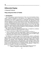
In this chapter, a panning procedure to isolate Ag specifi c MAb using a
modifi ed capture sandwich enzyme-linked immunosorbant assay (ELISA) are
described (Fig. 1). Sandwich ELISA uses two separate Abs for capture and
detection of Ags and is widely used for specifi c detection of target Ags from
crude preparations. A similar premise can be applied to a panning procedure, in
Ab-Guided Capture-Sandwich ELISA 195
which a crude Ag preparation can be used, if an appropriate Ab with a defi ned
specifi city against the Ag of interest is available. In this case, the Ab-displayed
phage library replaces the second detection Ab.
The advantages of this system are as follows:
1. Purifi cation of target Ag(s) is not necessary.
2. Abs against conformation-sensitive Ags can be selected because Ag denaturation
for direct coating to a plastic surface is not required.
3. By using capture Abs with varying specifi cities, MAbs against a variety of Ag
epitopes can be isolated from a single library. For instance, Abs specifi c for
functional determinants, e.g., neutralization, adhesion, and so on, can be selected
by using a capture Ab against nonfunctional determinants. Alternatively, MAbs
reactive with less immunogenic epitopes can be selected by using a capture Ab
against an immunodominant epitope. For example, the selection of human Fabs
196 Itoh and Suzuki
Fig. 1. Outline of the panning procedure for enrichment of Ag-specifi c phage Ab by
Ab-guided selection using a capture sandwich ELISA
against herpes simplex virus glycoproteins by utilizing MAbs with different
specifi cities, has been reported (3).
4. Both MAbs and polyclonal Abs can be used as capture Abs. Since polyclonal
Abs will recognize several epitopes on the Ag, polyclonal Ab-captured Ag
theoretically should present a variety of Ag epitopes accessible for panning,
depending on their abundance. We have isolated human Ab Fabs specifi c for
rotavirus VP6 protein using culture supernatants of virus-infected Vero cells as an
Ag and polyclonal Ab against human rotavirus Wa as a capture Ab (4).
2. Materials
1. ELISA plates: 96-well half-area plates (well vol 190 µL) (no. 3690, Costar,
Cambridge, MA). Regular area size ELISA plates or 60-mm plastic dishes can
also be used (see Note 1).
2. Capture Ab: either MAb or polyclonal Ab with defi ned Ag specifi city, diluted to
5–10 µg/mL in phosphate-buffered saline (PBS) (see Note 2).
3. 3% (w/v) and 1% (w/v) Bovine serum albumin (BSA) in PBS (PBS-BSA).
4. Ag: crude or partially purifi ed extract or purifi ed Ags from any source, e.g.,
culture supernatants, bacterial cell lysates, or tissue culture cell extracts are all
applicable. Ag should be diluted in PBS–1% BSA to a predetermined optimal
concentration (see Note 3).
5. Washing buffer: 0.05% (v/v) Tween-20 in PBS (PBS-T).
6. Ab phage library, constructed from bone marrow, lymph node, or peripheral
blood lymphocytes from patients or healthy individuals with high serum titer to
the Ag of interest. The library should be freshly amplifi ed and titered and diluted
to the appropriate concentration in PBS–1% BSA.
7. Elution buffer: 0.1 M glycine–HCl (pH 2.2).
8. Neutralization buffer: 2 M Tris-HCl.
9. Escherichia coli XL1-Blue or other suitable strain for amplifi cation.
3. Methods
1. Add 50 µL capture Ab into each well (see Note 4). Cover the plate with plastic
wrap or adhesive tape to prevent evaporation of the solution. Incubate overnight
at 4°C.
2. Discard the Ab solution and rinse the wells once with 150 µL of PBS. Fill the
wells with 150 µL PBS–3% BSA and incubate for 1 h at 37°C.
3. Discard the blocking solution and remove any residual solution by tapping the
plate onto a paper towel. Add 50 µL Ag solution into each well and incubate
for 1 h at 37°C.
4. Discard the unbound Ag solution and wash the wells 5× with PBS-T (see
Note 5).
5. Add 50 µL phage Ab library (typically containing 10
11
cfu) into each well and
incubate for 2 h at 37°C.
6. Discard the phage solution and wash the wells with PBS-T by pipeting vigorously
up and down (see Note 6).
Ab-Guided Capture-Sandwich ELISA 197
7. Add 50 µL of elution buffer to each well. Wait for 1 min, then pipet vigorously
up and down. Transfer the solution into an Eppendorf tube containing 3 µL
neutralization solution (see Note 7).
8. Infect the eluted phage into to a mid-log-phase bacterial culture (e.g., XL1-Blue)
and amplify overnight according to standard protocols.
9. Repeat the panning process for a further 3–4 rounds to enrich the Ag-specifi c
Ab phage population.
4. Notes
1. Half-area ELISA plates are used to minimize the amount of capture Ab and Ag
used. If using regular-size ELISA plates or 60-mm size Petri dishes, increase
the amount of capture Ab and Ag accordingly, corresponding to their surface
area.
2. Affi nity-purifi ed or Protein A/G-purifi ed Ab with no or minimal contamination,
should be used as the capture Ab. The optimal concentration of the capture Ab
should be predetermined by a direct ELISA. Briefl y aliquot the serial dilutions
of the capture Ab (twofold dilutions from 20 to 0.1 µg/mL) into the wells
and incubate overnight at 4°C. Detect binding of the coated Ab by using an
appropriate enzyme-labeled secondary Ab. Choose the capture Ab concentration
that correlates to approx 70% of total Ab binding as the optimal concentration
for plate coating.
3. The optimal concentration of Ag for plate coating, particularly for crude Ags,
should be predetermined by a sandwich ELISA using the optimized capture
Ab concentration as outlined in Note 2. If more than one Ab against the Ag is
available, e.g., from different species, detection of Ag-bound Ab with a secondary
Ab may be possible.
4. As the output of eluted phage increases with each panning round, the number of
wells used for each successive round of panning can be decreased. For example,
coat four wells with capture Ab for the fi rst three rounds of panning. Only two
panning wells would be required for a fourth and any subsequent panning round
(see Table 1).
5. Each wash consists of following four steps:
a. Add 150 µL PBS-T into the wells.
b. Pipet vigorously up and down 10×.
c. Leave for 1 min.
d. Discard the solution.
Remove the residual solution completely after the fi nal wash by tapping the
plate onto a paper towel.
6. Increase the number of washes with successive panning rounds, because the
Ag-specifi c Ab phage increase in frequency to become the majority of phage Ab
in later rounds (see Table 1). A recommended procedure is as follows: wash once
in round 1, 5× in rounds 2 and 3, and 10× in round 4 and any further rounds.
7. Confi rm that the eluted phage has effectively neutralized using a pH paper to
avoid loss of phage infectivity.
198 Itoh and Suzuki
References
1. Burton, D. R. and Barbas, C. F. (1994) Human antibodies from combinatorial
libraries. Adv. Immunol. 57, 191–280.
2. Winter, G., Griffi ths, A. D., Hawkins, R. E., and Hoogenboom, H. R. (1994)
Making antibodies by phage display technology. Ann. Rev. Immunol. 12, 433–455.
3. Sanna, P. P., Williamson, R. A., De Logu, A., Bloom, F. E., and Burton, D. R.
(1995) Directed selection of recombinant human monoclonal antibodies to herpes
simplex virus glycoproteins from phage display libraries. Proc. Natl. Acad. Sci.
USA 92, 6439–6443.
4. Itoh, K., Nakagomi, O., Suzuki, K., Inoue, K., Tada, H., and Suzuki, T. (1999)
Recombinant human monoclonal Fab fragments against rotavirus from phage
display combinatorial libraries. J. Biochem. 125, 123–129.
Table 1
Enrichment of Fab-Displayed Phage Library During Panning Against
Polyclonal Ab-Captured Ag (
see
ref.
4
)
Cap. Ab
Eluted phage titer (cfu/mL)
Round of coating Washing
panning (wells) Library O Library N (times)
1 4 2.9 × 10
5
(1) 7.2 × 10
6
(–) 11
2 4 3.9 × 10
5
(1.3) 3.8 × 10
5
(1) 15
3 4 7.6 × 10
5
(2.6) 8.9 × 10
5
(2.3) 15
4 2 5.7 × 10
6
(19.7) 1.1 × 10
6
(2.9) 10
5 2 8.9 × 10
6
(30.7) 1.1 × 10
7
(28.9) 10
Number in parentheses shows the enrichment of the Ag-specifi c Fab phage population in
the library.
Ab-Guided Capture-Sandwich ELISA 199
201
From:
Methods in Molecular Biology, vol. 178: Antibody Phage Display: Methods and Protocols
Edited by: P. M. O’Brien and R. Aitken © Humana Press Inc., Totowa, NJ
16
Proximity-Guided (ProxiMol) Antibody Selection
Jane K. Osbourn
1. Introduction
Cell surfaces provide a rich source of potential antigen (Ag) targets for
therapeutic and research reagent antibodies (Abs). However, in some circum-
stances, access to these targets may be diffi cult since it is technically challeng-
ing to purify individual Ags while retaining their native confi guration. One way
to circumvent the need for purifi cation is to use whole cells or cell membranes
as the basis for Ab selection. This has, in a number of cases, been successful,
but necessitates the need for large-scale screening processes because the
selection process will also generate many Abs that are not specifi c for the
target of interest, but which bind to other proteins on the cell surface. Proxim-
ity (ProxiMol) selection is a method of selection that enriches the selected
population for Abs that bind at or around sites on the cell surface of the target
Ag and so reduce the need for labor-intensive screening processes.
The selection process involves the use of catalyzed reporter enzyme deposi-
tion (CARD), which is a method of signal amplifi cation previously used in
enzyme-linked immunosorbant assay, immunocytochemistry, blotting, and
fl ow cytometry formats (1–5). CARD uses horseradish peroxidase (HRP)-
conjugated targeting reagents, such as Abs together with biotin tyramine. In the
presence of H
2
O
2
(the natural substrate of HRP), HRP catalyzes the formation
of biotin tyramine free radicals, which are highly reactive species capable of
covalently binding to proteins in the vicinity of the HRP. This reaction can form
the basis of a signal amplifi cation system by the addition of streptavidin–HRP,
which increases the number of enzyme moieties at the target site. This results
in signal enhancement when the enzyme is detected colorimetrically with no
detectable loss of resolution.
Proximity-Guided Ab Selection 201
This signal-enhancement procedure can be modifi ed for Ab phage display, in
which HRP and biotin tyramine are used to biotinylate phage particles that bind
around the site of the HRP activity. HRP can be targeted to specifi c sites on the
cell surface using Abs, natural ligands (such as growth factors or chemokines),
or any other molecule that is known to bind specifi cally to a target Ag. Only
phage that bind at, or close to, the site of enzyme activity are biotinylated and
these phage can be recovered on streptavidin-coated magnetic beads.
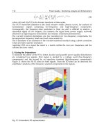
This chapter describes the use of ProxiMol selection to isolate phage Ab
against cell surface markers (Fig. 1). However, proximity selections need not be
restricted to cell surfaces: purifi ed Ags, cell extracts, or membrane preparations
may also be used. Selection of Abs that bind to a number of different target
Ags has been demonstrated using this technique, using either Abs or natural
ligands as guide molecules (6,7).
2. Materials
1. 16-Well chamber slides (Nunc).
2. Cell line for selection. Cells should be grown under normal culture conditions on
chamber slides to approx 80% confl uence (1 × 10
5
–1 × 10
6
cells/chamber).
Fig. 1. Flow chart for isolation of Abs against cell surface Ags by ProxiMol selection.
202 Osbourn
3. Cell fi xative, e.g., 0.1% glutaraldehyde in phosphate-buffered saline (PBS) (or
other appropriate fi xative).
4. Phage Ab library, freshly amplifi ed and titered (colony-forming units [cfu]/mL).
5. PBS–3% (w/v) skim milk powder (PBSM) (see Note 1).
6. PBS–0.1% (v/v) Tween-20.
7. Primary Ab to be used as a guide molecule, diluted as appropriate in PBSM
(see Note 2).
8. Secondary anti-species–HRP conjugate at an appropriate dilution (normally
1Ϻ1000–1Ϻ5000) in PBSM.
9. Biotin tyramine, stock concentration ~1 mg/mL (available as part of the Renais-
sance TSA kit) (NEN, Perkin Elmer Life Sciences, Boston, MA).
10. 50 mM Tris-HCl, pH 7.4, containing 0.03% H
2
O
2
(freshly made).
11. 1 M Tris-HCl, pH 7.4.
12. 100 mM Triethylamine, freshly diluted in H
2
O on day of use.
13. Streptavidin-coated magnetic beads with magnetic rack (Dynal).
14. Escherichia coli strain TG1, freshly grown exponential phase culture.
15. 2TY agar plates (243 × 243 mm) containing 100 µg/mL ampicillin and 2% (w/v)
glucose (or other appropriate antibiotics for recombinant Ab phage selection).
3. Methods
1. Using a pipet tip, remove the culture media from the chamber slides and wash
with 100 µL PBS.
2. Fix the cells with 100 µL 0.1% gluteraldehyde for 15 min at room temperature
(see Notes 3 and 4). Wash with 100 µL PBS as above.
3. Block the cells with 100 µL PBSM for 1–2 h at room temperature.
4. Gently wash the cells 3× by adding 100 µL PBS, then discarding.
5. Add 100 µL of the primary guide molecule and incubate for 1 h at room
temperature (see Note 5).
6. Wash the cells as in step 4.
7. Add 1 × 10
12
cfu Ab phage in 100 µL PBSM and incubate for 1–2 h at room
temperature.
8. Wash the cells as in step 4.
9. Add 100 µL secondary anti-species–HRP conjugate and incubate for 1 h at
room temperature.
10. Wash the cells as in step 4.
11. For each well, dilute 0.4 µL biotin tyramine stock solution in 100 µL 50 mM Tris-
HCl, pH 7.4–0.03% H
2
O
2
. Add to the wells and incubate at room temperature
for 10 min.
12. Wash the cells as in step 4.
13. Elute the bound phage Ab by adding 100 µL 100 mM triethylamine and incubate
at room temperature for 20 min.
14. Transfer the eluted phage to a 1.5 mL Eppendorf tube and neutralize immediately
with 50 µL 1 M Tris-HCl, pH 7.4.
Proximity-Guided Ab Selection 203
15. Take a 20-µL aliquot of the streptavidin-coated magnetic beads and remove
from the solution using the magnetic rack. Preblock the beads by resuspending
them in 50 µL PBSM.
16. Add the blocked beads to the eluted phage and incubate the suspension for
15 min at room temperature on a rotating platform.
17. Using the magnetic rack, remove the beads from the suspension and wash
them 3× in 1 mL PBS–0.1% Tween-20, followed by another three washes in
1 mL PBS.
18. Resuspend the beads in 100 µL PBS and use 50 µL of this suspension to
infect 5 mL exponentially growing culture of E. coli TG1. Incubate at 37°C for
30 min (no shaking), followed by a further incubation at 37°C for 30 min with
slow shaking (150 rpm).
19. Spin the bacteria at 2500g for 10 min and plate on 2TY agar–ampicillin–glucose
plates and incubate overnight at 30°C.
20. Use the resulting colonies to generate soluble Ab according to standard protocols
and screen for binding specifi city or activity in an appropriate assay (see Note 6).
4. Notes
1. It is advisable to remove large particulates from the PBSM solution by a 10 s
pulse in a microcentrifuge. If preferred, it is also possible to use PBS containing
0.5% BSA as a blocking reagent.
2. The primary guide Ab can be directly conjugated to HRP to avoid the use of an
anti-species HRP conjugate. HRP conjugation can be achieved using maleimide-
activated HRP, which is available in kit form from Pierce and Warriner (Chester,
UK). If a direct guide molecule–HRP conjugate is used, proceed directly to step
10, following incubation of the phage Ab library.
3. It is possible to use unfi xed cells in a proximity selection, although some loss
of cells may occur from the chamber slides after washing. If unfi xed cells are
used, washing steps must be carried out with the utmost care and all stages of the
selection process should be carried out at 4°C to reduce possible internalization
of target Ags.
4. Cells in solution can also be used for proximity selections. In this case, the
protocol should be modifi ed to include pelleting of the cells after each washing
step. If selections are to be carried out in solution, use approx 1 × 10
5
cells in a
volume of 200–500 µL PBS block solution.
5. Abs are just one example of the type of molecule that can be used as a guide
molecule. Any ligand that is known to specifi cally bind to the target Ag, and which
can be tagged in some way to allow HRP localization, can be used. Alternatives
include natural ligands. Biotinylated ligands can be used in combination with
streptavidin–HRP as long as a specifi c staining profi le is retained.
6. Any Abs selected using a ProxiMol selection will not bind to the epitope on the
target molecule to which the guide molecule binds since this particular epitope
will be blocked. It is possible, however, to carry out a second round of ProxiMol
selection, using the output from a fi rst as a guide population. In this way, Abs that
204 Osbourn
bind to the original guide-molecule binding site can be isolated. As an example
of this, Abs that block the binding of the chemokine macrophage inhibitory
protein 1-α (MIP-1α) have been isolated using MIP-1α as the original guide
molecule (7). The second round of selection was performed using the captured
biotinylated phage population output from the fi rst round as a guide population.
The phage were added to a fresh batch of cells without MIP-1α present and HRP
localization to the phage particles was achieved using streptavidin–HRP. The
output of this selection (referred to as a “step-back selection”) included clones
that blocked MIP-1α binding to the cells, along with other clones that bound to
the CD4
+
cells, but which did not inhibit binding. This general principle may to
applicable to many other selection targets.
References
1. Bobrow, M. N., Harris, T. D., Shaughnessy, K. J., and Litt, G. J. (1989) Catalyzed
reporter deposition, a novel method of signal amplifi cation. J. Immunol. Methods
125, 279–285.
2. Bobrow, M. N., Litt, G. J., Shaughnessy, K. J., Mayer, P. C., and Colon, J. (1992)
The use of catalyzed reporter deposition as a means of signal amplifi cation in a
variety of formats. J. Immunol. Methods 150, 145–149.
3. Merz, H., Malisius, R., Mannweiler, S., Zhou, R., Hartmann, W., Orscheschek,
K., Moubayed, P., and Feller, A. C. (1995) A maximised immunohistochemical
method for the retrieval and enhancement of hidden antigens. Lab. Invest. 73,
149–156.
4. Adams, J. C. (1992) Biotin amplifi cation of biotin and horseradish. J. Histochem.
Cytochem. 40, 1457–1463.
5. Earnshaw, J. C. and Osbourn, J. K. (1999) Signal amplifi cation in fl ow cytometry
using biotin tyramine. Cytometry 35, 176–179.
6. Osbourn, J. K., Derbyshire, E. J., Vaughan, T. J., Field, A. W., and Johnson,
K. S. (1998) Pathfi nder selection: in situ isolation of novel antibodies. Immunotech-
nology 3, 293–302.
7. Osbourn, J. K., Earnshaw, J. C., Johnson, K. S., Parmentier, M., Timmermans,
V., and McCafferty, J. (1998) Directed selection of MIP-1α neutralizing CCR5
antibodies from a phage display human antibody library. Nature Biotechnol. 16,
778–781.
Proximity-Guided Ab Selection 205
207
From:
Methods in Molecular Biology, vol. 178: Antibody Phage Display: Methods and Protocols
Edited by: P. M. O’Brien and R. Aitken © Humana Press Inc., Totowa, NJ
17
Isolation of Human Monoclonal Antibodies
Using Guided Selection with Mouse
Monoclonal Antibodies
Mariangela Figini and Silvana Canevari
1. Introduction
Repertoires of antibody (Ab) V genes derived from nonimmunized human
donors (1) or made synthetically (2,3) have been cloned for display on
fi lamentous bacteriophage as either scFvs or Fabs fused to the minor phage
coat protein (pIII) (4). Phage Ab repertoires can be subjected to multiple
rounds of panning on individual immobilized antigens (Ags) in order to isolate
individual Ag-binding clones. This approach has been successfully applied to
numerous purifi ed soluble molecules, yielding high-affi nity Abs (5). Selection
against almost any soluble Ag is now theoretically feasible.
Although conventional biochemistry and advanced biotechnological
approaches have led to the availability of a large variety of molecules in
purifi ed and/or recombinant form, such molecules often do not maintain the
correct conformation in soluble form and are easily disrupted by biochemical
manipulation. Thus, the use of whole living cells as a direct source of target
Ag is desirable to retain the physiological status of the molecule as much as
possible. Selection of Abs against unpurifi ed cell surface markers by panning
on whole cells is also desirable. Of particular interest is the generation of
human Abs against the surface Ags of human tumor cells since these reagents
have a potential application in immunotherapy. Unfortunately, panning on
whole cells has proven diffi cult because of the enormous number of different
Ags and the low abundance of many of them on the cell surface.
Thus, a general methodology was developed that is tailored to specifi c needs,
such as raising an Ab against a predefi ned epitope or a cell surface Ag not
Isolation of Human MAbs 207
available in purifi ed form. The procedure, originally called “epitope imprinting
selection,” and now defi ned as “guided selection” (6,7) uses one of the variable
chains of an available mouse monoclonal antibody (MAb) directed against the
target Ag to drive selection of a human Ab of corresponding-specifi city from a
preassembled repertoire of genes encoding the variable domains of human Ab
heavy chains (HCs) and light chains (LCs).
The combination of shuffl ing of V genes and selection on cells provides
a powerful tool for isolating human MAb reagents with potential clinical
application, including when the corresponding murine MAb is already in
clinical use. For example, guided selection using the murine complementarity
determining region 3 and panning on purifi ed recombinant Ag identifi ed a high-
affi nity human Ab against epithelial glycoprotein 2, a transmembrane protein
abundantly expressed on a variety of human carcinomas (8). This technique
also provides an additional approach for isolating human MAbs against Ags
present on human tumor cells (9). By combining guided selection and panning
on whole cells, we have selected human Fabs against the folate receptor, a cell
surface Ag overexpressed in many human carcinomas from phage Ab libraries
(6). Selection for other tumor-associated Ags is in progress.
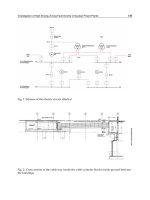
In this chapter we outline our strategy to isolate Abs against a cell-associated
Ag not available in purifi ed form. To produce and characterize such reagents,
the following procedures are carried out sequentially (Fig. 1):
1. Cloning of murine MAb V
L
C
L
gene.
2. Production of murine–human hybrid Ab library.
3. First selection by panning on cell monolayers.
4. First screening of selected Ab.
5. Production of fully human Fab library.
6. Second selection by panning on cell monolayers.
7. Second screening of selected Ab.
8. Characterization of Fab binders.
In the authors’ published procedure, the LC of a murine MAb was used to
guide HC pairings from a repertoire of human HCs (6), but either HC or LC can
be used. When soluble Ag is available for selection, the panning (steps 3 and 6)
is performed on immobilized or biotinylated Ag (see Chapters 9 and 10).
2. Materials
1. Libraries (see Note 1).
a. Library 1: Human HC repertoire (V
H
C
H
1) in a fd fi lamentous phage vector,
e.g., fdDOG (4) in Escherichia coli TG1, freshly amplified and titered
(colony-forming units [cfu]/mL) (see Note 2).
208 Figini and Canevari
Fig. 1. Schematic diagram of antibody-guided selection on cells (steps 2–8).
Isolation of Human MAbs 209
b. Library 2: Human LC repertoire (V
L
C
L
), expressed as an equimolar mixture
of phagemid expressing κ-chain and λ-chain as fusion proteins with the pIII
protein, e.g., pHEN (6) in E. coli TG1.
2. Total RNA extracted from the hybridoma cell line expressing a MAb against
the Ag of interest.
3. Cell line(s) expressing the surface Ag of interest.
4. Reagents for reverse transcriptase-polymerase chain reaction (PCR) e.g., Gene-
Amp RNA PCR Kit (Perkin-Elmer); appropriate restriction enzymes and T4
DNA ligase for cloning.
5. Oligonucleotides (Table 1; see Note 3).
6. Periplasmic expression vector for cloned V
L
C
L
e.g., pUC19SNMyc (6).
7. 8% Glutaraldehyde stock solution. Dilute to 0.2% in phosphate-buffered saline
(PBS) for fi xation.
8. PBS: PBS containing 2% (w/v) and 10% nonfat dry milk powder (PBSM);
PBS–0.1% (v/v) Tween-20; PBS–0.75% glycine–0.001% phenol red; PBS–1%
bovine serum albumin (BSA).
9. Freshly grown exponential culture of E. coli TG1.
10. 2TY medium: 2TY containing 100 µg/mL ampicillin (AMP); 2TY–AMP contain-
ing 10 µg/mL tetracycline (TET).
11. TYE agar plates: Add 15 g agar to 1 L 2TY medium, autoclave, when cool, add
glucose to 1% (w/v) and AMP and TET (as done previously).
12. Electrocompetent E. coli TG1 cells; Gene Pulser (e.g., Bio-Rad).
13. Reagents for amplifi cation of libraries (antibiotics, helper phage VCSM13, 20%
polyethylene glycol (PEG)–2.5 M NaCl solution).
14. Anti-M13 horseradish peroxidase-conjugated Ab (Pharmacia Biotech cat. no.
27-9411-01); fl uorescein-isothiocyanate-conjugated anti-sheep Ab; anti-M13 Ab
(Pharmacia Biotech cat. no. 27-9410-01).
15. Tetramethyl-benzidine dihydrochloride (TMB) solution (Sigma, T8665); 1 M
H
2
SO
4
.
16. Petri dishes and 96-well fl at-bottomed plates for cell culture; enzyme-linked
immunosorbent assay (ELISA) plates; large, square plastic Petri dishes (243
× 243 mm).
3. Methods
All of the following methods are based on the expression vectors and bacte-
rial strains that are used in our system. Variations may require modifi cation to
antibiotics and restriction enzymes used.
3.1. Cloning of Murine MAb V
L
C
L
Gene
This step allows cloning of the murine LC variable region (V
L
) gene,
together with the constant region (C
L
), into a plasmid vector that enables
secretion of the entire murine LC into the bacterial periplasm. This protocol
210 Figini and Canevari
describes a method for cloning into pUC19SNMyc, but can be adapted for
other suitable vectors.
1. Perform a cDNA reaction according to standard protocols using 1–2 µg hybrid-
oma RNA and 20 pmol VKBackSfi primer (Table 1).
2. Set up a 50 µL PCR reaction according to standard protocols using 50 ng cDNA
and 10 pM each of the VKBackSfi and MOCKForNot primers (Table 1).
3. Amplify by PCR using 30 cycles at 94°C for 1 min, 55°C for 1 min, and 72°C
for 2 min, followed by incubation at 72°C for 10 min.
4. Purify the PCR product (~600 bp) on an agarose gel and extract from the agarose
using standard protocols. Digest the PCR product and the expression vector using
the appropriate restriction enzymes (Sfi I and NotI for pUC19SNMyc). Inactivate
the enzymes and/or clean up the reaction as appropriate.
5. Ligate the PCR product into the plasmid vector using standard protocols (see
Chapters 2 and 3).
6. Transform the ligated DNA into E. coli TG1 by electroporation and plate out the
transformants on TYE–AMP plates. Incubate overnight at 37°C.
7. Using standard molecular biology methods, determine those clones with the
correct size insert and sequence (see Note 4).
3.2. Production of a Murine–Human Hybrid Ab Library
Bacteria bearing the plasmid with the LC are infected with recombinant
phage expressing the human V
H
C
H
1 repertoire. Phage particles produced by
these infected bacteria display hybrid Fabs (human V
H
CH–murine V
L
C
L
) on
their surface: the human HCs fused at their C-terminus to the phage pIII and
their murine LC partners associate spontaneously in the periplasmic space (2).
1. Grow a 10 mL culture from a fresh colony of the V
L
C
L
clone in TG1 in 2TY
containing the appropriate antibiotic selection (AMP for pUC19SNMyc) at 37°C
until optical density 600 nm reaches 0.5.
Table 1
Oligonucleotide Primers for PCR
Primer Sequence
G3LASCGTGBack GTCCTCGCAACTGGCGCGCCACAATTTCACAGTAAGG
AGGTTTAACTTGTGAAAAAATTATTATTCGCAATT
MOCKForNot CCAGCATTCTGCGGCCGCCTCATTCCTGTTGAAGCTC
TTGAC
VKBackSfi CATGACCACGCGGCCCAGCCGGCCATGGCCGACATTG
AGCTCACCCAGTCT
FdSEQ1 GAATTTTCTGTATGAGG
Isolation of Human MAbs 211
2. Mix the 10 mL log-phase culture of the V
L
C
L
clone with 10
12
cfu freshly
amplifi ed phage from the V
H
C
H
library and incubate for 30 min at 37°C without
shaking.
3. Take out a 0.5 mL aliquot and use to calculate the library titer (cfu/mL) by infect-
ing diluted aliquots in log-phase E. coli TG1 and plating on TYE–AMP–TET
plates.
4. Centrifuge the remaining 9.5 mL at 3300g for 10 min and resuspend the pellet
in 0.6 mL 2TY.
5. Plate the resuspended pellet on a TYE–AMP–TET large, square plate (243 ×
243 mm) and grow overnight at 30°C.
6. Harvest the cells by fl ooding the plate with 2–10 mL 2TY and detaching the
cells with a sterile scraper. Transfer the cells to a sterile polypropylene tube and
disperse clumps using a vortex.
7. Inoculate a 500 mL culture of 2TY–AMP–TET with an aliquot of this suspension
containing the number of bacteria that corresponds to at least 10× the library size
(where optical density of 1 at 600 nm is approx 8 × 10
8
bacteria/mL). Amplify
and isolate the recombinant phage by PEG precipitation according to standard
protocols (see Note 2).
3.3. First Selection by Panning on Cell Monolayers
The hybrid murine–human Fab phage repertoire is selected by panning on
a monolayer of cells overexpressing the target Ag. Phage that bind are used to
directly infect bacterial cells containing the LC and the bacteria are grown to
produce more Fab phage. The repertoire must be subjected to repeated rounds
of selection and infection to enrich the population for Ag binders. The fi rst
panning round is the most critical since selection for any abnormalities or
mistakes at this point will be amplifi ed during further panning. Usually, the
selection and reinfection must be carried out 2–3× in order to obtain a positive
signal in polyclonal ELISA (see Note 5).
1. Recover the cell line for panning (see Note 6) by detachment with trypsin–
ethylenediamine tetraacetic acid or by centrifugation, depending on their growth
characteristics (see Note 7).
2. Seed the cells in 100 mm cell culture Petri dishes at a concentration of
4 × 10
5
/mL in 20 mL standard growth medium and grow at 37°C until 80%
confl uent (1–2 d, depending on the duplication time of the cell line).
3. If necessary (i.e., when the attachment of cells to plastic is not strong enough),
and, if it is possible (i.e., Ag does not change conformation during the fi xation),
fi x the cells with gluteraldehyde by adding 4 mL 0.2% glutaraldehyde in PBS
and leave for 5 min only (NOT MORE).
4. Wash the cells 5–6× by adding 10 mL PBS briefl y swirling the plate, then
discarding.
5. Add 4 mL PBS–0.75% glycine–0.001% phenol red and leave for 5 min.
212 Figini and Canevari
6. Wash 5–6× with PBS.
7. Add 20 mL 2% PBSM and incubate for 2 h at 37°C to block any remaining
unsaturated binding sites on the plastic.
8. Discard the block solution and add ~10
12
cfu freshly amplifi ed phage library dis-
playing the hybrid Fabs in 10 mL 2% PBSM. Shake slowly at room temperature
for 1 h (see Note 8).
9. Wash the dish 5× with 10 mL PBS–0.1% Tween-20, then 10× with 10 mL PBS
alone. In each washing step, gently add the buffer, briefl y swirl in the plate, and
immediately remove (see Note 9).
10. Add 10 mL log-phase E. coli TG1 bearing the V
L
C
L
plasmid (see Subheading
3.1.) directly to the Petri dish (see Note 10). Incubate for 30 min at 37°C without
shaking.
11. Collect the bacterial suspension from the dish, take out a 0.5 mL aliquot,
and use to calculate the rescue titer (cfu/mL) by plate-diluted aliquots on
TYE–AMP–TET glucose plates.
12. Centrifuge the remaining 9.5 mL at 3300g for 10 min and resuspend the pellet
in 0.6 mL 2TY.
13. Plate the resuspended pellet on a TYE–AMP–TET large, square plate (243 ×
243 mm) and grow overnight at 30°C.
14. Harvest the cells by fl ooding the plate with 2–10 mL 2TY and detaching the
cells with a sterile scraper. Transfer the cells to a sterile polypropylene tube and
disperse clumps using a vortex.
15. Inoculate a 500 mL culture of 2TY–AMP–TET with an aliquot of this suspension,
corresponding to at least 10× the library size (see Subheading 3.2., step 7).
Amplify and isolate the recombinant phage by PEG precipitation according to
standard protocols (see Note 2).
16. Check the phage-binding specifi city by phage ELISA or fl uorescence-activated
cell sorting (FACS) analysis using the total amplifi ed selected phage (polyclonal)
or single clones (monoclonal).
17. Repeat the selection and infection 2–3×. After every round of infection, check the
titer of the phage to monitor the extent of enrichment and check the cell binding
by ELISA. Repeat the panning until a positive signal against Ag is detected.
3.4. Screening of Selected Phage Clones
Single clones (monoclonal) or the bulk amplifi ed (polyclonal) phage are
tested for binding both in ELISA and in FACS analysis on cells overexpressing
the Ag of interest and on Ag-negative cells.
3.4.1. Monoclonal Phage ELISA
1. Seed the target cells in 96-well flat-bottomed plates at a concentration of
1–3 × 10
5
cells/mL in 200 µL and grow until they reach confl uence (1–2 d
depending on the duplication time of cells).
Isolation of Human MAbs 213
2. Pick individual colonies from the last round of panning using a sterile toothpick
into wells of a 96-well sterile ELISA plate containing 200 µL 2TY–AMP–TET.
Grow with shaking (300 rpm) at 30°C for 16–20 h (see Note 11).
3. Centrifuge the bacterial plate at 3300g for 10 min.
4. Wash the plate containing the cell monolayer 3× by gently adding 200 µL of PBS
and aspirating off. Block the unsaturated binding sites on the plastic by adding
200 µL 2% PBSM to each well and incubate at 37°C for 2 h.
5. Aspirate off the 2% PBSM and add 25 µL 10% PBSM to each well.
6. Using a multichannel pipet, transfer 80 µL bacterial culture supernatant from
step 3 above to the ELISA plate, mix, and incubate at room temperature for 1 h.
7. Wash the wells 3× with PBS–0.1% Tween-20, then 3× with PBS.
8. Add 100 µL 1Ϻ5000 dilution of the anti-M13–horseradish peroxidase conjugate
Ab in 2% PBSM to each well. Incubate for 1 h at room temperature.
9. Wash the wells as in step 7.
10. Add 50 µL TMB substrate to each well and leave at room temperature for 10–20 min
or until a clear signal is seen.
11. Stop the reaction by adding 50 µL 1 M H
2
SO
4
to each well and read the
absorbance at 450 nm in an ELISA multichannel reader.
3.4.2. FACS Analysis Using Polyclonal Phage
1. Detach the adherent cell line expressing the Ag of interest from the fl ask as
described in Subheading 3.3., step 1. Wash the cells by adding 1 mL PBS and
centrifuging at 350g for 5 min, then count the cells. For each FACS sample,
aliquot 3 × 10
5
cells into a centrifuge tube and pellet by centrifugation described
previously.
2. Resuspend the cell pellet with ~5 × 10
9
polyclonal phage particles in 100 µL
PBS–1% BSA and incubate at room temperature for 1 h.
3. Wash the cells 3× with PBS, as in Subheading 3.4.1., step 7.
4. Add 100 µL anti-M13 Ab (1Ϻ1000 dilution in PBS–1% BSA) to each tube and
incubate on ice for 30 min.
5. Wash the cells 3× with PBS.
6. Add 100 µL fluorescein-isothiocyanate-conjugated anti-sheep Ab (1Ϻ1000
dilution in PBS–1% BSA) and incubate on ice for 30 min.
7. Wash the cells 3× with PBS.
8. Immediately analyze the cells using a FACScan. Alternatively, fi x the cells in
PBS–1% formaldehyde and store at 4°C (the cells will retain their fl uorescence
for about 2 wk).
At the end of 3–6 rounds of positive selection and screening, at least one
human V
H
C
H
1-bearing clone should be identifi able, which, in combination with
the original murine LC used as the guide probe, can bind to the target Ag.
214 Figini and Canevari
3.5. Selection of the Human LC
After screening and identifi cation of the best human V
H
, the second shuffl e
can be performed. To obtain a completely human Fab, the selected V
H
C
H
1
is amplified and inserted into the phagemid library containing precloned
repertoires of human κ and human λ LC genes. The resulting human Fab
phagemid repertoire is rescued by infection with helper phage and the phagemid
particles are selected on cell monolayers as previously mentioned.
1. Pick a fresh colony of the selected clone(s) into 10 mL 2TY–TET and grow
overnight at 37°C.
2. Extract the plasmid DNA using standard miniprep procedures.
3. Amplify the V
H
C
H
1 insert, using a 50-µL PCR reaction containing 100 ng V
H
C
H
1
plasmid DNA and 10 pM each of the Fdseq1 primer and G3LASCGTGBack
primers (Table 1) and perform 30 cycles at 94°C for 1 min, 55°C for 1 min, 72°C
for 2 min, followed by incubation at 72°C for 10 min.
4. Run out the reaction on a 1.5% agarose gel and purify the fragment using
standard protocols.
5. Digest 5 µg V
H
C
H
1 fragment and 6 µg plasmid containing the human V
L
C
L
library DNA with AscI and NotI. Inactivate the enzymes and/or clean up the
reaction as appropriate.
6. Using standard protocols, construct the fully human library by ligation. Amplify
the library using helper phage, VCSM13, and isolate recombinant phage using
PEG precipitation.
7. Perform a further 3–4 rounds of panning on the cell line as in Subheading 3.3.
and analyze the selected phage by phage ELISA (Fig. 1; see steps 6 and 7).
8. Characterize the selected fully human Fab clones, e.g., by sequencing and by
biological/biochemical analysis (Fig. 1; see step 8; ref. 10).
4. Notes
1. These libraries may be prepared using the protocol described in ref. 6, or may
be available in other laboratories. To obtain an update of the primers used
in amplifi cation of the immunoglobulin variable genes, consult the website
A critical
step in determining the success of the guided selection procedure is that these
libraries should be as large as possible, with a high number of functional inserts.
Since the aim of guided selection is to recapitulate all the properties of binding
specifi city and affi nity of the mouse Ab in its human equivalent, maximization
of library sizes during chain shuffl ing ensures that as many permutations as
possible of HC/LC pairings are available for Ag selection. The ease with which
a high-affi nity Ab can be isolated by phage display correlates with the size of
the starting repertoire (1,5).
Isolation of Human MAbs 215
2. The fdDOG vector is a phage, therefore, helper phage is not required for packag-
ing of this library. pHEN is a phagemid, and requires helper phage (VCSM13) for
amplifi cation. The fdDOG vector carries a tetracycline-resistance marker.
3. Table 1 lists the sequences of the universal primers, MOCKForNot and
VKBackSfi , which can be used to amplify the majority of murine VKCK. If these
primers fail to amplify the LC, try the primers designed by Pharmacia (cat. no.
27-1583-01). Also check that the LC is not from a rare murine family.
4. Many hybridomas also express nonfunctional mRNAs that encode abortive
immunoglobulin variable regions and that can be amplifi ed during PCR. It is
therefore advisable to construct scFvs from the hybridoma, before proceeding
to ensure the identifi cation of functional V
L
. Furthermore, for some Abs, the
V
H
alone is suffi cient to determine the binding specifi city to the target Ag (11).
Therefore, it is advisable to control for this possibility before proceeding to
guided selection with the LC.
5. Starting with 10
12
phage, the fi rst round of selection should yield at least 10
4
phage. Initial rounds of selection should be carried out under low stringency
(using cells with high Ag density and minimizing the number and duration of
washes), so as not to lose rare binders, then use more stringent conditions in later
rounds. Stringency conditions should be fi ne-tuned by the operator according to
the topobiology of the target Ag. All rescues should be checked for the presence of
insert (by PCR) and the number of positive clones should scored. If no enrichment
is obtained after 4–6 pannings, check the length of the insert, change the conditions
of panning (temperature, incubation time, fi xation of cells), use an alternative cell
line, if possible, and/or carry out a new selection from the beginning.
6. Maintain the cells expressing the target Ag in complete culture medium in
a humidified 5% CO
2
atmosphere at 37°C. Routinely confirm cell surface
expression of the Ag by FACS analysis or ELISA.
7. These procedures pertain to adherent cells. Similar procedures can be applied
to cells growing in suspension, using centrifugation for each wash step. The
major risk with using cells in suspension is their loss and consequently that of
the bound phage during centrifugation. Thus, a centrifugation speed must be
selected that is appropriate to the size and sensitivity of the cells, balancing
the risk of cell loss caused by fl otation (low speed) and centrifugation damage
(squashing) (high-speed). Alternatively, the cells can be attached to the plastic
substrate, using polylysine.
8. Culture plates are treated to ensure cell adhesion even in presence of excess pro-
tein; therefore, shaking is necessary to reduce nonspecifi c phage adherence
to plates.
9. If the cells are detergent-sensitive, wash with PBS alone, or, alternatively, use
fi xed cells.
10. Infection of bound phages by direct addition of the E. coli strain containing
the V
L
C
L
to the cells generally leads to an increased number of rescued phage
without an increase in background. Alternatively, bound phages can be eluted
216 Figini and Canevari
from the plate with 100 mM triethylamine or 50 mM glycine–HCl, pH 2.7,
150 mM NaCl.
11. At this stage, the culture is saturated, and should yield 10
10
cfu/mL. Aeration is
important at this stage of growth; aeration and yields can be increased by placing
the 96-well plate into a box without a lid.
References
1. Marks, J. D., Hoogenboom, H. R., Bonnert, T. P., McCafferty, J., Griffi ths, A. D.,
and Winter, G. (1991) By-passing immunization: human antibodies from V-gene
libraries displayed on phage. J. Mol. Biol. 222, 581–197.
2. Hoogenboom, H. R. and Winter, G. (1992) Bypassing immunization: human
antibodies from synthetic repertoires of germ line VH-gene segments rearranged
in vitro. J. Mol. Biol. 227, 381–388.
3. Nissim, A., Hoogenboom, H. R., Tomlinson, I. M., Flynn, G., Midgley, C., Lane,
D., and Winter, G. (1994) Antibody fragments from a ‘single pot’ phage display
library as immunochemical reagents. EMBO J. 13, 692–698.
4. Hoogenboom, H. R., Griffi ths, A. D., Johnson, K. S., Chiswell, D. J., Hudson, P.,
and Winter, G. (1991) Multi-subunit proteins on the surface of fi lamentous phage:
methodologies for displaying antibody (Fab) heavy and light chains. Nucleic Acids
Res. 19, 4133–4137.
5. Griffi ths, A. D., Williams, S. C., and Hartley, O. (1994) Isolation of high affi nity human
antibodies directly from large synthetic repertoires. EMBO J. 13, 3245–3260.
6. Figini, M., Marks, J. D., Winter, G., and Griffi ths, A. D. (1994) In vitro assembly
of repertoires of antibody chains on the surface of phage by renaturation. J. Mol.
Biol. 239, 68–78.
7. Jespers, L. S., Roberts, A., Mahler, S. M., Winter, G., and Hoogenboom, H. R.
(1994) Guiding the selection of human antibodies from phage display repertoires
to a single epitope of an antigen. Biotechnology 12, 899–903.
8. Beiboer, S. H. W., Reurs, A., Roovers, R. C., Arends, J. W., Whitelegg, N. R.,
Rees, A. R., and Hoogenboom, H. R. (2000) Guided selection of a pan carcinoma
specifi c antibody reveals similar binding characteristics yet structural divergence
between the original murine antibody and its human equivalent. J. Mol. Biol.
296, 833–849.
9. Kupsch, J. M., Tidman, N. H., Kang, N. V., Truman, H., Hamilton, S., Patel, N., et
al. (1999) Isolation of human tumor-specifi c antibodies by selection of an antibody
phage library on melanoma cells. Clin. Cancer Res. 5, 925–931.
10. Figini, M., Obici, L., Mezzanzanica, D., Griffi ths, A. D., Colnaghi, M. I., Winter,
G., and Canevari, S. (1998) Panning phage antibody libraries on cells: isolation of
human Fab fragments against ovarian carcinoma using guided selection. Cancer
Res. 58, 991–996.
11. Ward, E. S., Gussow, D. H., Griffi ths, A. D., Jones, P. T., and Winter, G. (1989)
Binding activities of a repertoire of single immunoglobulin variable domains
secreted for Escherichia coli. Nature 341, 544–546.
Isolation of Human MAbs 217
219
From:
Methods in Molecular Biology, vol. 178: Antibody Phage Display: Methods and Protocols
Edited by: P. M. O’Brien and R. Aitken © Humana Press Inc., Totowa, NJ
18
Selecting Antibodies to Cell-Surface Antigens
Using Magnetic Sorting Techniques
Don L. Siegel
1. Introduction
As described in other chapters, the selection of phage-displayed immuno-
globulin (Ig) fragments with desired specifi city can be accomplished through
successive rounds of panning on purifi ed antigen (Ag). However, for many
applications, the target Ag may not be able to be purifi ed because its identity
is unknown (e.g., a putative stem cell or tumor-specifi c marker) or because the
process of purifi cation destroys its native conformation (e.g., in the case of
some integral membrane proteins). In such experimental systems, methods that
select phage-displayed Ig directly on intact cell surfaces are required.
Compared to panning on purifi ed Ag, cell-surface selection must overcome a
number of technical hurdles, most notably the nonspecifi c adsorption of phage
by irrelevant protein, carbohydrate, or lipid structures expressed by the cell.
In some cases, several rounds of negative selection on Ag-negative cells can
be performed prior to positive selection on cells bearing the target molecules.
This strategy may be ineffi cient because only a small fraction of nonspecifi c
phage can be removed during each cycle of negative selection and one runs
the risk of losing the desired phage particles (initially present at low levels)
through nonspecifi c interactions with the Ag-negative cells.
More effi cient methods for cell-surface panning utilize simultaneous posi-
tive and negative selection. In this chapter, such a competitive cell-panning
approach is presented, in which target cells are precoated with magnetic beads
and mixed with an excess of unmodifi ed Ag-negative cells (1).Following
incubation of the cell mixture with a phage-display library, the target cells
bearing Ag-specifi c phage are rapidly separated on a magnetic column and
Selection Using Magnetic Techniques 219
washed, and the desired phage-displayed Igs are eluted from the cell surface
for subsequent amplifi cation. This approach has been used for the isolation of
human auto- and alloantibodies, particularly for the selection of large arrays of
anti-red blood cell (RBC) antibodies from human immune libraries (2). In the
protocols and notes that follow, representative sample data from these studies
are provided for reference.
2. Materials
1. Phage-display library, freshly amplifi ed and titered (typically at a concentration
of ~10
13
colony-forming units [cfu]/mL in phosphate-buffered saline [PBS]).
2. Ag-positive (target) cells and Ag-negative (absorber) cells. For RBCs, 3–4%
(v/v) suspensions of phenotyped cells are available from Gamma Biologicals,
Houston, TX. Approximately 10
8
target and absorber RBCs are needed per
experiment. For other cell types, see Note 1.
3. Sulfo-NHS-LC-biotin (Pierce, Rockford, IL). Prepare a 1 mg/mL solution (see
Note 2) in room temperature (RT) PBS immediately prior to use.
4. Streptavidin-coated paramagnetic beads; minicolumns for magnetically activated
cell sorting (MACS); magnet separation unit (MiniMACS) (Miltenyi Biotec,
Sunnyvale, CA).
5. 30-gauge × 1/2-in. hypodermic needle (Becton-Dickinson, Franklin Lakes, NJ).
6. 5× PBSM: 10% (w/v) nonfat dry milk in PBS, pH 7.4; PBSM: 5× PBSM diluted
to 1× with PBS and degassed before use.
7. PBS-BSA: bovine serum albumin (BSA) prepared as a 3% (w/v) solution in
PBS, pH 7.4.
8. Acidic phage elution buffer (76 mM citric acid, pH 2.4) and phage elution
neutralization solution (untitrated 2 M Tris-HCl base) (see Note 3).
9. Materials for phage amplifi cation (see Note 4).
10. Materials for assaying the binding of panned libraries and isolated phage clones
(see Note 5).
3. Methods
1. To prepare the target cells for surface biotinylation, wash the cells 5× with RT
PBS and resuspend to a fi nal volume that yields a 20% (v/v) cell suspension
(see Fig. 1 and Note 6). For experiments utilizing RBCs, 10
8
RBCs will provide
enough target cells for ~10 selection procedures (see Note 1).
2. Add an equal amount of freshly prepared biotin reagent solution to the cell
suspension, mix thoroughly by drawing up and down with a micropipetor,
and incubate for 40 min at RT on a laboratory rotator to maintain the cells in
suspension.
3. Wash the cells 5× with 400 µL RT PBS to remove the unreacted biotin reagent.
For RBC, pulse centrifugation for approx 4 s in a microcentrifuge set at full
speed is suffi cient to pellet the cells. After the fi nal wash, resuspend the cells in
PBS to their prebiotinylated volume as in step 1.
220 Siegel