differential display methods and protocols
Bạn đang xem bản rút gọn của tài liệu. Xem và tải ngay bản đầy đủ của tài liệu tại đây (11.85 MB, 294 trang )
1
Differential
Display
A General Protocol
Peng Liang and Arthur B. Pardee
1. Introduction
One of the greatest unsolved mysteries of life 1show the hundreds of thousands of genes embedded in the genome of an organism are selectively
expressed mto the mRNA and protems m a temporally and spatially regulated
manner that gives rise to different tissues and organs. The abnormality m this
intricate regulatory cn-cuitry IS beheved to be one of the underlmmg causes of
a variety of pathological alterations or disease states.The rsolation and characterization of differentially expressed genes becomes one of the first steps
toward the understanding of these important biological questions. Differential
display (1) and a related RAP-PCR method (2) were developed to more efficiently Identify and isolate these genes.
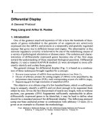
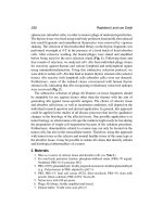
The general strategy for differential display (Fig. 1) IS based on a combmatton of three techniques brought together by a concept:
1 Reverse transcrlptlon of mRNA from anchored primers (see Note l),
2 Choice of arbitrary primers for setting lengths of cDNAs to be amplified by the
polymerase chain reaction (PCR), each corresponding to part of a mRNA (tags),
3 Sequencmg gels for high resolution of amplified cDNA
The objective IS to obtain a tag of a few hundred bases,which 1ssufficiently
long to uniquely identify a mRNA and yet short enough to be separated from
others by size. Given the fact that primers of nearly any length, with or without
anchors, can generate cDNA fingerprints sufticrently reproducible to allow
differentially expressed genes to be identified, it may be hard to define what
should be a standard protocol for differential display. The followmg protocol
using one-base anchored primer m combmatron with arbitrary 13-mers (3) IS
given as an example to illustrate the methodology.
From
Methods m Molecular Bology, Vol 8.5 D/fferenf/a/
Edlted by P Llang and A B Pardee Humana
3
Dsplay Methods and Protocols
Press Inc , Totowa, NJ
Liang and Pardee
L
Reverse banswiption
5’.AAGC3’
dNTPs
MMLV reverse tfanscnptase
.
WAA-A~
GVGAA
4
JI.
(H-TUG)
PcRamplificatloll
AAGCTTGATECC
I
5’-AAGCTTGAITGCC-3’
(H-AP-1 Ptimer)
5’44GC3
(H-TIIG)
dNTPs
a-(‘“S-dATP]
Ampli’lhq DNA poiymelase
.
GWGAA
A4GCiTGA’lTGCC
.
G-GU
IIL
Denaturing polyacrylamide gel
Fig. 1. Schemattc representation of one-base anchored differentral display
2. Materials
1. 5X RT buffer: 125 mA4TrwC1, pH 8 3,188 mMKCl,7.5
dithrothretol.
2 MMLV reverse transcrrptase (100 cl/@,)
3. dNTP (250 @I)
4. 5’-AAGCTTTTTTTTTTTG-3’
(2 /.&I).
5. 5’-AAGCTTTTTTTTTTTA-3’
(2 pA4)
6 5’-AAGCTTTTTTTTTTTC-3’
(2 /&I).
7 Arbitrary 13-mers (2 pM)
8. 10X PCR buffer.
mMMgC12, and 25 ti
Differential
9
10.
Il.
12.
13.
14.
15
16.
17
18.
19.
20.
2 1.
Display
5
dNTP (25 pJ4).
Glycogen (10 mg/mL).
Distilled water (dH,O).
DEPC-treated H,O
Loading dye
AmphTaq DNA polymerase, Perkin-Elmer Corporation (Norwalk, CT).
a-[33P]dATP (>2000 Wrnmole) or a-[35S]dATP (>l,OOO Ci/mmole) (see Note 2).
RNase-free DNase I (10 U/pL).
QIAEXrM DNA extraction kit (Qiagen, Chatsworth, CA).
pCR-TRAPTM clonmg system (GenHunter Corporation, Nashville, TN).
Thermocycler
6% denaturmg polyacrylamide gel.
DNA sequencing apparatus.
Although individual components may be purchased separately from varrous
suppliers, most of them can be obtained in kit forms from GenHunter Corporation.
3. Methods
3.1. DNase I Treatment of Total RNA
Purification polyadenylated RNAs is neither necessary nor helpful for differential display. The major pitfalls of using the polyadenylated mRNAs are
the frequent contammation of the oligo-dT primers, that give high background
smearing in the display and the difficulty m assessing the integrity of the
mRNAs templates (4). Total cellular RNAs can be easily purified with one-step
acid-phenol extraction method (5). However, no matter what methods are used
for the total RNA purification, a trace amount of chromosomal DNA contamination m the RNA sample could be amplified along with mRNAs thereby comphcating the pattern of displayed bands. Therefore removal of all contaminating
chromosomal DNA from RNA samples is essential before carrying out differential display.
1. Incubate 10-100 pg of total cellular RNA with 10 U of DNase I (RNase free) in
10 mMTris-Cl, pH 8.3, 50 mMKC1, 1.5 mMMgC& for 30 mm at 37°C.
2 Inactivate DNase I by adding an equal volume of phenolchloroform
(3: 1) to the
sample
3. Mix by vortexing and leave the sample on ice for 10 mm.
4 Centrifuge the samplefor 5 min at 4’C m an Eppendorf centrifuge.
5. Save the supernatant, and ethanol precipitate the RNA by adding 3 vol of ethanol
in the presence of 0.3MNaOAC, and incubate at -80°C for 30 mm.
6. Pellet the RNA by centrifuging at 4°C for 10 min.
7. Rinse the RNA pellet with 0.5 mL of 70% ethanol (made with DEPC-H20) and
redissolve the RNA in 20 pL of DEPC-treated HzO.
8. Measure the RNA concentration at ODS6s with a spectrophotometer by diluting
1 pL of the RNA sample in 1 mL of HzO.
Llang and Pardee
6
9 Check the integrity of the RNA samples before and after cleanmg wtth DNase I
by runnmg 1-3 ~18of each RNA on a 7% formaldehyde agarose gel
10 Store the RNA sample at a concentratton htgher then 1 pg/$ at -80°C before
using for differential dtsplay
3.2. Reverse
Transcription
of mRNA
1 Set up three reverse transcription
reactions for each RNA sample in three
microfuge tubes (0 5-mL), each contammg one of the three dtfferent anchored
ohgo-dT prrmers as follows. For 20 pL final volume 9 4 p.L of dH,O, 4 pI. of 5X
RT buffer, 1.6 pL of dNTP (250 I.&‘), 2 pL of DNA-free total RNA (freshly
diluted to 0 1 pg/pL wtth DEPC-treated H,O), and 2 pL of AAGCT, ,M (2 $4)
(M can be either G, A, or C)
2 Program your thermocycler to* 65°C for 5 mm, 37°C for 60 min, 75’C for 5 mm,
4°C (see Note 3)
3 1 pL MMLV reverse transcrlptase 1s added to each tube 10 mm after at 37°C and
mix well quickly by finger tipping
4. Continue mcubation and at the end of the reverse transcription reaction, spm the
tube briefly to collect condensation Set tubes on ice for PCR or store at -80°C
for later use.
3.3. PCR Amplification
1 Set up PCR reacttons at room temperature as follow* 20 p.L final volume for each
primer set combmation 10 pL of dH,O, 2 $ of 10X PCR buffer, 1.6 l.iL of dNTP
(25 pA4), 2 pL of arbitrary 13-mer (2 CUM),2 pL of AAGCT, ,M (2 CIM), 2 pL of
RT-mix from step 3 2 , 0 2 pL of a-[33P]-dATP (see Note 2), 0 2 p.L of AmpliTaq.
Mix well by pipetmg up and down (see Note 4)
2. Add 25 pI.. mineral oil if needed
3 PCR as follows 94°C for 30 s, 40°C for 2 mm, 72°C for 30 s for 40 cycles, 72°C for
5 mm, 4“C (For Perkm-Elmer’s 9600 thermocycler it is recommend that the denaturanon temperature be shortened to 15 s and the rest of parameters kept the same )
3.4. 6% Denaturing
1
2
3
4
Polyacrylamide
Gel Electrophoresis
Prepare a 6% denaturmg polyacrylamide gel m TBE buffer
Let it polymertze at least for more than 2 h before usmg
Prerun the gel for 30 mm
Mix 3.5 pL of each sample with 2 p.L of loading dye and incubate at 80°C for
2 mm immediately before loading onto a 6% DNA sequencmg gel (see Note 5)
5 Electrophorese for about 3 5 h at 60 W constant power (with voltage not to exceed
1700 V) until the xylene dye (the slower movmg dye) reaches the bottom Turn
off the power supply and blot the gel onto a piece of 3M Paper Cover the gel
with a plastic wrap and dry tt at 80°C for 1 h Do not fix the gel with methanol/
acetic acid (see Note 6)
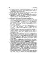
6. Orient the autoradiogram and dried gel wtth radioacttve mk or needle punches
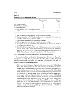
before exposing to a X-ray film. Figure 2 shows a representative differential dts-
7
Differential Display
H-T110
H-T110
H-TIIA
H-WA
H-TIIA
H-TIIC
H-TllC
H-AP3
H-APB
H-API
H-AP3
H-AP3
H-APl
H-AP3
Fig. 2. Differential display using one-base anchored oligo-dT primers (7). Four RNA
samples from non-transformed cell line Rat 1 and H-ras transformed cell lines rat 1 (ras),
T101-4 and Al-5 (lanes from left to right, respectively) were compared by differential
display using three one-base anchored oligo-dT primers, AAGCT, ,G, AAGCT, ,A and
AAGCT, ,C in combinations with three arbitrary 13-mers, H-API (AAGCTTGATTGCC),
HAP2 (AAGCTTCGACTGT)
and HAP3 (AAGC’l’l’l’GGTCAG).
The mob-l (ZO)
and mob-7 cDNA fragments were marked by the right and let? arrowheads, respectively.
play obtained with three one-base anchored oligo-dT primers in combinations
with three arbitrary 13-mers (3).
3.5. Reamplification
of cDNA Probe
1. After developing the film (overnight to 72-h exposure), orient the autoradiogram
with the gel.
2. Locate bands of interest (see Note 7) either by marking with a clean pencil from
underneath the film or punching through the film with a needle at the four corners
Liang and Pardee
3.
4.
5
6.
7.
8
9.
10.
11.
12
13.
14.
15
of each band of interest (Handle the dried gel with gloves and save it between
two sheets of clean paper)
Cut out the located band with a clean razor blade
Soak the gel slice along with the 3M paper in 100 pL dH,O for 10 mm.
Boil the tube with tightly closed cap (e.g., with parefilm) for 15 min.
Spm for 2 mm to collect condensatron and pellet the gel and paper debris Transfer the supernatant to a new micromge tube.
Add 10 pL of 3MNaOAC, 5 pL of glycogen (10 mg/mL) and 450 p.L of 100%
EtOH.
Let sit for 30 mm on dry ice or in a -8O’C freezer Spm for 10 mm at 4°C to
pellet DNA.
Remove supernatant and rinse the pellet with 200 pL we-cold 85% EtOH (you
will lose your DNA if less concentrated EtOH is used!).
Spm briefly and remove the resrdual ethanol.
Dissolve the pellet m 10 pL of PCR H,O and use 4 pL for reamplificatron.
Save the rest at -20°C in case of mishaps.
Reamplification should be done using the same primer set and PCR conditions
except the dNTP concentrations are at 20 piV (use 250 @4 dNTP stock) instead
of 2-4 pA4 and no rsotopes added. A 40-pL reaction is recommended for each
reactron: 20.4 of pL dH,O, 4 pL of 10X PCR buffer, 3.2 pL of dNTP (250 @4),
4 & of arbitrary 13-mer (2 @?), 4 $ AAGCT, ,M (2 @4) (M can be either G, C,
or A), 4 pL of cDNA template from step 3.2. and 0.4 pL of AmphTaq (5 U/pL).
Run 30 pL of the PCR sample on a 1.5% agarose gel stained with ethidmm bromide (More than 90% probes should be visible on the agarose gel ) Save the
remaining PCR samples at -2O’C for subclomng.
Check to see rf the size of your reamplified PCR products are consistent with
their size on the denaturing polyacrylamide gel.
3.6. Confirmation
of Differential
Gene Expression
1. Extract the reamplified cDNA probe from the agarose gel using QIAEX kit.
2. Use the extracted cDNA as a probe for Northern blot confirmation following the
standard protocol (ref. 5; see Note 8; Fig. 3)
3. Clone the cDNA probe using the pCR-TR4PTM cloning system (see Note 9).
4. Confirmation of differentially expressed cDNA probes can be also carried out
more efficiently by “Reverse Northern” dot blot or differential screening of
cloned cDNA probes by colony hybrtdization (ref. 6; Chapter 8 by H Zhang et
al. m this book).
5. Clone the full-length cDNA by screening a cDNA library followmg the standard
procedure (5).
4. Notes
1. The initial choice of usmg two-base anchored ohgo-dT primers (1) instead of
one-base anchored primers (3) were owing to a historical rather than scienttfic
reason. The cloned marine thymidine kmase (TK) cDNA originally used as a
Differential
9
Display
Mob-l
A
H-AP2
+
AAgc~~CTGTACAAAGG~~C~~T~A~~AC~~~~~
ATATGTAAGAACGTATGTATCAATGGGTAGITAAAGTlTACATAGG
CAAATGClTl-GAATGCTACATAlTACAAGATGTGlTGGATGGlllTCAMATAMAT
GTACTGTATTGAATGTAGTATGAGACCAAAAAA
GTAATAAAGTAATAATAACTGAC
ATGAAAAAAAAAAAGC-IT
4
H-T1 I C
Mob-7
B
H-AP2
*
AAgcttcGAcTGTAcAAA~GcGGAAcTccfGAATGTATTTT
ATAT~AAGAAClTGTGTGGTAAGTATGTATGTAfCAATGGGTAGlTAAA~ACATAGG
CAAATGCllTGAATGCTACATATTACAAGATGElTGGATGGlllTCAAAATAAAAT
GTACCCAAAAAAGTAATAAAGTAATAATAACTGAC
ATGAAATGCAAAAAAAAAAAGCTT
4
H-T1 I G
C
1234
Mob-7
rRNA
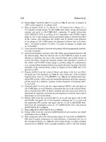
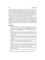
Fig. 3. Nucleotide sequences of mob-l (A) and mob-7 (B) cDNA fragments cloned
by differential display. The flanking primers are marked by arrow bars and the
polyadenylation site is underlined. Mob-7 differs from mob-l only by 6 base addition
at the 3’ end of the cDNA (see Note 10). Northern blot analysis with mob-7 cDNA probe
(C). The 253 bp mob-7 cDNA was used as a probe to confirm the differential expression of the gene using 20 pg of total RNA from Rat 1 and three transformed derivatives Rat 1 @as), T101-4 and Al-5 cells (lanes 1 to 4, respectively). The lower panel is
ethidium bromide staining of ribosomal RNAs as a control for equal sample loading.
10
2
3.
4
5
6
7.
8.
9.
Lang and Pardee
control cDNA template had only 11 As m its poly(A) tall It was found that onebase anchored prrmer Tl 1C fatled to amplify the TK 3’ termmus m combmatton
wtth an upstream primer specific to TK. Extension of one more base from the 3’
end instead of the 5’ end of the anchored primer was a logical. Interestingly,
Tl 1CA started to work successfully m PCR to amplify the expected TK cDNA
template (1) Later, longer one-base anchored primers that had mismatches at the
5’ ends of the prtmers were shown to be much more efficient for differential
display m subdividing the mRNA populations mto three groups (3) One-base
anchored primers have significant advantage over the two-base anchored primers
m that the former cuts down the redundancy of priming, elimmates the high background smearing problem for two-base anchored pnmers ending with the 3’ “T” and
reduce the number of reverse transcription reactions from 12 to 3 per RNA sample.
It has been observed that 35S labeled nucleottde origmally used for differential
display would leak through PCR reaction tubes (espectally when thin-walled
tubes are used) and 33P labeled nucleotide was recommended as the best alternative (9). 33P is not only safer to use but also gives better sensmvtty compared to 35S.
For the reverse transcrtption reaction, the mmal 65°C incubation is intended to
denature the RNA secondary structure. The final incubation at 75°C for 5 minis
to inactivate the reverse transcrlptase without denaturing the cDNA/mRNA
duplexes Therefore “hot start “PCR is neither necessary, nor helpful for the subsequent PCR reactions using cDNAs as templates
Make core mixes as much as possible to avoid ptpetmg errors (e g , aliquot
RT-mix and AP-primer mdrvidually)
Otherwise it would be difficult to pipet
0.2 pL of AmpliTaq. Mix well by pipetmg up and down
It is crucial that the urea in the wells be completely flushed right before loading
your samples For best resolution, flush every 4-6 wells each time during sample
loading while trying not to disturb the samples that have been already loaded.
DNA is acid labile, especially at high temperature when the gel is dried. This will
affect the subsequent PCR during the reamplificatton of the cDNA fragments to
be analyzed further
First tentatively identify those bands that appear to be differentially expressed on
the initial display gel. Then repeat the RT step and the PCR reactions for these
lanes and see if these differences are reproducible before pursumg further It 1s
recommended that bands bigger than 100 bp be selected. It has been generally
observed that shorter cDNA probes have higher probability of failing to detect
any signals on the Northern blot.
It IS recommended that the standard prehybrrdrzatron and hybridrzation condttton
at 42°C be used. Wash with 1X SSC, 0.1% SDS at room temperature for 15 min
twice followed by washing with 0.25X SSC, 0 1% SDS at 55-6O”C for 15-30 mm.
Do not go over 60°C Expose with intensifying screen at -80°C for overnight
to 1 wk.
pCR-TRAP cloning system is by far the most efficient cloning method for PCR
products that we have tested The pCR-TRAP clonmg system utilizes the third
generation cloning vector that features postttve-selection for DNA mserts Only
Different/al Display
II
the recombinant plasmtds confer the antibiotic resistance The prmclple of thts
unique clonmg system 1sbased on that the phage Lambda repressor gene c1 cloned
on the pCR-TRAP vector codes for a repressor protein. The repressor protein
binds to the Lambda right operators Or1 to Or3 of the cro gene, thereby turning
off the promoter that drives the TetR gene on the plasmtd Therefore, cloning of
the PCR products dtrectly, without any post-PCR purification, into the c1 gene
leads to the inactivation of the repressor gene, thus turnmg on the TetR gene The
cloned PCR insert can then be readily sequenced or retrieved as a probe by PCR
using primers flanking the cloning site of the vector.
10. It 1sknown that the poly(A) tail of a rnRNA is not always added at a fixed position downstream of the AATAAA polyadenylatton signal This 1s why both
mob-l and mob-7 correspondmg to the same mRNA were detected by the same
arbitrary primer m combmatton with different anchored primers
Acknowledgment
We thank GenHunter Corporation for the permtsslon of adapting its protocols for Message CleanTMkit and RNAlmage TMkit for differential display. The
work was supported m part by a Natlonal Institute of Health grant CA61232
awarded to Arthur B. Pardee and Peng Llang.
References
1 Liang, P. and Pardee, A B. (1992) Dtfferentlal display of eukaryotic messenger
RNA by means of the polymerase cham reaction. Science 257, 967-97 1.
2 Welsh, J , Chada, K , Dalal, S S , Cheng, R , Ralph, D., and McClelland, M
(1992) Arbitrarily primed PCR fingerprmtmg of RNA Nuclezc Aczds Res 20,
4965-4970
3 Liang, P. Zhu, W , Zhang, X., Guo, Z , O’Connell, R P., Averboukh, L , Wang,
F , and Pardee A B (1994) Differenttal display usmg one-base anchored ohgo dT
primers. Nucleic Acids Res 22, 5763,5764.
4 Ltang, P. Averboukh, L., and Pardee, A B (1993) Dtstrlbutton and cloning of
eukaryottc mRNAs by means of differential display* refinements and optimization Nuclezc Acids Res 21, 3269-3275.
5 Ausubel, F , Brent, R , Kingston, R. E , Moore, D. D., Seidman, J. G , Smith, J
A, and Struhl, K (1988) Current Protocols In Molecular Biology, Greene and
Wiley-Interscience, New York
6. Zhang, H , Zhang, R., and Ltang, P. (1996) Differential screening of gene expression difference enriched by differential display. Nuclezc Acids Res 24,2454-2456.
7 Trentmann, S M , Knaap, E., Kende, H., Ltang, P., and Pardee A. B (1995)
Alternatives to 35S as a label for the differential display of eukaryottc messenger
RNA. Sczence 267,1186,1187
Fingerprinting
by Arbitrarily
Primed PCR
Michael McClelland, Rhonda Honeycutt,
Francoise Mathieu-Daude, Thomas Vogt, and John Welsh
1. Introduction
PCR using primers of arbitrary sequence can generate a reproducible tingerprint of products from DNA (1,2). Differences m the fingerprint of products
generated from DNA of related organism are a result of polymorphisms. These
polymorphisms proved useful markers for genetic mappmg (3-7), population
biology (8-13), epidemiology (Z&19), and even the discovery of the mutator
phenotype m cancer (20). When applied to RNA the method is also capable of
detecting polymorphisms in expressed transcripts (21). However, more importantly, the method can detect a sample of differentially expressed genes (21,22).
This chapter will concentrate on recent efforts in our laboratory to extend
and improve the method and ascertain its limitations using relatively “lowtech” solutions available to most laboratories.
There are three vital steps common to all RNA tingerprmting experiments:
(1) the arbitrarily primed PCR amphfication of a sample of transcripts, (2) the
isolation and characterization of differentially amplified products, and (3) the
confirmation of differentially expressed products in the system of interest.
We will discuss issues pertaining to each of these steps. When performed with
the correct controls, with efficient analysis protocols, and taking into account
what it cannot do efficiently, the method is suitable for identifying a sample of
regulated genes m a wide variety of experimental systems.
It should be noted that our protocol (RNA arbitrarily primed PCR [RAPPCR]) differs from the Liang and Pardee Differential Display protocol m that
we use an arbitrary primer m both steps of the PCR reaction, rather than an
anchored oligo(dT) in the first step. The use of arbitrary primers to define both
ends of fingerprmt products allows internal RNA fragments to be sampled,
From
Methods m Molecular Bfology, Vol 85 D/fferent/a/
Edited by P Llang and A B Pardee Humana
13
Display Methods and Protocols
Press Inc , Totowa, NJ
14
McClelland et al.
including open reading frames. In addition, mRNAs that are not polyadenylated
can be sampled, such as some bacterial RNAs (23). We cannot be certain that all
the properties of these two protocols are the same,nevertheless, there 1sreason to
believe that the issues we discuss here are equally applicable
to both protocols.
2. Materials
1.
2
3.
4
5
6
7
8.
9
10
I 1.
12
13
14
15
16
17.
18
19.
20
21.
22.
Multipipetor for 5- to 200~pL volumes
RNeasy total RNA purificatton kit (QIAGEN, Chatsworth, CA)
DNase stock (10 U/uL) (Boehrmger Mannhelm Biochemicals, Indianapolis, IN)
RNasm RNase mhibitor, 40 II/& (BMB).
2X RT Buffer. 100 tiTris,
pH 8 3, 100 mA4KC1, 8 mMMgC12
MuLV-reverse transcrtptase 1200 U/pL (Promega, Madison, WI)
Stocks of all four dNTPs (5 mM)
Stocks of primers (100 w (Genosys, Woodlands, TX)
[a-32P] dCTP (3000 Ci/mmol; ICN, Costa Mesa, CA).
2X Tuq polymerase mixture 20 mM Tris, pH 8 3; 20 mM or 100 mA4 KCl;
8 n-&f MgC12
AmphTaq polymerase, 5 U/pL. (Perkm-Elmer-Cents, Norwalk, CT)
AmphTuq polymerase Stoffel fragment, 10 U/pL (Perkm-Elmer-Cetus).
GeneAmp PCR System 9600 thermocycler (Perkm-Elmer-Cetus)
Formamrde dye solution. 96% formamide, 0.1% bromophenol blue, 0 1% xylene
cyanol, 10 mA4 EDTA
Acrylamrde stock solutrons (40% 19.1 acrylamrde.bzs-acrylamide)
Urea
Ammonium persulfate (fresh 10% solution)
TEMED.
10X TBE buffer: 90 mM Tris-Borate, 20 mMNa2EDTA, pH 8.3.
Hydrolmk MDE gel solutions (J T Baker Inc , Phillipsburg, NJ).
NaOH dye solution. 96% formamide, 0 1% bromophenol blue, 0.1% xylene
cyanol, 10 mM NaOH.
Fluroimager or Aquasol scintillation fluid and a scinhllation counter.
3. Methods
3.1. Fingerprinting
1. Total RNA is purified usmg the RNeasy total RNA purification kit (QIAGEN,
Chatsworth, CA) Typically, lo6 mammalian cells from cell culture yield 5 pg of
RNA m 50 $ For bacterta, a hot phenol extraction may be used (23) This is
treated with 0 08 U/pL DNase (plus 0 24 U/$ of RNasm, an RNase inhibitor) at
37°C for 40 mm m 1X RT buffer The RNA is repurtfied using the RNeasy kit
The yield is estimated by spectrophotometry and the RNA is diluted to 200 ng/pL
in water If sufficient RNA is available, the quality and concentration of the RNA
is checked by agarose gel electrophoresis before bemg stored at -80°C (see Note
1 for comments on experrmental destgn)
Fmgerprin
ting
15
Reverse transcription 1s performed on total RNA at three concentrations per
sample (500, 250, and 125 ng per reactton) using an ohgonucleotrde primer of
arbitrary sequence of 10-20 nt m length (see Note 2) 5 ,LILof each RNA is mixed
with the same volume of RT reaction mrxture for a 10 pL final reaction contammg 50 mA4 Tris pH 8.3, 50 mA4 KCI, 4 mA4 MgCl,, 10 n&f DTT, 0 2 mM of
each dNTP, 2 @4 of first primer, and 16 U of MuLV-reverse transcrrptase
(see Notes 3-6).
The first strand cDNA synthesis reaction IS then ramped from room temperature
to 37°C over 5 min, held at 37’C for 1 h, then heated to 94°C for 5 mm to stop the
reaction Finally, the resultant first strand cDNA IS diluted fourfold by the addttion of 3 vol of water.
For second strand synthesis, the diluted cDNA (10 pL) 1s mixed with the same
volume of PCR mixture for a 20-pL final reaction contammg 10 mMTrts pH 8.3,
10 rnA4KC1,4 mA4MgC12, 0.2 mA4of each dNTP, 4 pA4of a second primer, 1 &I
[u-~~P] dCTP, and 4 U of AmphZrq polymerase Stoffel fragment (see Note 7)
Thermocyclmg 1sperformed using: 30 cycles of 94°C for 30 s, 35°C for 1 mm,
1 mm ramp to 72°C and 72°C for 2 mm.
Amphficatron products (5 pL) are mixed with 15 ,rrL of formamide dye solution,
denatured at 68°C for 10 mm, and 2 2 pL IS loaded onto a 5% acrylamrde-50%
urea gel, prepared m 1X TBE buffer. Electrophoresis 1s performed using a
sequencing apparatus at 58 W constant power (about 1500 V) until the xylene
cyan01 tracking dye reaches the bottom of the gel (approx 4 h).
After electrophoresrs the gel is transferred to Whatman 3MM paper, and dried
under vacuum
The drred gel 1s autoradrographed using Bromax film (Kodak) for 12 h-4 d. An
mtensrfymg screen 1snot used because this tends to blur the fine details of closely
packed PCR products. An example IS shown in Fig. 1,
3.2. Isolation
of Differentially
Amplified PCR Products
One of the major bottlenecks m using RAP-PCR or Differential Display is
the need to isolate and characterize the PCR fragments representmg differentially amplified PCR products. The initial step employed has usually been the
same as that used for AP-PCR of DNA (4), namely, cutting the band from the
denaturing polyacrylamide gel and reamplifymg the resultmg product. However, there is quite often a problem with this approach. The “Cot effect” (see
Note 6) preferentially amplifies the other products of a stmrlar srze that are
copurified with the band of interest. Although this IS not a problem in many
caseswhere the band of interest vastly predominates even after partial normalrzatton during PCR, it can lead to a lot of wasted effort for a subset of products
where contaminants predominate. We have used two approaches to address
this problem. One way to increase the probability of cloning the correct band
from the mixture is to simply mimmrze the number of cycles of PCR, thereby
limiting the mass of DNA made and thus the Cot effect. Alternatively, the
16
McClelland et al.
tel
di
mes
met
622 bp
527 bp
404 bp
307 bp ’
Fig. 1. Differential gene expression in the mouse brain. The following parts of the
mouse brain were dissected at 12 d postconception by Kiran Chada, UMDNJ: telencephalon, diencephalon, mesencephalon, and metencephalon. We prepared RNA
from these samples and 1000, 500, and 250 ng of each were reverse transcribed
with the primer Tryp 1- (S-GTGGCGTTGAT).
One-fourth of the first strand synthesis
was used in a second strand cDNA reaction after the addition of the primer OPN25
(S- GGGGCACCAG) and Stoffel fragment of Tuq polymerase. After PCR the products were resolved on a 5% denaturing acrylamide gel. Arrows indicate two differentially expressed genes.
fingerprinting
17
reamplified material can be separated by single-strand conformation polymorphisms (SSCP) resolved on a native polyacrylamlde
gel (24). This method
resolves the denatured reampllfied PCR product by virtue of secondary structure. Because the product of interest and the contaminants are of entirely different sequence, they migrate to different points on the gel. The level of
contammatton
can be assessed from these gels allowing
problematic
reampiificatlon
mixtures to be rejected for further study.
Finally, if enough PCR product is loaded in the initial fingerprmtmg
gel
then SSCP can be performed
on the eluted product of interest without
reamplification.
For this procedure we have found a 2-4 d autoradiography
with an intensifying screen is needed to vlsuahze the strands. However, this
strategy entirely avoids the problems associated with reampllficatlon
of the
initial PCR product until the product of interest is highly purified on the SSCP
gel. After removal from the SSCP gel the product(s) are reamplified
for 20
cycles or less and either used directly for sequencing, or for cloning then
sequencing. This latter protocol is presented here.
1. When products of Interest are identified the RAP-PCR reactions of interest and
suitable control RAP-PCR reactions are loaded in multiple adJacent lanes following the fingerprinting protocol m Section 3.1 , steps 6-8. The purpose of this
preparative gel is to resolve large quantities of the PCR products of interest (see
Note 8). RadIoactive ink or fluorescent markers are attached to the dried gel to
allow reorlentatlon of the film with the gel.
2. The band of interest IS excised from this preparative gel, and the gel re-exposed
to X-ray film to reveal a clear swathe where the band was excrsed.
3 The piece of dried acrylamide containing the band of Interest IS placed in 100 &
TE, and heated to 68°C for an hour then left overnight at room temperature to
allow the PCR product to diffuse out.
4. The eluted material IS ethanol precipitated. The incompletely polymerized
acrylamide acts as an effective carrier
5. The radioactive pellet is directly dissolved m 4 pL 10 mA4 NaOH dye solution,
denatured for 2 min at 94”C, placed on ice for 5 mm, then loaded on an MDE gel
for resolution of single strand conformation polymorphisms
6 After electrophoresls, the SSCP gel IS dried (see steps 6, and 7 in Section 3.1,)
and autoradlographed using an intenslfymg screen. Because the band of interest
IS of higher intensity than background bands, the darkest band on the SSCP gel
corresponds to the desired product Frequently, this band will resolve mto two
strands. Background bands, of relatively low intensity and of an entirely dlfferent sequence, will resolve to many positions on the gel, and are often not visible
after short autoradiographic exposure Usually double stranded product runs
much faster than the single stranded products of interest An example is shown
in Frg 2
7. Once resolved by SSCP, the product can be excised and eluted from the gel, PCR
amplified for 20 cycles or less, and used for direct sequencing or for cloning
18
McClelland et al.
SlS2S3S4S5S6S
*rl
Fig. 2. SSCP of products isolated from a RAP-PCR gel. Products of interest were
cut from the dried fingerprinting gel shown above. The products were eluted overnight, ethanol precipitated, and the entire pellet was mixed with 10 mM NaOH buffer,
denatured, and loaded on a 1X MDE native acrylamide gel (JT Baker Inc., Phillipsburg,
NJ). A denatured MspI digest of pBR322 was used as a marker. After electrophoresis
the gel was dried and exposed to X-ray film using an intensifying screen at -80°C.
Subsequently each of the single stranded products was isolated, reamplitied, cloned,
sequenced, and deposited in the GenBank database.
19
Fingerprinting
3.3. Confirmation
of Differential
Expression
Not all products that are characterized from RNA fingerprmtmg gels are
actually differentially expressed. The sources of false positives m&de “sporadic” PCR products generated during fingerprmtmg, although these are usually ehmmated by comparmg two RNA concentrations per sample. In addition,
the wrong product may reampltfy efficiently from the excised fragment of
interest, although this IS usually elrmmated by performing SSCP before
reamplrficatton. Some independent method such as Northern blotting or
RT-PCR must be used to confirm the product arises from a differentially
expressed transcript
We have developed streamlined ways to confirm differential expression
One method that has proved both effective and relatively simple IS based on
RT-PCR
A patr of spectfic primers with melting temperatures of 60°C or more (typically
18 bases or longer) are derived from the sequence of the RAP-PCR product
of interest
The spectftc prtmers are both used at a concentration of 2 l&J for first strand
reverse transcrtptton for each of the RNAs where differential expression needs to
be examined (usmg the buffer mcubatton condtttons m Section 3 l., steps 2 and 3)
PCR is performed using low stringency (35°C) annealing, m a manner similar to
RAP-PCR, at two template concentrattons (typically, 100 and 200 ng). The buffer
condtttons and amphficatton profile m Section 3 1 , steps 4 and 5 are used except
that the primers are at a concentratton of 1 5 cul/%
PCR 1sperformed m separate
tubes for 15,20, and 25 cycles to ensure that the spectfic product of interest can
be easrly detected, regardless of mtttal abundance.
Electrophorests and autoradtography are as m Section 3.1 , steps 6-8
Thts protocol generates a product of the expected size for the transcript of mterest. However, it also generates other products from arbttrartly prtmed PCR Most
of these arbitrary products are derived from RNAs that are not dtfferenttally
expressed. Such products act as a convenient internal control for the level of
amphfication and the quality of the PCR reaction The desired PCR products and
a swathe of control products are each cut from each lane of the gel and counted m
scmtillation fluid If a fluortmager is available this would be the preferred tool.
When the mass of the PCR product from the gene of interest is normahzed against
the mternal controls an estimate of the relative expresston of the gene m the different samples is obtained One caveat m this method IS that the Cot effect (see
Note 6) causes differences observed between products to underestimate the true
differences between the startmg RNA samples However, this discrepancy is
reduced at lower cycle numbers so the amplificatton IS best sampled at a number
of PCR cycles or at a number of starting RNA concentrations The most useful
data is then derived for the lowest number of cycles or lowest RNA concentratton
that 1scompatible wtth the method employed for measuring product formation.
20
McClellana et al.
4. Notes
1. Experimental design: There are multiple steps needed to detect and characterize
a differentially expressed transcript and sampling is biased towards more abundant transcripts (see Note 4). Thus, for many questions regarding differential
gene expression there may be other methods that are better suited, such as differential screening or subtractive hybridization. However, unlike these methods,
RAP-PCR allows many RNA samples to be compared in parallel (25). If eight
different RNA samples are compared and the levels of each transcript is
unchanged or up- or downregulated in each RNA sample then there are almost 3*
(more than 6000) possible permutations of gene expression that are surveyed.
This calculation does not take into account the further division of expression
profiles to account for large versus small increase or decreases in gene expression. Most of the vast number of observable regulatory categories will not exist
but, nevertheless, examples of genes that fall into rather sophisticated regulatory
categories can be searched for. Furthermore, genes that fall into totally unexpected categories may be found.
2. Longer primers can be used. For an 1%mer primer the annealing temperature
during PCR is changed to 45°C and Tuq polymerase Stoffel fragment is replaced
by AmpliTaq. One advantage of longer primers is that they can easily accommodate restriction sites for subsequent cloning or can encode “motif’ sequences that
may direct priming to transcripts encoding conserved amino acid sequences (26).
Even 10-mer primers can encode short motifs. An example is the Tryp 1- primer
(5’-GTGGCGTTGAT)
in Fig. I that is a conceptual translation of a tyrosine phosphatase conserved amino acid motif.
3. Fingerprinting multiple RNA concentrations: As is true for DNA fingerprints, a
few products in each RNA fingerprint can be sensitive to RNA concentration and
quality. For this reason we perform RNA fingerprints using at least two RNA
concentrations that differ by twofold. This strategy allows those PCR products
that are not reproducible at both RNA concentrations to be eliminated from
further consideration, Fingerprinting duplicates of each RNA from each experimental condition at the same concentration is less effective because this does not
necessarily control for variation that is concentration- and quality-dependent. Fingerprinting separate RNA preparations for each sample condition is also less
effective because differences in RNA quality between different experimental conditions are not controlled.
4. Sampling efficiency: One of the primary limitations of arbitrarily primed RNA
fingerprinting methods is the fact that the probability of being able to visualize a
PCR product derived from a particular transcript is a function of both the quality
of the match of the primer with two sites in the template and the abundance of the
transcript. For example, if two templates have the same match and are equally
efficiently amplified but differ by loo-fold in abundance, the ratio of these products will remain 100: 1 during most of the reaction. As a consequence, less abundant transcripts will be more difficult to see on a fingerprint or may not be visible
at all. Calculations that assume each transcript can be sampled with equal effi-
Fingerprinting
ctency do not take into account the fact that rarer transcripts will be on average
much more difficult to vtsualize. Thus, a calculation of the number of fingerprints needed to sample, say, 95% of the transcripts that assumed normalized
sampling and equal visibility of products derived from rare transcripts would be
a considerable underestimate of the actual number of fingerprints needed One
logical response to these considerations is to ensure that any expertment that
is to be performed using this method does not require efficient coverage of
rarer transcripts.
5. One method we have used in an attempt to improve sampling of rarer transcripts
is to reamplify the RNA fingerprint with a primer that contains an extra arbitrarily selected base at the 5’ end (or more than one such base) (26). The Idea is to
select a subset of the initial fingerprint for reamplification, Including PCR products, which are not visible on the initial fingerprint because they originate from
the complex class of rare transcripts. When primers of 18 bases m length are used
for the initial fingerprinting and nestmgs, this method can generate a new fingerprint when the “nesting” includes up to three bases However, the fingerprint
becomes less reliable as the nesting length is increased, presumably because products of extension from poorer and less efficient matches are also amplified from
the background of the fingerprint. Interestingly, lo-mer primers are too short for
nesting presumably because annealing must take place at so low a temperature
that extension occurs from all of the initial fingerprint products, regardless of the
3’ match The method is also useful if the RNA used for fingerprmtmg is so
limited that the generation of further fingerprints from the first fingerprint would
be desirable to conserve precious RNA.
6. The “Cot effect”: In later cycles of PCR the concentration of the more abundant
PCR products is sufficiently high that there is considerable product self-annealing that occurs in the lower temperature steps of the PCR cycles. This has the
effect of preferentially slowing the amplification efficiency of the more abundant
PCR products. Two consequences can be expected. First, less abundant products
have a chance to gain some ground and become visible. This advantage of rarer
products may slightly mitigate against the fact that rarer products are otherwise
less easily seen in fingerprints. Second, differences visible between different
RNA samples may be partially erased, particularly for the more abundant products. Thus, surprismgly, differences visible between lanes may actually underestimate the true differences in abundances of the transcripts. We have called this
phenomenon of product self-annealing the Cot effect in reference to the dependence of annealing on the initial concentration and time.
It is interesting to note that intentionally increasing the Cot effect could be
desirable in some circumstances. For example, if one is interested in “normahzing” a mtxture of PCR products. Possible application would include sampling the
entire complexity of genotypes for an environmental sample. A class of genes of
interest would be PCR amplified under conditions where homologs would
rehybndize, allowing other amplified genes from rarer organisms m the sample
to become relatively enriched in the sample. Another example might be the nor-
McClelland et al.
22
maltzatton of a cDNA library, although crossover PCR might be a concern In
both cases the PCR reaction would be held at 60-85°C for many mmutes, or
perhaps even hours, at each cycle to block amphficatton of the more abundant
products
7 About 0 5 l&f of the first primer 1scarried over into the second reaction Adding
4 @4 of a second primer for the second strand syntheses results prtmartly m PCR
products that have the first primer at the 3’ end of the sense strand of the transcript and the second primer at the 5’ end Thus, the sense ortentatton of the products 1sgenerally known Thts phenomenon is accentuated by the observatron that
PCR products that have the same primer at each end seem to be out-competed by
products that have different primers at each end (27)
The use of an arbitrary primer m the RT reaction allows PCR products to be
derived from internal parts of a transcript, mcludmg the protein coding region. This
can help later when determinmg the nature of the transcript Also, transcripts that
are not polyadenylated, such as bactertal RNAs, can be sampled by thus method.
8 If the origmal fingerprmt IS no longer radioactive then It can be made radioactive
again by reamphficatron of 1 pL of the reaction for a further five PCR cycles m
10 & of fresh RAP-PCR amplification mixture
Acknowledgments
Thts work was supported in part by grants A134829, NS33377,
and
CA68822 from the National Institutes of Health and by a generous gift from
Sidney Kmnnel.
References
Welsh, J and McClelland, M. (1990) Fingerprintmg genomes using PCR wtth
arbttrary primers Nuclezc Aczds Res 18, 7213-72 18
Wtlltams, J G , Kubehk, A R , Ltvak, K J , Rafalskt, J. A , and Tmgey, S. V
(1990) DNA polymorphtsms amplified by arbttrary primers are useful as genettc
markers Nuclezc Acids Res 18, 653 l-6535
Rerter, R. S , Willrams, J. G., Feldmann, K A., Rafalskt, J A , Tingey, S. V , and
Scolmk, P A. (1992) Global and local genome mappmg m Arabzdopszs thalzana
by using recombinant inbred lines and random amphfied polymorphic DNAs
Proc Nat1 Acad Sa USA 89,1477-1481
Welsh, J , Petersen, C , and McClelland, M (1991) Polymorphtsms generated by
arbttrartly prrmed PCR in the mouse. application to strain rdenttficatton and
genetic mappmg Nuclezc Aczds Res 19, 303-306
Al Janabt, S M , Honeycutt, R J , McClelland, M., and Sobral, B W. (1993) A
genetic linkage map of Saccharum spontaneum L ‘SES 208’ Genetics 134,
1249-1260
Birkenmeter, E. H , Schneider, U , and Thurston, S J (1992) Fingerprmtmg
genomes by use of PCR with prrmers that encode protem mottfs or contam
sequences that regulate gene expression [published erratum appears m Mamm
Genome 1993,4(2) 1331. Mamm Genome 3,537-545
Fingerprinting
23
7 Mlchelmore, R. W , Paran, I , and Kesseh, R V (1991) Identtficatlon of markers
lmked to disease-resistance genes by bulked segregant analysts* a rapid method to
detect markers m specific genomic regions by usmg segregating populattons
Proc Natl Acad. Scz. USA 88,9828-9832.
8 Mathteu-Daude, F , Stevens, J., Welsh, J., Tibayrenc, M , and McClelland, M.
(1995) Genetic diversity and population structure of Trypanosoma brucet
clonality versus sexuality. Mol Blochem Parasltol 72, 89-101
9. O’Rourke, M. and Sprat& B. G (1994) Further evidence for the non-clonal population structure of Nezsserza gonorrhoeae extensive genettc dtverstty within ISOlates of the same electrophoretlc type Mzcrobzology 140, 1285-1290
10 Fukatsu, T and Ishtkawa, H (1994) Dtfferenttatlon of aphid clones by arbttrarlly
primed polymerase cham reaction (AP-PCR) DNA fingerprmtmg Mel Ecol 3,
187-192
11. Levttan, D R. and Grosberg, R K (1993) The analysts of paternity and mater-t-my
m the marme hydrozoan Hydvactznla symbzolonglcarpus usmg randomly amplltied polymorphic DNA (RAPD) markers MoZ Ecol 2,3 15-326
12 Tamate, H B., Shlbata, K , Tsuchtya, T , and Ohtalshi, N (1995) Assessment of
genettc variations within populations of Sika deer m Japan by analysts of randomly amplified polymorphic DNA (RAPD) Zoolog Scz 12,669-673
13 Chapco, W., Ashton, N W , Martel, R K , Antomshyn, N., and Crosby, W L
(1992) A feaslblllty study of the use of random amplified polymorphtc DNA m
the population genetics and systematics of grasshoppers Genome 35, 569-574.
14 Fang, F C , McClelland, M , Gumey, D G , Jackson, M M , Hartstem, A I,
Morthland, V H , Davis, C E , McPherson, D C , and Welsh, J (1993) Value of
molecular epldemlologrc analysts in a nosocomtal methlclllm-resistant
Staphylococcus aureus outbreak [see comments] JAMA 270, 1323-1328.
15 van Belkum, A , van Leeuwen, W , Kluytmans, J., and Verbrugh, H (1995)
Molecular nosocomtal epidemiology* high speed typmg of mlcroblal pathogens
by arbitrary primed polymerase chain reaction assays Infect Control Hasp
Epldemzol
16,658466
16 Tang, Y. J., Houston, S T , Gumerlock, P. H , Mulhgan, M E , Gerdmg, D. N ,
Johnson, S , Fekety, F R., and Silva, J J. (1995) Comparison of arbitrarily primed
PCR with restrmtton endonuclease and lmmunoblot
analyses for typmg
Clostrldlum d&f?clle isolates J Clan Mlcroblol 33, 3169-3173
17. Coelho, A., Vicente, A. C , Baptista, M A , Momen, H , Santos, F A , and Salles,
C A. (1995) The distinction of pathogenic Vtbrto cholerae groups using arbltrarlly primed PCR fingerprints. Res Mlcroblol. 146, 67 l-683
18. van Belkum, A , Kluytmans, J , van Leeuwen, W., Bax, R , Qumt, W., Peters, E ,
Fluit, A , Vandenbroucke Grauls, C , van den Brule, A , Koeleman, H , et al
(1995) Multtcenter evaluation of arbitrarily primed PCR for typmg of Staphylococcus aureus strains J Clwz. Mlcroblol 33, 1537-1547
19. Madico, G., Akopyants, N. S , and Berg, D E (1995) Arbitrarily primed PCR
DNA fingerprmtmg of Escherzchza co110157.H7 strams by using templates from
boiled cultures J Clrn Mzcroblol 33, 1534-1536
McClelland et al.
24
20. Perucho, M., Welsh, J., Peinado, M. A., Ionov, Y., and McClelland, M. (1995)
Fingerprinting of DNA and RNA by arbitrarily primed polymerase chain reaction: apphcatlons m cancer research Methods Enzymol 254,275-290
21. Welsh, J., Chada, K., Dalal, S S., Cheng, R., Ralph, D., and McClelland, M.
(1992) Arbitrarily primed PCR tingerprmting of RNA. Nucleic Acids Res 20,
4965-4970.
22 Liang, P. and Pardee, A. B. (1992) Differential drsplay of eukaryotic messenger
RNA by means of the polymerase chain reaction [see comments] Science 257,
967-971.
23. Wong, K. K. and McClelland, M. (1994) Stress-inducible gene of Salmonella
typhlmurlum identified by arbitrarily primed PCR of RNA Proc Natl. Acad SCL
USA 91,639-643
24. Hayashi, K. and Yandell, D W. (1993) How sensitive is PCR-SSCP? Hum Mutat
2,338-346
25. McClelland, M., Ralph, D., Cheng, R., and Welsh, J. (1994) Interactions among
regulators of RNA abundance characterized using RNA tingerprintmg by arbitrarily primed PCR. Nuclezc Acids Res 22,44 19-443 1
26. Ralph, D., McClelland, M., and Welsh, J. (1993) RNA fingerprmtmg usmg arbltrarlly primed PCR identifies drfferentially regulated RNAs in mink lung (Mv 1Lu)
cells growth arrested by transforming growth factor beta 1 Proc Nut1 Acad Sci.
USA 90, 10,71O-10,714
27. Welsh, J. and McClelland, M. (1991) Genomic tingerprmting using arbitrarily
primed PCR and a matrix of painvlse combinations of primers. Nuclex Acids Res
19,5275-5279.
3
Differential Display
Using Random Hexamer-Primed cDNA,
Motif Primers, and Agarose Gel Electrophoresis
Patrick J. Donohue, Debbie K. W. Hsu,
and Jeffrey A. Winkles
I, Introduction
Polypeptide growth factors stimulate cellular proliferation by bmdmg to the
extracellular domam of transmembrane receptors and thereby activating mtracellular signal transduction pathways. One cellular response to mitogenic
stimulation is the sequential transcriptional induction of specific nuclear genes
encoding proteins of diverse functions (reviewed in refs. 1 and 2). Many of
these proteins are likely to be required for DNA replication and cellular division.
As an approach to identify novel gene products involved in polypeptide growth
factor signaling, we are studying tibroblast growth factor (FGF)- 1-inducible
gene expression m murine NIH-3T3 cells. FGF-1 is a member of a family of
structurally-related mitogens than can promote cellular proliferation, migration and differentiation (reviewed in refs. 3’ and 4). Its biological effects are
mediated by protein tyrosine kinase cell surface receptors present on most cell
types. FGF-1 is likely to be involved in the pathogenesis of several human
diseases, including atherosclerosis and cancer.
We reported in 1993 that a differential display approach using agarose gel
electrophoresis could be used successfully to identify genes expressed following FGF-1 treatment of serum-starved NEH-3T3 fibroblasts (5). This approach
is conceptually similar to the mRNA differential display and RNA fingerprmtmg
methods described by other groups (reviewed in refs. 6 and 7). However, in
comparison to these approaches, in our method (1) cDNA is synthesized usmg
random hexamer primers; (2) polymerase chain reaction (PCR) assaysare performed using sense and antisense oligonucleotide primers, usually degenerate
From
Methods m Molecular Bology, Vol 85 D/fferentral
Edlted by P Llang and A B Pardee Humana
25
D/splay Methods and Protocols
Press Inc , Totowa, NJ
Donohue, Hsu, and Winkles
26
m sequence, which are designed to amplify cDNA templates encoding proteins
with particular structural motifs; and (3) ampllficatlon
products are displayed
using agarose gel electrophoresls and ethldmm bromide staining (reviewed m
ref. 8). This method does not require the use of radlolsotopes nor the potent
neurotoxms, acrylamlde and bu-acrylamlde.
However, It is hmlted by the relatively low resolution of agarose gels and the Inability of fluorescent dye staining
to detect amplification
products derived from relatively rare cDNA templates. In
our mltlal series of experiments, 30 cDNA fragments were isolated and 25 of
these were successfully reamphfied and cloned mto a plasmld vector. When used
as probes m Northern blot hybridization
experiments,
15 of the 25 cDNAs
detected transcripts that were expressed at an increased level m FGF- 1-stimulated
cells. DNA sequence analysis revealed that 13 of the 15 cDNAs were unique and
that four of the 13 cDNAs were amplified when a single ollgonucleotlde
functioned as both a sense and antisense primer. Furthermore, although our initial
goal was to use the motif primers to enrich for differentially expressed members
of particular gene families, the majority of the FGF- l-inducible genes characterized to date do not encode protems with the targeted motifs ($9-11). This 1s
because, under the PCR condltlons used, many of the motif primers were able to
anneal to and prime cDNA templates with a relatively low degree of sequence
identity. However, m at least one case, the targeting aspect of the approach descrtbed
here was successful (12). In this report, we outline our basic differential display technique and note vanatlons performed by us and also described by others.
2. Materials
2. I. RNA Isolation
from Tissue Culture Cells
1 RNA STAT-60 (Tel-Test “B,” Friendswood, TX). This solution should be stored at
4°C up to 9 mo and 1slight-sensitive. It contains phenol and guamdlmum thlocyanate
and should be handled wearing gloves and a lab coat. Avold breathmg vapor.
2 Chloroform. This should also be handled using the precautions described m item 1
3. Isopropanol.
4. Ethanol
2.2. cDNA Synthesis
1. Moloney murme leukemia virus (M-MLV) reverse transcrlptase (Life Technologies, Galthersburg, MD), 200 U/pi. Store at -20°C (not m a frost-free freezer)
2. 5X Reverse transcrlptase buffer: 250 mM Tns-HCl, pH 8.3, 375 mM KCl,
15 mMMgC1,
3. Dlthiothreltol, O.lM.
4 dNTP mix: 1 25 mM of each dNTP (Boehringer Mannhelm, Indianapolis, IN)
5 RNasm ribonuclease inhibitor (Promega, Madison, WI), 33 U/@ Store at -2O’C
(not In a frost-free freezer).
6 Random hexamer (pd[N]& primers (Boehringer Mannhelm), 50 ng/&
Differential D/splay Vauatlons
Table 1
Motif Oligonucleotide
Motif
Protein tyrosme
kmase
Zmc linger
Leucme zipper
Src homology-2
Primers
Ammo acid
sequence
IHRDL
DVWSFG
GQKPYEC
HQRIHTG
LEEKATQL
LEEKATQL
LEEKATQL
LEEKATQL
FLVRESET
VKHYKIR
FLVRESET
VKHYKIR
27
Used for PCR Amplification
DNA
strand
Sense
Antisense
Sense
Antisense
Sense
Sense
Antisense
Antisense
Sense
Sense
Antisense
Antisense
Primer sequencea b
CGGATCCACMGNGAYYT
GGAATTCCAWAGGACCASACRTC
GGNGAGAARCCCTWYGARTG
CCHGTGTGARTCCTCTGRTG
CTGGAGGAGAAGGYGRCCCAGCT
CTGGARGMNVAGRHSRMSMMGCT
AGCTGGGYCRCCTTCTCCTCCAG
AGCKKSKYSDYCTBNKCYTCCAG
TTCCTGGTGCGGGAGTCTGAGACC
GTGAAGCACTACAAGATCCGG
GGTCTCAGACTCCCGCACCAGGAA
CCGGATCTTGTAGTGCTTCAC
aPrlmer sequences are 5’ to 3’, addltlonal nucleotldes used for restrxtlon enzyme recogmtlon
are m bold
6Degenerate bases m the primers are abbreviated as recommended by a nomenclature
committee (21)
2.3. PCR
1. Taq DNA polymerase (Boehrmger Mannhelm), 5 U/pL. Store at -20°C (not m a
frost-free freezer)
2 10X PCR buffer 100 mMTris-HCl,
pH 8 3,500 mM KCI, 15 mMMgC1,
3. dNTP mix. 1 25 mA4 of each dNTP (Boehrmger Mannhelm)
4 Motif ohgonucleotide primers (Table 1); 0 5 ug/pL
2.4. Agarose
Gel EIectrophoresis
1 Agarose (Life Technologies)
2. 1OX Tris-acetate (TAE) buffer 400 mA4 Tris-acetate, 10 mM EDTA
3. 10X DNA gel loading buffer 50% glycerol, 0 2% bromophenol blue, 0.2%
xylene cyan01
4. Ethtdmm bromide (Sigma, St. Louis, MO), 10 mg/mL This fluorescent dye is
stored at 4°C and is light-sensitive. It is a mutagen and may be carcmogemc/
teratogemc, therefore, it should be handled wearing gloves and ethidmm
bromide-contammg solutions should be disposed of properly
2.5. PCR Product Isolation,
Reamplification,
and Cloning
1 PCR and agarose gel electrophoresis reagents described above m Sections 2 3
and 2.4
2 Plasmid pCRI1 (Invitrogen, San Diego, CA), 25 ng/pL