Laboratory Exercises in Microbiology - part 2 pot
Bạn đang xem bản rút gọn của tài liệu. Xem và tải ngay bản đầy đủ của tài liệu tại đây (1.55 MB, 45 trang )
Harley−Prescott:
Laboratory Exercises in
Microbiology, Fifth Edition
II. Bacterial Morphology
and Staining
6. Negative Staining
© The McGraw−Hill
Companies, 2002
Review Questions
1. When is negative staining used?
2. Name three stains that can be used for negative staining.
a.
b.
c.
3. Why do the bacteria remain unstained in the negative staining procedure?
4. What is an advantage of negative staining?
5. Why didn’t you heat-fix the bacterial suspension before staining?
6. Why is negative staining also called either indirect or background staining?
7. When streaking with the second slide, why must it be held at a 45° angle?
36 Bacterial Morphology and Staining
Harley−Prescott:
Laboratory Exercises in
Microbiology, Fifth Edition
II. Bacterial Morphology
and Staining
7. Smear Preparation and
Simple Staining
© The McGraw−Hill
Companies, 2002
Materials per Student
24- to 48-hour tryptic soy broth or agar slants of
Bacillus subtilis (ATCC 6051),
Corynebacterium pseudodiphtheriticum
(ATCC 7091), Micrococcus luteus (ATCC
9341), and Spirillum volutans (ATCC 19554)
microscope
clean microscope slides
bibulous paper
inoculating loop and needle
sterile distilled water
Bunsen burner
Loeffler’s alkaline methylene blue
crystal violet (1% aqueous solution)
Ziehl’s carbolfuchsin
wax pencil
immersion oil
lens paper and lens cleaner
slide holder or clothespin
slide warmer
Learning Objectives
Each student should be able to
1. Learn the proper procedure for preparing a
bacterial smear
2. Do several simple staining procedures
Suggested Reading in Textbook
1. Fixation, section 2.3.
2. Dyes and Simple Staining, section 2.3.
3. Size, Shape, and Arrangement, section 3.1; see
also figures 3.1 and 3.2.
Pronunciation Guide
Bacillus subtilis (bah-SIL-lus sub-til-us)
Corynebacterium pseudodiphtheriticum (koh-rye-nee-
back-TIR-ee-um soo-doh-dif-theh-RIT-ee-cum)
Micrococcus luteus (my-kro-KOK-us LOO-tee-us)
Spirillum volutans (spy-RIL-lum VOL-u-tans)
Why Are the Above Bacteria Used
in This Exercise?
The same three cultures (B. subtilis, M. luteus, and S volu-
tans) that were used for the negative staining exercise will
continue to be used in this exercise. The new bacterium is
Corynebacterium pseudodiphtheriticum. C. pseudodiph-
theriticum (M.L. n, pseudodiphtheriticum, relating to false
diphtheria) is a straight or slightly curved slender rod 0.5 to
2.0 Ȗm in length that has tapered or sometimes clubbed
ends. Cells are arranged singly or in pairs, often in a “V”
formation or in palisades of several parallel cells. C. pseu-
dodiphtheriticum is primarily an obligate parasite of mu-
cous membranes or the skin of mammals. By using Loef-
fler’s alkaline methylene blue, crystal violet, and Ziehl’s
carbolfuchsin, the student gains expertise in using some
simple stains to observe the morphology and characteristics
of four different bacteria.
Principles
While negative staining is satisfactory when making
simple observations on bacterial morphology and size,
more specific stains are necessary if bacterial detail is
37
EXERCISE
Smear Preparation and Simple Staining
7
SAFETY CONSIDERATIONS
Always use a slide holder or clothespin to hold glass
slides when heat-fixing them. Never touch a hot slide
until it cools. If a glass slide is held in the flame too
long, it can shatter. Be careful with the Bunsen burner
flame. If the stains used in this experiment get on your
clothing, they will not wash out. Always discard slides
in a container with disinfectant.
Harley−Prescott:
Laboratory Exercises in
Microbiology, Fifth Edition
II. Bacterial Morphology
and Staining
7. Smear Preparation and
Simple Staining
© The McGraw−Hill
Companies, 2002
to be observed. One way of achieving this detail in-
volves smear preparation and simple staining. A bac-
terial smear is a dried preparation of bacterial cells
on a glass slide. In a bacterial smear that has been
properly processed, (1) the bacteria are evenly spread
out on the slide in such a concentration that they are
adequately separated from one another, (2) the bacte-
ria are not washed off the slide during staining, and
(3) bacterial form is not distorted.
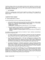
In making a smear, bacteria from either a broth
culture or an agar slant or plate may be used. If a slant
or plate is used, a small amount of bacterial growth is
transferred to a drop of water on a glass slide (figure
7.1a) and mixed. The mixture is then spread out
evenly over a large area on the slide (figure 7.1b).
One of the most common errors in smear prepara-
tion from agar cultures is the use of too large an in-
oculum. This invariably results in the occurrence of
large aggregates of bacteria piled on top of one an-
other. If the medium is liquid, place one or two loops
of the medium directly on the slide (figure 7.1c) and
spread the bacteria over a large area (figure 7.1d).
Allow the slide to air dry at room temperature (figure
7.1e). After the smear is dry, the next step is to attach
the bacteria to the slide by heat-fixing. This is accom-
plished by gentle heating (figure 7.1f ), passing the
slide several times through the hot portion of the
flame of a Bunsen burner. Most bacteria can be fixed
to the slide and killed in this way without serious dis-
tortion of cell structure.
The use of a single stain or dye to create contrast
between the bacteria and the background is referred to
as simple staining. Its chief value lies in its simplicity
and ease of use. Simple staining is often employed
when information about cell shape, size, and arrange-
ment is desired. In this procedure, one places the heat-
fixed slide on a staining rack, covers the smear with a
small amount of the desired stain for the proper
amount of time, washes the stain off with water for a
few seconds, and, finally, blots it dry. Basic dyes such
as crystal violet (20 to 30 seconds staining time),
carbolfuchsin (5 to 10 seconds staining time), or
methylene blue (1 minute staining time) are often
used. Once bacteria have been properly stained, it is
usually an easy matter to discern their overall shape.
Bacterial morphology is usually uncomplicated and
limited to one of a few variations. For future reference,
the most common shapes are presented in figure 7.2.
Procedure
Smear Preparation
1. With the wax pencil, mark the name of the
bacterial culture in the far left corner on each of
three slides.
2. For the broth culture, shake the culture tube and,
with an inoculating loop, aseptically (see figure
14.3) transfer 1 to 2 loopfuls of bacteria to the
center of the slide. Spread this out to about a d-inch
area. When preparing a smear from a slant or plate,
place a loopful of water in the center of the slide.
With the inoculating needle, aseptically pick up a
very small amount of culture and mix into the drop
of water. Spread this out as above. (Three slides
should be prepared; one each of B. subtilis or C.
pseudodiphtheriticum, M. luteus, and S. volutans.)
38 Bacterial Morphology and Staining
Figure 7.1 Bacterial Smear Preparation.
1 drop
of water
Air dry
Heat-fix
(f)
(e)
Spread out
water-bacteria
mixture
Spread out
broth culture
mixture
(b) (d)
(a) (c)
1 needle
of bacterial
growth
Inoculating
needle
Inoculating
loop
1-2 loops
of bacteria
From solid medium From liquid medium
Harley−Prescott:
Laboratory Exercises in
Microbiology, Fifth Edition
II. Bacterial Morphology
and Staining
7. Smear Preparation and
Simple Staining
© The McGraw−Hill
Companies, 2002
3. Allow the slide to air dry, or place it on a slide
warmer (figure 7.3).
4. Pass the slide through a Bunsen burner flame
three times to heat-fix and kill the bacteria.
Simple Staining
1. Place the three fixed smears on a staining loop or
rack over a sink or other suitable receptacle
(figure 7.4a).
2. Stain one slide with alkaline methylene blue for
1 to 1d minutes; one slide with carbolfuchsin for
5 to 10 seconds; and one slide with crystal violet
for 20 to 30 seconds.
3. Wash stain off slide with water for a few seconds
(figure 7.4b).
4. Blot slide dry with bibulous paper (figure 7.4c).
Be careful not to rub the smear when drying the
slide because this will remove the stained
bacteria.
5. Examine under the oil immersion lens and
complete the report for exercise 7.
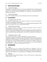
6. You may want to treat smears of the same
bacterium with all three stains in order to compare
them more directly. It is also instructive to cover
bacterial smears for varying lengths of time with a
given stain in order to get a feel for how reactive
they are and the results of overstaining or
understaining a slide preparation. See figure
7.5a–c for examples of bacteria stained with
crystal violet.
Smear Preparation and Simple Staining 39
Figure 7.2 Common Bacterial Shapes.
Shape
coccus
(pl., cocci)
Arrangement
diplococcus
(pairs)
staphylococcus
(random or
grapelike clusters)
micrococcus
(square groups
of four cells)
bacillus
(pl., bacilli)
spirillum
(pl., spirilla)
vibrio
(pl., vibrios)
pleomorphic
Spherical
Rod-shaped
Spiral
Incomplete
spiral
streptococcus
(chains)
sarcina
(cubical packets
of eight cells)
streptobacillus
(chains)
Irregular or
variable
shape
Figure 7.3 A Typical Slide Warmer Used to Speed Up the
Drying of Slides.
Figure 7.4 Simple Staining Procedure.
(c)
Gentle blotting
(b)
Wash bottle
Water
Staining bottle
(a)
Stain
Staining loop
Sink or suitable receptacle
Harley−Prescott:
Laboratory Exercises in
Microbiology, Fifth Edition
II. Bacterial Morphology
and Staining
7. Smear Preparation and
Simple Staining
© The McGraw−Hill
Companies, 2002
40 Bacterial Morphology and Staining
(a) (c)
(b)
Figure 7.5 Bacteria Stained with Crystal Violet. (a) Bacillus subtilis (×1,000). (b) Spirillus volutans (×1,000). (c) Micrococcus luteus
(×1,000).
HINTS AND PRECAUTIONS
(1) When heat-fixing a smear, always make sure that the
smear is on the top of the slide as you pass it through
the flame. (2) Bacteria growing on solid media tend to
cling to each other and must be dispersed sufficiently by
diluting with water. If this is not done, the smear will be
too thick and uneven. Be careful not to use too much
paste in making the smear. It is easy to ruin your results
by using too many bacteria. (3) Always wait until the
slide is dry before heat-fixing. (4) Fixing smears with
an open flame may create artifacts. (5) The inoculating
loop must be relatively cool before inserting it into any
broth. If the loop is too hot, it will spatter the broth and
suspend bacteria into the air. Always flame the inoculat-
ing loop after using it and before setting it down. (6)
When rinsing with water, direct the stream of water so
that it runs gently over the smear.
Harley−Prescott:
Laboratory Exercises in
Microbiology, Fifth Edition
II. Bacterial Morphology
and Staining
7. Smear Preparation and
Simple Staining
© The McGraw−Hill
Companies, 2002
41
Name:
———————————————————————
Date:
————————————————————————
Lab Section:
—————————————————————
Laboratory Report
7
Smear Preparation and Simple Staining
1. Complete the following drawings and table for the simple staining procedure.
C. pseudodiphtheriticum
M. luteusB. subtilis S. volutans
Drawing of
representative
field
Bacterium ______________________ ______________________ ______________________ ______________________
Magnification ______________________ ______________________ ______________________ ______________________
Stain ______________________ ______________________ ______________________ ______________________
Cell form (shape) ______________________ ______________________ ______________________ ______________________
Cell color ______________________ ______________________ ______________________ ______________________
Background color ______________________ ______________________ ______________________ ______________________
Cell grouping ______________________ ______________________ ______________________ ______________________
Harley−Prescott:
Laboratory Exercises in
Microbiology, Fifth Edition
II. Bacterial Morphology
and Staining
7. Smear Preparation and
Simple Staining
© The McGraw−Hill
Companies, 2002
Review Questions
1. What are the two purposes of heat fixation?
a.
b.
2. What is the purpose of simple staining?
3. Why are basic dyes more successful in staining bacteria than acidic dyes?
4. Name three basic stains.
a.
b.
c.
5. Why is time an important factor in simple staining?
6. How would you define a properly prepared bacterial smear?
7. Why should you use an inoculating needle when making smears from solid media? An inoculating loop from
liquid media?
42 Bacterial Morphology and Staining
Harley−Prescott:
Laboratory Exercises in
Microbiology, Fifth Edition
II. Bacterial Morphology
and Staining
8. Gram Stain
© The McGraw−Hill
Companies, 2002
Materials per Student
18- to 24-hour tryptic soy broth cultures of
formalinized (1 ml of concentrated formalin
per 10 ml of culture) Staphyloccus aureus
(ATCC 25923), Escherichia coli (ATCC
25922), and a mixture of S. aureus and E. coli
solutions of crystal violet, Gram’s iodine (2 g
potassium iodide in 300 ml distilled water plus
1 g iodine crystals), 95% ethanol and/or
isopropanol-acetone mixture (3:1 v/v), and
safranin
Bismark brown stain (for color-blind students)
clean glass slides
inoculating loop
Bunsen burner
bibulous paper
microscope
lens paper and lens cleaner
immersion oil
Hyphomonas (Hyphomicrobium) neptunium
(ATCC 15444) grown in marine broth (Difco)
slide warmer
staining rack
Bacto Gram Stain Reagents from Difco for the
three-step Gram stain
Learning Objectives
Each student should be able to
1. Understand the biochemistry underlying the Gram
stain
2. Understand the theoretical basis for differential
staining procedures
3. Perform a satisfactory Gram stain
4. Differentiate a mixture of bacteria into gram-
positive and gram-negative cells
Suggested Reading in Textbook
1. Differential Staining, section 2.3; see also figures
2.14 and 2.15.
2. Gram-Positive Cell Walls, section 3.5.
3. Gram-Negative Cell Walls, section 3.5.
4. The Mechanism of Gram Staining, section 3.5.
5. Budding and/or Appendaged Bacteria, section 22.1;
see also figures 22.4 and 22.5.
Pronunciation Guide
Escherichia coli (esh-er-I-ke-a KOH-lee)
Hyphomonas (Hyphomicrobium) neptunium (hi-fo-
MO-nas nep-TU-ne-um)
Staphylococcus aureus (staf-il-oh-KOK-us ORE-ee-us)
43
EXERCISE
Gram Stain
8
SAFETY CONSIDERATIONS
Be careful with the Bunsen burner flame. Volatile and
flammable liquids (ethanol, isopropanol-acetone) are
used in this experiment. Do not use them near an open
flame. If the stains used in this experiment get on your
clothing, they will not wash out. Discard slides in a con-
tainer with disinfectant. Hold all slides with forceps or a
clothespin when heat-fixing. Gram crystal violet,
safranin, and iodine can cause irritation to the eyes, res-
piratory system and skin. Avoid contact with skin and
eyes. Do not breathe spray. Wear suitable protective
gloves. Always keep the containers tightly closed.
Harley−Prescott:
Laboratory Exercises in
Microbiology, Fifth Edition
II. Bacterial Morphology
and Staining
8. Gram Stain
© The McGraw−Hill
Companies, 2002
Why Are the Following Bacteria
Used in This Exercise?
The major objective of this exercise is to enable the student
to correctly use the Gram stain to differentiate a mixture of
bacteria into gram-positive and gram-negative cells. The
classical standards for this differentiation are Staphylococ-
cus aureus and Escherichia coli. S. aureus (L. aureus,
golden) cells are spherical, 0.5 to 1.0 Ȗm in diameter, oc-
curring singly, in pairs, and in irregular clusters. This bac-
terium is gram-positive, nonmotile, and nonsporing. S. au-
reus is mainly associated with the skin and mucous
membranes of warm-blooded vertebrates but is often iso-
lated from food products, dust, and water. E. coli (Gr.
colon, large intestine) cells are straight rods, 2.0 to 6.0 Ȗm
in length, occurring singly or in pairs. This bacterium is
gram-negative. E. coli occurs as part of the normal flora in
the lower part of the intestine of warm-blooded animals.
Hyphomonas (Hyphomicrobium) neptunium is a rod-
shaped, oval, or bean-shaped cell (1 to 3 Ȗm in length) with
a polar prostheca of varying length. This bacterium is
gram-negative and provides the student the opportunity to
Gram stain a large bacterium that differs in its morphology
and reproduction. H. neptunium is widely distributed in
freshwater, marine, and soil habitats.
Medical Application
Gram staining is the single most useful test in the clinical
microbiology laboratory. It is the differential staining pro-
cedure most commonly used for the direct examination of
specimens and bacterial colonies because it has a broad
staining spectrum. The Gram stain is the first differential
test run on a bacterial specimen brought into the laboratory
for specific identification. The staining spectrum includes
almost all bacteria, many fungi, and parasites such as Tri-
chomonas, Strongyloides, and miscellaneous protozoan
cysts. The significant exceptions include Treponema, My-
coplasma, Chlamydia, and Rickettsia, which are too small
to visualize by light microscopy or lack a cell wall.
Principles
Simple staining depends on the fact that bacteria differ
chemically from their surroundings and thus can be
stained to contrast with their environment. Bacteria
also differ from one another chemically and physically
and may react differently to a given staining procedure.
This is the principle of differential staining. Differen-
tial staining can distinguish between types of bacteria.
The Gram stain (named after Christian Gram,
Danish scientist and physician, 1853–1938) is the
most useful and widely employed differential stain in
bacteriology. It divides bacteria into two groups—
gram negative and gram positive.
The first step in the procedure involves staining
with the basic dye crystal violet. This is the primary
stain. It is followed by treatment with an iodine solu-
tion, which functions as a mordant; that is, it in-
creases the interaction between the bacterial cell and
the dye so that the dye is more tightly bound or the
cell is more strongly stained. The smear is then decol-
orized by washing with an agent such as 95% ethanol
or isopropanol-acetone. Gram-positive bacteria retain
the crystal violet-iodine complex when washed with
the decolorizer, whereas gram-negative bacteria lose
their crystal violet-iodine complex and become color-
less. Finally, the smear is counterstained with a basic
dye, different in color than crystal violet. This coun-
terstain is usually safranin. The safranin will stain the
colorless, gram-negative bacteria pink but does not
alter the dark purple color of the gram-positive bacte-
ria. The end result is that gram-positive bacteria are
deep purple in color and gram-negative bacteria are
pinkish to red in color (figure 8.1).
The Gram stain does not always yield clear results.
Species will differ from one another in regard to the
ease with which the crystal violet-iodine complex is re-
moved by ethanol. Gram-positive cultures may often
turn gram negative if they get too old. Thus, it is al-
ways best to Gram stain young, vigorous cultures rather
than older ones. Furthermore, some bacterial species
are gram variable. That is, some cells in the same cul-
44 Bacterial Morphology and Staining
Figure 8.1 Gram Stain. Light micrograph (×900) of a Gram-
stained mixture of gram-positive Staphylococcus aureus (purple cocci)
and gram-negative Escherichia coli (pink rods).
Harley−Prescott:
Laboratory Exercises in
Microbiology, Fifth Edition
II. Bacterial Morphology
and Staining
8. Gram Stain
© The McGraw−Hill
Companies, 2002
ture will be gram positive and some, gram negative.
Therefore, one should always be certain to run Gram
stains on several cultures under carefully controlled
conditions in order to make certain that a given bacte-
rial “strain” is truly gram positive or gram negative.
Indistinct Gram-stain results can be confirmed by
a simple test using KOH. Place a drop of 10% KOH
on a clean glass slide and mix with a loopful of bacte-
rial paste. Wait 30 seconds, then pull the loop slowly
through the suspension and up and away from the
slide. A gram-negative organism will produce a mu-
coid string; a gram-positive organism remains fluid.
In most introductory microbiology laboratories,
the bacteria that are used in staining exercises are
normally relatively small gram-negative or gram-
positive cocci and rods. One usually does not have
the opportunity to observe larger bacteria or those
with differences in morphology and reproduction.
Part of the Gram-staining exercise has been designed
to help alleviate this deficiency by introducing you to
a less typical bacterium, Hyphomonas (Hyphomicro-
bium) neptunium.
Hyphomicrobia are widely distributed in fresh-
water, marine, and soil habitats. Of particular concern in
this Gram-stain exercise is the unique morphology and
morphogenic cycle (figure 8.2) of these procaryotes.
A small, nonmotile swarmer cell about 0.5 Ȗm in
diameter matures into an ovoid cell, measuring 0.5 by
1.0 Ȗm. This cell grows a stalk (hypha) about 0.3 Ȗm
wide and about 3.0 Ȗm long. The stalk is just thick
enough to be seen under the oil immersion lens, and
success in viewing it provides a good test of one’s
ability to Gram stain correctly and focus the micro-
scope. Through the tip of a growing hypha, a bud is
formed, which grows a single flagellum. Completing
the cycle, the bud separates from the parent and swims
away (to later differentiate into a stalked cell itself),
while the mother cell continues to generate more buds.
All morphological forms are gram negative.
Procedure for Traditional Gram-Stain
Technique
1. Prepare heat-fixed smears of E. coli, S. aureus, and
the mixture of E. coli and S. aureus (see figure 7.1).
2. Place the slides on the staining rack.
3. Flood the smears with crystal violet and let stand
for 30 seconds (figure 8.3a).
4. Rinse with water for 5 seconds (figure 8.3b).
5. Cover with Gram’s iodine mordant and let stand
for 1 minute (figure 8.3c).
6. Rinse with water for 5 seconds (figure 8.3d).
7. Decolorize with 95% ethanol for 15 to 30 seconds.
Do not decolorize too long. Add the decolorizer
drop by drop until the crystal violet fails to wash
from the slide (figure 8.3e). Alternatively, the
smears may be decolorized for 30 to 60 seconds
with a mixture of isopropanol-acetone (3:1 v/v).
8. Rinse with water for 5 seconds (figure 8.3f).
9. Counterstain with safranin for about 60 to 80
seconds (figure 8.3g). Safranin preparations vary
considerably in strength, and different staining
times may be required for each batch of stain. (If
you are color-blind, use Bismark brown stain
rather than safranin.)
10. Rinse with water for 5 seconds (figure 8.3h).
11. Blot dry with bibulous paper (figure 8.3i) and
examine under oil immersion. Gram-positive
organisms stain blue to purple; gram-negative
organisms stain pink to red. There is no need to
place a coverslip on the stained smear. See figure
8.1 for an example of gram-positive and gram-
negative bacteria.
Control Procedure
1. Prepare two heat-fixed slides of the mixed culture
of E. coli and S. aureus.
2. Stain one with crystal violet only (steps 3 to 6).
Gram Stain 45
Figure 8.2 Hyphomonas (Hyphomicrobium) neptunium.
Morphological forms of the life cycle: (1) nonmotile swarmer;
(2) mature cell; (3) stalked cell with bud; (4) stalked cell with
flagellated bud; (5) stalked cell; (6) motile swarmer.
2
1
3
6
5
4
Harley−Prescott:
Laboratory Exercises in
Microbiology, Fifth Edition
II. Bacterial Morphology
and Staining
8. Gram Stain
© The McGraw−Hill
Companies, 2002
stain on a clinical specimen, particularly when the
results will be used as a guide to the selection of a
therapeutic agent, such a control system furnishes
assurance that the iodine solution is providing
proper mordant activity and that decolorization was
performed properly.
3. Carry the second slide through the decolorizing
process (steps 3 to 8).
4. Examine these two slides and compare with the
mixed culture slide that was carried all the way
through the staining procedure (steps 1 to 10).
Your observations should help you understand
how the Gram stain works.
Hyphomonas (Hyphomicrobium) neptunium
1. Gram stain this bacterium according to standard
procedures (figure 8.3a–i).
Procedure for Three-Step Gram Stain
Difco Laboratories has introduced reagents for a three-
step Gram stain. The advantages include less reagent
usage versus conventional stains, reduced chance of
overdecolorization, and saved time. The procedure rec-
ommended by the company is as follows:
1. Flood smear with gram crystal violet primary
stain and stain for 1 minute.
2. Wash off the crystal violet with cold water.
3. Flood the slide with Gram’s iodine mordant and
let sit for 1 minute.
4. Wash off the mordant with safranin
decolorizer/counterstain solution. Then add more
decolorizer/counterstain solution to the slide and
stain for 20 to 50 seconds.
5. Wash off the decolorizer/counterstain with cold
water.
6. Either blot or air dry.
If the three-step Gram-stain reagents are avail-
able, this new procedure may be used in place of the
traditional approach.
Regardless of which procedure is used, run
known cultures or controls. Smears of known cul-
tures are available commercially (figure 8.4) or can
be prepared in the laboratory. It is very important
that controls be included in each staining run,
preferably on the same slide using Staphylococcus
aureus (ATTC 25923) and Escherichia coli (ATCC
25922). Both of these are also available from Difco
as Bactrol™ Disks. When performing the Gram
46 Bacterial Morphology and Staining
Figure 8.3 Gram-stain Procedure.
(a) Crystal violet; 30 seconds (b) Rinse for 5 seconds
(c) Cover with Gram's iodine
for 1 minute
(d) Rinse with water for
5 seconds
Water
Safranin
Water
Decolorizer
Gram's
iodine
Water
Crystal
violet
Water
(e) Decolorize for 15–30
seconds
(f) Rinse with water for
5 seconds
(h) Rinse for 5 seconds(g) Counterstain with safranin
for about 60–80 seconds
(i) Blot dry with bibulous paper
Harley−Prescott:
Laboratory Exercises in
Microbiology, Fifth Edition
II. Bacterial Morphology
and Staining
8. Gram Stain
© The McGraw−Hill
Companies, 2002
Gram Stain 47
HINTS AND PRECAUTIONS
(1) Don’t make your smears too thick. (2) Thick
smears will require more time to decolorize than thin
ones. (3) Decolorization has occurred when the solu-
tion flows colorlessly from the slide. If you cannot tell
accurately when the solution becomes colorless, try
decolorizing with isopropanol-acetone mixture for
about 30 to 40 seconds. (4) Some common sources of
Gram-staining errors are (a) the inoculating loop was
too hot, (b) excessive heat was used during the heat-
fixing procedure, and (c) the decolorizing alcohol was
left on the slide too long.
Figure 8.4 Gram Stain Control Slide. Notice the positive
control at the top and negative control at the bottom. Each area
contains a known Gram-positive and Gram-negative bacterium.
Harley−Prescott:
Laboratory Exercises in
Microbiology, Fifth Edition
II. Bacterial Morphology
and Staining
8. Gram Stain
© The McGraw−Hill
Companies, 2002
49
Name:
———————————————————————
Date:
————————————————————————
Lab Section:
—————————————————————
Laboratory Report
8
1. Draw the Gram-stained bacteria in the following circles.
2. Control Gram-stain results.
3. Gram stain of H. neptunium illustrating the different stages in its life cycle.
Stage _____
_____ _____
S. aureus E. coli Mixed culture
(E. coli + S. aureus)
Steps 3–6 Steps 3–8
Bacterial
color
Harley−Prescott:
Laboratory Exercises in
Microbiology, Fifth Edition
II. Bacterial Morphology
and Staining
8. Gram Stain
© The McGraw−Hill
Companies, 2002
Review Questions
1. What is the difference between a simple and differential stain?
2. Name the reagent used and state the purpose of each of the following in the Gram stain:
a. mordant
b. primary stain
c. decolorizer
d. counterstain
3. Which step is the most crucial or most likely to cause poor results in the Gram stain? Why?
4. Why must young cultures be used when doing a Gram stain?
5. Why was H. neptunium Gram stained?
6. What is meant by gram variable?
7. What part of the bacterial cell is most involved with Gram staining, and why?
50 Bacterial Morphology and Staining
Harley−Prescott:
Laboratory Exercises in
Microbiology, Fifth Edition
II. Bacterial Morphology
and Staining
9. Acid−Fast Staining
(Ziehl−Neelsen and
Kinyoun) Procedures
© The McGraw−Hill
Companies, 2002
Materials per Student
tryptic soy broth culture of Escherichia coli
(ATCC 11229) and nutrient agar slant culture
of Mycobacterium smegmatis (ATCC 19420)
or Mycobacterium phlei (ATCC 354)—5-day-
old cultures
Ziehl’s carbolfuchsin
carbolfuchsin prepared with either Tergitol No. 4
(a drop per 30 ml of carbolfuchsin) or Triton-X
(2 drops per 100 ml of carbolfuchsin). Tergitol
No. 4 and Triton-X act as detergents,
emulsifiers, and wetting agents.
alkaline methylene blue
acid-alcohol
clean glass slides
commercial slides showing acid-fast
Mycobacterium tuberculosis (Carolina
Biological Supply, Wards)
inoculating loop
hot plate
microscope
bibulous paper
paper toweling
lens paper and lens cleaner
immersion oil
staining racks
1-ml pipettes with pipettor
Learning Objectives
Each student should be able to
1. Understand the biochemical basis of the acid-fast
stain
2. Perform an acid-fast stain
3. Differentiate bacteria into acid-fast and non-acid-
fast groups
Suggested Reading in Textbook
1. Differential Staining, section 2.3.
2. The Mycobacteria, section 24.5; see also figure 24.9.
3. Tuberculosis, section 39.1.
4. Leprosy, section 39.3.
Pronunciation Guide
Cryptosporidium (krip-toe-spoh-RED-jee-um)
Escherichia coli (esh-er-I-ke-a KOH-lee)
Mycobacterium phlei (mi-ko-bak-TE-re-um fee-ii)
M. smegmatis (M. smeg-MEH-tis)
M. tuberculosis (M. too-ber-ku-LO-sis)
Nocardia (no-KAR-dee-ah)
51
EXERCISE
Acid-Fast Staining
(Ziehl-Neelsen and Kinyoun) Procedures
9
SAFETY CONSIDERATIONS
A volatile and flammable liquid (acid-alcohol) is used
in this experiment. Do not use near an open flame. If the
carbolfuchsin or methylene blue get on your clothing,
they will not wash out. Note: when carbolfuchsin is
heated, phenol is driven off. Phenol is poisonous and
caustic. Thus, always use a chemical hood with the ex-
haust fan on for the hot plate or boiling water bath set-
up. Discard slides in a container with disinfectant. No
mouth pipetting. Mycobacteria should be handled in a
safety cabinet to prevent dissemination in case the
human pathogen Mycobacterium tuberculosis should
occur among the cultures. Infected material should be
disinfected by heat because mycobacteria are relatively
resistant to chemical disinfectants.
Harley−Prescott:
Laboratory Exercises in
Microbiology, Fifth Edition
II. Bacterial Morphology
and Staining
9. Acid−Fast Staining
(Ziehl−Neelsen and
Kinyoun) Procedures
© The McGraw−Hill
Companies, 2002
Why Are the Above Bacteria Used
in This Exercise?
One of the major objectives of this exercise is to give the
student expertise in acid-fast staining. To allow the student
to differentiate between acid-fast and non-acid-fast bacte-
ria, the authors have chosen one of the cultures from the
last exercise, Escherichia coli. E. coli is a good example of
a non-acid-fast bacterium. Mycobacterium smegmatis and
M. phlei are nonpathogenic members of the genus My-
cobacterium. These bacteria are straight or slightly curved
rods, 1 to 10 Ȗm in length, acid-fast at some stage of
growth, and not readily stained by Gram’s method. They
are also nonmotile, nonsporing, without capsules, and slow
or very slow growers. The mycobacteria are widely distrib-
uted in soil and water; some species are obligate parasites
and pathogens of vertebrates.
Medical Application
In the clinical laboratory, the acid-fast stain is important in
identifying bacteria in the genus Mycobacterium; specifi-
cally, M. leprae (leprosy) and M. tuberculosis (tuberculo-
sis). This differential stain is also used to identify members
of the aerobic actinomycete genus Nocardia; specifically,
the opportunistic pathogens N. brasiliensis and N. aster-
oides that cause the lung disease nocardiosis. The water-
borne protozoan parasite Cryptosporidium that causes diar-
rhea in humans (cryptosporidiosis) can also be identified by
the acid-fast stain.
Principles
A few species of bacteria in the genera Mycobacterium
and Nocardia, and the parasite Cryptosporidium do not
readily stain with simple stains. However, these microor-
ganisms can be stained by heating them with carbol-
fuchsin. The heat drives the stain into the cells. Once the
microorganisms have taken up the carbolfuchsin, they
are not easily decolorized by acid-alcohol, and hence are
termed acid-fast. This acid-fastness is due to the high
lipid content (mycolic acid) in the cell wall of these mi-
croorganisms. The Ziehl-Neelsen acid-fast staining
procedure (developed by Franz Ziehl, a German bacte-
riologist, and Friedrich Neelsen, a German pathologist,
in the late 1800s) is a very useful differential staining
technique that makes use of this difference in retention
of carbolfuchsin. Acid-fast microorganisms will retain
this dye and appear red (figure 9.1a, b). Microorganisms
that are not acid-fast, termed non-acid-fast, will appear
blue or brown due to the counterstaining with methylene
blue after they have been decolorized by the acid-alco-
hol. A modification of this procedure that employs a wet-
ting agent (Tergitol No. 7) rather than heat to ensure stain
penetration is known as the Kinyoun staining proce-
dure (developed by Joseph Kinyoun, German bacteriol-
ogist, in the early 1900s).
Procedure
Ziehl-Neelsen (Hot Stain) Procedure
1. Prepare a smear consisting of a mixture of E. coli
and M. smegmatis.
52 Bacterial Morphology and Staining
Figure 9.1 Ziehl-Neelsen Stain of Mycobacterium Acid-fast Rods. (a) Mycobacterium smegmatis stained red (×1,000). (b) In this
photomicrograph, Mycobacterium smegmatis stains red and the background cells blue-brown.
(a) (b)
Harley−Prescott:
Laboratory Exercises in
Microbiology, Fifth Edition
II. Bacterial Morphology
and Staining
9. Acid−Fast Staining
(Ziehl−Neelsen and
Kinyoun) Procedures
© The McGraw−Hill
Companies, 2002
2. Allow the smear to air dry and then heat-fix (see
figure 7.1).
3. Place the slide on a hot plate that is within a
chemical hood (with the exhaust fan on), and
cover the smear with a piece of paper toweling
that has been cut to the same size as the
microscope slide. Saturate the paper with Ziehl’s
carbolfuchsin (figure 9.2a). Heat for 3 to 5
minutes. Do not allow the slide to dry out, and
avoid excess flooding! Also, prevent boiling by
adjusting the hot plate to a proper temperature. A
boiling water bath with a staining rack or loop
held 1 to 2 inches above the water surface also
works well. (Instead of using a hot plate to heat-
drive the carbolfuchsin into the bacteria, an
alternate procedure is to cover the heat-fixed slide
with a piece of paper towel. Soak the towel with
the carbolfuchsin and heat, well above a Bunsen
burner flame.)
4. Remove the slide, let it cool, and rinse with water
for 30 seconds (figure 9.2b).
5. Decolorize by adding acid-alcohol drop by drop
until the slide remains only slightly pink. This
requires 10 to 30 seconds and must be done
carefully (figure 9.2c).
6. Rinse with water for 5 seconds (figure 9.2d).
7. Counterstain with alkaline methylene blue for
about 2 minutes (figure 9.2e).
8. Rinse with water for 30 seconds (figure 9.2f).
9. Blot dry with bibulous paper (figure 9.2g).
10. There is no need to place a coverslip on the
stained smear. Examine the slide under oil
immersion and record your results in the report
for exercise 9. Acid-fast organisms stain red; the
background and other organisms stain blue or
brown. See figure 9.1 for an example of the
Ziehl-Neelsen stain.
11. Examine the prepared slide of Mycobacterium
tuberculosis.
Kinyoun (Cold Stain) Procedure
(This may be used instead of or in addition to the
Ziehl-Neelsen procedure.)
1. Heat-fix the slide as previously directed.
2. Flood the slide for 5 minutes with carbolfuchsin
prepared with Tergitol No. 7 (heat is not
necessary).
3. Decolorize with acid-alcohol and wash with tap
water. Repeat this step until no more color runs
off the slide.
4. Counterstain with alkaline methylene blue for 2
minutes. Wash and blot dry.
5. Examine under oil. Acid-fast organisms stain red;
the background and other organisms stain blue.
Acid-Fast Staining (Ziehl-Neelsen and Kinyoun) Procedures 53
Figure 9.2 Acid-fast Staining Procedure.
(a) Apply carbolfuchsin to
saturate paper and heat
for 5 minutes in an
exhaust hood
Carbol-
fuchsin
Water
Water
Water
Acid-
alcohol
Methylene
blue
(b) Cool and rinse with water
for 30 seconds
(d) Rinse with water for
5 seconds
(c) Decolorize with acid-
alcohol until pink
(10–30 seconds)
(e) Counterstain with
methylene blue for
about 2 minutes
(f) Rinse with water for
30 seconds
(g) Blot dry with
bibulous paper
HINTS AND PRECAUTIONS
(1) Light (diaphragm and condenser adjustments) is criti-
cal in the ability to distinguish acid-fast-stained micro-
organisms in sputum or other viscous background materi-
als. (2) If the bacteria are not adhering to the slide, mix
the bacteria with sheep serum or egg albumin during
smear preparation. This will help the bacteria adhere to
the slide.
Harley−Prescott:
Laboratory Exercises in
Microbiology, Fifth Edition
II. Bacterial Morphology
and Staining
9. Acid−Fast Staining
(Ziehl−Neelsen and
Kinyoun) Procedures
© The McGraw−Hill
Companies, 2002
55
Name:
———————————————————————
Date:
————————————————————————
Lab Section:
—————————————————————
Laboratory Report
9
Acid-Fast Staining (Ziehl-Neelsen and Kinyoun) Procedures
1. Complete the following table with respect to the acid-fast stain and draw representative specimens.
2. Are you satisfied with your results? __________ If not, what can you do to improve your technique the next
time you prepare an acid-fast stain from a broth culture?
E. coli M. smegmatis M. phlei
Magnification ____________________ ____________________ ____________________
Bacterium other
than above ____________________ ____________________ ____________________
Bacterial shape ____________________ ____________________ ____________________
Cell color ____________________ ____________________ ____________________
Acid-fast ____________________ ____________________ ____________________
×× ×
Harley−Prescott:
Laboratory Exercises in
Microbiology, Fifth Edition
II. Bacterial Morphology
and Staining
9. Acid−Fast Staining
(Ziehl−Neelsen and
Kinyoun) Procedures
© The McGraw−Hill
Companies, 2002
Review Questions
1. What is the purpose of the heat during the acid-fast staining procedure?
2. What is the function of the counterstain in the acid-fast staining procedure?
3. Are acid-fast bacteria gram positive or gram negative? Explain your answer.
4. For what diseases would you use an acid-fast stain?
5. What makes a microorganism non-acid-fast?
6. What chemical is responsible for the acid-fast property of mycobacteria?
7. Is a Gram stain an adequate substitute for an acid-fast stain? Why or why not?
56 Bacterial Morphology and Staining
Harley−Prescott:
Laboratory Exercises in
Microbiology, Fifth Edition
II. Bacterial Morphology
and Staining
10. Endospore Staining
(Schaeffer−Fulton or
Wirtz−Conklin)
© The McGraw−Hill
Companies, 2002
Materials per Student
24- to 48-hour nutrient agar slant cultures of
Bacillus megaterium (ATCC 12872) and
Bacillus macerans (ATCC 8244), and old
(more than 48 hours) thioglycollate cultures of
Clostridium butyricum (ATCC 19398) and
Bacillus circulans (ATCC 4513)
clean glass slides
microscope
immersion oil
wax pencil
inoculating loop
hot plate or boiling water bath with staining rack
or loop
5% malachite green solution
safranin
bibulous paper
paper toweling
lens paper and lens cleaner
slide warmer
forceps
Learning Objectives
Each student should be able to
1. Understand the biochemistry underlying
endospore staining
2. Perform an endospore stain
3. Differentiate between bacterial endospore and
vegetative cell forms
Suggested Reading in Textbook
1. Staining Specific Structures, section 2.3.
2. The Bacterial Endospore, section 3.8; see also
figures 3.40–3.44, 23.5, 23.6, 23.8.
3. Anthrax, section 39.3.
4. Tetanus, section 39.3.
Pronunciation Guide
Bacillus megaterium (bah-SIL-us meg-AH-ter-ee-um)
B. macerans (ma-ser-ANS)
B. circulans (sir-KOO-lanz)
Clostridium butyricum (klos-STRID-ee-um bu-TER-
a-cum)
Why Are the Above Bacteria Used
in This Exercise?
Because the major objective of this exercise is to provide ex-
perience in endospore staining, the authors have chosen sev-
eral bacteria that vary in the size and shape of their en-
dospores. Bacillus megaterium (M. L. n. megaterium, big
beast) is a cylindrical to oval or pear-shaped cell about 1.2 to
1.5 Ȗm in diameter and 2 to 5 Ȗm long; it tends to occur in
short, twisted chains. The spores are central and vary from
short oval to elongate. Spores occur in the soil. Bacillus mac-
erans (L. macerans, softening by steeping, rotting) is an
elongated cell 0.5 to 0.7 Ȗm wide and 2.5 to 5 Ȗm in length
with terminal spores. Spores are relatively scarce in the soil.
Bacillus circulans (L. circulans, circling) is an elongate cell
2 to 5 Ȗm in length and 0.5 to 0.7 Ȗm wide. In most strains,
the spore is terminal to subterminal; it is central in a spindle-
shaped sporangium if the bacillus is short. In many strains,
deeply stainable material persists on the surface of the free
spores. The spores are found in the soil. Clostridium bu-
tyricum (Gr. butyrum, butter) is a straight or slightly curved
rod, 2.4 to 7.6 Ȗm in length and 0.5 to 1.7 Ȗm wide, with
rounded ends. The cells occur singly, in pairs, in short
chains, and occasionally as long filaments. They are motile
with peritrichous flagella. Spores are oval and eccentric to
subterminal and are found in the soil and animal feces.
57
EXERCISE
Endospore Staining
(Schaeffer-Fulton or Wirtz-Conklin)
10
SAFETY CONSIDERATIONS
Be careful with the Bunsen burner flame and boiling
water bath. If either malachite green or safranin get on
your clothes, they will not wash out. Discard slides in a
container with disinfectant.
Harley−Prescott:
Laboratory Exercises in
Microbiology, Fifth Edition
II. Bacterial Morphology
and Staining
10. Endospore Staining
(Schaeffer−Fulton or
Wirtz−Conklin)
© The McGraw−Hill
Companies, 2002
Medical Application
Only a few bacteria produce endospores. Those of medical
importance include Bacillus anthracis (anthrax), Clostrid-
ium tetani (tetanus), C botulinium (botulism), and C. per-
fringens (gas gangrene). In the clinical laboratory, the loca-
tion and size of endospores vary with the species; thus, they
are often of value in identifying bacteria.
Principles
Bacteria in genera such as Bacillus and Clostridium
produce quite a resistant structure capable of surviv-
ing for long periods in an unfavorable environment
and then giving rise to a new bacterial cell (figure
10.1). This structure is called an endospore since it
develops within the bacterial cell. Endospores are
spherical to elliptical in shape and may be either
smaller or larger than the parent bacterial cell. En-
dospore position within the cell is characteristic and
may be central, subterminal, or terminal.
Endospores do not stain easily, but, once stained,
they strongly resist decolorization. This property is the
basis of the Schaeffer-Fulton (Alice B. Schaeffer and
MacDonald Fulton were microbiologists at Middlebury
College, Vermont, in the 1930s) or Wirtz-Conklin
method (Robert Wirtz and Marie E. Conklin were bacte-
riologists in the early 1900s) of staining endospores. The
endospores are stained with malachite green. Heat is used
to provide stain penetration. The rest of the cell is then
decolorized and counterstained a light red with safranin.
Procedure
1. With a wax pencil, place the names of the respective
bacteria on the edge of four clean glass slides.
2. As shown in figure 14.3, aseptically transfer one
species of bacterium with an inoculating loop to
each of the respective slides, air dry (or use a
slide warmer), and heat-fix.
3. Place the slide to be stained on a hot plate or
boiling water bath equipped with a staining loop
or rack. Cover the smear with paper toweling that
has been cut the same size as the microscope slide.
4. Soak the paper with the malachite green staining
solution. Gently heat on the hot plate (just until
the stain steams) for 5 to 6 minutes after the
malachite green solution begins to steam. Replace
the malachite green solution as it evaporates so that
the paper remains saturated during heating (figure
10.2a). Do not allow the slide to become dry.
5. Remove the paper using forceps, allow the slide
to cool, and rinse the slide with water for 30
seconds (figure 10.2b).
6. Counterstain with safranin for 60 to 90 seconds
(figure 10.2c).
7. Rinse the slide with water for 30 seconds (figure
10.2d).
58 Bacterial Morphology and Staining
Figure 10.2 Endospore Staining Procedure.
(c) Counterstain with safranin
for 60–90 seconds
Safranin
Water
Malachite
green
(a) Apply malachite green to
saturate paper and steam
for 5 minutes
(b) Remove paper, cool, and
rinse with water for
30 seconds
Water
(d) Rinse with water for
30 seconds
(e) Blot dry with
bibulous paper
Figure 10.1 The Life Cycle of Endospore-forming Bacteria.
Sporogenesis
Endospore
Vegetative
cell
Vegetative
cell
Growth of
spore
Free
spore
Germination
Harley−Prescott:
Laboratory Exercises in
Microbiology, Fifth Edition
II. Bacterial Morphology
and Staining
10. Endospore Staining
(Schaeffer−Fulton or
Wirtz−Conklin)
© The McGraw−Hill
Companies, 2002
Endospore Staining (Schaeffer-Fulton or Wirtz-Conklin) 59
HINTS AND PRECAUTIONS
(1) Do not boil the stain—always steam gently.
(2) After steaming the slide, cool it before flooding it
with cold water. If the slide is not cooled, it may shatter
or crack when rinsed with cold water.
Figure 10.3 Examples of Endospores. (a) Central spores of Bacillus stained with malachite green and counterstained with safranin
(×1,000). Notice that the cells are rod-shaped and straight, often arranged in pairs or chains, with rounded squared ends. The endospores are
oval and not more than one spore per cell. (b) Clostridium tetani showing round, terminal spores that usually distend the cell (×1,000). Notice
that the cells are rod-shaped and are often arranged in pairs or short chains with rounded or sometimes pointed ends. (c) Bacillus megaterium
showing short oval to elongate spores.
(a) (b)
(c)
8. Blot dry with bibulous paper (figure 10.2e) and
examine under oil immersion. A coverslip is not
necessary. The spores, both endospores and free
spores, stain green; vegetative cells stain red.
Draw the bacteria in the space provided in the
report for exercise 10. See figure 10.3a–c for an
example of endospore staining.
Harley−Prescott:
Laboratory Exercises in
Microbiology, Fifth Edition
II. Bacterial Morphology
and Staining
10. Endospore Staining
(Schaeffer−Fulton or
Wirtz−Conklin)
© The McGraw−Hill
Companies, 2002
61
Name:
———————————————————————
Date:
————————————————————————
Lab Section:
—————————————————————
Laboratory Report
10
Endospore Staining (Schaeffer–Fulton or Wirtz–Conklin)
1. Make drawings and answer the questions for each of the bacterial endospore slides.
2. Are you satisfied with the results of your endospore stain? ______ If not, how can you improve your results
the next time you prepare an endospore stain?
Bacterium __________________ __________________ __________________ __________________
Magnification __________________ __________________ __________________ __________________
Bacterium other than above __________________ __________________ __________________ __________________
Spore color __________________ __________________ __________________ __________________
Color of vegetative cell __________________ __________________ __________________ __________________
Location of endospore (central,
terminal, subterminal) __________________ __________________ __________________ __________________
××××
B. megaterium B. macerans B. circulans C. butyricum
Harley−Prescott:
Laboratory Exercises in
Microbiology, Fifth Edition
II. Bacterial Morphology
and Staining
10. Endospore Staining
(Schaeffer−Fulton or
Wirtz−Conklin)
© The McGraw−Hill
Companies, 2002
Review Questions
1. Why is heat necessary in order to stain endospores?
2. Where are endospores located within vegetative cells?
3. In the Schaeffer–Fulton endospore stain, what is the primary stain? The counterstain?
4. Name two disease-causing bacteria that produce endospores.
a.
b.
5. What is the function of an endospore?
6. Why are endospores so difficult to stain?
7. What do endospore stains have in common with the acid-fast (Ziehl–Neelsen) stain?
62 Bacterial Morphology and Staining
Harley−Prescott:
Laboratory Exercises in
Microbiology, Fifth Edition
II. Bacterial Morphology
and Staining
11. Capsule Staining
© The McGraw−Hill
Companies, 2002
Materials per Student
18-hour skim milk cultures of Klebsiella
pneumoniae (ATCC e13883) and Alcaligenes
denitrificans (ATCC 15173)
Tyler’s crystal violet (1% aqueous solution) or
Gram’s crystal violet (1% aqueous solution)
20% (w/v) solution of copper sulfate
(CuSO
4
и 5H
2
O)
microscope
immersion oil
lens paper and lens cleaner
clean glass slides
wax pencil
bibulous paper
inoculating loop
Bon Ami
70% ethyl alcohol
India ink (Higgins no. 4465 black or Pelikan
Drawing ink No. 17 black for technical pens)
or SpotTest India ink ampules from Difco
safranin stain
Learning Objectives
Each student should be able to
1. Understand the biochemistry of the capsule stain
2. Perform a capsule stain
3. Distinguish capsular material from the bacterial
cell
Suggested Reading in Textbook
1. Capsules, Slime Layers, and S Layers, section
3.6; see also figure 3.27.
Pronunciation Guide
Alcaligenes denitrificans (al-kah-LIJ-e-neez de-ni-tri-
fi-KANS)
Klebsiella pneumoniae (kleb-se-EL-lah nu-MO-ne-EYE)
Why Are the Above Bacteria Used
in This Exercise?
One of the major objectives of this exercise is to give the
student experience in capsule staining. To help accomplish
this objective, the authors have chosen one capsulated and
one noncapsulated bacterium. Klebsiella pneumoniae (Gr.
pneumonia, pneumonia) is a nonmotile, capsulated rod, 0.6
to 6 Ȗm in length, and is arranged singly, in pairs, or short
chains. Cells contain a large polysaccharide capsule and
give rise to large mucoid colonies. There are more than 80
capsular (K) antigens that can be used to serotype klebsiel-
lae. K. pneumoniae occurs in human feces and clinical spec-
imens, water, grain, fruits, and vegetables. Alcaligenes deni-
trificans (are able to reduce NO
3
–
to NO
2
–
and N
2
) occurs as
a rod, a coccal rod, or a coccus; is 0.5 to 2.6 Ȗm in length;
and usually occurs singly in water and soil. It is motile with
1 to 4 peritrichous flagella. No capsule is present.
Medical Application
Many bacteria (e.g., Bacillus anthracis [anthrax], Streptococ-
cus mutans [tooth decay], Streptococcus pneumoniae [pneu-
monia]) and the fungus Cryptococcus neoformans [crypto-
coccosis] contain a gelatinous covering called a capsule. In
the clinical laboratory, demonstrating the presence of a cap-
sule is a means of diagnosis and determining the organism’s
virulence, the degree to which a pathogen can cause disease.
63
EXERCISE
Capsule Staining
11
SAFETY CONSIDERATIONS
Be careful with the Bunsen burner flame. If India ink,
crystal violet, or safranin get on your clothes, they will
not wash out. Seventy percent ethyl alcohol is flamma-
ble—keep away from open flames. Discard slides in a
container with disinfectant.