Adaptive Motion of Animals and Machines - Hiroshi Kimura et al (Eds) Part 4 doc
Bạn đang xem bản rút gọn của tài liệu. Xem và tải ngay bản đầy đủ của tài liệu tại đây (420.82 KB, 20 trang )
54 S. Mori, F. Mori, K. Nakajima
reflexes and fundamental movements [2,4]. From a phylogenetical point of
view, the motor pathways descending from the brainstem to the spinal cord
are the earliest developing ones [5]. In contrast, the motor cortices estab-
lish functional connections postnatally first with the cervical MNs innervat-
ing the fore-limbs and then the lumbar MNs innervating the hind-limbs. In
the macaque monkey, full myelination (maturation) of corticospinal axons in
the spinal cord occurs at around 36 months of age [6]. Such a rostrocaudal
development of cortico-motoneuronal (CM) connections is well reflected in
the postnatal developmental pattern of posture and movements in both the
human [1] and non-human primates [7]. In parallel with the growth of the
musculoskeletal system and the CNS, locomotor learning from daily practice
and experience is necessary for the acquisition of the skill of Bp locomotion.
Locomotor practice and experience help the development of CM connections
to distally located muscles of the foot, and build up and storage of ‘locomotor
memory’ and/or reference centers [2,8].
To advance understanding of CNS control of Bp standing and Bp walk-
ing, we have been analyzing the unrestrained normal quadrupedal (Qp) and
operantly-trained Bp locomotor behavior of a non-human primate, the Japanese
monkey, M fuscata [9-13]. Japanese monkeys are originally Qp, but with long-
term locomotor training, they acquire the novel strategy of walking bipedally
on the surface of a moving treadmill belt. To describe the functional signifi-
cance of our findings, the present report addresses four major aspects relat-
ing to the elaboration of Bp locomotion: (a) our concept of locomotor control
CNS mechanisms including anticipatory and reactive control mechanisms, (b)
emergence, acquisition and refinement of Bp locomotion in juvenile Japanese
monkeys, and integration of posture and locomotion (c) common and dif-
ferent control properties of Qp and Bp locomotion, and (d) similarity and
difference in the kinematics of lower limbs during Bp walking in our monkey
model and in the human. The last section addresses a future perspective for
understanding “brain-locomotor behavior” relationships.
2 Locomotor control CNS mechanisms including
anticipatory and reactive control mechanisms
We have recently proposed a new concept of CNS mechanisms related to
locomotor control [2]. As shown conceptually in Figure 1, we hypothesize that
descending commands from the cognitive and emotive portions of the higher
CNS, and activity of both locomotion evoking centers and posture control
centers are constantly compared with that of the reference centers, with their
collective output sent to the integration centers. Such a system incorporates
both anticipatory and reactive control processes [14]. Critical components of
the reference centers are the postural and locomotor memory that is built up
by daily walking practice and experience. Its other component includes the
postural body scheme or the reference frame of bodily configuration essential
Higher Nervous Control of Quadrupedal vs Bipedal Locomotion 55
for Bp locomotion [2,15]. The integration centers participate in a comparative
function: comparing top-down locomotor command feedforward signals with
bottom-up feedback signals revealing the current state of locomotion, and
minimizing impairments of posture and locomotion. The integration center’s
efferent output is distributed by way of executing centers. The latter’s concern
is that motor signals must be sent to a number of different muscle control
systems such that the multiple motor segments they control are activated in
a coordinated manner.
Major elements of motor control units are ‘interneuronal circuits includ-
ing the central pattern generator (CPG)’, spinal MN columns and motor
segments [2,16]. Output signals arising from the execution centers are carried
to the spinal cord by the phylogenetically old reticulospinal (RS) and vestibu-
lospinal (VS) pathways, and ensure that appropriate and timely forces are
applied to relevant limb joints, the result being a smooth execution of locomo-
tion, with correctly phased limb movements and adequate levels of postural
muscle tone [2,4]. Output signals arising from the higher CNS, such as the
primary motor area (M1) and supplementary motor area (SMA), are also
carried to the MNs of motor control units by way of phylogenetically recent
corticospinal and cortico-reticulospinal pathways, and contribute to the re-
finement of limb movements such as to avoid obstacles on the walking path
[2].
During Bp standing and Bp walking, changes in body configuration are
first registered by both the labyrinthine and proprioceptive receptors em-
bedded in the motor segments. Changes in the external world are perceived
by telereceptors, such as the eyes and ears [3]. By continuous reception and
processing of multi-modal interoceptive and exteroceptive afferent inputs, the
integration centers can compare the body’s moment-to-moment configuration
relative to the immediate and distant environment. When both quadrupeds
and bipeds encounter unexpected obstacles, they adopt preparatory or antic-
ipatory postures to avoid them. When they fail to clear the obstacles, they
take reactive and/or defensive postures to minimize and compensate for the
impairments to ongoing locomotion [14]. The central feedback from the inte-
gration center combined with peripheral feedback at the cerebral cortical level
enables the animal conscious perception of its kinesthetic aspects of volitional
(anticipatory) and automatic (reactive) adjustments to locomotion [2]. An-
ticipatory control mechanisms are probably stored at a high CNS levels such
as the visual cortex, SMA and M1 and interconnecting networks, whereas
reactive control mechanisms are probably stored at low CNS levels such as
the cerebellum, brainstem and spinal cord and interconnecting networks [2].
56 S. Mori, F. Mori, K. Nakajima
Environment
reactive control
Locomotion
multiple
motor
control
units
execution
centers
locomotion
evoking centers
reference
centers
posture control
centers
Cognitive brain
anticipatory
control
Emotive
brain
reactive control
integration
centers
Fig. 1. A conceptualization of the overall integrated control of posture and lo-
comotion including anticipatory and reactive control. From the left to right, the
CNS structures and their proposed processes include: cognitive processing, emotive
processing, locomotion evoking centers, posture control centers, reference centers,
integration centers, execution centers, and multiple motor control units. Open and
closed arrowheads represent the ascending and descending flow of signals. Modified
from reference [2].
3 Emergence, acquisition and refinement of Bp
locomotion in Juvenile Japanese monkeys
Genetically Qp young Japanese monkey, M. fuscata, can acquire a novel ca-
pability of Bp walking on the surface of a moving treadmill belt [13]. The
operant-conditioning methods with which monkey learned to walk quadrupedally
and/or bipedally are described in detail elsewhere [10, 12]. After sufficient
physical growth and locomotor learning (12 to 24 months), young monkeys
(estimated age: 1.6 to 2.4 years) gradually acquired a more upright and a
more stable posture, a more stable (less variable) cyclic patterns of joint an-
gles in the lower limbs and coupling among the neighboring joints, and also
faster speeds of Bp walking [13]. It was also found that stability of kinematic
patterns developed in a rostro-caudal direction, i.e. in the same direction
as observed in developing human infants [1]. Our findings demonstrated for
the first time the basic principles of the developing monkey to integrate the
neural and musculoskeletal mechanisms required for sufficient coordination
of upper (head, neck, trunk) and lower (hind-limbs) motor segments so that
Bp standing could be maintained and Bp walking elaborated.
Once the monkeys acquired Bp walking capability, they still could walk
bipedally even after a few weeks of cessation of locomotor training. This
suggests that the monkeys stored a postural body scheme or the reference
Higher Nervous Control of Quadrupedal vs Bipedal Locomotion 57
frame of bodily configuration necessary for Bp walking. We also found that
the Qp walking monkey on the moving treadmill belt could right its posture
and continue Bp locomotion [17]. The transition from Qp to Bp walking
always began when the left (L) or right (R) hind-limb initiated a stance (ST)
phase of the step. For example, at the time when the imaginary position
of the monkey’s center of body mass (CoM) projected to the supporting
L hind-limb, the monkey began an upward excursion of the angle of the
weight-bearing hip joint. The L forelimb was then freed from the constraints
of weight bearing. With further upward excursion of the hip joint angle,
the monkey started to right its posture and initiate reaching and grasping
movements, extending the freed fore-limb forward to attain the reward and to
eat it ad libitum. This suggests that the monkey’s CNS can rapidly select and
combine integrated subsets of posture- and locomotor-related neural control
mechanisms appropriate for the elaboration of a required task. Our animal
model thus provides a unique opportunity to compare the kinematics of Qp
and Bp locomotion in a single animal.
During the transitional period from Qp to Bp locomotion, the monkey
coordinated sequentially independent movements of multiple motor segments
such as eyes, head, neck, trunk, fore and hind-limbs, in order to satisfy the
dual purpose of freeing the forelimbs from the constraints of weight-bearing
and adopting Bp walking. The locomotion conversion process involved the
rapid and smooth succession of targeting, orienting, and righting. Targeting
requires the coordinated activity of head, neck, trunk and fore-limbs, and
righting that of head, neck, trunk and hind-limb. Kinematics of eye-head
position, body axis, and major joint angles of the hind-limbs revealed the
significance of a hip maneuver strategy for the monkey’s conversion from
stable Qp to similarly stable Bp locomotion [17]. Each of these processes
includes visuo-motor and vestibulo-motor coordination. The latter is based
on interactions of vestibular information with sensory information arising
from SW and ST limbs and thus ensuring a good postural stability and
postural orientation over a wide range of environmental condition [18]. It is
conceivable that spinal reflexes play a crucial role in the coordination of SW
and ST limbs. According to Zehr and Stein, generally cutaneous reflexes act
to alter SW limb trajectory to avoid stumbling and falling. Stretch reflexes
act to stabilize limb trajectory and assist force production during ST. Load
receptor reflexes have an effect on both ST phase body weight support and
step cycle timing [19].
We have previously proposed that the fastigial nucleus (FN) in the cere-
bellum is importantly involved in the initiation of Qp locomotion, and in ad-
dition in the rapid and smooth succession of targeting, orienting, and righting
necessary for the conversion from Qp to Bp walking [2, 17]. In a high decer-
ebrate cat, we have demonstrated that train-pulse microstimulation of the
hook bundle of Russell at its midline (cerebellar locomotor region, CLR),
through which the crossed fastigiofugal fibers pass, evokes Qp locomotion on
58 S. Mori, F. Mori, K. Nakajima
the surface of a moving treadmill belt [20,21]. Descending fastigiofugal fibers
projecting contralaterally include fastigio-RS, fastigio-VS, fastigiospinal and
fastigio-tecto-RS fibers [2]. In both cats [4,22] and monkeys [23,24], command
signals related to righting and walking are mediated to the spinal cord by the
RS and VS pathways. Command signals carried by fastigiospinal pathway
contribute to the control of neck extensor muscles (targeting), whereas those
carried by fastigio-tecto-RS pathway contribute to the coordinated control of
head, neck, and body movements (orienting)[2]. Presumably, the command
signals descend in parallel from a number of interconnected CNS regions, and
the weighting function of each CNS site may vary depending on the external
and internal requirements for the execution and purpose of locomotion.
It is important to note that the FN is under the control of the cerebellar
vermis, to which visual, vestibular, prorpioceptive and exteroceptive afferents
converge [25,26]. In the FN, there is an additional group of cells, which project
to the SMA and M1 via the fastigiothalamic projection [27]. These cells in
the FN may conceivably participate even in the volitional control aspect of
locomotion [2]. In Sherrington’s classic 1906 monograph he described interac-
tions between posture and movements as “posture follows movements like a
shadow” [3]. In parallel command signals arising from the FN will certainly
contribute to the control and integration of posture and locomotor-related
neuronal subsystems in the CNS.
4 Common and different control properties of Qp and
Bp locomotion
During monkey’s Qp walking, there were periods in which the CoM was
supported by either three or two diagonal limbs. At treadmill speeds of 0.4
and 0.7 m/s, for example, the body mass was supported by the L fore-limb,
R hind-limb and R fore-limb when the monkey lifted the L hind-limb from
the treadmill belt initiating the ‘swing (SW) phase’. At treadmill speeds
of 1.0 and 1.3m/s, the body mass was supported mainly by the fore- and
hindlimbs on a diagonal axis. During this period, the two other diagonal
limbs were often lifted from the treadmill surface and were in ‘SW phase’.
With an increase in treadmill speed, the period of double support phase (ST
phase) by the diagonal limbs was shortened so that these two limbs promptly
initiated the next SW phase. In addition, the monkey considerably increased
‘stride length’ of the fore- and hind-limbs by increasing ‘mobile ranges’ of hip
joint angle. Such changes in the stride length were accompanied by marked
dorsi- and plantar flexion of fore- and hind-limb’ toes during SW and ST
phases, respectively [28].
As during the human Bp walking, M. fuscata showed Bp walking charac-
terized by double and/or single support phases of the L and R hind-limbs.
During the SW phase of the L hind-limb, the weight of the body mass was
fully supported by the R hind-limb alone (single support phase). The stance
Higher Nervous Control of Quadrupedal vs Bipedal Locomotion 59
R hind-limb soon became the swing limb. However, ‘stride length’ of the
Bp hind-limbs was considerably shorter than that of Qp hind-limbs due to
kinematic reconfigurations of the hind-limbs, presumably related to biome-
chanical constraints of Bp standing. These included smaller mobile ranges
of the hip and ankle joints, and shorter ST phase. Interestingly, the profile
of angular changes of the knee joint was similar for Qp and Bp locomotion,
except for a slight change at the ST phase. At faster speed of Bp walking, the
monkey inclined its body axis maximally during the period of double support
phase. Marked dorsi- and plantar flexion of hind-limb toes were also observed
during SW and ST phases, respectively [28].
The SW and ST phases and step cycle frequency are interactive parame-
ters during Bp walking in the human [29]. In two adult monkeys, we compared
the changes in these interactive parameters during Qp and Bp walking as the
treadmill speeds were increased from 0.4 to 1.5 m/s. As forward speed in-
creased from 0.4 to 1.5 m/s, the average duration of the ST phase for the two
animals during Qp locomotion reduced from ∼0.9 to ∼0.4s, whereas the SW
phase remained at ∼0.3 s. The associated increase in step cycle frequency
was ∼0.9 to 1.5 Hz. During Bp locomotion, the corresponding changes were:
ST phase, 0.7 to 0.3; SW phase, constant at ∼0.2 s; and step cycle frequency,
∼1.1 to ∼2.0 Hz. These results show that M. fuscata increased the speed
of its trained Bp locomotion by an increase in the stepping frequency of the
hind-limbs whereas it increased the speed of its Qp locomotion by an increase
in the total excursion distance of the fore- and hind-limbs. Similar changes in
these interactive parameters suggest that our monkeys used the same overall
CNS strategy for both Qp and Bp locomotion.
5 Similarity and difference in the kinematics of lower
limbs during Bp walking between our monkey model
and the human
The bipedal striding gait is uniquely human, and is a most efficient way
of moving overground [30]. With Bp walking overground, there is a heel-
strike at start of the ST phase and push-off by the big toe at the end. The
hip joint extends steadily from approximately 160
o
at initial foot contact
to approximately 180
o
at the end of the ST phase, whereas the knee joint
shows initial flexion (∼20
o
) and extension (∼15
o
) at mid-ST phase followed
by major flexion (∼45
o
) at the latter half of this phase. The mobile ranges
of the hip and knee joints were estimated to be approximately 50
o
and 70
o
,
respectively [31]. In five species of non-human primates (chimpanzee, gibbon.
baboon, Japanese macaques and spider monkey) walking overground, Okada
found that, at foot contact, the joint angles of hip and knee operated in
mobile ranges far from a completely stretched position (i.e., 180
o
) [32]. Hip
extension was delayed until the latter half of the ST phase, and the knee
joint flexed steadily from the beginning to the end of this phase. All the
60 S. Mori, F. Mori, K. Nakajima
non-human primates excepting the spider monkeys walked with a bent-hip,
bent-knee posture.
From the above findings, Okada suggested that the propulsive force which
carries the CoM forward is contributed largely by the movement of hip joint
during human Bp walking, whereas the knee joint has this function in the
non-trained, non-human primates [32]. In our trained adult monkey, the Bp
walking pattern was quite different from the “bent-hip, bent-knee” walking
pattern. We have not observed, however, a heel-strike at the start of ST
phase but we found push-off by the toes, probably including the big toe,
at the end of this phase. During Bp walking, the mobile ranges of hip and
knee joints were approximately 50
o
(∼120
o
−∼170
o
)and60
o
(∼95
o
−∼155
o
),
respectively. The general pattern of hip extension and flexion was comparable
to the pattern in Bp walking humans. It was also noteworthy that at mid-
ST phase, knee joint angle changed from a decrease (flexion) to an increase
(extension). This flexion and extension pattern was also comparable to that
in humans. Our results suggest that, for Bp walking, M. fuscata acquired a
new hip and ankle joint motion appropriate for the generation of propulsive
force in a fashion similar to that of the human.
Our suggestion has been reinforced by results related to anticipatory and
reactive control of Bp locomotion in the human [33,34]. To study the anticipa-
tory and reactive control capabilities of Bp walking monkey, it was necessary
to elicit walking on the treadmill belt on which a rectangular block was at-
tached as an obstacle (block height: 3, 5 or 7 cm) (14 and F Mori et al.,
in this volume). We have found that the monkey cleared the obstacle with
larger than usual flexion of hip and knee joints so that the trailing hind-limb
produced enough clearance space over the obstacle while the leading limb
alone supported the weight of the body mass. Even before encountering the
obstacle, the monkey adopted this “hip and knee flexion strategy” indicat-
ing the recruitment of “anticipatory control mechanisms”. The observed “hip
and knee flexion strategy” in the monkey was essentially the same as that
in the human [33]. When it failed to clear the obstacle, the monkey adopted
a defensive posture to compensate for the perturbed posture, indicating the
recruitment of “reactive control mechanisms”.
6 Summary and discussion
In the study of Qp and Bp locomotion of non-human primates, most previ-
ous studies were by anthropologists and biologists seeking to elucidate their
kinematics and the relationships between morphology and species-specific
locomotor behavior. Recently, D’Aoˆut et al., studied kinesiological features
of bonobo (Pan panicus) walking, the extant great apes, because of their
phylogenetical and morphological similarities with early hominids [35]. They
compared spatio-temporal characteristics of natural Bp and Qp walking over-
ground, especially of hind-limb joint movements, and found that they differ
Higher Nervous Control of Quadrupedal vs Bipedal Locomotion 61
strongly from the human patterns as characterized by “bent-hip, bent-knee”
walking. In relation to the heel, they found it was being lifted relative to the
toe tips throughout ST phase.
The control mechanisms of Bp human locomotion have been the sub-
ject of studies since Marey’s first study in 1894 [36]. A series of photograph
was taken of human Bp walking by Muybridge [37]. Bernstein depicted stick
figures of body movements from such photographs [38]. Herman et al. mea-
sured angular displacement of the hip, knee and ankle joints during human Bp
walking and revealed a precise spatio-temporal ordering between them [29].
Nilsson and Thorstensson recorded three orthogonal ground reaction force
components in the weight bearing limbs during Bp walking and running, and
found complex interaction between the vertical and horizontal forces needed
for propulsion and equilibrium [39]. Patla studied and discussed the impor-
tance of visual information for “avoidance strategies” and “accommodation
strategies” related to planning and execution of changes in gait patterns when
safe travel is threatened [34]. For six species of anthropoid primates including
the human, Yamazaki calculated muscular forces acting at the joints during
Bp walking using computer simulation [40]. Using SPECT (Single Photon
Emission Computed Tomography), Fukuyama et al. identified several brain
regions where activity increased during Bp walking in human [41].
The change from Qp walking to Bp walking must have required a re-
design of the CNS along with reconfiguration of the musculoskeletal system.
In Eccles’s 1989 monograph he mentioned that much of the evolution from
the simpler mammalian brains had already been accomplished in the higher
primates [30]. From an evolutional point of view, he also summarized sev-
eral anatomical changes specific to humans. These included elongation of the
hind-limb relative to the fore-limb; shortening and broadening of the pelvis;
reshaping of the foot; a forward curvature of the vertebral column in the lum-
bar region (lordosis) with a forward rotation of the iliac portion of the pelvis.
The movements of human Bp walking based on such anatomical changes
clearly demonstrate that there had been a transformation in the operation
of the neural machinery of the brain, but far fewer studies have been under-
taken from a movement neuroscience perspective, and our knowledge of the
neuronal machinery involved in Bp standing and/or Bp walking, and causal
relationships between CNS activity and the control mode of the multiple
motor segments is still inadequate.
Our group’s long-term goal is to elucidate CNS mechanisms in the non-
human primate that contribute to the control of Bp standing and Bp walking,
and especially of the adaptability of locomotor movements to meet the envi-
ronmental demands. This adaptability is one of the most important charac-
teristics of human Bp walking [34]. In this model animal, non-invasive studies
of the CNS and functional inactivation are feasible. Our preliminary study
using PET (Positron Emission Tomography) has already revealed that the
activity of the M1, SMA, visual cortex and the cerebellum increased in par-
62 S. Mori, F. Mori, K. Nakajima
allel, with some intriguing differences noted between Bp and Qp walking [42].
Inactivation of the M1 [43] and SMA by microinjection of muscimol into each
area [44] also resulted, respectively, in focal and general impairments of the
Bp standing and Bp walking. With the newly developed Bp walking monkey
model, we are now at the beginning of a long-term investigation to compare
and extrapolate such discovered mechanisms to those that might operate in
the human. We plan to continue such investigations on M. fuscata,inthe
hope that our multi-disciplinary approach will help understanding “brain-
locomotor behavior” relationships by providing definitive information about
the role and operation of higher CNS structure in the integrated control of Bp
standing and Bp walking. Within this spectrum of experimental approaches,
there is a clear and important role for approaches that feature use of the
theory, modeling/simulation and techniques of system neuroscience.
Acknowledgments
The author expresses sincere appreciation to Dr. Edger Garcia-Rill for his
critical review and to Dr. D.G. Stuart for his editing of the original version
of this manuscript. This study was supported by: a Grant-in Aid for General
Scientific Research to S.M., from the Ministry of Education, Science, Sports,
Culture and Technology of Japan; and a Grant in Aid on Comprehensive
Research on Aging and Health to S.M. from the Ministry of Health and
Welfare of Japan.
References
1. McGraw, M. B. 1940. Neuromuscular development of the human infant as ex-
emplified in the achievement of erect locomotion, J. Pediatrics 17:747–771.
2. Mori, S., Nakajima, K., Mori, F. 2003. Integration of multiple motor segments
for the elaboration of locomotion: the role of fastigial nucleus of the cerebellum.
Brain Mechanisms for the Integration of Posture and Movement. S. Mori, M.
Wiesendanger and D.G. Stuart Eds, Elsevier, Amsterdam, pp. 335–345.
3. Sherrington, C. S. 1906. The Integrative Action of the Nervous System, Yale
Univ. Press, Yale.
4. Mori, S. 1987. Integration of posture and locomotion in acute decerebrate cats
andinawake,freelymovingcats.Prog. Neurobiol. 26:161–195.
5. Brodal, A. 1981. Neurological Anatomy in relation to Clinical Medicine, Oxford
Univ. Press, London.
6. Olivier, E., Edgley, S.A., Armand, J., and Lemon, R. 1997. An electrophysio-
logical study of the postnatal development of the corticospinal system in the
macaque monkey, J. Neurosci. 17:267–276.
7. Hildebrand. M., 1967. Symmetrical gaits of primates, Am. J. Phys. Anthropol.
26:119–130.
8. Brooks, V. B. 1986. The Neural Basis of Motor Control, Oxford Univ. Press.
Oxford.
Higher Nervous Control of Quadrupedal vs Bipedal Locomotion 63
9. Mori, S., Miyashita, E., Nakajima, K., and Asanome, M. 1996. Quadrupedal lo-
comotor movements in monkeys (M. fuscata) on a treadmill: kinematic analyses,
Neuroreport 7:2277–2285.
10. Mori, S., Matsuyama, K., Miyashita, E., Nakajima, K., and Asanome, M. 1996.
Basic neurophysiology of primate locomotion, Folia Primatol. 66:192–203.
11. Mori, F., Nakajima, K., Gantchev, N., Matsuyama, K., and Mori, S. 1999
A new model for the study of the neurobiology of bipedal locomotion: The
Japanese monkey, M. fuscata. From Basic Motor Control to Functional Recov-
ery, N. Gantchev and G.N. Gantchev Eds, Academic Publishing House, Sofia,
pp. 47–51.
12. Mori, F., Tachibana, A., Takasu, C., Nakajima, K., and Mori, S. 2001. Bipedal
locomotion by the normally quadrupedal Japanese monkey, M. fuscata:Strate-
gies for obstacle clearance and recovery from stumbling, Acta Physiol. Pharma-
col. Bulg. 26:147–150.
13. Tachibana, A., Mori, F., Boliek, C. A., Nakajima, K., Takasu, C., and Mori,
S. 2003. Acquisition of operant-trained bipedal locomotion in juvenile Japanese
monkeys (Macaca fuscata): a longitudinal study, Motor Control 7: 395–420.
14. Mori, F., Nakajima, K., Tachibana, A., Takasu, C., Mori, M., Tsujimoto, T.,
Tsukada, H., and Mori, S. 2003. Reactive and anticipatory control of posture
and bipedal locomotion in a non-human primate. Brain Mechanisms for the in-
tegration of Posture and Movement, S. Mori, M. Wiesendanger and D.G. Stuart
Eds, Elsevier, Amsterdam, pp. 191–198.
15. Massion, J. 1992. Movement, posture and equilibrium: interaction and coordi-
nation, Prog. Neurobiol. 38:35–56.
16. Grillner, S. 2003. The motor infrastructure: from ion channels to neuronal net-
work. Nature Rev. Neurosci. 4:573–586.
17. Nakajima, K., Mori, F., Takasu, C., Tachibana, A., Okumura, T., Mori, M.,
and Mori, S. 2001. Integration of upright posture and bipedal locomotion in
non-human primates. Sensorimotor Control, R. Dengler and A.R. Kossev Eds,
IOS Press, Amsterdam, pp. 95–102.
18. Horak, F.B., Mirka, A., and Shupert, C.L. 1989. The role of peripheral vestibu-
lar disorders in postural dyscontrol in the elderly. The Development of posture
and gait across the lifespan, A. Woollacott and A. Shumway-Cook Eds. Univ. of
South Carolina Press, Columbia. pp. 253–279.
19. Zehr,E.P., and Stein, R.B. 1999. What functions do reflexes serve during human
locomotion? Prog. Neurobiol. 58:185–205.
20. Mori, S., Matsui, T., Kuze, B., Asanome, M., Nakajima, K., and Matsuyama,
K. 1999. Stimulation of a restricted region in the midline cerebellar white matter
evokes coordinated quadrupedal locomotion in the decerebratre cat. J. Neuro-
physiol. 82:290–300.
21. Mori, S., Matsui, T., Mori, F., Nakajima,, K., and Matsuyama, K. 2000. Instiga-
tion and control of treadmill locomotion in high decerebrate cats by stimulation
of the hook bundle of Russell in the cerebellum, Can. J. Physiol. Pharmacol.
78:945–957.
22. Shik, M.L., and Orlovsky, G.N. 1976. Neurophysiology of locomotor automa-
tism. Physiol. Rev. 56: 465–501.
23. Lawrence, D. G., and Kuypers, H. G. J. M. 1968. The functional organization
of the motor system in the monkey. I. The effects of bilateral pyramidal tract
lesion. Brain 91:1–14.
64 S. Mori, F. Mori, K. Nakajima
24. Lawrence, D. G., and Kuypers, H. G. J. M. 1968. The functional organization
of the motor system in the monkey. II. The effects of lesions of the descending
brain-stem pathways. Brain 91:15–36
25. Arshavsky, Y. I., Gelfand, I. M., and Orlovsky, G. N. 1986. Cerebellum and
Rhythmical Movements. Springer-Verlag. Berlin.
26. Armstrong, D. M. 1986. Supraspinal contribution to the initiation and control
of locomotion in the cat. Prog. Neurobiol. 26:273–361.
27. Steriade, M. 1995. Two channels in the cerebellothalamocortical system. J.
Comp. Neurol. 354:57–70.
28. Nakajima, k., Mori, F., Takasu, C., Mori, M., Matsuyama, K., and Mori, S.
2003 Biomechanical constraints in hindlimb joints during the quadrupedal vs.
bipedal locomotion of M. fuscata. Brain Mechanisms for the Integration of Pos-
ture and Movement. S. Mori, M. Wiesendanger and D.G. Stuart Eds, Elsevier,
Amsterdam. pp. 183–190.
29. Herman, R. M., Wirta, R., Bampton, S., and Finley, F. R. 1976. Human so-
lutions for locomotion. I. Single limb analysis. Neural Control of Locomotion,
Advances in Behavioral Biology 18, R.M. Herman, S. Grillner, P.S.G. Stein and
D.G. Stuart Eds, Plenum Press, New York, pp.13–49.
30. Eccles, J. C. 1989. Evolution of the Brain: Creation of the Self, Routledge,
London and New York.
31. Kondo, S. 1985. Locomotor Analyses and human Bipedalism, Tokyo Univ. Press,
Tokyo.
32. Okada. M. 1985. Primate bipedal walking: Comparative kinematics. Primate
Morphophysiology, Locomotor Analyses and human Bipedalism, S. Kondo Ed,
Tokyo Univ. Press, Tokyo, pp. 47–58.
33. McFadyen, B. J., and Winter, D. A. 1991. Anticipatory locomotor adjustments
during obstructed human walking, Neurosci. Res. Commun. 9:37–44.
34. Patla. A. E. 1991, Visual control of human locomotion. Adaptability of Human
Gait, A.E. Patla Ed, Elsevier Science Publishers B.V. North-Holland. pp. 55–97.
35. D’Aoˆut, K., Aerts, P., De Clercq, D., De Meester, K., and Van Elsacker, L.
2002. Segment and joint angles of hind limb during bipedal and quadrupedal
walking of the Bonobo (Pan paniscuc). Am. J. Physical Anthropol. 119:37–51.
36. Marey, E. J. 1894. Le Movement , G. Masson, Paris.
37. Muybridge, E. 1957. Animals in Motion,Dover,NewYork.
38. Bernstein, N. 1967. The Co-ordination and Regulation of Movements. Perga-
mon Press. Oxford.
39. Nilsson, J., and Thorstensson, A. 1989. Ground reaction forces at different
speeds of human walking and running, Acta Physiol. Scan. 136:217–227.
40. Yamazaki, N. 1985. Primate bipedal walking: Computer simulation. Primate
Morphophysiology, Locomotor Analyses and human Bipedalism, S. Kondo Ed,
Tokyo Univ. Press. Tokyo, pp. 105–130.
41. Fukuyama, H., Ouchi, Y., Matsuzaki, S., Nagahama, Y., Yamauchi, H., Ogawa,
M., Kimura, J., and Shibasaki, H. 1997. Brain functional activity during gait in
normal subject: a SPECT study. Neurosci. Lett. 228:183–186.
42. Mori, F, Nakajima, K., Tachibana, A., et al., 2002. “High-order CNS control
mechanisms during bipedal locomotion in the Japanese monkey,” Program No.
854.9, Abstract Viewer/Itinerary Planner, Washington, DC: Society for Neuro-
science, CD-ROM
Higher Nervous Control of Quadrupedal vs Bipedal Locomotion 65
43. Nakajima, K., Mori, F., Tachibana, A., Nambu, A., and Mori, S. 2003. Cortical
mechanisms for the control of bipedal locomotion in Japanese monkeys: I. Local
inactivation of the primary motor cortex (M1). Neurosci. Res. 46(suppl. 1):S156.
44. Mori, F., Nakajima, K., Tachibana, A., Nambu, A., and Mori, S. 2003. Cortical
mechanisms for the control of bipedal locomotion in Japanese monkeys: II. Local
inactivation of the supplementary motor area (SMA). Neurosci. Res. 46(suppl.
1):S157.
Part 2
Adaptive Mechanics
Interactions between Motions of the Trunk
and the Angle of Attack of the Forelimbs in
Synchronous Gaits of the Pika (Ochotona
rufescens)
Remi Hackert
1
, Hartmut Witte
1,2
and Martin S. Fischer
1
1
Institut f¨ur Spezielle Zoologie und Evolutionsbiologie,
Friedrich-Schiller-Universit¨at Jena, Erbertstraße 1, D-07743 Jena, Germany,
2
Fachgebiet Biomechatronik, Technische Universit¨at Ilmenau, Pf 100565,
D-98684 Ilmenau,
Abstract. During half-bound gait on a treadmill pikas (Ochotona rufescens: Lago-
morpha) show a preference in the choice of the trailing limb (“handedness”). Du-
ration of steps shows significantly higher variation in the trailing limb than in the
leading limb. This observation motivated calculations of the position of the center of
mass (CoM) in the body frame of the pika during half-bound cycles. CoM is aligned
with first of the ulna of the trailing and second of the leading limb during major
parts of the forelimbs‘ stance phase. Referring to our large cineradiographic data
base on the kinematics of the legs we could note that the horizontal motion of the
CoM in the body is mainly determined by flexion and extension of the back. This
observation underlines the determinant role of the trunk as the main engine for fast
locomotion. Using high-speed video films we measured the angle of attack (defined
as the angle between the ulna and the ground at touch down). We couldn’t observe
any significant change with speed during half-bound, indicating the important role
of self-stabilising mechanisms on the choice of kinematics.
Fig. 1. Pika (Ochotona rufescens) in half-bound. Cineradiography with 150 fps. Six
events during one motion cycle in time intervals of 25 ms are shown. The hindlimbs
move synchronously, while the forelimb show a phase difference (leading vs. trailing
limb). Please note especially the flexion of the spine.
70 R. Hackert, H. Witte, M. S. Fischer
1 Introduction
Synchronous gaits, where the feet within a pair of fore- or hindlimbs touch the
ground with only slight time differences, gain growing interest in robotics. In
comparison to the machines using symmetrical gaits (where feet are placed in
diagonals - definition of gaits cf. Hildebrand 1965, 1977), programming work
is simplified considerably. In the extreme case, the Buehler hopper shows a
pure “bound“, with no phase difference within each pair of legs. Animals
are as well able to produce a pure bound (e. g. “bouncing“ goats), but the
common synchronous locomotor mode of small (and ancestral) mammals is
“half-bound“. The hindlimbs are moved synchronously, while the forelimbs
show a fluctuating phase lag. The leg which touches the ground first is called
“trailing limb“, the other one shows the greater cranial excursion and thus is
called the (spatially) “leading limb“ (fig. 1).
We performed analyses on four male pikas (Ochotona rufescens: Lagomor-
pha), small tailless mammals living in the steppes of central Asia. They own
body weights of 150-200 g, crown-rump-lengths of 140 mm and heights of the
CoM over ground of 45 mm. Kinematics have been described in detail in [4].
Pikas are performing half-bound at speeds between 1.2 m/s and 2.4 m/s.
2 Preliminiary question: do pikas prefer one forelimb
as trailing limb?
We filmed the animals in lateral view with a high speed video system (Micromac
R
Camsys
R
+ Zoom lenses Fujinon
R
2,0/12.5-75.5 mm) at 500 Hz. The dura-
tion of each session accounted for about one hour. The pikas ran 30 seconds
and were filmed 15 seconds at each sequenc followed by a recreation period of
3 minutes. Speed was staged between 1.2 m/s and 2.2 m/ with a step width
of about 0.1 m/s. The effective treadmill speed was controlled via tracking
the movements of markers disposed along the treadmill belt. The results of
the experiment clearly indicate that pikas systematically prefer one of their
forelimbs as the trailing one (fig. 2). With increasing speed this preference be-
comes more evident but the differences between medium and fast half-bound
not always were significant.
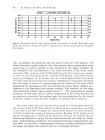
The step duration of a pika is described by a decreasing power like func-
tion of speed (Fischer and Lehmann, 1998). In the range of half-bound speed,
this function may be linearized (cf. fig. 3). At very high speeds
(> 1.75 m/sec), in the individuals under study here we noted that the stan-
dard deviation of the step duration measured from touch-down of the leading
limb to the next touch-down was significantly smaller than the standard de-
viation of the step duration measured for the trailing limb.
These results indicate that even in animals using their “hands” (fore feet)
for running a handedness exists, which even in a small group of animals
shows differences between individuals what concerns the preferred side. May
Interactions between Motions of the Trunk and the Angle of Attack 71
this be an indicator of a body side specific specialization of the extremities (in
mechanical performance and/or control), even without profound knowledge
about the bases of this effect it indicates that “the” motion scheme of “the”
pika does not exist – in so far pikas are real individuals.
Fig. 2. The four individuals under study systematically preferred one of their fore-
limbs for the first touch down in a motion cycle of half-bound (trailing forelimb).
Fig. 3. Step duration of the pika (Ochotona rufescens) in half-bound. At speeds >
1.75 m/sec, the S.D. of the step duration is significantly higher (p < 0.05) for the
leading limb than that for the trailing limb. At each speed n = 20 motion cycles
were analysed.
72 R. Hackert, H. Witte, M. S. Fischer
3 Trajectories of the centre of mass of pikas in
half-bound gait
3.1 Method: Videoradiography.
The animals were filmed at a frequency of 1,000 fps, half-bounding on a
treadmill at a speed of 2.0 m/s. At this speed, the step frequency is about 8
cycles per second. At 1,000 fps the high speed cameras provide a resolution of
256 x 64 pixels. The treadmills belt is twice as wide as a pika’s body width.
One camera was used to film the pikas from the lateral side (the ground
appears to be a line on the screen). To control the permanence of speed, this
lateral zoom-camera was adjusted with the maximal focus length (75 mm)
in such a way that the picture just covered the length of the animal when
it was maximally extended. A second camera documented the front view, to
ensure that the pika was running straight forward.
3.2 Method: Digitization
To control the effects of optical distorsion, a reference grid (mesh width 10 ±
0.05 mm, steel balls of 1 ± 0.01 mm in diameter) was filmed and served as a
control for linearization means. The outline of the body was digitised in the
global frame with 35 points alternately distributed on the dorsal and on the
ventral border of the sagittal projection of the animal. Limb segments were
incorporated into the body shape proximally of the elbow and knee joints.
The background of the picture (grid of the Faraday‘s cage of our laboratory)
was filled with vertical lines spaced approx. 1 cm. We took advantage from
these lines to get an equal distribution of the digitisation points along the
body contour. 90% of the animal’s mass is included in this digitised area.
The number of points used for digitising the body outline arose to be a
good compromise between the needs for the binding line between two even
following points to stay near to the contour line and the wish to limit the
expense for the digitising work.
3.3 Method: Weighing of triangle segments of trunk elements
The distribution of the points on the body outline defined a series of triangles,
the areas and centers of which were computed from their corner coordinates.
To take account of the mass distribution, we weighed a series of 14 transversal
slices of a pika cadaver frozen in its extended position (fig. 4). These values
were the base for the computational weight distribution onto the triangles.
We thus implicitely neglected the effect of oscillating masses, or seen the
other way round, since the thickness of the zone defined by the base of the
triangle is about 1 cm, this means that the masses have been considered to
oscillate locallly in this volume.
Interactions between Motions of the Trunk and the Angle of Attack 73
3.4 Results
Motion of the center of mass in the body:
• The CoM is located underneath the lung base. It is closer to the ventral
outline than to the dorsal one (40:60) (fig. 4).
• The position of the center of mass relatively to the nose (which is a
representative for the rather unaccelerated head) is not constant. The
horizontal excursion of the CoM is in fixed phase coupling with the motion
of the back. During spinal extension, which takes place during the stance
phase of the hindlimbs, and at the beginning of the forelimbs’ stance phase
the CoM moves in the cranio-caudal direction. During spinal bending the
CoM moves in the caudo-cranial direction. This excursion equals about
10 % of the animals’ length (fig. 5).
Fig. 4. Left: Mass distribution of the trunk of a pika (Ochotona rufescens) including
the upper arm (proximally of the elbow joint) and the thigh. Right: position of the
center of mass at touch down of the forelimbs (extended back) and of the hindlimbs
(bended back). The radius of the circle corresponds to the strength of the interval
of confidence.
Vertical motions of the CoM in the global frame:
• The amplitude of the motion of the CoM at 2 m/sec accounts for about
6 mm (10% of the animal’s height) (fig. 5).
• During the extension of the back the CoM globally moves down, during
the bending of the back it globally moves up (fig. 5).
• The pattern of the CoM vertical motion has more than two extrema.
74 R. Hackert, H. Witte, M. S. Fischer
Fig. 5. Motions of the CoM during half-bound of a pika (Ochotona rufescens).
Left: horizontal excursions relative to the nose
Right: Vertical excursions with corresponding footfall patterns
Position of the CoM relative to the forelimbs.
• The angle wrist-elbow-CoM of the trailing forelimb is about 180˚ during
that part of its stance phase when no other ground contacts exist (fig. 6).
• After the leading forelimb touches the ground, the weight is transferred
to it: the alignment CoM-trailing ulna decreases while the alignment with
the leading ulna becomes almost complete.
4 Does the angle of attack couple with speed?
The angle of attack is defined as the angle formed by the connection line of
CoM and the ground contact point versus ground. To quantify the variation
of the angle of attack with speed we took advantage of the above described ef-
fect that at touch down of the trailing limb the ulna points is in the direction
of the CoM. The orientation of the ulna does not coincide exactly with the
direction defined by the connection line of the ground contact point (under-
neath the metatarso-phalangial joint) and the CoM. This error is systematic
and accounts for + 5˚.
4.1 Methods
The high speed X-Ray camera accessible to us provided 150 fps. This frame
rate is insufficient to determine significant values for the angle of attack, since
a pika at observation speed may run up to 8 cycles per second. Consecutively
we shaved the forelimbs of a pika and filmed the half-bounding animal on the
treadmill with the high speed video system (500 fps, resolution of 256x256
pixels).
The camera field was adjusted to cover one pika length. This enables a
rigorous control of pika speed.
Interactions between Motions of the Trunk and the Angle of Attack 75
4.2 Results
1. The angle of attack does not variate strongly with increasing speed (fig.7).
2. The angle ulna/ground equals about 50˚, consecutively the angle of at-
tack is about 45˚.
Fig. 6. The angle wrist-elbow-CoM of the trailing forelimb is about 180˚ during
that part of its stance phase when no other ground contacts exist. During late mid
stance the leading forelimb takes over and its ulna points to the CoM. Alignment of
the shank (kinematically eqivalent to the upper arm) mainly occurs during aerial
phases.
5 Conclusions
The small mammal’s limb is a four segmented flexed structure, which may be
compared to a pantograph [5]. It effectively allows for compensation of small
irregularities of the ground. It also plays the role of a spring-damper system
as the pika runs or trots. The occurance of elastic phenomena during legged
locomotion is commonly accepted in biology (cf. [6], [7], [8] and succeeding
publications). The movement of the human CoM during running may be
described using spring-mass models [9] [10]. The vertical excursion of the
CoM of a half-bounding pika (about 5-6 mm) relatively to the leg length
(70 mm) is quite comparable to the excursion of the CoM in human running
(about 10 %) [11]. From this point of view (in addition to many others), it
also seems promising to extend these templates to quadrupedal locomotion