Neuronal Control of Eye Movements - part 6 pdf
Bạn đang xem bản rút gọn của tài liệu. Xem và tải ngay bản đầy đủ của tài liệu tại đây (177.62 KB, 21 trang )
Disconjugate Eye Movements 95
In a stereoblind patient with strabismus, the Listing’s planes of the two eyes
were normal in shape, i.e. relatively planar, but changed their orientation
depending on which eye was fixating [62]. This effect was most probably due to
accommodation-induced vergence.
Asymmetric Vergence Movements and Hering’s Law
Hering’s law of equal innervation implies that equal version and vergence
commands are sent to both eyes and that the binocular motor output represents
the sum of the two signals. The analysis of asymmetric vergence movements
(fig. 2) can give some indication whether Hering’s law holds [63, 64] or
whether the two eyes are independently controlled, as advocated by Helmholtz
[65, 66]. As we will see, there are arguments for both theories.
During static convergence on a target in front of one eye, i.e. asymmetric
convergence, only the inferior oblique muscle contracts in this eye, as demon-
strated with MRI; contraction of the same muscle, apart from contractile
changes in the lateral and medial rectus muscles, is also seen in the fellow eye,
which is directed inward [47]. During rapid gaze shifts along the line of sight of
one eye, which calls for asymmetric vergence, the horizontal peak accelerations
of the two eyes are similar, despite different position trajectories [67]. This find-
ing suggests equal saccadic pulses for each eye, according to Hering’s law,
together with an additional vergence signal. After human subjects were trained
to have a vertical vergence component during symmetric horizontal vergence,
the vertical vergence component could also be demonstrated during smooth
pursuit of targets in depth both along the line of sight of one eye [68]. Thus
symmetric smooth pursuit seems to be combined with vergence to produce
Symmetric Asymmetric
Fig. 2. Top view of both eyes during symmetric and asymmetric convergence move-
ments. The visual target moves from far to near (arrow).
Straumann 96
asymmetric slow eye movements, which speaks against monocular control of
these movements.
Some subjects are able to initiate smooth asymmetrical ‘saccade-free’ con-
vergence movements when changing gaze from a far to a near target [69]. Thus,
during binocular viewing, the ocular motor system is able to generate eye
movements that do not adhere to Hering’s law of equal innervation. Similarly,
the initial monocular smooth pursuit response to a target that moves in depth
solely depends on target motion and is independent of the response of the other
eye [70].
The firing rate of abducens motoneurons for a given eye position is higher
with than without convergence, but, paradoxically, lateral rectus force (and sim-
ilarly medial rectus force) is not increased [70a]. This finding still awaits an
explanation. A reanalysis of single neuron recordings during eye movements
that included vergence revealed that neural signals in abducens motoneurons,
abducens interneurons, and medial rectus motoneurons encode the position of
both eyes, not just one eye [71]. On the other hand, premotor neurons in the
paramedian pontine reticular formation encode saccadic velocity signals for
only one eye, not both [72]. These findings speak against a neural implementa-
tion of Hering’s law.
Saccade-Associated Vergence Movements
Peak vergence velocity increases when vergence is combined with a sac-
cade, an effect that is more pronounced in divergence than convergence [73].
Vice versa, when saccades occur with vergence movements, the peak velocity
of the saccades is reduced, more prominently so with convergence than diver-
gence [74]. These findings suggest a nonlinear interaction between conjugate
and disconjugate premotor systems; the omnipause neurons probably represent
the crucial neural structure for gating saccade-related horizontal vergence [75].
This would also explain why saccadic oscillations occur, when saccades end
during ongoing vergence [76–78]. Note that even horizontal and vertical sac-
cades between far targets are associated with small transient vergence compo-
nents, but these are probably related to mechanical differences between
adducting and abducting muscles [75, 79]. Horizontal saccades also produce
small torsional transients out of Listing’s plane, which are not equal in ampli-
tude; hence, the eyes cycloverge somewhat shortly after the beginning of each
saccade [80].
Saccades in patients with one deeply amblyopic eye are nonconjugate, i.e.
Hering’s law seems to rely on intact binocular vision [81]. Subjects with ani-
sometropic spectacles show saccades with different amplitudes in both eyes and
Disconjugate Eye Movements 97
asymmetric postsaccadic drift [82]. When saccades are made between targets at
different distances, a presaccadic vergence movement along the isovergent line
of the initial target appears [83]. This observation speaks for separate version
and vergence channels contributing to fast eye displacements. A similarly
strong coupling between version and vergence is found during incorrect sac-
cades evoked by two targets appearing simultaneously in 3-D space [84].
Conversely, when targets are placed at closer distances from the eyes, no pre-
saccadic convergence and only a small presaccadic divergence is observed, and
postsaccadic vergence is usually asymmetric [85]. The latter finding speaks
against a balanced interaction between the vergence and version systems during
the saccade, and therefore against a Hering-type implementation of such move-
ments. Such saccades are dominated by one eye, so that a least one of the two
eyes is on target in time.
Binocular vertical displacements between near targets in front of one eye
require different vertical amplitudes of each eye to maintain binocular align-
ment. In downward movements, a major portion of the required disconjugacy
takes place during the saccades, while in upward movements the intrasaccadic
portion amounts to about half [86]. Dynamic dissociations between saccadic
and vergence movements can also be observed during vertical saccades
between targets in the midsagittal plane at different depth [87].
Binocular Adaptation
Phoria Adaptation
Normal binocular fixation of a near target in a tertiary position requires a
vertical vergence component, when eye positions are expressed in a head-fixed
coordinate system. This component appears to be independent of whether sub-
jects are viewing monocularly or binocularly [88]. Eight hours of monocular
occlusion leads to excyclophoria and hyper- or hypophoria [89]. If an eye is
covered and passively rotated away from the position of the fellow eye with a
scleral suction lens during a few minutes, ocular misalignment persists up to
10 min or until binocular viewing is permitted [90].
When short-term phoria adaptation is performed with a vertical disparity
at a single location, phoria becomes uniform for all gaze directions. Upon two
vertical disparities at opposite gaze directions and with opposite sign, adapted
phoria shows a gradient along the line between the two stimuli [91, 92]. Phoria
adaptation to opposite vertical disparities is also effective along the depth axis
[93] or to multiple vertical disparities at different near and far locations [94].
Human subjects are also able to adapt vertical phoria to different prism-induced
vertical disparities that vary with head position [95] or with head and gaze
Straumann 98
position [96]. When monkeys are trained to synchronize vergence eye move-
ments in synchrony with vestibularly evoked eye movements upon pitch oscilla-
tions, these oscillations evoked vergence eye movements even in the dark [97, 98].
Adaptation to discrete increments of refraction along a horizontal prism is
also possible, but adapted vergence changes only gradually when crossing the
prism edges [99]. After 30–150 s of cyclovergence evoked by incyclo- or excy-
clodisparity, the eyes do not tort back to their previous torsional positions, even
in the presence of a visual stimulus [100]. Most likely, this torsional hysteresis
is the result of fast phoria adaptation.
Phoria adaptation with a vertical prism over one eye is often impaired in
patients with cerebellar disease. Thus the cerebellum seems to be decisively
involved in phoria adaptation [101].
Adaptation of Listing’s Plane
Three days of vertical disparity with prisms induces, besides vertical pho-
ria, reorientations of Listing’s planes; Listing’s plane of the higher eye is rotated
up and Listing’s plane of the lower eye rotated down [102]. Phoria adaptation to
different cyclodisparities along the vertical axis also modifies the orientation of
Listing’s planes [103].
Binocular Saccade Adaptation
Intrasaccadic displacement of a visual target leads to rapid binocular sac-
cade adaptation. If the displacement is only presented to one eye, while the tar-
get is unchanged for the other eye, short-term adjustments are again conjugate,
which suggests that there is no mechanism for fast disconjugate saccade adap-
tation [104]. Dichoptically presented random-dot patterns with local disparities
representing a 3-D object lead to immediate position-dependent saccadic dis-
conjugacies that persist during subsequent monocular viewing [105]. Similar
immediate disconjugacies of saccades can be observed when disparities are
introduced by dichoptical images that differ in size [106].
Subjects with anisometropic spectacles show saccades with different
amplitudes and postsaccadic drifts between both eyes, even during monocular
viewing [82, 107]. Already an image size inequality of 2% leads to disconjugate
horizontal and vertical saccades, which persist after a short training period
when tested in the absence of normal binocular visual targets [108]. Placing an
afocal magnifier in front of one eye leads to disconjugate memory-guided sac-
cades, which outlasts the removing of the magnifier after the training period,
when subjects are viewing monocularly [109, 110]. Dichoptically presented
patterns that are displaced at the end of each vertical saccade induce amplitude
disconjugacy, but only little disconjugate postsaccadic drift [111]. Apparently,
this effect does not require foveal fusion since microstrabismic patients adapt as
Disconjugate Eye Movements 99
well [112]. When vertical saccades are disconjugately adapted, smooth pursuit
movements remain conjugate and vice versa [113]. Thus, the two classes of eye
movements have separate mechanisms for binocular adaptation.
In patients with trochlear nerve palsy, saccades become more conjugate
after strabismus surgery, an effect that is more pronounced in patients with con-
genital than in patients with acquired trochlear nerve palsy [114]. In rhesus mon-
keys with one surgically weakened extraocular muscle, the paretic eye shows
postsaccadic drift with the normal eye viewing. Deafferenting the paretic eye
leaves postsaccadic drift unchanged; thus, proprioception from the paretic eye
does not play a role in the adaptation of postsaccadic drift [115]. Proprioceptive
deafferentation alone impairs ocular alignment and saccade conjugacy [116].
Disconjugate Eye Movements Evoked by Vestibular Stimulation
Vergence eye movements are elicited by linear motion in the dark with or
without visual targets [117]. The gain of the translational vestibulo-ocular
reflex (VOR) during heave ( ϭ up-down) and sway ( ϭ left-right) whole-body
oscillation increases with increasing convergence [118, 119]. During surge
( ϭ fore-aft) oscillation, the gain of the translational VOR increases with both
increasing gaze eccentricity and increasing convergence, which is qualitatively
accurate for foveal stabilization of both eyes [120–122]. Such vergence respon-
ses are enhanced by the presence of visual stimuli [123]. During visual fixation
upon isovergence targets along the horizontal meridian and concurrent rapid
oscillations in various directions in the horizontal plane, both eyes move in the
geometrically correct direction needed to stabilize the targets on the two foveae;
the gain of the version component (average velocity of both eyes divided target
velocity), however, amounts to only around 0.5, while the gain of the vergence
component (right eye velocity minus left eye velocity) ranges around unity
[124]. This finding might reflect the fact that for visual acuity it is more impor-
tant to stabilize the relative orientation of the lines of sight than binocular posi-
tion. Vergence also modifies the gain of the angular VOR for gaze stabilization.
For example, the gain of the VOR elicited on a horizontal turntable anticipates
the vergence angle by about 50 ms [125].
Ocular counterroll elicited by head or whole-body roll interferes with
stereopsis. This geometric incompatibility increases further with decreasing tar-
get distance. It is therefore advantageous that ocular counterroll decreases
strongly during convergence [126, 127]. In the presence of ocular counterroll,
binocular movements from a far to a near target show unequal torsion; the
required torsion for the undermost eye is larger than for the uppermost eye,
since convergence is associated with extorsion. Such torsional disconjugacy,
Straumann 100
however, cannot be demonstrated for divergent eye movements [128]. Static
head roll also leads to excyclovergent eye positions [129]. This phenomenon
can be explained by a static hysteresis that differs between the eyes contra- and
ipsilateral to head roll [130]. Probably, ocular torsional hysteresis is introduced
at the level of the otolith pathways because the direction-dependent torsional
position lag of the eyes was related to head roll position, not eye position.
Asymmetric binocular torsion evoked by hypo- or hypergravity may be a pre-
dictor for space sickness [131–133].
During position steps of head roll, the eyes show dynamic binocular coun-
terrolling and skewing. While the gain of dynamic binocular torsion is larger in
upright than in supine position, dynamic skewing is unaffected by the addi-
tional otolith input that appears in upright position [134]. Constant rotation
about an off-vertical axis causes horizontal vergence movements [135]. During
oscillatory head roll, the ocular rotation axes of the two eyes are convergent
both in the dark and when fixating upon a far light dot; when subjects fix upon
a near light dot, the convergence of binocular rotation axes exceeds the conver-
gence of binocular positions [136]. The Bielschowsky head-tilt sign in unilat-
eral trochlear nerve palsy, i.e. increased vertical and torsional divergence with
the head tilted towards the affected eye, can be explained by inward tilt of the
rotation axis of the covered eye during head oscillation about the naso-occipital
axis [137]. This ‘convergence’ of ocular rotation axes is the result of decreased
force by the SO of the covered paretic eye or, according to Hering’s law,
increased force parallel to the paretic SO in the covered unaffected eye. The
gain of the VOR in an eye with trochlear nerve palsy is reduced in all directions,
but especially towards intorsion, depression and abduction, in accordance with
the 3-D pulling direction of the SO [138]. In patients with peripheral abducens
nerve palsy, the gain of the horizontal VOR in the affected eye is reduced in
both directions, when tested in the dark. In the light, horizontal gains normalize
in patients with mild or moderate palsy [139]. The gain of the torsional VOR is
reduced in both the healthy and the affected eye [140].
The orientation of ocular rotation axes as a function of eye position depends
on the gain of the torsional VOR; the lower the torsional gain, the more the axes
tilt with eccentric gaze position [141]. As the torsional gain decreases further
with increasing convergence, average 3-D eye positions scatter closely around
the temporally rotated Listing’s plane, which is advantageous for binocular reti-
nal stabilization [142]. Head roll in patients with peripheral abducens nerve
palsy leads to a hyperdeviation of the ipsilateral eye, independent of which eye is
affected. In patients with central abducens palsy, the same eye (healthy or
affected) hyperdeviates when rolling the head to the left or the right side [143].
At low frequencies, the horizontal and vertical VOR can be cancelled by
visually fixing upon head-fixed targets. During head oscillations about the
Disconjugate Eye Movements 101
naso-occipital axis visual suppression of the elicited torsional VOR is incom-
plete, but the lines of sight of the two eyes remain on target [144]. If subjects
during head roll fix upon head-fixed eccentric horizontal targets at near distance,
the eyes also show vertical movement components, even if one eye is covered
[145]. These components are required to keep the lines of sight pointed to the
targets. Thus, the vergence system correctly modifies the eye movements that are
not visually cancelled to prevent horizontal and vertical retinal slip in either eye.
Disconjugate Eye Movements and Blinks
Initial eye movements during voluntary blinks are extorsional, downward,
and inward, consistent with an early pulse-like innervation of the inferior rectus
muscle [146]. Thus, during this early phase of blinking, the eyes converge and
excyclodiverge. Blinks modify the kinematics and dynamics saccade-vergence
and slow vergence eye movements [147, 148]. Besides mechanical factors of
the eye plant, the found changes might reflect the blink-induced decrease in
omnipause neuron activity.
Pathological Disconjugate Eye Movements
Normally, vergence eye movements in response to steps of a visual stimuli
become slower with age, which has to be taken into account when evaluating
patients with suspected vergence disorders [149].
Binocular positions in patients with cerebellar dysfunction are usually
esophoric or even esotropic. In addition, there is a hypertropia that varies as a
function of horizontal eye position, so-called alternating skew deviation with
the abducting eye higher. The patients show both conjugate and disconjugate
saccadic abnormalities that are also eye position dependent [150]. The mecha-
nism of alternating skew deviation in patients with cerebellar disease could be
due to a lost correction of changed eye muscle pulling directions, which is
required when animals become frontal eyed. If, in addition, one assumes an
imbalance of graviceptive-ocular pathways responding to head pitch, alternat-
ing skew deviation can be explained by this mechanism [151].
Dissociated vertical divergence (DVD) includes the following ocular
motor phenomena [152]: Upon occlusion of either eye, a horizontal and
cyclovertical latent nystagmus develops. This is quickly followed by cyclover-
sion/vertical vergence, with the fixing eye intorting and tending to move down-
ward and the covered eye extorting and moving up. Simultaneously, upward
versions occur for the maintenance of fixation. This, in turn, leads to further
Straumann 102
upward movement of the covered eye and, at the same time, to a reduction of the
cyclovertical component of the latent nystagmus. Thus, a possible ‘purpose’ of
this cycloversion and vertical vergence is to damp the cyclovertical nystagmus
that occurs when one eye is covered [153]. Brodsky hypothesized that DVD is a
dorsal light reflex that occurs when binocular vision is impaired in infancy
[154]. Since patients with DVD only transiently perceive a tilt of the subjective
visual vertical when one eye is covered, it was speculated that the cancellation
of SVV tilt in these patients is the main function of DVD [155].
Binocular eye movements in patients with convergent-divergent pendular
nystagmus are conjugate in the vertical direction, but phase shifted by 180Њ in
the horizontal and torsional directions [156]. The lesion is usually localized
within neural structures of the vergence system. If horizontal saccades or
smooth pursuit eye movements are pathologically coupled with convergence,
the abducting eye will appear paretic despite an intact abducens nerve. This so-
called pseudo-abducens palsy is caused by lesions of convergence pathways
near the midbrain-diencephalic junction and is frequently associated with
upgaze palsy and convergence-retraction nystagmus [157]. Paramedian thala-
mic infarctions without involvement of the midbrain may lead to a selective
bilateral pseudo-abducens palsy [158]. Convergence-retraction nystagmus,
however, is due to a mesencephalic lesion [159] and represents a disorder of the
vergence system [160]. Pathologically disconjugate eye movements with the
vergence system intact, is typical of internuclear ophthalmoparesis [161]. Mild
internuclear ophthalmoparesis, in which the adducting eye is only slightly
slower than the abducting eye, is often missed by clinicians, as demonstrated by
infrared oculography [162].
Ocular bobbing, which rarely appears after infratentorial lesions, but oth-
erwise has no localizing value, may be disconjugate [163]. Disconjugate verti-
cal and torsional ocular movements, resembling seesaw nystagmus, have been
observed in a patient with locked-in syndrome after large infarction of the pons
[164]. Smaller lesions in the ventral pons involving the nucleus reticularis
tegmenti pontis lead to impairment of slow vergence movements to ramp tar-
gets [165]. On the other hand, fast vergence movements to step targets are
affected by lesions of upper pontine nuclei [166].
References
1 Sheliga BM, Miles FA: Perception can influence the vergence responses associated with open-
loop gaze shifts in 3D. J Vis 2003;3:654–676.
2 Cornell ED, MacDougall HG, Predebon J, Curthoys IS: Errors of binocular fixation are common
in normal subjects during natural conditions. Optom Vis Sci 2003;80:764–771.
3 Francis EL, Jiang BC, Owens DA, Tyrrell RA: Accommodation and vergence require effort-to-
see. Optom Vis Sci 2003;80:467–473.
Disconjugate Eye Movements 103
4 Stevenson SB, Lott LA, Yang J: The influence of subject instruction on horizontal and vertical ver-
gence tracking. Vision Res 1997;37:2891–2898.
5 Semmlow JL, Yuan W, Alvarez TL: Evidence for separate control of slow version and vergence eye
movements: support for Hering’s Law. Vision Res 1998;38:1145–1152.
6 van Leeuwen AF, Collewijn H, Erkelens CJ: Dynamics of horizontal vergence movements: inter-
action with horizontal and vertical saccades and relation with monocular preferences. Vision Res
1998;38:3943–3954.
7 Hung GK, Zhu H, Ciuffreda KJ: Convergence and divergence exhibit different response charac-
teristics to symmetric stimuli. Vision Res 1997;37:1197–1205.
8 Alvarez TL, Semmlow JL, Pedrono C: Divergence eye movements are dependent on initial stimu-
lus position. Vision Res 2005;45:1847–1855.
9 Semmlow JL, Hung GK, Horng JL, Ciuffreda K: Initial control component in disparity vergence
eye movements. Ophthalmic Physiol Opt 1993;13:48–55.
10 Semmlow JL, Hung GK, Horng JL, Ciuffreda KJ: Disparity vergence eye movements exhibit pre-
programmed motor control. Vision Res 1994;34:1335–1343.
11 Semmlow JL, Yuan W: Adaptive modification of disparity vergence components: an independent
component analysis study. Invest Ophthalmol Vis Sci 2002;43:2189–2195.
12 Masson GS, Yang DS, Miles FA: Version and vergence eye movements in humans: open-loop
dynamics determined by monocular rather than binocular image speed. Vision Res 2002;42:
2853–2867.
13 Horng JL, Semmlow JL, Hung GK, Ciuffreda KJ: Dynamic asymmetries in disparity convergence
eye movements. Vision Res 1998;38:2761–2768.
14 Horng JL, Semmlow JL, Hung GK, Ciuffreda KJ: Initial component control in disparity vergence:
a model-based study. IEEE Trans Biomed Eng 1998;45:249–257.
15 Alvarez TL, Semmlow JL, Yuan W: Closely spaced, fast dynamic movements in disparity ver-
gence. J Neurophysiol 1998;79:37–44.
16 Alvarez TL, Semmlow JL, Yuan W, Munoz P: Disparity vergence double responses processed by
internal error. Vision Res 2000;40:341–347.
17 Howard IP, Fang X, Allison RS, Zacher JE: Effects of stimulus size and eccentricity on horizontal
and vertical vergence. Exp Brain Res 2000;130:124–132.
18 Busettini C, Miles FA, Krauzlis RJ: Short-latency disparity vergence responses and their depen-
dence on a prior saccadic eye movement. J Neurophysiol 1996;75:1392–1410.
19 Busettini C, FitzGibbon EJ, Miles FA: Short-latency disparity vergence in humans. J Neurophysiol
2001;85:1129–1152.
20 Masson GS, Busettini C, Miles FA: Vergence eye movements in response to binocular disparity
without depth perception. Nature 1997;389:283–286.
21 Busettini C, Masson GS, Miles FA: Radial optic flow induces vergence eye movements with ultra-
short latencies. Nature 1997;390:512–515.
22 Coubard O, Daunys G, Kapoula Z: Gap effects on saccade and vergence latency. Exp Brain Res
2004;154:368–381.
23 Luu CD, Abel L: The plasticity of vertical motor and sensory fusion in normal subjects.
Strabismus 2003;11:109–118.
24 Hara N, Steffen H, Roberts DC, Zee DS: Effect of horizontal vergence on the motor and sensory
components of vertical fusion. Invest Ophthalmol Vis Sci 1998;39:2268–2276.
25 Betts GA, Curthoys U, Todd MJ: The effect of roll-tilt on ocular skew deviation. Acta Otolaryngol
Suppl 1995;520:304–306.
26 Enright JT: Unexpected role of the oblique muscles in the human vertical fusional reflex. J Physiol
1992;451:279–293.
27 Cheeseman EW Jr, Guyton DL: Vertical fusional vergence: the key to dissociated vertical devia-
tion. Arch Ophthalmol 1999;117:1188–1191.
28 van Rijn LJ, Collewijn H: Eye torsion associated with disparity-induced vertical vergence in
humans. Vision Res 1994;34:2307–2316.
29 Mudgil AV, Walker M, Steffen H, Guyton DL, Zee DS: Motor mechanisms of vertical fusion in
individuals with superior oblique paresis. J AAPOS 2002;6:145–153.
Straumann 104
30 Howard IP, Allison RS, Zacher JE: The dynamics of vertical vergence. Exp Brain Res 1997;116:
153–159.
31 Howard IP, Fang X, Allison RS, Zacher JE: Effects of stimulus size and eccentricity on horizontal
and vertical vergence. Exp Brain Res 2000;130:124–132.
32 van Rijn LJ, Vandersteen J, Collewijn H: Instability of ocular torsion during fixation – cyclover-
gence is more stable than cycloversion. Vision Res 1994;34:1077–1087.
33 Hooge IT, van den Berg AV: Visually evoked cyclovergence and extended Listing’s law.
J Neurophysiol 2000;83:2757–2775.
34 van Rijn LJ, Vandersteen J, Collewijn H: Visually induced cycloversion and cyclovergence. Vision
Res 1992;32:1875–1883.
35 Howard IP, Zacher JE: Human cyclovergence as a function of stimulus frequency and amplitude.
Exp Brain Res 1991;85:445–450.
36 Howard IP, Sun L, Shen X: Cycloversion and cyclovergence: the effects of the area and position of
the visual display. Exp Brain Res 1994;100:509–514.
37 Minken AW, Gielen CC, van Gisbergen JA: An alternative three-dimensional interpretation of
Hering’s equal-innervation law for version and vergence eye movements. Vision Res 1995;35:
93–102.
38 Mok D, Ro A, Cadera W, Crawford JD, Vilis T: Rotation of Listing’s plane during vergence. Vision
Res 1992;32:2055–2064.
39 van Rijn LJ, van den Berg AV: Binocular eye orientation during fixations: Listing’s law extended
to include eye vergence. Vision Res 1993;33:691–708.
40 Somani RA, DeSouza JF, Tweed D, Vilis T: Visual test of Listing’s law during vergence. Vision Res
1998;38:911–923.
41 Minken AW, van Gisbergen JA: A three-dimensional analysis of vergence movements at various
levels of elevation. Exp Brain Res 1994;101:331–345.
42 Tweed D: Visual-motor optimization in binocular control. Vision Res 1997;37:1939–1951.
43 Bruno P, van den Berg AV: Relative orientation of primary positions of the two eyes. Vision Res
1997;37:935–947.
44 Hepp K: Theoretical explanations of Listing’s law and their implication for binocular vision.
Vision Res 1995;35:3237–3241.
45 Tweed D: Visual-motor optimization in binocular control. Vision Res 1997;37:1939–1951.
46 Schreiber K, Crawford JD, Fetter M, Tweed D: The motor side of depth vision. Nature 2001;410:
819–822.
47 Demer JL, Kono R, Wright W: Magnetic resonance imaging of human extraocular muscles in con-
vergence. J Neurophysiol 2003;89:2072–2085.
48 Steffen H, Walker MF, Zee DS: Rotation of Listing’s plane with convergence: independence from
eye position. Invest Ophthalmol Vis Sci 2000;41:715–721.
49 Kapoula Z, Bernotas M, Haslwanter T: Listing’s plane rotation with convergence: role of disparity,
accommodation, and depth perception. Exp Brain Res 1999;126:175–186.
50 Mikhael S, Nicolle D, Vilis T: Rotation of Listing’s plane by horizontal, vertical and oblique
prism-induced vergence. Vision Res 1995;35:3243–3254.
51 Minken AW, van Gisbergen JA: Dynamical version-vergence interactions for a binocular imple-
mentation of Donders’ law. Vision Res 1996;36:853–867.
52 Tweed D, Vilis T: Geometric relations of eye position and velocity vectors during saccades. Vision
Res 1990;30:111–127.
53 Porrill J, Ivins JP, Frisby JP: The variation of torsion with vergence and elevation. Vision Res
1999;39:3934–3950.
54 Ivins JP, Porrill J, Frisby JP: Instability of torsion during smooth asymmetric vergence. Vision Res
1999;39:993–1009.
55 Migliaccio AA, Cremer PD, Aw ST, Halmagyi GM, Curthoys IS, Minor LB, Todd MJ: Vergence-
mediated changes in the axis of eye rotation during the human vestibulo-ocular reflex can occur
independent of eye position. Exp Brain Res 2003;151:238–248.
56 Straumann D, Steffen H, Landau K, Bergamin O, Mudgil AV, Walker MF, Guyton DL, Zee DS:
Primary position and Listing’s law is acquired and congenital trochlear nerve palsy. Invest
Ophthalmol Vis Sci 2003;44:4282–4292.
Disconjugate Eye Movements 105
57 Migliaccio AA, Cremer PD, Aw ST, Halmagyi GM: Vergence-mediated changes in Listing’s plane
do not occur in an eye with superior oblique palsy. Invest Ophthalmol Vis Sci 2004;45:3043–3047.
58 Wong AMF, Sharpe JA, Tweed D: Adaptive neural mechanism for Listing’s law revealed in
patients with fourth nerve palsy. Invest Ophthalmol Vis Sci 2002;43:1796–1803.
59 Wong AMF, Tweed D, Sharpe JA: Adaptive neural mechanism for Listing’s law revealed in
patients with sixth nerve palsy. Invest Ophthalmol Vis Sci 2002;43:112–119.
60 Somani RA, Hutnik C, DeSouza JF, Tweed D, Nicolle D, Vilis T: Using a synoptophore to test
Listing’s law during vergence in normal subjects and strabismic patients. Vision Res 1998;38:
3621–3631.
61 van den Berg AV, van Rijn LJ, de Faber JT: Excess cyclovergence in patients with intermittent
exotropia. Vision Res 1995;35:3265–3278.
62 Melis BJ, Cruysberg JR, van Gisbergen JA: Listing’s plane dependence on alternating fixation in
a strabismus patient. Vision Res 1997;37:1355–1366.
63 Moschovakis AK: Are laws that govern behavior embedded in the structure of the CNS? The case
of Hering’s law. Vision Res 1995;35:3207–3216.
64 Mays L: Has Hering been hooked? Nat Med 1998;4:889–890.
65 King WM, Zhou W: Neural basis of disjunctive eye movements. Ann N Y Acad Sci 2002;956:
273–283.
66 Dell’Osso LF: Evidence suggesting individual ocular motor control of each eye (muscle). J Vestib
Res 1994;4:335–345.
67 Ramat S, Das VE, Somers JT, Leigh RJ: Tests of two hypotheses to account for different-sized sac-
cades during disjunctive gaze shifts. Exp Brain Res 1999;129:500–510.
68 Maxwell JS, Schor CM: Symmetrical horizontal vergence contributes to the asymmetrical pursuit
of targets in depth. Vision Res 2004;44:3015–3024.
69 Enright JT: Slow-velocity asymmetrical convergence: a decisive failure of ‘Hering’s law’. Vision
Res 1996;36:3667–3684.
70 King WM, Zhou W: Initiation of disjunctive smooth pursuit in monkeys: evidence that Hering’s law
of equal innervation is not obeyed by the smooth pursuit system. Vision Res 1995;35:3389–3400.
70a Miller JM, Bockisch CJ, Pavlovski DS: Missing lateral rectus force and absence of medial rectus
co-contraction in ocular convergence. J Neurophysiol 2002;8:2421–2433.
71 King WM, Zhou W, Tomlinson RD, McConville KM, Page WK, Paige GD, Maxwell JS: Eye posi-
tion signals in the abducens and oculomotor nuclei of monkeys during ocular convergence.
J Vestib Res 1994;4:401–408.
72 Zhou W, King WM: Premotor commands encode monocular eye movements. Nature 1998;393:
692–695.
73 Maxwell JS, King WM: Dynamics and efficacy of saccade-facilitated vergence eye movements in
monkeys. J Neurophysiol 1992;68:1248–1260.
74 Collewijn H, Erkelens CJ, Steinman RM: Voluntary binocular gaze-shifts in the plane of regard:
dynamics of version and vergence. Vision Res 1995;35:3335–3358.
75 Zee DS, FitzGibbon EJ, Optican LM: Saccade-Vergence interactions in humans. J Neurophysiol
1992;68:1624–1641.
76 Ramat S, Somers JT, Das VE, Leigh RJ: Conjugate ocular oscillations during shifts of the direc-
tion and depth of visual fixation. Invest Ophthalmol Vis Sci 1999;40:1681–1686.
77 Bhidayasiri R, Somers JT, Kim JI, Ramat S, Nayak S, Bokil HS, Leigh RJ: Ocular oscillations
induced by shifts of the direction and depth of visual fixation. Ann Neurol 2001;49:24–28.
78 Ramat S, Leigh RJ, Zee DS, Optican LM: Ocular oscillations generated by coupling of brainstem
excitatory and inhibitory saccadic burst neurons. Exp Brain Res 2005;160:89–106.
79 Collewijn H, Erkelens CJ, Steinman RM: Trajectories of the human binocular fixation point dur-
ing conjugate and non-conjugate gaze-shifts. Vision Res 1997;37:1049–1069.
80 Straumann D, Zee DS, Solomon D, Lasker AG, Roberts DC: Transient torsion during and after
saccades. Vision Res 1995;35:3321–3334.
81 Maxwell GF, Lemij HG, Collewijn H: Conjugacy of saccades in deep amblyopia. Invest
Ophthalmol Vis Sci 1995;36:2514–2522.
82 Lemij HG, Collewijn H: Long-term nonconjugate adaptation of human saccades to anisometropic
spectacles. Vision Res 1991;31:1939–1954.
Straumann 106
83 Collewijn H, Erkelens CJ, Steinman RM: Trajectories of the human binocular fixation point dur-
ing conjugate and non-conjugate gaze-shifts. Vision Res 1997;37:1049–1069.
84 Chaturvedi V, van Gisbergen JA: Specificity of saccadic adaptation in three-dimensional space.
Vision Res 1997;37:1367–1382.
85 Enright JT: Monocularly programmed human saccades during vergence changes? J Physiol 1998;
512:235–250.
86 Ygge J, Zee DS: Control of vertical eye alignment in three-dimensional space. Vision Res
1995;35:3169–3181.
87 Kumar AN, Han Y, Dell’Osso LF, Durand DM, Leigh RJ: Directional asymmetry during combined
saccade-vergence movements. J Neurophysiol 2005;93:2797–2808.
88 Schor CM, Maxwell JS, Stevenson SB: Isovergence surfaces: the conjugacy of vertical eye
movements in tertiary positions of gaze. Ophthalmic Physiol Opt 1994;14:279–286.
89 Graf EW, Maxwell JS, Schor CM: Changes in cyclotorsion and vertical eye alignment during
prolonged monocular occlusion. Vision Res 2002;42:1185–1194.
90 Gauthier GM, Vercher JL, Zee DS: Changes in ocular alignment and pointing accuracy after
sustained passive rotation of one eye. Vision Res 1994;34:2613–2627.
91 Maxwell JS, Schor CM: Mechanisms of vertical phoria adaptation revealed by time-course and
2-dimensional spatiotopic maps. Vision Res 1994;34:241–251.
92 Schor CM, Gleason G, Maxwell J, Lunn R: Spatial aspects of vertical phoria adaptation. Vision
Res 1993;33:73–84.
93 Schor CM, McCandless JW: An adaptable association between vertical and horizontal vergence.
Vision Res 1995;35:3519–3527.
94 Schor CM, McCandless JW: Context-specific adaptation of vertical vergence to correlates of eye
position. Vision Res 1997;37:1929–1937.
95 Maxwell JS, Schor CM: Adaptation of vertical eye alignment in relation to head tilt. Vision Res
1996;36:1195–1205.
96 Maxwell JS, Schor CM: Head-position-dependent adaptation of nonconcomitant vertical skew.
Vision Res 1997;37:441–446.
97 Akao T, Kurkin S, Fukushima K: Latency of adaptive vergence eye movements induced by ver-
gence-vestibular interaction training in monkeys. Exp Brain Res 2004;158:129–132.
98 Sato F, Akao T, Kurkin S, Fukushima J, Fukushima K: Adaptive changes in vergence eye move-
ments induced by vergence-vestibular interaction training in monkeys. Exp Brain Res 2004;156:
164–173.
99 Oohira A, Zee DS: Disconjugate ocular motor adaptation in rhesus monkey. Vision Res 1992;32:
489–497.
100 Taylor MJ, Roberts DC, Zee DS: Effect of sustained cyclovergence on eye alignment: rapid
torsional phoria adaptation. Invest Ophthalmol Vis Sci 2000;41:1076–1083.
101 Kono R, Hasebe S, Ohtsuki H, Kashihara K, Shiro Y: Impaired vertical phoria adaptation in
patients with cerebellar dysfunction. Invest Ophthalmol Vis Sci 2002;43:673–678.
102 Steffen H, Walker M, Zee DS: Changes in Listing’s plane after sustained vertical fusion. Invest
Ophthalmol Vis Sci 2002;43:668–672.
103 Schor CM, Maxwell JS, Graf EW: Plasticity of convergence-dependent variations of cyclover-
gence with vertical gaze. Vision Res 2001;41:3353–3369.
104 Albano JE, Marrero JA: Binocular interactions in rapid saccadic adaptation. Vision Res 1995;35:
3439–3450.
105 Eggert T, Kapoula Z: Position dependency of rapidly induced saccade disconjugacy. Vision Res
1995;35:3493–3503.
106 Kapoula Z, Eggert T, Bucci MP: Immediate saccade amplitude disconjugacy induced by unequal
images. Vision Res 1995;35:3505–3518.
107 Oohira A, Zee DS, Guyton DL: Disconjugate adaptation to long-standing, large-amplitude, spec-
tacle-corrected anisometropia. Invest Ophthalmol Vis Sci 1991;32:1693–1703.
108 Bucci MP, Gomes M, Paris S, Kapoula Z: Disconjugate oculomotor earning caused by feeble
image-size inequality: differences between secondary and tertiary positions. Vision Res 2001;41:
625–637.
Disconjugate Eye Movements 107
109 Paris S, Bucci MP, Kapoula Z: Disconjugate vertical memory-guided saccades to disparate targets.
Exp Brain Res 2000;135:267–274.
110 Donnet SP, Kapoula Z, Bucci MP, Daunys G: Vertical memory-based disconjugate learning for
downward saccades at a viewing distance of 70 cm: relation to horizontal vergence and to vertical
phoria. Exp Brain Res 2002;146:474–480.
111 Kapoula Z, Eggert T, Bucci MP: Disconjugate adaptation of the vertical oculomotor system.
Vision Res 1996;36:2735–2745.
112 Kapoula Z, Bucci MP, Eggert T, Zamfirescu F: Fast disconjugate adaptations of saccades in
microstrabismic subjects. Vision Res 1996;36:103–104.
113 Schor CM, Gleason J, Horner D: Selective nonconjugate binocular adaptation of vertical saccades
and pursuits. Vision Res 1990;30:1827–1844.
114 Lewis RF, Zee DS, Repka MX, Guyton DL, Miller NR: Regulation of static and dynamic ocular
alignment in patients with trochlear nerve pareses. Vision Res 1995;35:3255–3264.
115 Lewis RF, Zee DS, Goldstein HP, Guthrie BL: Proprioceptive and retinal afference modify post-
saccadic ocular drift. J Neurophysiol 1999;82:551–563.
116 Lewis RF, Zee DS, Gaymard BM, Guthrie BL: Extraocular muscle proprioception functions in the
control of ocular alignment and eye movement conjugacy. J Neurophysiol 1994;72:1028–1031.
117 Paige GD, Tomko DL: Eye movement responses to linear head motion in the squirrel monkey. II.
Visual-vestibular interactions and kinematic considerations. J Neurophysiol 1991;65:1183–1196.
118 Schwarz U, Miles FA: Ocular responses to translation and their dependence on viewing distance.
I. Motion of the observer. J Neurophysiol 1991;66:851–864.
119 Paige GD, Tomko DL: Eye movement responses to linear head motion in the squirrel monkey. II.
Visual-vestibular interactions and kinematic considerations. J Neurophysiol 1991;65:1183–1196.
120 Paige GD, Tomko DL: Eye movement responses to linear head motion in the squirrel monkey. II.
Visual-vestibular interactions and kinematic considerations. J Neurophysiol 1991;65:1183–1196.
121 McHenry MQ, Angelaki DE: Primate translational vestibuloocular reflexes. II. Version and ver-
gence responses to fore-aft motion. J Neurophysiol 2000;83:1648–1661.
122 Ramat S, Zee DS: Binocular coordination in fore/aft motion. Ann N Y Acad Sci 2005;1039:36–53.
123 Kodaka Y, Wada Y, Kawano K: Vergence responses to forward motion in monkeys: visual modula-
tion at ultra-short latencies. Exp Brain Res 2003;148:541–544.
124 Angelaki DE, Hess BJM: Direction of heading and vestibular control of binocular eye movements.
Vision Res 2001;41:3215–3228.
125 Snyder LH, Lawrence DM, King WM: Changes in vestibulo-ocular reflex (VOR) anticipate
changes in vergence angle in monkey. Vision Res 1992;32:569–575.
126 Misslisch H, Tweed D, Hess BJ: Stereopsis outweighs gravity in the control of the eyes. J Neurosci
2001;21:RC126.
127 Ooi D, Cornell ED, Curthoys IS, Burgess AM, MacDougall HG: Convergence reduces ocular
counterroll (OCR) during static roll-tilt. Vision Res 2004;44:2825–2833.
128 Mandelli MJ, Misslisch H, Hess BJ: Static and dynamic properties of vergence-induced reduction
of ocular counter-roll in near vision. Eur J Neurosci 2005;21:549–555.
129 Pansell T, Ygge J, Schworm HD: Conjugacy of torsional eye movements in response to a head tilt
paradigm. Invest Ophthalmol Vis Sci 2003;44:2557–2564.
130 Palla A, Bockisch CJ, Bergamin O, Straumann D: Dissociated hysteresis of static ocular counter-
roll in humans. J Neurophysiol 2006;95:2222–2232.
131 Diamond SG, Markham CH: Ocular torsion as a test of the asymmetry hypothesis of space motion
sickness. Acta Astronaut 1992;27:11–17.
132 Markham CH, Diamond SG: Further evidence to support disconjugate eye torsion as a predictor
of space motion sickness. Aviat Space Environ Med 1992;63:118–121.
133 Diamond SG, Markham CH: Prediction of space motion sickness susceptibility by disconjugate
eye torsion in parabolic flight. Aviat Space Environ Med 1991;62:201–205.
134 Kori AA, Schmid-Priscoveanu A, Straumann D: Vertical divergence and counterroll eye movements
evoked by whole-body position steps about the roll axis of the head in humans. J Neurophysiol
2001;85:671–678.
Straumann 108
135 Dai M, Raphan T, Kozlovskaya I, Cohen B: Modulation of vergence by off-vertical yaw axis rota-
tion in the monkey: normal characteristics and effects of space flight. Exp Brain Res 1996;111:
21–29.
136 Jauregui-Renaud K, Faldon ME, Gresty MA, Bronstein AM: Horizontal ocular vergence and the
three-dimensional response to whole-body roll motion. Exp Brain Res 2001;136:79–92.
137 Weber KP, Landau K, Palla A, Haslwanter T, Straumann D: Ocular rotation axes during dynamic
Bielschowsky head-tilt testing in unilateral trochlear nerve palsy. Invest Ophthalmol Vis Sci
2004;45:455–465.
138 Wong AMF, Sharpe JA, Tweed D: The vestibulo-ocular reflex in fourth nerve palsy: deficits and
adaptation. Vision Res 2002;42:2205–2218.
139 Wong AM, Tweed D, Sharpe JA: Adaptations and deficits in the vestibulo-ocular reflex after sixth
nerve palsy. Invest Ophthalmol Vis Sci 2002;43:99–111.
140 Wong AM, Tweed D, Sharpe JA: Adaptations and deficits in the vestibulo-ocular reflex after sixth
nerve palsy. Invest Ophthalmol Vis Sci 2002;43:99–111.
141 Misslisch H, Tweed D: Neural and mechanical factors in eye control. J Neurophysiol 2001;86:
1877–1883.
142 Misslisch H, Hess BJ: Combined influence of vergence and eye position on three-dimensional
vestibulo-ocular reflex in the monkey. J Neurophysiol 2002;88:2368–2376.
143 Wong AMF, Tweed D, Sharpe JA: Vertical misalignment in unilateral sixth nerve palsy.
Ophthalmology 2002;109:1315–1325.
144 Misslisch H, Tweed D, Fetter M, Dichgans J, Vilis T: Interaction of smooth pursuit and the vestibu-
loocular reflex in three dimensions. J Neurophysiol 1996;75:2520–2532.
145 Bergamin O, Straumann D: Three-dimensional binocular kinematics of torsional vestibular nys-
tagmus during convergence on head-fixed targets in humans. J Neurophysiol 2001;86:113–122.
146 Bergamin O, Bizzarri S, Straumann D: Ocular torsion during voluntary blinks in humans. Invest
Ophthalmol Vis Sci 2002;43:3438–3443.
147 Rambold H, Sprenger A, Helmchen C: Effects of voluntary blinks on saccades, vergence eye
movements, and saccade-vergence interactions in humans. J Neurophysiol 2002;88:1220–1233.
148 Rambold H, Neumann G, Sprenger A, Helmchen C: Blink effect on slow vergence. Neuroreport
2002;13:2041–2044.
149 Rambold H, Neumann G, Sander T, Helmchen C: Age-related changes of vergence under natural
viewing conditions. Neurobiol Aging 2006;27:163–172.
150 Versino M, Hurko O, Zee DS: Disorders of binocular control of eye movements in patients with
cerebellar dysfunction. Brain 1996;119:1933–1950.
151 Zee DS: Considerations on the mechanisms of alternating skew deviation in patients with cerebel-
lar lesions. J Vestib Res 1996;6:395–401.
152 Guyton DL: Dissociated vertical deviation: etiology, mechanism, and associated phenomena.
Costenbader Lecture. J AAPOS 2000;4:131–144.
153 Guyton DL, Cheeseman EW Jr, Ellis FJ, Straumann D, Zee DS: Dissociated vertical deviation: an
exaggerated normal eye movement used to damp cyclovertical latent nystagmus. Trans Am
Ophthalmol Soc 1998;96:389–424; discussion 424–429.
154 Brodsky MC: Dissociated vertical divergence: a righting reflex gone wrong. Arch Ophthalmol
1999;117:1216–1222.
155 Brodsky MC: Dissociated vertical divergence: perceptual correlates of the human dorsal light
reflex. Arch Ophthalmol 2002;120:1174–1178.
156 Averbuch-Heller L, Zivotofsky AZ, Remler BF, Das VE, Dell’Osso LF, Leigh RJ: Convergent-
divergent pendular nystagmus: possible role of the vergence system. Neurology 1995;45:509–515.
157 Pullicino P, Lincoff N, Truax BT: Abnormal vergence with upper brainstem infarcts: pseudoab-
ducens palsy. Neurology 2000;55:352–358.
158 Wiest G: Abnormal vergence with upper brainstem infarcts: pseudoabducens palsy. Neurology
2001;56:424–425.
159 Pullicino P, Lincoff N, Truax BT: Abnormal vergence with upper brainstem infarcts: pseudoab-
ducens palsy. Neurology 2000;55:352–358.
160 Rambold H, Kömpf D, Helmchen C: Convergence retraction nystagmus: a disorder of vergence?
Ann Neurol 2001;50:677–681.
Disconjugate Eye Movements 109
161 Zee DS: Internuclear ophthalmoplegia: pathophysiology and diagnosis. Baillieres Clin Neurol
1992;1:455–470.
162 Frohman TC, Frohman EM, O’Suilleabhain P, Salter A, Dewey RB Jr, Hogan N, Galetta S, Lee
AG, Straumann D, Noseworthy J, Zee D, Corbett J, Corboy J, Rivera VM, Kramer PD: Accuracy of
clinical detection of INO in MS: corroboration with quantitative infrared oculography. Neurology
2003;61:848–850.
163 Gaymard B: Disconjugate ocular bobbing. Neurology 1993;43:2151.
164 Park SH, Na DL, Kim M: Disconjugate vertical ocular movement in a patient with locked-in syn-
drome. Br J Ophthalmol 2001;85:497–498.
165 Rambold H, Neumann G, Helmchen C: Vergence deficits in pontine lesions. Neurology 2004;62:
1850–1853.
166 Rambold H, Sander T, Neumann G, Helmchen C: Palsy of ‘fast’ and ‘slow’ vergence by pontine
lesions. Neurology 2005;64:338–340.
Dominik Straumann
Neurology Department
Zurich University Hospital
CH–8091 Zurich (Switzerland)
Tel. ϩ41 44 255 4407, Fax ϩ41 44 255 5564, E-Mail
Straube A, Büttner U (eds): Neuro-Ophthalmology.
Dev Ophthalmol. Basel, Karger, 2007, vol 40, pp 110–131
The Eyelid and Its Contribution to
Eye Movements
C. Helmchen, H. Rambold
Department of Neurology, University of Lübeck, Lübeck, Germany
Abstract
Lid and electromyographic recordings have contributed significantly to our under-
standing of clinical lid disorders. Tonic lid disorders (e.g. ptosis, blepharospasm, lid retrac-
tion, blepharocolysis) can be distinguished from dynamic lid disorders (lid lag) and from
specific deficits of eye-lid coordination (e.g. lid nystagmus). Electromyographic recordings
allow the identification of specific lid disorders that benefit from effective therapeutic inter-
ventions, e.g., botulinum toxin injections. Rapid lid closure (blink), which exerts substantial
neural influence on oculomotor systems without obscuring vision, can be used for the diag-
nosis of brainstem disease.
Copyright © 2007 S. Karger AG, Basel
Whereas clinicians often use peripheral eyelid disorders for a topologic
diagnosis, supranuclear eyelid disorders have received little attention. Over the
past 15 years, considerable progress has been made in our understanding of the
supranuclear control of eyelid function. Moreover, several lines of evidence
indicate a strong interaction between the neural control of eyelid and eye move-
ments. Therefore, this chapter has three aims. First, the current knowledge of
the anatomic and physiologic basis of eyelid movements will be reviewed, with
particular emphasis on the supranuclear control of eyelid movements and eye-
lid coordination. Subsequently, the recent evidence for substantial interaction
between eyelid and eye movements will be given (e.g. saccades and smooth
pursuit eye movements) and the clinical implications. Finally, a variety of clini-
cal eyelid disorders will be discussed.
The Eyelid and Its Contribution to Eye Movements 111
Neural Control of the Eyelid
Although the position of the upper eyelid is actively controlled by several
muscles, eyelid closure (blink) is itself a passive movement of the eyelid. It
occurs when innervation of the levator palpebrae muscle (LPM) ceases [1]. In
addition to the LPM inhibition, connective tissue (canthal tendons, superior
transverse ligament) serves as an elastic force that is stretched during upgaze
and released during downgaze. Voluntary firm closure of the eyelid is supplied
by the orbicularis oculi (OO) muscles, which are innervated by the facial nerve
[2]. The OO muscle is, however, not active during lid movements that accom-
pany vertical eye movements [2, 3]. Eyelid opening is largely controlled by the
strong LPM, which is innervated by the superior branch of the third cranial
(oculomotor) nerve. In contrast, the superior tarsal (Müller) muscle is supplied
by sympathetic efferents and regulates the width of the palpebral fissure. The
LPM contains singly (but not multiply) innervated fibers that enable tonic
activity [4]. Both fast-twitch and slow-twitch fibers of the LPM are rich in
mitochondria and help to resist fatigue. In addition, the frontal muscle helps to
retract the lid in maximal upgaze.
The motoneurons of the LPM lie in the central caudal nucleus (CCN) of
the oculomotor nucleus complex in the midbrain. This uniquely unpaired
nucleus is located midline between the caudal pole of the oculomotor nucleus
and the rostral pole of the trochlear nucleus [5]. Since motoneurons of both
LPMs intermingle within the CCN, any lesion of the CCN affects both eyelids.
Lid-Eye Coordination
Eyelid and vertical eye movements are tightly coupled to avoid visual dis-
turbances on upward gaze and to protect the eye on downward gaze.
Accordingly, the neuronal activity of LP and superior rectus motoneurons [6]
and also the dynamic properties of lid and eye saccades are very similar in their
temporal profile, which is also reflected in electromyographic (EMG) record-
ings. The gain and phase shift of the eye and lid movement are similar during
sinusoidal smooth pursuit. In contrast, during saccades the lid starts about 5 ms
later than the eye but reaches the peak velocity at about the same time as the eye
[7]. Lid movements that accompany saccadic eye movements between the
straight ahead position and the lower visual field are larger than lid movements
that accompany saccadic eye movements between the straight ahead position
and the upper visual field [7]. Lid saccades are not as conjugate as saccades [8].
During fixation periods, lid position is quite unstable; the lids perform idiosyn-
cratic eye movements that can amount to up to 5Њ [7]. The tight coupling of lid-
eye coordination may be changed by additional factors. The magnitude of
OO-EMG activity is reduced, when a saccade is made to a previously cued
Helmchen/Rambold 112
spatial location. Thus, the modulation of gaze-evoked OO-EMG activity does
not appear to depend on the presence of visual information per se, but results
from an extraretinal signal [9]. Moreover, the tonic lid position and the tonic
activity of the LPM depend on the state of alertness. The lid involuntarily low-
ers with increasing fatigue [10].
Levator motoneurons discharge at a steady rate. This increases linearly
with the elevating lid position. Upward lid saccades are caused by a burst of
activity in the LP motoneurons. Lid velocity increases with amplitude, saturat-
ing at about 450Њ/s [11]. LPM pause in firing during downward lid saccades,
which are entirely due to the elastic forces.
The eye and lid movement dissociate during a blink, and eye-lid coupling
is discontinued. In contrast to superior rectus motoneurons, LP motoneurons
cease firing [6]. Additional inhibition of the basal tonic LPM activity is
required. This inhibition is presumably received from the nucleus of the poste-
rior commissure (nPC) [12]. Physiologically, the inhibition of LPM precedes
and outlasts the OO activation by about 10 ms [7]. Only during forced voluntary
eye closure does OO activity precede LPM inhibition [13].
Due to the tight coupling of eye-lid coordination, the supranuclear areas
for vertical eye movements are likely to also be involved, e.g. the interstitial
nucleus of Cajal (iC) and the rostral interstitial nucleus of the medial longitu-
dinal fascicle (riMLF). A small region, the M group, has been identified to be
a supranuclear center of eye-lid coordination, at least for saccades. It is caudal
and medial from the riMLF in the cat [14], monkey, and human [15]; from
there, it projects to the superior rectus and the inferior oblique subnuclei. For
this reason, the M group is thought to control the eyelids and eyes bilaterally
[15], thus allowing close synchronicity of both eyelids. The CCN receives
input from the nPC, the riMLF, and the superior colliculus (SC). Accordingly,
disorders of eye-lid coordination in the absence of LPM or superior rectus
paresis are likely to be caused by lesions of the M group or the nPC (see
below).
The nPC is located bilaterally adjacent to the posterior commissure [16].
Experimental and clinical nPC lesions elicit vertical upward gaze palsy and lid
disorders [17]. Lid retraction is the most frequent sign [18–21]. Single vertical
saccade-related neurons have been identified in the nPC [22], but their relation
to lid movements has not yet been investigated. The nPC receives afferents from
the frontal eye field (FEF) and SC, and projects to the neural integrator for ver-
tical and torsional eye movements (iC) [23, 24], the riMLF, SC, and the para-
median pontine reticular formation (PPRF) [16]. It has reciprocal connections
with the M group [25] and lesions involving the nPC [21] or the M group [26]
may impair supranuclear inhibition of the CCN, leading to lid retraction and
discoupling of eye-lid coordination.
The Eyelid and Its Contribution to Eye Movements 113
The saccade-related medium-lead burst neurons in the riMLF represent the
neural substrate for vertical saccades [27]. They receive input from the omni-
pause neurons (OPNs) in the pontine reticular formation (nucleus raphe inter-
positus), which control their activity [28], and the SC. The rostral SC in turn
exerts tonic excitation of the OPNs to suppress unwanted saccades, whereas the
caudal SC provides the motor command to the pontine saccade-related burst
neurons. The activity of SC neurons is reduced during blinks [29], but it
remains unknown whether their activity is related to lid movements. Lesion
experiments have not yet described lid disorders or deficits in lid-eye coordina-
tion [30]. The SC underlies the cortical control of the FEF, the parietal fields,
and the subcortical control of the basal ganglia, e.g. the caudate nucleus and the
substantia nigra (pars reticulata). Accordingly, disorders of eyelid movements
are found in (right-sided) cortical lesions involving the FEF [31–34] and
parkinsonian syndromes (see below).
The cortical control of voluntary blinking involving the OO muscles has
recently been identified by retrograde tracing experiments in the monkey [12].
Cortical afferents in OO motoneurons were obtained from multiple motor and
sensory areas, e.g. the motor cortex (M1), FEF, supplementary motor area, cin-
gulate motor area, and lateral prefrontal areas. Functional imaging techniques
have demonstrated activation in the FEF, the supplementary eye field, the dor-
solateral prefrontal cortex, and the posterior parietal eye field during voluntary
blinking [35, 36]. Cortical efferents might use anatomic projections to the pon-
tine and mesencephalic brainstem [37–39], but the precise efferent pathways of
the cortical control of the supranuclear centers of lid and eye-lid coordination in
the brainstem remain largely unknown.
Physiology of the Interaction between Eyelid and Eye Movements
The quantitative recording of eyelid movements with the search coil sys-
tem in a magnetic field [11, 40–42] allows a precise analysis of lid-eye coordi-
nation. Eyelid movements are classified as spontaneous, passive (following eye
saccades), reflectory (elicited by tone, air puff, visual signals), and acquired, for
example when they are learned during classic conditioning procedures [40].
Thus, apart from lid-eye coordination, the eyelid motor system has become an
excellent experimental tool for investigating learned motor behavior in experi-
mental [43, 44] and clinical studies [43, 44]. Reflexive blink responses have a
slightly shorter duration (200 ms) than spontaneous or voluntary blinks [2, 45].
They have a latency of 9–16 ms in the cat [3, 46] and 12 ms in humans [47];
their amplitude is smaller, and they only cover the pupil by a smaller degree
than voluntary blinks. During voluntary blinks, only the pretarsal portion of the
Helmchen/Rambold 114
OO is involved [48]. Several blink-related aspects have to be considered in
experimental studies: (a) voluntary blinks are distinctly different from reflec-
tory blinks [2, 3, 49], (b) blinks interfere to some degree with visual perception,
and (c) blinks elicit small amplitude eye movements. They will be discussed in
more detail.
Visual Consequences of Blinks
While blinks can interrupt vision for a considerable amount of time, one is
unaware of this type of blanking [49–54]. Visual sensitivity is reduced during
blinks [53]. This reduction in sensitivity was found to be closely related to the
duration of pupil occlusion during the blink [53]. Visual suppression during
blinks is incomplete compared to that of saccades [51, 52, 55]. Blinks applied
during saccades did not cause blanking of the target. Recent functional mag-
netic resonance imaging demonstrated a change in blink-related activity in the
visual cortex and in areas of parietal and prefrontal cortex [56, 57]. This indi-
cates active top-down modulation of visual processing during blinking, sug-
gesting a possible mechanism by which blinks go unnoticed.
Blink-Associated Eye Movements
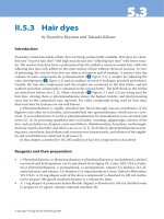
Long-lasting eye closure causes an upward eye drift known as Bell’s
phenomenon [58, 59]; short eye closure with blinks induces distinctly different
eye movements in humans (fig. 1) [2, 42, 45, 47, 59, 60]. During a blink, there
is an early inward, downward, [45, 59, 61] and ex-torsional movement of the
eyes [62]. The amplitude and direction of these eye movements depend on the
initial eye position [45, 59]. During adduction and downward gaze, the ampli-
tudes of the blink-associated eye movement components are minimal. The
horizontal amplitude increases during abduction, and the vertical amplitude
during upward gaze [45]. The horizontal, vertical, and torsional components of
the blink-associated eye movement start before lid movement onset [53, 60,
62], and the movement is completed before blink termination [47, 59]. Blink-
associated eye movements are slower than saccades; they do not obey the saccadic
main sequence [59] and Listing’s law [63]. Blink-associated eye movements are
caused by cocontraction of all eye muscles [1, 13, 62, 64]. Bergamin et al. [62]
showed in humans that during the early phase of eyelid closure of voluntary
blinks the eye moves in a 3-D direction that can best be explained by a pulse-
like activation of the inferior rectus muscle.
Blink-associated eye movements reflect an active process, i.e. they are not
caused by mechanical eye-lid interaction [59], and they are important for the
protection of the cornea [2, 40, 45]. Blink-associated eye movements are not
only found during fixation but are superimposed on all kinds of eye move-
ments, e.g. smooth pursuit, saccades, and vergence eye movements [41, 65–68].
The Eyelid and Its Contribution to Eye Movements 115
Effect of Blinks on Eye Movements
Blinks affect eye movements in at least two ways: (1) by superimposing
blink-associated eye movements and (2) by modifying the neuronal premotor
activity in brainstem circuits, which change their dynamic properties. This sec-
ond aspect will be discussed in more detail below.
Blinks and Saccades
Voluntary and reflexive blinks influence horizontal and vertical visually
guided saccades in monkeys and in humans [30, 42, 47, 69]. Blinks reduce hor-
izontal saccade velocity, acceleration, deceleration, and increase saccade dura-
tion, but they do not change saccade amplitude (fig. 2) [42, 47]. The effect of
the blink on the saccade is time dependent. The maximum effect is observed
with blinks elicited about 150 ms before saccade onset [42, 69]. If blinks are
elicited later during the saccade, dynamic overshoots may occur [47]. Blinks
reduce saccade latency [69] when they are elicited shortly after but not before
[67] stimulus onset.
This influence of blinks can be explained by the blink-induced changes of
the neuronal oculomotor circuits in the brainstem [29, 30, 42, 47]. Medium-lead
burst neurons of the PPRF and the riMLF provide the premotor saccadic com-
mand to the extraocular motoneurons [70]. Several lines of evidence indicate
that OPNs in the nucleus raphe interpositus [71] control saccadic burst neurons
[72]. OPNs, which discharge spontaneously, cease firing during saccades [70,
con
div
left
right
up
down
open
100 ms
0
1
close
Lid VeIV
200º/s 200º/s 200º/s
VeIH Verg
Fig. 1. Effect of blinks on eye velocity
components at gaze straight ahead in a nor-
mal subject. Mean eye vergence velocity
(Verg), horizontal (VelH) and vertical
(VelV) version velocity, as well as relative
eyelid position (Lid) are averaged (5 blinks)
and aligned to blink onset. Note that the
blink duration exceeds the eye movement
duration; modified after Rambold et al.
[42], with permission.