Medical Microbiology made memorable - part 7 pot
Bạn đang xem bản rút gọn của tài liệu. Xem và tải ngay bản đầy đủ của tài liệu tại đây (519.59 KB, 11 trang )
M E D I C A L
MICROBIOLOGY
80
Clinical features
The repeated rounds of
erythrocyte invasion and rupture release toxins
that cause bouts of high fever. Classic symptoms
include:
•
cycles of shaking chills followed by fever and
profuse sweating
•
anaemia and jaundice due to erythrocyte
destruction
•
dark pigmented urine ('blackwater fever')
from erythrocyte destruction
•
liver and spleen enlargement and renal failure.
The time between these fever episodes can be
characteristic of the infecting
Plasmodium
species
(Table 33.1). With
P. falcipa rum
this is every
36-48 h (malignant tertian malaria) compared
with 72 h for P.
malariae
(
quartan
malaria). The
cycle is also 48 h for
P. vivax
but the symptoms
are less severe
(benign tertian malaria).
Cerebral involvement is a serious consequence
of falciparum malaria. The high levels of
parasitaemia lead to the schizont-containing
erythrocytes blocking brain capillaries. The
resulting hypoxia causes confusion, coma
and death.
The dormant liver hypnozoites formed in ovate
and vivax malaria can result in relapse many
years after the initial infection.
Diagnosis
Malaria should be suspected in any
case of fever associated with travel to endemic
areas. Diagnosis is made by clinical symptoms
and microscopic examination of blood to
identify the erythrocytic forms. This permits
the differentiation of
Plasmodium
species which
is vital in the correct choice of treatment.
Treatment
Malaria can normally be cured by
antimalarial drugs (Table 33.1). Chloroquine
is the drug of choice, although resistance by
P. falciparum
has restricted its effectiveness in
many parts of the world. Alternative drugs are
quinine, mefloquine and the combination of
sulfadoxine plus pyrimethamine. However,
resistance to these agents is also being reported.
Primaquine is included in ovate and vivax
malaria to destroy the liver hypnozoites.
Prevention
Vaccines are being developed but
are not yet available against malaria. Travellers
to endemic areas must protect themselves
from infection and seek expert advice about
antimalarial prophylaxis before embarking.
The regimen for drug prophylaxis depends
on whether resistance is present in the area.
Examples include chloroquine, fansidar,
pyrimethamine plus dapsone and chloroquine
plus proguanil. Preventing mosquito bites by
covering limbs, using insect repellents and
sleeping under mosquito nets is also essential.
Stagnant water, the breeding ground of
mosquitoes, should also be avoided.
Malaria is an infection of liver and red blood
cells caused by protozoan parasites of the genus
Plasmodium.
Malaria is one of the most serious
health problems facing humanity today, affecting
four hundred million people world-wide and
causing 2 million deaths each year. Four species
infect man:
P. falciparum
(the most common and
dangerous),
P.
malariae, P. ovate
and
P. vivax.
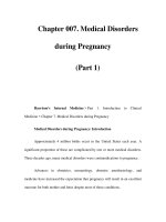
Life cycle
Malaria is spread by the bite of an infected
Anopheles
mosquito. Only females of the species
bite humans and transmit the disease. The
parasite has a complex life cycle involving
sexual reproduction in the mosquito and
asexual reproduction in liver parenchymal cells
and erythrocytes (red blood cells) in humans
(Fig. 33.1):
1.
In the mosquito:
(a)
Male and female gametocytes from
an infected human are ingested when
mosquito feeds.
(b)
Gametocytes undergo sexual reproduction
in stomach to form an oocyst containing
sporozoites.
(c)
Oocyst penetrates the gut wall and the
sporozoites enter salivary glands.
(d) Sporozoites infect human when mosquito
next feeds.
2. In humans:
(e)
Sporozoites enter the blood and infect
parenchymal liver cells.
(f)
Asexual reproduction
(schizogony)
forms
schizonts in which thousands of
merozoites
develop.
(g)
Merozoites rupture from the liver
schizonts and enter erythrocytes.
(h)
Ring-form trophozoites, then sporozoites
and finally merozoites develop.
(i)
Merozoites rupture from the cells to
invade other erythrocytes.
(j)
Some merozoites form gametocytes that
infect the female mosquito at next feed
and continue life cycle.
In
P. ovate
and P.
vivax
infection, some
sporozoites remain dormant as
hypnozoites
in
the parenchymal cells, only starting the process
of schizogony months or years later.
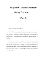
Epidemiology
In spite of intensive control
measures, malaria remains widely distributed
in the tropics and subtropics of Africa, Asia
and Latin America (Fig. 33.2).
P. falciparum
and
P. vivax account for 95% of all malaria cases,
and 80% of these occur in tropical Africa.
•
P. falciparum
and
P.
malariae
are the
predominant species in the tropics.
•
P. vivax
is common in the tropics, subtropics
and some temperate regions.
•
P. ovate
is common in West Africa.
Malaria
8
1
FIG 33.1
Generalised life ccle of
Plasmodium
I
FIG 33.2
Global occurrence of malaria
Species
Distribution
I
ncubation period
Duration of fever
(liver cycle)
(erythrocytic cycle)
Clinical
condition
Major
complications
Treatment
P falciparum
West, East and Central
7-14 days
36-48 h
Malignant tertian
Cerebral malaria,
Chloroquine,
Africa, Middle East,
malaria
haemolytic anaemia
quinine, mefloquine,
Far East, South America
('blackwater fever'),
sulfadoxine and
P vivax
I
ndia, North and East
1
2-17 days
48 h
Benign tertian
j
aundice, hypoglycaemia
Relapse due to liver
pyrimethamine
Chloroquine
Africa, South America,
(
with relapse
malaria
hypnozoites
with primaquine
P malariae
Far East
Tropical Africa, India,
up to 3 years)
13-40 days
72 h
Quartan malaria
Nephrotic syndrome
Chloroquine
Far East
(
with rare relapse)
with primaquine
P ovate
Tropical Africa
9-18 days (with
48 h
Ovale tertian
Relapse due to liver
Chloroquine
relapse up to 20 years)
malaria
hypnozoites
with primaquine
M E D I C A L
MICROBIOLOGY
84
Other tropical infections
Tropical infections affect millions of people
world-wide, causing considerable human
suffering and economic hardship. Far from
declining, the incidence of many tropical
infections is increasing throughout the world.
The impact of human immunodeficiency virus
(
HIV) and AIDS has seen the emergence of 'new'
opportunistic pathogens as well as the increased
prevalence of other recognised types. Climatic
changes induced through global warming have
aided the spread of many diseases, whilst
starvation and the breakdown in sanitation that
accompanies war has seen the re-emergence of
others. In addition, the development of drug
resistance has also dramatically influenced the
ability to treat and control many diseases, notably
parasitic infections.
Clinicians in the West will encounter tropical
infections. The ease and speed with which the
globe can be traversed by air travel and the quest
for ever more exotic holiday destinations means
patients can become infected and return home
before symptoms have developed. Refugees and
i
mmigrants can also import infections into the
country or acquire them on visits home.
Tropical infections may be broadly classified as
those causing fever, diarrhoea and skin diseases.
They are caused by a variety of bacteria, viruses
and parasites as summarised in Table 34.1.
More common examples are described below.
Fever
Malaria
is the prime suspect in any patient
presenting with fever after returning from a risk
area (e.g. the tropics and subtropics of Africa, Asia
and Latin America). A blood film examination for
the parasite is an urgent investigation.
Typhoid and paratyphoid fevers
are highly
infectious bacterial causes and present as fever
with abdominal discomfort or vague abdominal
pains, rose spots on the trunk and splenomegaly.
Infected persons may become asymptomatic
excretors of the organism.
Tuberculosis
(TB) in the United Kingdom is
30-200 times as common in immigrants as in the
indigenous population. This is probably because
of increased susceptibility to the infection and
can take a form unfamiliar to doctors not
educated in third-world countries. HIV infection
greatly increases the risk of TB, and the AIDS
pandemic has seen a resurgence of the disease.
Other causes of subacute or chronic imported
fever include:
•
viruses:
viral hepatitis, Lassa fever, rabies
•
bacteria:
tick typhus, brucellosis, relapsing fever
•
parasites:
amoebic liver abscess, early
schistosomiasis, visceral leishmaniasis,
African and American trypanosomiasis.
Diarrhoea
Diarrhoea is a common complaint after foreign
travel, and acute cases will be caused by
food-poisoning organisms found also in the
West (e.g. salmonella, shigella, campylobacter,
enteric viruses). Tropical causes are likely to be
protozoa or helminths. Infections are usually
asymptomatic in the native population but can
be severe when acquired by the non-indigenous
visitor.
Bacteria Cholera
causes severe diarrhoea that
may be fatal because of extensive electrolyte and
water depletion. It is endemic where standards of
sanitation and hygiene are low. Up to 10% of the
patient's body weight can be lost in a few hours
through the 'rice-water stool' that arises from the
infection.
Protozoa and helminthsAmoebiasis and
giardiasis are the commonest infective causes
of chronic diarrhoea. Persistent eosinophilia
i
mplicates a worm infection, most commonly
filariasis, schistosomiasis and occasionally
strongyloidiasis. Other causes are:
•
ascariasis (the large roundworm)-heavy
infections may cause a variety of complications,
including intestinal obstruction
•
trichuriasis (whipworm)-chronic bloody
diarrhoea, anaemia and rectal prolapse when
very large numbers are present
•
hookworms-can cause significant blood loss
resulting in iron deficiency anaemia
•
tapeworms-infections are common,
but autoinfection by the pig tapeworm
(cysticerosis) can be life-threatening.
Skin conditions
Ulcers are the most common skin lesions in the
tropics. The cause is usually unknown, although
Vincent's organisms (a fusiform and a spirochaete)
and (3-haemolytic streptococci are often isolated
on culture. Mycobacteria, corynebacteria and
the protozoan parasite
Leishmania
are also
i
mportant causes.
Leprosy is caused by an acid-fast mycobacterium
and spread by person-to-person contact. The
condition is characterised by a variety of
symptoms, but the most important is thickening
of peripheral nerves leading to localised areas
of anaesthesia in the affected tissues. Some ten
million people are affected world-wide.
8 5
Disease
Organism
Symptoms
Mode of transmission Distribution Treatment
Bacteria
Cholera
Vibrio
cholera
Copious watery diarrhoea
Faecal-oral from
World-wide: India,
Fluid + electrolyte
Bubonic plague
Yersinia
pestis
('rice-water stool')
Fever, swollen lymph nodes
contaminated water
Rodent fleas
South-east Asia and
South America
South-western USA,
replacement; oral
tetracycline
Streptomycin +
('Black Death')
('buboes'), pneumonia, black
Africa and Asia
tetracycline
Endemic typhus
Rickettsia
typhi
skin necrosis
Fever, flu-like symptoms, rash,
Louse bite
World-wide
Tetracycline
Leprosy
Mycobacterium
meningoencephalitis, coma
Lepromatous: (progressive)
Person-to-person contact Africa, India, South-east Dapsone + rifampicin
leprae
skin nodules,
nerve
i
nvolvement
Asia and South America
Tropical ulcer
Mycobacterium
Tuberculoid: skin lesions
(
benign), severe nerve and
tissue destruction
Buruli ulcer: gross, necrotising
Unknown
Tropical areas in all Clofazimine or rifampicin
ulcerans
ulceration of the skin
continents
Tuberculosis
Mycobacterium
Pulmonary: cough, chest pain, Person to person through
World-wide
Ethambutol, isoniazid,
tuberculosis
fever, dyspnoea, haemoptysis
respiratory secretions; milk
rifampicin, pyrazinamide
and weight loss
from infected cattle
(in combination)
Typhoid and
Salmonella typhi
Glandular involvement
(in tropics) associated with
HIV infection
Fever and systemic infection
Faecal-oral
World-wide
Co-trimoxazole,
paratyphoid fever
and
paratyphi
from invasion of bloodstream
ciprofloxacin, ceftriaxone
M E D I C A L
MICROBIOLOGY
8 6
Examples of tropical infections
Disease
Organism
Symptoms
Mode of transmission
Distribution
Treatment
Viruses
Rabies
Ebola
Lassa fever
Yellow fever
Rhabdovirus
Filovirus
Arenavirus
Flavivirus
Severe pain at bite, hydrophobia,
muscle spasms, laryngospasm,
extreme excitability
Fever, headache, malaise, chest
discomfort, diarrhoea and vomiting
Fever, haemorrhage, renal
failure
Fever, jaundice, haemorrhage
Saliva via bite, scratch,
or abrasion
Person to person
Rat excreta contamination
of skin abrasions, food,
water, or airborne
Aedes mosquito
World-wide
(some exceptions)
Northern Zaire and
southern Sudan
West Africa
Central and South
America and Africa
None
None
None
None
Protozoa
Amoebiasis
Balantidiasis
Malaria
African
t
rypanosomiasis
American
t
rypanosomiasis
Leishmaniasis
Entamoeba
histolytica
Balantidium coli
Plasmodium
species
Trypanosoma
gambiense
and
rhodesiense
T cruzii
Leishmania
Bloody diarrhoea and
occasionally liver infection
Mild to severe diarrhoea
Liver, blood and CNS infection
General febrile illness followed
by CNS invasion
Fever, lymphadenopathy,
hepatosplenomegaly, cardiac
and CNS involvement
Skin sores (cutaneous) nose,
mouth, palate destruction
(
mucocutaneous)
Faecal-oral via cysts
i
n food and water
Faecal-oral from cysts
i
n food and water
Mosquito
Tsetse fly
Triatomid bug
Sandfly
Common in tropics
Common in tropics
Africa, Asia and
Latin America
East and west Africa
Mexico, Central and
South America
North Africa,
I
ndia (cutaneous);
Mexico, Central
and South America
(
mucocutaneous)
Metronidazole,
tinidazole
Tetracycline, iodoquinol
Chloroquine, quinine,
mefloquine, sulfadoxine
+ pyrimethamine,
primaquine
Pentamidine,
melarsoprol
Nifurtimox and
benznidazole
Stibogluconate,
meglumine antimonate,
amphotericin B or
pentamidine
M E D I C A L
MICROBIOLOGY
87
Disease
Organism
Symptoms
Mode of transmission
Distribution
Treatment
Helminths
Hookworms and
Ancylostoma
Gut, lungs and heart infection;
Larval infection
Mediterranean, Mebendazole,
Strongyloidiasis
duodenalis,
malnutrition, pneumonitis, through skin
southern USA, Central
albendazole;
Necator americanus
anaemia
and South America,
thiabendazole,
and Strongyloides
Africa, Asia
i
vermectin
stercoralis
(strongyloidiasis)
Ascariasis Ascaris
As for hookworms
Faecal-oral ingestion
Southern USA, Central
Mebendazole or
('roundworm')
lumbricoides
of eggs
and South America,
albendazole
Trichuris
Trichuris trichiura
Gut infection, malnutrition
Faecal-oral ingestion
Africa, Asia, Australia
As for
Ascaris
Albendazole or
('
whipworm')
of eggs
mebendazole
Bancroftian Wuchereria
Fever and lymphangitis leading
Mosquito
South America,
I
vermectin or DEC
filariasis
bancrofti
to obstruction of the lymphatics Central Africa,
Onchocerciasis
Onchocerca
Lymphadenopathy in groin and Blackfly
Far East
Central America,
Ditto
('river blindness')
volvulus
axilla, intradermal oedema and
Central Africa and
pachyderma, keratitis,
the Yemen
Loaiasis
Loa loa
retinochoroiditis
Migration of worm in eyelid,
Mango fly Central and West Africa
Ditto
('eyeworm')
vitreous and anterior chamber
Taeniasis
Taenia saginata Asymptomatic or abdominal
I
ngestion of cysticerci World-wide
Praziquantil or
(beef tapeworm)
pain, diarrhoea and weight loss
i
n beef
niclosamide
Cysticercosis Taenia solium
Cysticercosis (pork tapeworm):
I
ngestion of cysticerci
South and Central
Ditto
(pork tapeworm)
l
arvae penetrate gut and form
i
n pork
America, China,
cysticerci in muscles
I
ndonesia
Schistosomiasis
Schistosoma
Liver and bladder
Burrowing into skin of
South America,
Praziquantel
('bilharzia')
(
S.
haematobium)
or rectum
schistosome
cercariae
West Indies, Africa,
(
S.
mansoni, S. japonicum)
from aquatic snails Middle East, Egypt,
Far East
Examples of tropical infections
M E D I C A L
MICROBIOLOGY
88
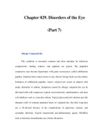
Pyrexia of unknown origin
The majority of patients present with
symptom/physical sign complexes compatible
with only a few diseases, and they require little
investigation. Other clinical presentations, such
as pyrexia or
fever of unknown origin
(PUO or
FUO), are more difficult because they have few
signs and symptoms, so that the list of differential
diagnoses is large and the need for investigation
correspondingly greater. 'Classic' PUO has three
features:
•
an illness of more than 3 weeks' duration
•
a temperature greater than 38.3°C (101°F) on
several occasions
•
no specific diagnosis after a week of hospital
inpatient investigation.
With the changing pattern in hospital admissions,
shorter inpatient times and more use of
community and outpatient services, and also
the development of a greater range of powerful
diagnostic procedures, the third criterion may
be replaced by a minimum set of investigations
(Table 35.1) rather than a timed period in
hospital. There are over two hundred reported
diverse causes of PUO, which vary slightly
according to age (Fig. 35.1). As the number of
conditions that has to be considered is so large,
some clinicians divide patients into further
categories such as
neutropaenic PUO, nosocomial
PUO
and
HIV associated PUO,
to focus on
particular causes and therefore streamline their
investigation.
It is important to get a prompt diagnosis, as
this may improve the prognosis for the patient
through early treatment, and also prevent
the risk of transmission to others in the case of
communicable infections such as tuberculosis.
Investigation is expensive both in time and
resources, and it is equally important to ensure
that the patient does indeed have a pyrexia and
that it is not a factitious fever. Body temperature
is normally higher in the evening than the
morning, but some healthy individuals have
an exaggerated circadian temperature rhythm.
Others may invent physical diseases to gain
medical attention
(
Munchausen syndrome),
and an unexplained temperature is one means
of doing this, either by manipulating the
temperature recording device or even injection
of contaminated materials.
I
nvestigation
Every case requires a
comprehensive history,
careful
and
repeated
physical examination, as well as a range of
diagnostic tests and procedures. History should
include a thorough systems review with
particular care concerning travel, occupational
history and hobbies, pets and animal contact,
drug prescriptions and other drug intake,
familial diseases, previous illness and alcohol
consumption.
A complete examination should include
examination of the teeth, ears, fundoscopy and
review of the skin in good light for faint rashes.
This must be repeated at frequent intervals to
spot important developing or fleeting physical
signs. Temperature should be recorded
methodically, although the great majority of
patients never display the characteristic patterns
of fever described in the textbooks.
Investigation may include: samples sent for
laboratory testing; non-invasive tests such as
diagnostic radiology and ultrasound and
radionuclide scanning; skin testing, essentially
the
tuberculin
test for infection with
Mycobacterium tuberculosis;
and invasive testing
such as biopsy, endoscopy and surgical
exploration. A possible minimum set of
investigations is listed in Table 35.1; further
investigation will depend upon what has already
been done, and clues that may be obtained from
the history and examination, working through
all the possible differential diagnoses.
Causes
Some well-recognised causes of PUO
are given in Table 35.2. However, in the majority
of cases, the cause is a familiar disease with an
unusual presentation, rather than a rare disorder:
•
Infections are the single most common cause
of PUO, particularly in the young. They may
be difficult to diagnose because the patient
was on antibiotics when the sample was
taken, because the site of infection is hidden
or because the infectious agent is difficult or
i
mpossible to culture in the laboratory.
•
Neoplasms are an important cause of PUO,
particularly in the elderly. Certain tumours
seem to cause pyrexia themselves, others may
produce it because of necrosis or secondary
infection.
•
Collagen-vascular disease.
•
Miscellaneous.
•
Undiagnosed: this category is largely made
up of patients who recovered from a benign
febrile illness before a specific diagnosis
was made.
Management
There is no treatment for the
clinical presentation of PUO itself; success lies in
finding the cause of the PUO and then managing
that condition.
89
Some causes of PUO
I
nfection
Localised infection
Specific infections
•
Viral
•
Bacterial
•
Fungal
•
Parasitic
Abscess: abdominal, dental, pelvic, intracranial
Endocarditis and mycotic aneurysm, osteomyelitis, pyelonephritis, sinusitis,
mast oiditis
Cytomegalovirus, Epstein-Barr virus, hepatitis viruses, HIV, parvovirus B19
Mycobacterium tuberculosis
(tuberculosis),
Brucellae
abortus or
melitensis
(brucellosis),
Legionella pneumophila
(Legionnaire's disease),
Bartonella
henselae
(cat scratch fever),
Chlamydia psitta
(
psittacosis),
Coxiella burn etti
(
Q fever),
Salmonella typhi
(typhoid),
Campylobacter, Leptospira, Borrelia
recurrent is
(relapsing fever) and
burgdorferi
(
Lyme disease),
Treponema
pallidum
(syphilis)
Candida, Aspergillus, Cryptococcus neoformans,
Histoplasma,
Coccidioides,
Blastomyces, Sporothrix
Malaria, giardiasis, toxocariasis, toxoplasmosis, trypanosomiasis,
schistosomiasis, leishmaniasis
Neoplastic
Many tumours but especially lymphomas, leukaemias, renal-cell carcinoma
and atria) myxoma
Collagen-vascular
disease
Still's disease, rheumatoid arthritis, systemic lupus erythematosus (SLE),
Reiter's syndrome, rheumatic fever, Felty's syndrome
Various vasculit ides
Miscellaneous
Haematoma, recurrent pulmonary embolism
Drug fever, metal poisoning
Crohn's disease, ulcerative colitis, sarcoidosis
Familial fevers, cyclical neutropaenia
Undiagnosed
???
Possible minimum diagnostic evaluation for PUO
•
Comprehensive history
•
Repeated
and complete
physical examination
•
Complete blood count, including differential and platelet count
•
`
Routine' blood chemistry, including lactate dehydrogenase, bilirubin
and liver enzymes
•
Urinalysis, including microscopic examination
•
Chest X-ray
•
Erythrocyte sedimentation rate (ESR)
•
Antinuclear antibodies
•
Rheumatoid factor
•
Angiotensin converting enzyme
•
Multiple blood films (if any possibility of malaria)
•
Blood cultures (3 sets) whilst not
receiving
antibiotics
•
Cytomegalovirus IgM antibodies or virus detection in blood
•
Heterophile antibody tests or EBV serology (children and young adults)
•
Tuberculin skin test
•
CT of abdomen/radionuclide scan
•
HIV serology or virus detection assay
•
Further evaluation of any abnormality detected by above
FIG 35.1
Causes of PUO at different ages
M E D I C A L
MICROBTOLOGY
90
There has been considerable success in combating
infection in the developed world over the last
fifty years, through advances in nutrition and
hygiene as well as the development of drugs and
vaccines. The world-wide eradication of smallpox
was a notable success, but 'new' infections are
constantly being described
(Appendix
6, p. 130),
and there is always the problem of established
i
nfections becoming resistant to current therapies
(
Table 36.1). And there are few diseases that can
have such a dramatic effect on human health as
those that are caused by infection (Fig. 36.1).
New diseases
Viruses are potentially the most rapidly evolving
of all infectious agents and therefore the greatest
future threat. Bacteria may acquire or generate
new virulence genes and become able to cause
new infections. Alternatively the micro-organisms
may not change but, because of changes in the
environment that surrounds us, they may
suddenly start to cause human disease.
•
Escheriehia eoli 0157
and
Haemophilus influenzae
biogroup Aegyptius are probably 'new'
pathogens.
•
Legionella pneumophila
(related to widespread
airconditioning) and probably bovine
spongiform encephalopathy/variant
Creutzfeld-Jacob disease (probably related to
changes in animal rendering) are pathogens
resulting from changes in the environment.
Established diseases
of unknown cause
When the aetiology of a disease is first discovered
there may be a greater appreciation of its
significance, although the disease itself is neither
new nor the numbers necessarily increasing.
A number of newly identified pathogens are
listed below with the method of their detection,
but there are likely to be many other diseases
which may have an infectious aetiology, possibly
even including conditions like sarcoidosis,
multiple sclerosis and bipolar depression:
•
Borrelia burgdorferi, Cam pylobaeter spp.
and
Helicobacter pylori
(laboratory culture)
•
Cryptosporidium spp.
and
Cyclospora spp.
(
microscopy)
•
Tropheryma whip pelii
and
Bartonella henselae
(
molecular biology).
Re-emerging diseases
The incidence of many infections fluctuates with
known periodicity over time - either due to
changes in the physical environment, such as the
peaks of food-borne illness associated with the
warm summer months, or probably due to
changes in levels of immunity within the
population, such as the 9 yearly cycle of
parvovirus infections. However, for some
diseases, these changes may be unpredictable
and dramatic. Pandemics of influenza virus
infection, killing millions of people, have
occurred in the past and may well do so again.
Human
Salmonella
infections (Fig. 36.2) have
varied considerably because of changing patterns
of food consumption. The resurgence in cases
of
Mycobacterium tuberculosis
infection in the
USA, including cases with multiple-antibiotic
resistance, results from running-down of public
health facilities, immigration, and also HIV
i
nfection as it has increased the number of
susceptible individuals who then form an
increased reservoir of infection to be passed
on to others.
The future?
As our civilisation develops, it is possible to
appreciate a number of changes which benefit
society but which may also be potential threats
to world health from infectious disease,
including:
•
rapid, global transport, especially air travel,
might allow a problem to be disseminated
before it is recognised
•
changes in food production - new methods
may create new problems, but also, with the
increasing industrialisation of production,
problems with a single producer may affect
vast numbers of people
•
exploration and use of unknown, potentially
threatening environments such as the rain
forest (and, possibly, outer space)
•
xenotransplantation
and the risk of
modifying animal diseases to infect humans
•
increasing size and density of urban
populations, and also an ever-increasing
number of immunosuppressed
individuals
•
the effects of global warming.
It is clearly important that there should be
sufficient infection surveillance in the population
to recognise and to react quickly to any new
threat; the American government and World
Health Organisation have already set up groups
specifically for this. There should be ongoing
development of treatments so that they can be
adapted to novel situations quickly and
effectively.
New and re-emerging
i
nfectious diseases
Emerging problems of drug resistance
FIG 36.1
Leading causes of death in US males aged 25-44 years
9
1
I
nfectious agent
Resistance problem
Herpes simplex virus
Acyclovir
Human immunodeficiency virus
Zidovudine and others
Methicillin-resistant
Staphylococcus aureus
(
MRSA)
3-lactams (and other antibiotics)
Vancomycin-resistant S.
aureus
(VISA or VRSA) MRSA now also resistant to vancomycin
Penicillin-resistant
Streptococcus pneumoniae
(3-lactams (and other antibiotics)
Neisseria gonorrhoeae
Penicillin, tetracycline, quinolones
Glycopeptide-resistant
Enterococcus
spp. (GRE)
Multi-resistance including glycopeptides
Multi-drug-resistant
Mycobacterium tuberculosis
(
MDR-TB)
I
soniazid, pyrazinamide, rifampicin and others
Gram-negatives with `extended spectrum
(3-lactamases' (ESBL)
(3-lactams (and other antibiotics)
Candida
spp.
Fluconazole
Plasmodium
spp.
Chloroquine
Scabies
Lindane
FIG 36.2
I
solates of Salmonella in England and Wales, 1981-96
Principles of hospital
i
nfection control
Hospital infection increases patient morbidity,
mortality, length of stay and treatment costs.
It is estimated that about five thousand patients
die as a result of hospital-acquired infection in
Britain each year. Control of infection is therefore
an essential element of hospital practice.
Definitions
Infections acquired by patients or staff in hospital
are termed hospital-acquired or nosocomial
(Figs 37.1 and 37.2). The source of infection may
be other people (cross-infection), the patient's
own organisms (endogenous infection) or via
contaminated food, fluids equipment or the
environment. In England the prevalence of
hospital-acquired infection is approximately
10%, whereas the annual incidence is nearer 5%.
The sources of hospital-acquired infections are
shown in Fig. 37.3.
•
Staff should have health checks before
employment and have necessary
i
mmunizations.
•
Staff should report infections (e.g. diarrhoea)
and needlestick injuries.
II:
Prevent transmission Blocking routes of
transmission may reduce the risk of hospital-
acquired infection. Organisms may be transferred
by the airborne route or by contact spread. Few
organisms exist as isolated particles in air but
many are carried on dust particles, which largely
consist of skin scales. Staphylococci and other
Gram-positive bacteria may be spread by air,
particularly from patients or staff with infected
lesions or those who shed increased numbers of
epithelial cells. The airborne route may also
spread respiratory viruses and bacteria during
coughing or sneezing. Airborne spread may be
controlled in two ways:
Control of hospital infection
Each hospital has an infection control officer
(usually a consultant microbiologist) and
infection control nurse(s) who together constitute
the infection control team. This team advises on
the prevention and control of hospital infection,
utilising policies provided and updated by the
hospital infection control committee. The team
also monitors infection by surveillance -
typically this involves tracking so-called alert
organisms from sites such as wounds (e.g. Staph.
aureus (including MRSA) and Strep. pyogenes),
faeces (e.g. Salmonella spp. and Clostridium
difficile) and respiratory tract (e.g. Mycobacterium
tuberculosis). Another important function of the
infection control team is education for all groups
of staff to update them on new issues and
i
mprove compliance with policies.
The three principles of infection control are:
I:
Exclude the source of infection Inanimate
sources of infection can be identified and in many
cases excluded from hospitals by ensuring the
provision of:
•
sterile instruments, dressings and
intravenous fluids
•
clean linen
•
uncontaminated food and drink
•
cleaned and disinfected or disposable
equipment
•
a safe and clean environment including water
and air of appropriate quality
•
clear policies for safe disposal of hospital waste.
Both patients and staff may act as a source of
infection, so:
•
Infected and colonised patients should be
identified by adequate microbiological
investigations and infections treated with
appropriate antimicrobials.
•
the use of filtered air in ventilation systems,
e.g. operating theatres
•
isolation rooms or wards.
Patients with organisms that pose a risk for others
are placed in source isolation to minimise spread.
Protective isolation is provided for highly
susceptible patients (e.g. immunosuppressed
patients immediately following organ
transplants). Ideally, isolation rooms should
have pressurised ventilation systems which
direct airflow in (for source isolation) or out of
the room (for protective isolation).
Contact spread is transmission of organisms by
direct contact between the patient and equipment
or staff. This route is minimised by aseptic
technique, taking particular care when handling
dressings, secretions and excretions that may
transmit organisms directly by hands or via
contaminated equipment. However, any clinical
contact with infected or colonised patients
(or their immediate environment) may transfer
organisms to the hands of staff. It is therefore
very important that all staff are aware of the
i
mportance of hand hygiene - the single most
i
mportant aspect of infection control (Table 37.1
and rig. 37.4).
III: Improve the patient's resistance to
i
nfection This may be achieved by:
•
meticulous technique during surgery -
haemostasis, removal of dead tissue and foreign
bodies, avoiding wound drains where possible
•
care of invasive devices, e.g. intravascular
lines, urinary catheters and endotracheal tubes
•
control of underlying disease, e.g. diabetes
•
enhancing immunity by immunisation,
e.g. tetanus
•
avoiding unnecessary antimicrobial treatment
•
appropriate use of antimicrobial prophylaxis.