Sinusitis From Microbiology to Management - part 4 docx
Bạn đang xem bản rút gọn của tài liệu. Xem và tải ngay bản đầy đủ của tài liệu tại đây (453.34 KB, 49 trang )
relative contribution of anaerobic bacteria has been debated, and the large
variations in rates of isolation have been attributed to culture techniques.
Brook reported that up to 50% of cases of CRS were culture-positive for anae-
robic bacteria, with the predominance of Prevotella, Fusobacterium, and
Peptostreptococcus spp. (56,57). In adults, infectious CRS is commonly polymi-
crobial, and both gram-positive and gram-negative aerobic and anaerobic bac-
teria are frequently isolated. A wide variety of aerobic bacteria, such as
coagulase negative Staphylococcus, S. aureus, Streptococcus viridans, P.aerugi-
nosa, Klebsiella pneumoniae, Proteus mirabilis,andEnterobacter spp. have been
isolated. Also, several different anaerobic species have been demonstrated,
including Prevotella, Fusobacterium, and Peptostreptococcus spp. (56–64).
Biofilms. Biofilms are sessile bacterial microcolonies that are enclosed
in a highly hydrated polysaccharide matrix with interstitial voids in which
nutrients and signaling molecules can be circulated. The structural and func-
tional heterogeneity of bacterial cells within these communities protects
them against the body’s natural defenses and provides them with antimicro-
bial resistance. Through genetic alterations, bacteria in biofilms are also able
to transition to the mobile planktonic form, which has been the traditional
model for studying bacterial diseases (65,66). Bacterial biofilms have been
demonstrated on many areas of mucosa in the human body, including the
ear mucosa and tympanostomy tubes removed from patients with chronic
effusions and infections (67,68). It has been hypothesized that biofilms
may play an important role in cases that are refractory to antibiotic therapy,
and antibiotic resistance has been demonstrated to be up to 1000-fold
greater in bacteria in the biofilm form versus the planktonic form
(66–70). Similarities between chronic otitis media and CRS exist. Both of
these disease processes take place in the ciliated respiratory epithelium
and are largely associated with an infectious etiology. The presence of bac-
terial biofilms in CRS patien ts with culture-positive Pseudomonas has been
demonstrated using scanning electron microscopy (71) (Fig. 7).
Although further work in this area is required, knowledge of the pre-
sence, structural characteristics, and pathological mechanism of biofilms in
CRS may help to identify new treatment modalities.
Superantigens. Another new area of interest in infectious CRS involves
a group of potent mitogens termed superantigens sags. Sags are most com-
monly associated with bacteria, particularly S. aureus and S. pyogenes species,
but can also be produced by viruses and fungi. Unlike conventional antigens
whose activation requires multiple steps in only a limited number of T-lym-
phocytes, sags can directly stimulate a multitude of different T-lymphocytes.
Figure 7 (Facing page) Biofilms inHuman CRS. Source: Cyer J, Schipor I, Perloff JR,
Palmer JN. Densely coated sinonasal epithelium with tower-like structures (white
arrows) visible near the top edge of the specimen. Source: From Ref. 71.
J
Pathophysiology of Sinusitis 125
In the traditional pathway, the antigen is phagocytosized by an antigen-
presenting cell (APC), degraded into numerous peptide fragments, which
are then processed for cell surface display in conjunction with a major histo-
compatibility complex (MHC) II receptor. A compatible T-helper cell then
recognizes this MHC II/peptide complex, and an inflammatory response is
initiated. Sags are able to bypass these processing and presenting steps and
bind directly to the outside surfaces of the HLA-DR alpha domain of MHC
class II and V beta domain of the T-cell receptors (picture) (72–75). Through
this mechanism, they are able to stimulate a massive expression of IL-2 at
femtomolar concentrations (76). In turn, IL-2 stimulates the production of
other cytokines such as TNF-a, IL-1, Il-8, and platelet activating factor
(PAF), leading to an overwhelming inflammatory response. Additionally, sags
also act as traditional antigens, as well as stimulate the production of anti-
superantigen antibodies.
Recently, upregulation of IgE sags antibodies have been demonstrated
in patients with chronic obstructive pulmonary disease (COPD) exacerbation
(77). Likewise, a study by Basher et al. found increased levels of sags in
patients with NP versus control patients (78). Evidence of the roles of super-
antigen-producing bacterial strains in the pathologic mechanism of Kawasaki
disease, atopic dermititis, and rheumatoid arthritis has also been reported, and
a pathophysiological mechanism in which microbial persistence and superan-
tigen-induced T-cell inflammatory responses in CRS has also been proposed
(79). Further studies in this area, as well as in other areas of CRS, may provide
new diagnostic and treatment modalities.
Fungal infections: Fungal species play a variety of roles in chronic
sinusitis from colonization to invasive, life-threatening disease. Invasive
disease is characterized by histopathological evidence of hyphal forms
within the sinus mucosa, submucosa, blood vessels, or bone, and has been
associated with either fulminate or a more indolent chronic course of fungal
rhinosinusitis. In addition, chronic invasive disease may or may not be asso-
ciated with a giant cell response. The pathophysiology of these different
disease courses has been attributed primarily to the host’s immune response
to the fungus, although the fungal species also appears to play some role in
the disease course. Fungal species associated with fulminate forms of fungal
sinusitis include Absidia, Aspergillus, Basidobolus, Mucor, and Rhizopus
spp., and most often occur in immunocompromised patients (80). Species
associated with chronic invasive fungal sinusitis include Aspergillus, Mucor,
Alternaria, Curvularia, Bipolaris, and Candida spp., Sporothrix schenckii ,
and Pseudallescheria boydii, and can occur in both immunocompetent and
immunocompromised patients (81,82).
Two major forms of non-invasive fungal sinusitis—allergic fungal
sinusitis and sinus mycetoma—exist, with allergic fungal rhinosinusitis
(AFS) forming a distinct subcategory of CRS. Diagnostic criteria for AFS
126 Jackman and Kennedy
include the demonstration of five characteristics as defined by Bent and Kuhn:
gross production of eosinophilic mucin containing non-invasive fungal
hyphae, nasal polyposis, characteristic radiographic findings, immunocompe-
tence, and allergy to fungus (83). AFS is characterized by a sustained eosino-
philic inflammatory response to colonizing fungi. Mucus secretions, termed
allergic mucin, in AFS are characterized as being highly viscous and contain
branching non-invasive fungal hyphae within sheets of eosinophils and Char-
cot–Leyden crystals (84–88) (Fig. 8).
A non-IgE-dependent association of fungus with CRS has also been
proposed. I n 1999, Ponikau et al. reported a fungal colonization in 96% of con-
secutive patients with CRS, using an ultra-sensitive method of fungal identifica-
tion. Additionally, certain fungi were demonstrated to elicit an upregulation of
IL-5 and IL-13 and a resulting eosinophilic inflammatory response. This eosino-
philic response was IgE, and therefore, allergy-independent, which was thought
to indicate a broader role of fungus in CRS than previously hypothesized (89).
Allergy
Environmental allergens are frequently considered as important environmen-
tal factors in CRS, and atopy is identified as a prominent systemic host factor
in CRS. However, the exact contribution of allergy to the development of
CRS is still under investigation. Both pediatric and adult patients with allergic
Figure 8 Hematoxylin and eosin stained nasal tissue demonstrating fungal hyphae,
eosinophils and Charcot-Leyden crystals. Source: Diagnosis of chronic rhinosinusi-
tis. Lanza DC. Annals of Otology, Rhinology, & Laryngology – Supplement.
2004; 193:10–14.
Pathophysiology of Sinusitis 127
rhinitis are more commonly affected with CRS than non-allergic patients (90).
Furthermore, these individuals have been reported to respond more poorly
to medical management and to more frequently undergo endoscopic sinus
surgery (91,92). Inflammatory changes contribute to the development of
CRS in allergic patients. They are stimulated by the production of cytokines,
allergic mediators, and neurogenic stimulation. More specifically, allergen
stimulation of T
H
2 cells leads to the production of IL-4, which in turn causes
B-cell activation and IgE antibody production. Subsequent allergen exposure
causes IgE cross-linking and release of inflammatory mediators, such as his-
tamine, leukotrienes, and tryptase, and results in the later phase response–
eosinophil infiltration, mucus hypersecretion, and mucosal edema. Continued
allergen activation, referred to as ‘‘priming,’’ further increases the concentra-
tion and magnitude of action of inflammatory cells such as eosinophils and
neutrophils and their associated cytokines. Furthermore, an IgE response
to staphylococcal antigens has been implicated in the development of NPs
in CRS, and this relationship is currently under investigation (8,12,93–95).
Environmental Pollutants
A number of other environmental factors can be linked to the development of
CRS. In a study of 5300 Swedish children, Andrae et al. found a significantly
higher rate of asthma and hay fever in children living near polluting factories
(96). Futhermore, Suonpaa reported an increased incidence of acute sinusitis
and nasal polyposis in southwestern Finland over a decade, which provides
additional evidence for the presence of an environmental impact in CRS
(97). Dust, ozone, sulfur dioxide, volatile organic compounds, and smoke
are just a few of the pollutants that have been implicated in CRS. The major-
ity of these chemicals share a similar pathologic mechanism: they act as nasal
irritants causing dryness and local inflammation with an influx of neutrophils
(98,99). In addition to this mechanism, environmental tobacco smoke has
been shown to cause secondary ciliary disorders, which consist primarily of
microtubular defects (100). Occupational exposure to nickel, leather, or wood
dust has been associated with epithelial metaplasia as well as carcinoma (101).
SUMMARY
Maintenance of key functional components—ostiomeatal patency, muco-
ciliary clearance, an d normal mucus production—of the paranasal sinus is
essential for prevention and recovery from CRS. CRS is a complex disease
process that can result from a single or multiple independent etiologies,
as well as from multiple independent or interdependent etiologies (Fig. 9).
The factors contributing to this disease process can be divided into
systemic host, local host, and environmental factors. Systemic host factors,
such as genetic and autoimmune diseases, are important to identify so that
appropriate treatment modifications can be made, if available. Likewise,
128 Jackman and Kennedy
local host factors such as anatomic abnormalities and environmental factors
such as infection, allergy, and pollution need to be recognized and appropri-
ately managed.
There is a clear need for further research into the pathophysiology of
this disorder. Current research on biofilms, sags, and osteitis will hopefully
provide us with a better underst anding of the role of infection in CRS. Like-
wise, research on allergic CRS and other noninfectious etiologies of CRS
will help to better elucidate the role inflammation plays in this disorder.
A better understanding of both infectious and inflammatory mechanisms
of CRS will provide us with more effective and individualized therapies.
REFERENCES
1. Majima, Y. Mucoactive medication and airway disease. Paediatr Respir Rev
2002; 3:104–109.
2. Lehrer RI, Lichenstein AK, Ganz T. Defensins: antimicrobial and cytotoxic
peptides of mammalian cells. Annu Rev Immunol 1993; 11:105–128.
3. Martin E, Ganz T, Lehrer RI. Defensins and other endogenous peptide
antibiotics of vertebrates. J Leukoc Biol 1995; 58:128–136.
4. Lee SH, Kim JE, Lee HM, Choi JO. Antimicrobial defensin peptides of the
human nasal mucosa. Ann Otol Rhino Laryngol 2002; 111(2):135–141.
5. Goldman MJ, Anderson GM, Stolzenberg ED, Kari UP, Zasloff M, Wilson
JM. Human beta-defensin-1 is a salt sensitive antibiotic that is inactivated in
cystic fibrosis. Cell 1997; 88:553–560.
6. Knowles MR, Bouchers RC. Mucus clearance as a primary innate defense
mechanism for mammalian airways. J Clin Investig 2002; 109:571–577.
Figure 9 Etiological factors in the pathological cycle of CRS.
Pathophysiology of Sinusitis 129
7. Lanza DC, Kennedy DW. Adult rhinosinusitis defined. Otolaryngol Head
Neck Surg 1997; 117:S1–S7.
8. Benninger MS, Ferguson BJ, Hadley, JA, Hamilos DL, Jacobs M, Kennedy
DW, Lanza DC, Marple BF, Osguthrope JD, Stankiewicz JA, Anon J,
Denvey J, Emanuel I, Levine H. Adult chronic rhinosinusitis: definitions, diag-
nosis, epidemiology, and pathophysiology. Otolaryngol Head Neck Surg 2003;
129:S1–S32.
9. Wald ER, Miloe GJ, Bowen A, Ledesma-Medina J, Salamon N, Bluestone
CD. Acute maxillary sinusitis in children. N Engl J Med 1981; 304:749–754.
10. Wald ER, Byers C, Guerra N, Casselbrandt M, Beste D. Subacute sinusitis in
children. J Pediatr 1989; 115:28–32.
11. Wald ER, Reilly JS, Casselbrant M, Ledesma-Medina J, Milmore GJ,
Bluestone CD, Chipows D. Treatment of acute maxillary sinusitis in childhood:
a comparative study of amoxicillin and cefaclor. J Pediatr 1984; 104:297–302.
12. Meltzer EO, Hamilos DL, Hadley JA, Lanza DC, Marple BF, Nicklas RA.
Rhinosinusitis: establishing definitions for clinical research and patient care.
Otolaryngol Head Neck Surg 2004; 131:S1–S62.
13. Evans FO Jr, Sydnor JB, Moore WE, Manwariring JZL, Brill AH, Jackson RT,
Hanna S, Skaar JS, Holdeman LV, Fitz-Hugh S, Sunde MA, Gwaltney JM, Jr.
Sinusitis of maxillary antrum. N Engl J Med 1975; 293(15):735–739.
14. Berg O, Carenfelt C, Kronvall G. Bacteriology of maxillary sinusitis in
relation to character of inflammation and prior treatment. Scand J Infect
Dis 1988; 20:511–516.
15. Pederson M, Sakakura Y, Winther B, Brofeldt S, Mygind N. Nasal mucocili-
ary transport, number of ciliated cells, and beating pattern in naturally
acquired common colds. Eur J Respir Dis Suppl 1983; 128:355–365.
16. Hinni ML, McCaffrey TV, Kasperbauer JL. Early mucosal changes in experi-
mental sinusitis. Otolaryngol Head Neck Surg 1992; 107:537–548.
17. Marks SC. Acute sinusitis in the rabbit model: histologic analysis. Laryngo-
scope 1998; 108:320–325.
18. Rudack C, Hauser U, Wagenmann M, Bachert C, Ganzer U. Cytokine pattern
in various forms of sinusitis. Laryngorhinootologie 1998; 77:34–37.
19. Repka-Ramirez S, Naranch K, Park YJ, Clauw D, Baraniuk JN. Cytokines in
nasal lavage fluids from acute sinusitis, allergic rhinitis, and chronic fatigue
syndrome subjects. Allergy Asthma Proc 2002; 23:185–190.
20. Berger G, Kattan A, Bernheim J, Ophir D. Polypoid mucosa with eosinophilia
and glandular hyperplasia in chronic sinusitis: a histopathological and immu-
nohistochemical study. Laryngoscope 2002; 112:738–745.
21. Kramer MF, Ostertag P, Pfronger E, Rasp G. Nasal Interleukin-5, immuno-
globin E, eosinophilic cationic protein, and soluble intercellular adhesion
molecule-1 in chronic sinusitis, allergic rhinitis, and nasal polyposis. Laryngo-
scope 2000; 110:1056–1062.
22. Bachert C, Gevaert P, Holtappels G, Johansson SG, van Cawenberge P. Total
and specific IgE in nasal polyps is related to local eosinophilic inflammation.
J Allergy Clin Immunol 2001; 107:607–614.
23. Rhyoo C, Sanders SP, Leopold DA, Proud D. Sinus mucosal IL-8 gene expres-
sion in chronic rhinosinusitis. J Allergy Clin Immunol 1999; 103:395–400.
130 Jackman and Kennedy
24. Nonoyama T, Harada T, Shinogi J, Yoshimura E, Sakakura Y. Immunohis-
tochemical localization of cytokines and cell adhesion molecules in maxillary
sinus mucosa in chronic sinusitis. Auris Nasis Larynx 2000; 27:51–58.
25. Bachert C, Wagemann M, Rudack C, Hopken KZ, Hillebrandt M, Wang D,
van Cawenberge P. The role of cytokines in infectious sinusitis and nasal poly-
posis. Allergy 1998; 53:2–13.
26. Rudack C, Stoll W, Bachert C. Cytokines in nasal polyposis, acute and
chronic sinusitis. Am J Rhinol 1998; 12:383–388.
27. Bachert C, Gevaert P, van Cauwenberge P. Staphylococcus aureus enterotox-
ins: a key in airway disease? Allergy 2002; 57:480–487.
28. Hamilos DL, Leung DY, Huston DP, Kamil A, Wood R, Hamid Q.
GM-CSF, Il-5, and RANTES immunoreactivity and mRNA expression in
chronic hyperplastic sinusitis with nasal polyposis. Clin Exp Allergy 1998; 28:
1145–1152.
29. Coste A, Girodon E, Louis S, Pruliere-Escabasse V, Goossens M, Peynegre R,
Escudier E. Atypical sinusitis in adults must lead to looking for cystic fibrosis
and primary ciliary dyskinesia. Laryngoscope 2004; 144:839–843.
30. Varlotta L. Management and care of the newly diagnosed patient with cystic
fibrosis. Curr Opin Pulm Med 1998; 4:311–318.
31. Hulka GF. Head and neck manifestations of cystic fibrosis and ciliary dyski-
nesia. Otolaryngol Clin North Am 2000; 33:1333–1341.
32. Doull JM. Recent advances in cystic fibrosis. Arch Dis Child 2001; 85:62–66.
33. Gysin C, Alothman GA, Papsin BC. Sinonasal disease in cystic fibrosis:
clinical characteristics, diagnosis, and management. Pediatr Pulmonol 2000;
30:481–489.
34. Tandon R, Derkay C. Contemporary management of rhinosinusitis and cystic
fibrosis. Curr Opin Otolaryngol Head Neck Surg. 2003; 11:41–44.
35. Thaler ER. Postoperative care after endoscopic sinus surgery. Arch Otolaryn-
gol Head Neck Surg 2002; 128:1204–1206.
36. Meeks M, Bush A. Primary ciliary dyskinesia. Pediatr Pulm 2000; 29:307–316.
37. Bianchi E, Savasta S, Carligaro A, Beluffi G, Poggi P, Tinelli M, Mevio E,
Martinetti M. HLA haptlotype segregation and ultrastructure study in familial
immotile cilia syndrome. Hum Genet 1992; 89:270–274.
38. Pan Y, McCaskill CD, Thompson K, Hicks J, Casey B, Shaffer LG, Craigen
WJ. Paternal isodisomy of chromosome 7 associated with complete situs inver-
sus and immotile cilia. Am J Hum Genet 1998; 62:1551–1555.
39. Witt M, Wang Y-F, Wang S, Sun C-E, Pawlik J, Rutkiewicz E, Zebrak J,
Diehl SR. Exclusion of chromosome 7 for Kartenger syndrome but suggestion
of linkage in families with other forms of primary ciliary dyskinesia. Am J
Hum Genet 1998; 64:313–318.
40. Berlinger NT. Sinusitis in immunodeficient and immunosuppressed patients.
Laryngoscope 1985; 95:29–33.
41. Rubin J, Honigsberg R. Sinusitis in patients with acquired immunodeficiency
syndrome. Ear Nose Throat J 1990; 69:460–463.
42. Friedman M, Landsberg R, Tanyeri H, Schults RA, Kelanic S, Caldarelli DD.
Endoscopic sinus surgery in patients with HIV. Laryngoscope 2000; 110:
1613–1616.
Pathophysiology of Sinusitis 131
43. Long CM, Smith TL, Loehrl TA, Komorowski RA, Tookill TJ. Sinonasal
disease in patients with sarcoidosis. Am J Rhinol 2001; 15:211–215.
44. Hewins P, Tervaert JW, Savage CO, Kallenberg CG. Is Wegner’s granuloma-
tosis and autoimmune disease? Curr Opin Rheumatol 2000; 12:3–10.
45. Watts RA. Wegener’s granulomatosis: unusual presentations. Hosp Med 2000;
61:250–253.
46. Settipane RA, Lieberman P. Update on nonallergic rhinitis. Ann Allergy
Asthma Immunol 2001; 86:494–507.
47. Beers RF. Intolerance to aspirin. Ann Intern Med 1968; 68:975–983.
48. Jones NS. CT of paranasal sinuses: a review of the correlation with clinical,
surgical, and histopathologic findings. Clin Otolaryngol 2002; 27:11–17.
49. Alho OP. Paranasal sinus bony structures and sinus functioning during viral
colds in subjects with and without a history of recurrent sinusitis. Laryngo-
scope 2003; 113:2163–2168.
50. Richtsmeier WJ. Top 10 reasons for endoscopic maxillary sinus surgery
failure. Laryngoscope 2001; 111:1952–1956.
51. Som PM, Shapiro MD, Biller HF, Sasaki C, Lawson W. Sinonasal tumors and
inflammatory tissue differentiation with MR imaging. Radiology 1988; 167:
803–808.
52. Naranch K, Park YJ, Repka-Ramirez MS, Velarde A, Clauw D, Baranick JN.
A tender sinus does not always mean rhinosinusitis. Otolaryngol Head Neck
Surg 2002; 127:387–397.
53. Kennedy DW, Senior BA, Gannon FH, Montone KT, Hwang P, Lanza DC.
Histology and histomorpholometry of ethmoid bone in chronic rhinosinusitis.
Laryngoscope 1998; 108:502–507.
54. Perloff JR, Gannon FH, Bolger WE, Montone KT, Orlandi R, Kennedy DW.
Bone involvement in sinusitis: an apparent pathway for the spread of disease.
Laryngoscope 2000; 110:2095–2099.
55. Khalid AN, Hunt J, Perloff JR, Kennedy DW. The role of bone in chronic
sinusitis. Laryngoscope 2002; 112(11):1951–1957.
56. Brook I. Sinusitis—overcoming bacterial resistance. Int J Pediatr Otorhinolar-
yngol 2001; 58:27–36.
57. Brook I, Yoeum P, Shah K. Aerobic and anaerobic bacteriology of concurrent
chronic otitis media with effusion and chronic sinusitis. Arch Otolaryngol
Head Neck Surg 2000; 126:174–176.
58. Doyle PW, Woodham JD. Evaluation of the microbiology of chronic ethmoid
sinusitis. J Clin Microbiol 1991; 29:2396–2400.
59. Hoyt WH III. Bacterial patterns found in patients with chronic sinusitis. J Am
Osteopath Assoc 1992; 92:209–212.
60. Hsu J, Lanza, DC, Kennedy DW. Antimicrobial resistance in bacterial chronic
sinusitis. Am J Rhinol 1998; 12:243–248.
61. Biel MA, Brown CA, Levinson RM, Gavis GE, Paisner HM, Sigel ME,
Tedford TM. Evaluation of the microbiology of chronic maxillary sinusitis.
Ann Otol Rhinol Laryngol 1998: 107:942–945.
62. Brook I, Frazier EH. Correlation between microbiology and previous sinus
surgery in patients with chronic maxillary sinusitis. Ann Otol Rhinol Laryngol
2001; 110:148–151.
132 Jackman and Kennedy
63. Jiang RS, Lin JF, Hsu CY. Correlation between bacteriology of the middle
meatus and e thmoid s inus i n c hronic sinusitis. J Lary ngol Otol 20 02; 116:443–446.
64. Finegold SM, Flynn MJ, Rose FV, et al. Bacteriologic findings associated with
chronic bacterial maxillary sinusitis in adults. Clin Infect Dis 2002; 35:428–433.
65. Costerton JW, Vech R, Shirtlift M, Pasmore M, Post JC, Erlich G. The
application of biofilm science to the study and control of chronic bacterial
infections. J Clin Invest 2003; 112:1446–1477.
66. Stewart PS, Costerton JW. Antibiotic resistance of bacterial biofilms. Lancet
2001; 358:135–138.
67. Post CJ. Direct evidence of bacterial biofilms in otitis media. Laryngoscope
2001; 111(12):2083–2094.
68. Ehrlich GD, Vech R, Wang X, Costerton JW, Hayes JD, Hu FZ, Daigle BJ,
Erlich MD, Post JC. Mucosal biofilm formation on middle-ear mucosa in the
chinchilla model of otitis media. JAMA 2002; 287:1710–1715.
69. Costerton JW, Stewart PS, Greenberg EP. Bacterial biofilms: a common cause
of persistent infections. Science 284:318–322.
70. Hoyle BD, Costerton WJ. Bacterial resistance to antibiotics: the role of
biofilms. Prog Drug Res 1991; 37:91–105.
71. Cryer J, Schipor I, Perloff JR, Palmer JN. Evidence of bacterial biofilms in
human chronic sinusitis. ORL 2004; 66:155–158.
72. Choi YW, Kotzin B, Heron L, Callahan J, Marrack P, Kappler J. Interaction
of Staphylococcus aureus toxin ‘‘superantigen’’ with human T cells. Proc Natl
Acad Sci USA 1989; 86:8941–8945.
73. Kappler J, Kotzin B, Herron L, Gelfand EW, Bigler RD, Boylston A, Carrel
S, Posnett DN, Choi Y, Marrack P. V beta-specific stimulation of human T
cells by staphylococcal toxins. Science 1989; 244:811–813.
74. Fraser JD. High-affinity binding of staphylococcal enterotoxin A activated
human T cells. J Immunol 1989; 144:4663–4669.
75. Hong-Geller H, Gupta G. Therapeutic approaches to superantigen-based
diseases: a review. J Mol Recogn 2003; 16:91–101.
76. Carlsson R, Sjogren HO. Kinetics of IL-2 and interferon-gamma production,
expression of IL-2 receptors, and cell proliferation in human mononuclear
cells exposed to staphylococcal entertoxin A. Cell Immunol 1985; 96:175–183.
77. Rodhe G, Gevaert P, Holtappels G, Borg I, Wiethege A, Arinir U, Schultze-
Werninghaus G, Bachert C. Increased IgE-antibodies to Staphylococcus aureus
enterotoxins in patients with COPD. Respir Med 2004; 98:858–864.
78. Bachert C, Gevart P, Holtappels G, Johansson SG, van Crauwenberge P.
Total and specific IgE in nasal polyps is related to local eosinophilic inflamma-
tion. J Allergy Clin Immunol 2001; 107:607–614.
79. Schubet MS. A superantigen hypothesis for the pathogenesis of chronic hyper-
trophic rhinosinusitis, allergic fungal sinusitis, and related disorders. Ann
Allergy Asthma Immunol 2001; 87:181–188.
80. deShazo RD, O’Brien M, Chapin K, Soto-Aguilar M, Gardner L, Swain R.
A new classification and diagnostic criteria for invasive fungal sinusitis. Arch
Otolaryngol Head Neck Surg 1997; 123:1181–1188.
81. Schell WA. Unusual fungal pathogens in fungal rhinosinusitis. Otolaryngol
Clin North Am 2000; 33:375–387.
Pathophysiology of Sinusitis 133
82. Stringer S, Ryan MW. Chronic invasive fungal rhinosinusitis. Otolaryngol
Clin North Am 2000; 33:375–387.
83. Bent JP III, Kuhn FA. Diagnosis of allergic fungal sinusitis. Otolaryngol Head
Neck Surg 1994; 111:580–588.
84. Miloshev B, Davidson CM, Gentles JC, Sandison AT. Aspergilloma of
paranasal sinuses and orbit in Northern Sudanese. Lancet 1966; 1:746–747.
85. Stevenson DD, Simon RA, Mathison DA, Christiansen SC Monteleukast is
only partially effective in inhibiting aspirin responses in aspirin-sensitive asth-
matics. Ann Allergy Asthma Immunol 2000; 85:477–482.
86. Miller J, Johnston A, Lamb D. Allergic aspergillosis of the maxillary sinuses.
Proc Scot Thor Soc 1981; 36:710–715.
87. Lamb D, Miller J, Johnston A. Allergic aspergillosis of the paranasal sinuses.
J Pathol 1982; 137.
88. Katzenstein AL, Sale SR, Greenberger PA. Allergic aspergillosis sinusitis:
a newly recognized form of sinusitis. J Allergy Clin Immunol 1983; 72:89–93.
89. Ponikau JU, Sherris DA, Kern EB, et al. The diagnosis and incidence of
allergic fungal sinusitis (AFS). Mayo Clin Proc 1999; 74:877–884.
90. Smith LF, Brindley PC. Indications, evaluation, complications, and results of
functional endoscopic sinus surgery in 200 patients. Otolaryngol Head Neck
Surg 1993; 108:688–696.
91. Benninger MS. Rhinitis, sinusitis and their relationship to allergy. Am J
Rhinol 1992; 6:37–43.
92. Emaneul I, Shah S. Chronic rhinosinusitis: allergy and sinus computed tomo-
graphic relationships. Otolaryngol Head Neck Surg 2000; 123:687–691.
93. Baroody FM, Suh SH, Naclerio RM. Total IgE serum levels correlate with
sinus mucosal thickness on CT. J Allergy Clin Immunol 1997; 100:563–568.
94. Naclerio RM, deTineo ML, Baroody FM. Ragweed allergic rhinitis and the
paranasal sinuses. A computed tomographic study. Arch Otolaryngol Head
Neck Surg 1997; 123:193–196.
95. Suzuki M, Watanabe T, Suko T, Mogi G. Comparison of sinusitis with and
without allergic rhinitis: characteristics of paranasal sinus effusion and
mucosa. Am J Otolaryngol 1999; 20:143–150.
96. Andrae S, Axelson O, Bjorksten B, Fredriksson M, Kjellman NJ. Symptoms
of bronchial hyperreactivity and asthma in relation to environmental factors.
Arch Dis Child 1988; 63:473–478.
97. Suonpaa J, Antila J. Increase of acute frontal sinusitis in southwestern
Finland. Scand J Infect Dis 1990; 22:563–568.
98. Bascom R. Air pollution. In: Mygind N, Naclerio RM, eds. Allergic and
Nonallergic Rhinitis. Copenhagen: Munksgaard, 1993:33–86.
99. Graham D, Henderson F, House D. Neutrophil influx measured in nasal
lavages of humans exposed to ozone. Arch Environ Health 1988; 43:228–233.
100. Afzlius B. Immotile cilia syndrome and ciliary abnormalities induced by infec-
tion and injury. Am Rev Respir Dis 1981; 124:107–109.
101. Zeiger RS. Differential diagnosis and classification of rhinosinusitis. In: Schatz
M, Zeiger RS, Settipane GA, eds. Nasal Manifestations of Systemic Diseases.
Providence, RI: Oceanside Publications, 1991.
134 Jackman and Kennedy
7
Infective Basis of Acute and Recurrent
Acute Sinusitis
Ellen R. Wald
Department of Pediatrics and Otolaryngology, University of Pittsburgh School of
Medicine, Allergy, Immunology, and Infectious Diseases,
Pittsburgh, Pennsylvania, U.S.A.
INTRODUCTION
Sinusitis is a common complication of viral upper respiratory infection and
allergic inflammation. Although the paranasal sinuses are believed to be
sterile under normal circumstances, the upper respiratory tract—specifically
the nose and nasopharynx—are heavily colonized by normal flora. Despite
differences in normal nasal flora, the acute bacterial pathogens that cause
acute sinusitis are similar in adults and children.
OBTAINING SPECIMENS
To determine the infective basis of acute or recurrent acute sinusitis, a sample
of sinus secretions must be obtained from one of the paranasal sinuses with-
out contamination by normal respiratory or oral flora (1). The maxillary sinus
is the most accessible of the paranasal sinuses. There are two non-endoscopic
approaches to the maxillary sinus: via either the canine fossa or the inferior
meatus. Both the canine fossa and the nasal vestibule are colonized by patho-
genic bacteria. Accordingly, sterilization of the nasal vestibule and the
mucosa beneath the inferior nasal turbinate or of the mucosa overlying the
canine fossa is recommended if an aspirate of the maxillary sinus is planned.
SECTION III. MICROBIOLOGY
135
To avoid misinterpretation of culture results, acute infection is defined
as the recovery of a bacterial species in high density, that is, a colony count
of at least 10
3
–10
4
colony-forming units per milliliter (cfu/mL). This quan-
titative definition increases the probability that organisms recovered from
the maxillary sinus aspirate truly repres ent in situ infection and not contam-
ination from either the mucosa overlying the canine fossa or beneath the
inferior turbinate. In fact, most sinus aspirates from acutely infected sinuses
are associated with colony counts in excess of 10
4
cfu/mL. If quantitative
cultures cannot be performed, Gram stain of the aspirated specimens
affords semiquantitative data. If bacteria are readily apparent on a Gram
stain, the approximate bacterial density is 10
5
cfu/mL. The Gram stain is
especially helpful if bacteria are seen on the smear and the specimen fails
to grow when using standard aerobic culture techn iques. Anaerobic organ-
isms or other fastidious bacteria, such as a bacterial biofilm or partially anti-
biotic-treated infections, should be suspected. Performance of a Gram stain
will also permit an assessment of the local inflammatory response. The pre-
sence of many white blood cells in association with a positive bacterial cul-
ture in high density makes it likely that a bacterial infection is present.
Alternatively, a paucity or absence of white blood cells in association with
the presence of a positive culture in low density suggests that these bacteria
have contam inated the culture rather than have caused infection.
Endoscopic Cultures in Children and Adults
Recently there has been interest in and enthusiasm for obtaining cultures of
the middle meatus endoscopically, as a surrogate for cultures of a sinus aspi-
rate. The endoscopically obtained culture is less invasive and associated with
less morbidity (2). In normal children, unfortunately, the middle meatus has
been shown to be colonized by the same bacterial species such as Streptococcus
pneumoniae, Haemophilus influenzae, and Moraxella catarrhalis, as are com-
monly recovered from children with sinus infection (3). Accordingly, middle
meatus cultures are not interpretable. This technique cannot be recommended
for a precise bacterial diagnosis in children with sinus infections.
In three recent studies, cultures of the middle meatus have been
obtained endoscopically from normal adults. The bacterial species recovered
were coagulase-negative staphylococci in 35 to 50% of cultures, Cor ynebac-
terium spp. in 16 to 23% and Staphylococcus aureus in 8 to 20% (4–6).
The only overlap between commensals and potential pathogens is S. aureus.
While several studies in adults have shown a good correlation between
cultures of the middle meatus and the sinus aspirate in patients with acute
sinusitis (7,8), others have not (9,10). In a retrospective review of the litera-
ture between 1950 and 2000, Benninger et al. concluded that the data were
insufficient to recommend substitution of cultures of the middle meatus for
maxillary sinus aspirates in patients with acute rhinosinusitis (11).
136 Wald
Occasionally, neither a sinus aspirate nor a specimen obtained endos-
copically is sufficient for the diagnosis of a sinus infection. This is especially
true of patients with very protracted symptoms. In this instance, biopsy of
the sinus mucosa for culture and appropriate stains may be required.
MICROBIOLOGY OF ACUTE SINUSITIS IN CHILDREN
The microbiology of paranasal sinus infection can be anticipated according
to the age of the patient, clinical presentation, and immunocompetency of
the host. Despite the substantial prevalence and clinical importance of sinu-
sitis in childhood, study of the microbiology of acute and subacute sinusitis
in children has been relatively limited. Using a study design similar to the
one described by investigators at the University of Virginia (12), an investi-
gation of the microbiology of acute sinusitis in pediatric patients was
reported by the Children’s Hospital of Pittsburgh in 1981 (13). Patients were
eligible for this study if they were between 2 and 16 years of age and pre-
sented with one of two clinical pictures: onset with either ‘‘persistent’’ or
‘‘severe’’ respiratory symptoms.
Sinus radiographs were performed on eligible children. When a
maxillary sinus aspirate was performed on children presenting with clinical
symptoms and significantly abnormal sinus radiographs, bacteria in high
density were recovered from 70% (14). The bacterial isolates in their relative
order of preval ence are shown in Table 1. S. pneumoniae was most common,
followed closely by H. influenzae and M. catarrhalis. No staphylococci were
recovered. Mixed infection with heavy growth of two bacterial species was
occasionally found. In 25% of patients with bilateral maxillary sinusitis,
there were discordant bacterial culture results. In some cases, one sinus
aspirate was positive, while the other was negative. In the remaining cases,
different bacterial species were recovered from each aspirate.
Table 1 Bacterial Species Cultured from 79 Sinus Aspirates in 50 Children
Single isolates Multiple isolates Total
Streptococcus pneumoniae 14 8 22
Moraxella catarrhalis 13 2 15
Haemophilus influenzae 10 5 15
Eikenella corrodens 101
Group A streptococcus 1 0 1
Group C streptococcus 0 1 1
a-Streptococcus 1 1 2
Peptostreptococcus 011
Moraxella spp. 1 0 1
Source: Adapted from Ref. 13.
Acute and Recurrent Acute Sinusitis 137
Viral cultures were also performed on the maxillary sinus aspirates.
Because many children were evaluated after 10 or more days of symptoms,
viruses were recovered infrequently. Adenovirus as the only isolate was
grown from the aspirate of one subject; parainfluenza virus in combination
with a bacterial isolate was recovered from a second (13). In studies of
adults with acute sinusitis, other viruses, including influenza and rhinovirus,
have been recovered from approximately 10% of sinus aspirates (12).
Nucleic acid amplification technology was not available at the time of these
investigations (12,13).
MICROBIOLOGY OF ACUTE COMMUNITY-ACQUIRED
SINUSITIS IN ADULTS
Acute Maxillary Sinusitis
The most elegant work detailing the microbiology of acute sinusitis has been
done at the University of Virginia in Charlottesvile since 1975 (12). Informa-
tion is derived mainly from cultures of specimens obtained by aspiration of
the maxillary sinus because of the accessibility of this particular sinus. In
general, a sinus infection is caused by a single bacterial isolate in high den-
sity. In 25% of cases, two bacterial species, each in high density, will be
recovered.
The two most important causes of acute community-acquired sinusitis in
adults are S. pneumoniae and non-typeable H. influenzae (Table 2) (15,16). One
remarkable change observed by Gwaltney et al. between 1975 and 1991 was
the increase in the prevalence of beta-lactamase producing H. influenzae (16).
Next in frequency were anaerobic bacterial species and streptococci
other than pneumococci. The role of anaerobes in acute community-
acquired disease has been variable. Although anaerobic bacteria have a more
remarkable role in chronic rather than acute sinus disease, they account for
7% of acute cases, some of which arise from a primary dental pathology.
Moraxella and S. aureus account for 4% and 3% of cases, respectively.
Table 2 Community-Acquired Acute Sinusitis in Adults
Streptococcus pneumoniae 41%
Haemophilus influenzae 35%
Anaerobes 7%
Streptococcal species 7%
Moraxella catarrhalis 4%
Staphylococcus aureus 3%
Other 4%
Source: Adapted from Ref. 16.
138 Wald
Acute Sp henoid Sinusitis
Most of the stu dy of the microbiology of acute and recurrent acute sinusitis has
focused on the maxillary sinus. There have been several reports on the micro-
biology o f sphen oid sinusitis (17,18), inclu ding a recent study of 2 3 patients
who were cared for between 1975 and 2000 (19). Most of the patients were
adults. All of the specimens for culture were obtained at the time of surgery,
suggesting that the population of patients studied had serious disease. The most
common aerobic isolate in patients with acute disease was S. aureus. Strepto-
coccal species (viridans streptococci, microaerophilic streptococci, S. pne umo-
niae, Group F streptococci, and Streptococcus pyogenes) were next most
common. The predominance of gram-positive coccal species is consistent across
all reports (17–19). There were two isolates of H. influenzae. Anaerobes were
recovered from several patients (Peptostreptococcus spp., Propionibacterium
acnes, Fusobacterium nucleatum,andPrevotella melaninogenica).
Acute Frontal Sinusitis
The microbiology of frontal sinusitis has been evaluated in three studies
(20–22). In a recent review of Brook’s experience over a 26-year period,
28 cases of frontal sinusitis were described microbiologically (15 acute and
13 chronic) (22). The primary isolates in patients with acute frontal sinusitis
were S. pneumoniae, H. influenzae ,andM. catarrhalis. There was an occa-
sional isolation of anaerobes. These results are similar to those described
by other authors (22,21).
Recurrent Acute Bacterial Sinusitis
There has been relatively little study of the microbiology of recurrent acute
sinusitis. One small series, recently published, reviewed the results for eight
patients (23). Specimens were obtained via maxilla ry sinus endoscopy under
local anesthesia, through the middle meatus, with calcium alginate–tipped
microswabs. The swabs were placed in 1 mL of media and shaken vigorously
for two minutes, serially diluted, and inoculated. Only bacteria found in
numbers greater than 10
4
/mL were considered to be pathogens. Not surpris-
ingly, the isolates recovered were S. pneumoniae, H. influenzae, and M. cat-
arrhalis. There was only a single isolate of S. aureus.
Viruses as a Cause of Acute sinusitis
Although we commonly consider acute sinusitis to be a complication of viral
upper respiratory tract infections, several investigators have shown that
radiographic and other imaging abnormalities are very common in both
children and adults with the common cold, suggesting the presence of early
viral sinusitis (24,25). In a study by Puhakka et al., 200 young adults with
Acute and Recurrent Acute Sinusitis 139
the common cold were followed for 21 days. Plain radiographs were
performed on days 1, 7, and 21 of the common cold (26). Patients recorded
their symptoms on a diary card for 20 days, rating symptoms such as watery
rhinitis, purulent rhinitis, nasal congestion, nasal irritation, headache,
cough, sputum, sore throat, an d fever on a severity scale of zero to three
(ranging from absent to severe). The etiologic role of 10 viruses (rhino-
virus, adenovirus, coronavirus, enterovirus, influenza A and B viruses,
parainfluenza virus types 1, 2, and 3, and respiratory syncytial virus) was
investigated by virus culture, antigen detection, serology, and rhinovirus
polymerase chain reaction (PCR). Antibody concentrations to five bacter ia
(Chlamydia pneumoniae, H. influenzae , M. catarrhalis, Mycoplasma pneumo-
niae and S. pneumoniae) were assayed. Altogether, 57% of the patients had
sinus abnormalities (mucosal thickening, total opacity, air–fluid level, cyst,
or polyp) during the 21 days of the common cold. This compares to 87%
of adult patients with an uncomplicated common cold demonstrating
significant abnormalities when evaluated by computed tomography (24).
Antimicrobial treatment was not provided in this study and all patients
recovered spontaneously, suggesting that there was no substantial compo-
nent of bacterial superinfection (26).
The etiology of the common cold was determined in 69.5% of the sub-
jects. Viral infection was detected in 81.6% of the patients with sinusitis and
in 63.3% of the patients without sinusitis. Rhinovirus was the most frequent
cause of infection, detected in 55.3% and in 48.3% of subjects, respectively.
No significantly increased levels of antibodies to bacteria were detected in
the sinusitis group.
Support for the likelihood that these cases of radiologic ‘‘sinusitis’’
represent actual virus infection of the paranasal sinuses is found in a study
by Pitkaranta et al. (27). Twenty adult patients with a diagnosis of acute
community-acquired sinusitis were studied between May and July of 1996.
All patients had purulent rhinorrhea, nasal obstruction, and abnormal
radiographs. A nasal swab was obtained from each patient at the area of
puncture below the inferior turbinate. After puncture with a needle, a
bronchoscope brush was passed through the needle into the sinus and
rotated. Cultures and PCR for virus were performed on the nasal swab
and the bronchial brush specimen. Rhinovirus was detected in specimens
from 10 of the patients, including maxillary samples from eight and nasal
swabs from nine by reverse transcription–PCR (RT–PCR). These findings
suggest that viral invasion of the sinus cavity itself may be a common event
during uncomplicated upper respiratory infections. However, a positive
PCR may also have been caused by the presence of virions in the sinus or
viral RNA produced by replication elsewhere in the upper respiratory tract
epithelium an d introduced during coughing or sneezing, or potentially even
by RNA from human rhinovirus introduced into the sinus at the time of
puncture.
140 Wald
Fungal Sinusitis
Most cases of fungal sinusitis, especially the allergic forms of fungal
sinusitis, present with very protracted clinical symptoms and therefore are
not consider ed under the heading of either acute or recurrent acute sinusitis.
The only type of fungal sinusitis likely to present as acute disease is locally
or systemically invasi ve fungal sinusitis in immunoincompetent patients.
Patients particularly prone to fungal infections of the paranasal
sinuses include diabetics, patients with leukemia and solid malignancies
who are febrile and neutropenic (most of whom will have received broad-
spectrum antimicrobial therapy), patients on high-dose steroid therapy (e.g.,
for connective tissue disease, transplant recipients), and patients with severe
impairment of cell-mediated immunity (e.g., transplant recipients, persons
with congenital T-cell immunodeficiencies) (28).
The most common cause of fungal sinusitis in immunosuppressed
patients is aspergillus. Much less commonly, acute or chronic sinusitis
may be caused by Cand ida spp. or Mucor spp; the latter agent most
frequently affects diabetic patie nts. In addition, Pseudallescheria boydii ,
Alternaria spp., Exserohilum spp., and Bipolaris spp. have been observed
to cause sinusitis in the immunosuppressed. These infections will be covered
in more detail in the chapters on sinusiti s in the immunocompromised host
and fungal sinusitis.
Protozoa
Although protozoan species have not been described as a cause of acute or
chronic sinusitis in normal individuals, a case of acute sinusitis caused by
cryptosporidium has been reported in a 17-yea r-old boy with congenital
hypogammaglobulinemia, who presented with a three-week history of
increasingly severe headaches (29). Physical examination showed turbi d
nasal discharge, friable nasal mucosa, and facial tenderness over the maxil-
lary sinuses. CT revealed pansinusitis. The maxillary sinus aspirate con-
tained a moderate number of neutrophils and rare Cryptosporidium
oocysts. Extensive culturing for other microbiologic species was negative.
The patient’s headache improved after therapy with oral spiramycin and
intravenous 2 difloro-methylornithine HCl-monohydrate.
CONCLUSION
Most cases of clinically important acu te and recurrent acute sinusitis are
caused by the bacterial species S. pneum oniae, H. influenzae, and M.
catarrhalis. The most common predisposing event is a viral upper respira-
tory tract infection. Coinfection by viruses and bacteria is likely, as is self-
limited viral infection alone.
Acute and Recurrent Acute Sinusitis 141
REFERENCES
1. American Academy of Pediatrics. Subcommittee on Management of Sinusitis
and Committee on Quality Improvement. Clinical practice guideline: manage-
ment of sinusitis. Pediatrics 2001; 108:798–808.
2. Talbot GH, Kennedy DW, Scheld WM, Granito K. Rigid nasal endoscopy
versus sinus puncture and aspiration for microbiologic documentation of acute
bacterial maxillary sinusitis. Clin Infect Dis 2001; 33:1668–1675.
3. Gordts F, Abu Nasser I, Clement PA, Pierad D, Kaufman L. Bacteriology of
the middle meatus in children. Int J Pediatr Otorhinolaryngol 1999; 48:163–167.
4. Gordts F, Harlewyck S, Pierard D, Kaufman L, Clement PA. Microbiology
of the middle meatus: a comparison between normal adults and children. J
Laryngol Otol 2000; 114:184–188.
5. Klossek JM, Dubreuil L, Richet H, Richet B, Sedallian A, Beutter P. Bacteriol-
ogy of the adult middle meatus. J Laryngol Otol 1996; 110:847–849.
6. Nadel DM, Lanza DC, Kennedy DW. Endoscopically guided cultures in
chronic sinusitis. Am J Rhinol 1998; 12:233–241.
7. Gold SM, Tami TA. Role of middle meatus aspiration culture in the diagnosis
of chronic sinusitis. Laryngoscope 1997; 107:1586–1589.
8. Vogan JC, Bolger WE, Keyes AS. Endoscopically guided sinonasal cultures: a
direct comparison with maxillary sinus aspirate cultures. Otolaryngol Head
Neck Surg 2000; 122:370–373.
9. Winther B, Vicery CL, Gross CW, Hendley O. Microbiology of the maxillary
sinus in adults with chronic sinus disease. Am J Rhinol 1996; 10:347–350.
10. Kountakis SE, Skoulas IG. Middle meatal vs. antral lavage cultures in intensive
care unit patients. Otolaryngol Head Neck Surg 2002; 126:377–381.
11. Benninger MS, Appelbaum PC, Denneny JC, Osguthorpe DJ. Maxillary sinus
puncture and culture in the diagnosis of acute rhinosinusitis: the case for pursu-
ing alternative culture methods. Otolaryngol Head Neck Surg 2002; 127(1):
7–12.
12. Evans FO Jr, Sydnor JB, Moore WE, Moore GR, Manwaring JL, Brill AH,
Jackson RT, Hanna S, Skaar JS, Holdeman LV, Fitz-Hugh S, Sande MA,
Gwaltney JM Jr. Sinusitis of the maxillary antrum. N Engl J Med 1975;
293:735–739.
13. Wald ER, Milmoe GJ, Bowen AD, Ledesma-Medina J, Salmon N,
Bluestone CD. Acute maxillary sinusitis in children. N Engl J Med 1981; 304:
749–754.
14. Wald ER, Reilly JS, Casselbrant M, Ledesma-Medina J, Milmoe GJ,
Bluestone CD, Chiponis D. Treatment of acute maxillary sinusitis in childhood.
A comparative study of amoxicillin and cefaclor. J Pediatr 1984; 104:297–302.
15. Anon JB, Jacobs MR, Poole MD, Ambrose PG, Benninger MS, Hadley JA,
Craig WA. Sinus and allergy health partnership. Antimicrobial treatment
guidelines for acute bacterial rhinosinusitis. Otolaryngol Head Neck Surg
2004; 130(suppl 1):1–45.
16. Gwaltney JM Jr. Acute community-acquired sinusitis. Clin Infect Dis 1996;
23:1209–1225.
17. Lew D, Southwick FS, Montgomery WW, Weber AL, Baker AS. Sphenoid
sinusitis. A review of 30 cases. N Engl J Med 1983; 309:1149–1154.
142 Wald
18. Ruoppi P, Seppa J, Pukkila M, Nuutinen J. Isolated sphenoid sinus diseases:
report of 39 cases. Arch Otolaryngol Head Neck Surg 2000; 126:777–781.
19. Brook I. Bacteriology of acute and chronic sphenoid sinusitis. Ann Otol Rhinol
Laryngol 2002; 111:1002–1004.
20. Suonpaa J, Antila J. Increase of acute frontal sinusitis in southwestern Finland.
Scand J Infect Dis 1990; 22:563–568.
21. Ruoppi P, Seppa J, Nuutinen J. Acute frontal sinusitis: etiological factors and
treatment outcome. Acta Otolaryngol (Stockh) 1993; 113:201–205.
22. Brook I. Bacteriology of acute and chronic frontal sinusitis. Arch Otolaryngol
Head Neck Surg 2002; 128:583–585.
23. Brook I, Frazier EH. Microbiology of recurrent acute rhinosinusitis. Laryngo-
scope 2004; 114:129–131.
24. Gwaltney JM Jr, Phillips CG, Miller RD, Riker DK. Computed tomographic
study of the common cold. N Engl J Med 1994; 330:25–30.
25. Glasier CM, Mallory GB Jr, Steele RW. Significance of opacification of the
maxillary and ethmoid sinuses in infants. J Pediatr 1989; 114:45–50.
26. Puhakka T, Makela MJ, Alanen A, Kallio T, Korsoff L, Arstila P,
Leinonen M, Pulkkinen M, Suonpaa J, Mertsola J, Ruuskanen O. Sinusitis
in the common cold. J Allergy Clin Immunol 1998; 102:408.
27. Pitkaranta A, Arruda E, Molinberg H, Hayden FG. Detection of rhinovirus in
sinus brushings of patients with acute community-acquired sinusitis by reverse
transcription-PCR. J Clin Microbiol 1997; 35:1791–1793.
28. Wald ER. Microbiology of acute and chronic sinusitis in children and adults.
Am J Med Sci 1998; 31:13–20.
29. Davis JJ, Heyman MB. Cryptosporidiosis and sinusitis in an immunodeficient
adolescent. J Infect Dis 1988; 158:649.
Acute and Recurrent Acute Sinusitis 143
8
Infectious Causes of Sinusitis
Itzhak Brook
Departments of Pediatrics and Medicine, Georgetown University School
of Medicine, Washington, D.C., U.S.A.
INTRODUCTION
The upper respiratory tract, including the nasopharynx, serves as the reservoir
for pathogenic bacteria that can cause respiratory infections including sinusi-
tis (1). During a viral respiratory infection, potential pathogens can relocate
from the nasopharynx to the sinus cavity, causing sinusitis (2). Establishment
of the correct microbiology of all forms of sinusitis is of primary importance
as it can serve as a guide for choosing adequate antimicrobial therapy. This
chapter presents the current information regarding the microbiology of all
forms of sinusitis.
THE ORAL CAVITY NORMAL FLORA
The human mucosal and epithelial surfaces are covered with aerobic and
anaerobic microorganisms (3). The organisms that reside at these sites are
predominantly anaerobic and are actively multiplying. The trachea, bronchi,
esophagus, stomach, and upper urinary tract are not normally colonized by
indigenous flora. However, a limited number of transient organisms may
be present at these sites from time to time. Microflora also vary in different
sites within the body system, as in the oral cavity; the microorganisms
present in the buccal folds differ in their concentration and types of strains
from those isolated from the tongue or gingi val sulci. However, the organ-
isms that prevail in one body system tend to belong to certain major
145
bacterial species, and their presence in that system is predictable. The rela-
tive and total counts of organisms can be affected by various factors, such
as age, diet, anatomical variations, illness, hospitalization, and antimicro-
bial therapy. However, these sets of bacterial flora, remain stable through
life, with predictable patterns, despite their subjection to perturbing factors.
Anaerobes outnumber aerobic bacteria in all mucosal surfaces, and
certain organisms predominate in the different sites. The number of ana-
erobes at a site is generally inversely related to the oxygen tension (3). Their
predominance in the skin, mouth, nose, and throat, which are exposed to
oxygen, is explained by the anaerobic microenvironment generated by the
facultative bacteria that consume oxygen.
Knowledge of the composition of the flora at certain sites is useful for
predicting which organisms may be involved in an infection adjacent to that
site and can assist in the selection of a logical antimicrobial therapy, even
before the exact microbial etiology of the infection is known.
The normal flora is not just a potenti al hazard for the host, but also a
beneficial partner. Normal body flora also serves as protector from coloni-
zation and subsequent invasion by potentially virulent bacteria. In instances
where the host defenses are impaired or a breach occurs in the mucus
membranes or skin, however, the members of the normal flora can cause
infections.
Microbial Composition
The formation of the normal oral flora is initiated at birth. Certain organisms
such as lactobacilli and anaerobic streptococci, which establish themselves at
an early date, reach high numbers within a few days. Actinomyces, Fusobac-
terium, and Nocardia are acquired by the age of six months. Subsequently,
Prevotella, Porphyromonas spp., Leptotrichia, Propionibacterium, and Can-
dida also become part of the oral flora (3). Fusobacterium populations attain
high numbers after dentition and reach maximal numbers at age one year.
The most predominant group of facultative microorganisms native to
the oropharynx are the alpha-hemolytic streptococci, which include Strepto-
coccus mitis, Streptococcus milleri, Streptococcus sanguis, Streptococcus inter-
medius, Streptococcus salivarius, and several others (4). Other groups of
organisms native to the oropharynx are Moraxella catarrhalis and Haemo-
phillus influenzae that are capable of producing beta-lactamase and may
spread to adjacent sites causing otitis, sinusitis, or bronchitis. Encapsulated
H. influenzae also induces serious infections such as meningitis and bactere-
mia. The oropharynx also contains Staphylococcus aureus and Staphylococcus
epidermidis that can also produce beta-lactamase and take part in infections.
The normal oropharynx is seldom colonized by gram-negative
enterobacteriaceae. In contrast, hospitalized patients are generally heavily
colonized with these organisms. The reasons for this change in microflora
146 Brook
are not known, but may be related to changes in the glycocalyx of the phar-
yngeal epithelial cells or because of selective processes that occur following
the administration of antimicrobial therapy (5). The shift from predomi-
nantly gram-positive to gram-negative bacteria is thought to contribute to
the high incidence of sinus infection caused by gram-negative bacteria in
patients with chronic illnesses.
Anaerobic bacteria are present in large numbers in the oropharynx,
particularly in patients with poor dental hygiene, caries, or periodontal
disease. They outnumber their aerobic counterpart in a ratio of 10:1 to
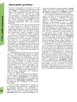
100:1 (Fig. 1). Anaerobic bacteria can adhere to tooth surfaces and contri-
bute, through the elaboration of metabolic by-products, to the development
of both carie s and periodontal disease (4). The predominant anaerobes
are anaerobic streptococci, Veillonella spp., Bacteroides spp., pigmented
Prevotella, and Porphyromonas spp. (previously called Bacteroides melanino-
genicus group), and Fusobacterium spp. (4). These organisms are a potential
source of a variety of chronic infections including otitis and sinusitis, aspira-
tion pneumonia lung abscesses, and abscesses of the oropharynx and teeth.
The microflora of the oral cavity is complex and contains many kinds
of obligate anaerobes. The distribution of bacteria within the mouth seems
to be a function of their ability to adhere to the oral surfaces. The differences
in numbers of the anaerobic microflora probably occur because of consider-
able variations in the oxygen concentration in parts of the oral cavity.
Figure 1 Oropharyngeal flora.
Infectious Causes of Sinusitis 147
For example, the maxillary and mandibular buccal folds contain 0.4%
and 0.3% oxygen, respectively, whereas the anterior and posterior tongue
surfaces contain 16.4% and 12.4%. The en vironment of the gingival sulcus
is more anaerobic than the buccal folds, and the periodontal pocket is the
most anaerobic area in the oral cavity. The ratio of anaerobic bacteria to
aerobic bacteria in saliva is approximately 10:1. The total count of anaero-
bic bacteria is 1.1 Â 10
8
/mL (Fig. 1). The predominant anaerobic bacteria
that colonize the anterior nose are P. acnes. Fusobacterium nucleatum is
the main species of Fusobacterium present in the oral cavity. Anaerobic
gram-negative bacilli found in the oral cavity include pigmented Prevotella
and Porphyromonas (previously called black-pigmented Bacteroides),
Porphyromonas gingivalis, Prevotella oralis, Prevotella orisbuccae (rumini-
cola), Prevotella disiens, and Bacteroides ureolytic us.
Fusobacteria are also a predominant part of the oral flora (6), as the
treponemas (7). Pigmented Prevotella and Porphyromonas represent <1%
of the coronal tooth surface, but constitute 4% to 8% of gingival crevice
flora. Veillonellae represent 1% to 3% of the coronal tooth surface, 5% to
15% of the gingival crevice flora, and 10% to 15% of the tongue flora. Micro-
aerophilic streptococci predominate in all areas of the oral cavity, and they
reach high numbers in the tongue and cheek (8). Other anaerobes prevalent
in the mouth are Actinomyces (9), Peptostreptococci, Leptotrichia buccalis,
Bifidobacterium, Eubacterium,andPropionibacterium (10).
Pigmented Prevotella, Porphyromonas, and Fusobacterium species can
also produce beta-lactamase (11). The recovery rate of aerobic and anaero-
bic beta-lactamase producing bacteria (BLPB) in the oropharynx has
increased in recent years, and these organisms were isolated from more than
half of the patients with head and neck infections including sinusitis (11).
BLPB can be involved directly in the infection, protecting not only them-
selves from the activity of penicillins but also penicillin-susceptible organ-
isms. This can occur when the enzyme beta-lactamase is secreted into the
infected tissue or abscess fluid in sufficient quantities to break the penicillin’s
beta-lactam ring before it can kill the susceptible bacteria (12) (Fig. 2).
The high incidence of recovery of BLPB in upper respiratory tract
infections may be because of the selection of these organisms following
antimicrobial therapy with beta-lactam antibiotics. Emergence of penicillin-
resistant flora can occur following only a short course of penicillin (13,14).
Obtaining Appropriate Sinus Content Cultures while Avoiding
the Normal Flora
If a patient with sinusitis develops severe infection, is immunocompromised
or fails to show significant improvement or shows signs of deterioration
despite treatment, it is important to obtain a culture, preferably through
sinus puncture, as this may reveal the presence of causative bacteria.
148 Brook
However, obtaining a culture through sinus endoscopy is an alternative
approach.
Sinus aspirates for culture must be obtained free of contamination so
that saprophytic organisms or normal flora are excluded and culture results
can be interpreted correctly. As indigenous aerobic and anaerobic bacteria
are present on the nasopharyngeal mucous membranes in large numbers,
even minimal contamination of a specimen with the normal flora can give
misleading results. The use of sinus puncture is the ‘‘gold standar d’’ method
of obtaining such specimens (15). There is, however, data that supports the
use of endoscopically obtained cultures in assessing the microbiology of
infected sinuses (16a–23).
Sinus Puncture
Obtaining sinus aspirates by puncture is the traditional method of specimens
collection. The maxillary sinus is the most accessible of all of the paranasal
sinuses. There are two approaches to the maxillary sinus that use puncture:
via either the canine fossa or the inferior meatus. The nasal vestibule is
often heavily colonized with pathogenic bacteria, mostly S. aureus. There-
fore, sterilization of the nasal vestibule and the area beneath the inferior
nasal turbinate is suggested.
Contamination with nasal flora may, however, occur. To prevent
misinterpretation of the culture results, an infec tion is defined as the reco-
very of a bacterial species in high density [i.e., a colony count of at least
10
3
to 10
4
colony forming units per milliliter (cfu/mL)]. This quantitative
definition increases the probability that microorganisms isolated from the
sinus aspirate truly repres ent in situ infection and not contaminati on. Most
Figure 2 Protection of penicillin-susceptible bacteria from penicillin by beta-
lactamase-producing bacteria.
Infectious Causes of Sinusitis 149