Biomedical Engineering From Theory to Applications Part 4 potx
Bạn đang xem bản rút gọn của tài liệu. Xem và tải ngay bản đầy đủ của tài liệu tại đây (632.03 KB, 30 trang )
Biomedical Engineering – From Theory to Applications
80
R. R. Harrison, P. T. Watkins, R. J. Kier, R. O. Lovejoy, D. J. Black, B. Greger, F. Solzbacher,
“A Low-Power Integrated Circuit for a Wireless 100-Electrode Neural Recording
System,” IEEE Journal of Solid State Circuits, Vol. 42, No. 1, Jan. 2007.
A. S. Sedra and A. C. Smith. Microelectronic Circuits. New York: Oxford UP, 2010.
5
Column Coupling
Electrophoresis in Biomedical Analysis
Peter Mikuš and Katarína Maráková
Faculty of Pharmacy, Comenius University,
Slovakia
1. Introduction
Biomedical analysis is one of the most advanced areas solved in analytical chemistry due to
the requirements on the analyzed samples (analyte vs. matrix problems) as well as on the
overall analytical process regarding automatization and miniaturization of the analyses.
Separation methods for the biomedical analysis are requested to provide high resolution
power, high separation efficiency and high sensitivity. This is connected with such conditions
that analytes are present in the samples in very low (trace) amounts and/or are present in
multicomponent matrices (serum, plasma, urine, etc.). These complex matrices consist from
inorganic and organic constituents at (very) differing concentrations and these can overlap the
analyte(s) peak(s) due to migration and detection interferences. In addition, a column
overloading can occur in such cases. It can be pronounced especially for the microscale
separation methods such as the capillary electrophoresis (CE). Hence, it is obvious that there is
the need for the sample preparation: (i) preconcentration – lower limits of detection and
quantification; (ii) purification of the sample and isolation of analytes – elimination of sample
matrix; (iii) derivatization – improvement of physical and/or chemical properties of the
analytes, before the CE analysis in these situations to reach relevant analytical information.
Sample pretreatment can be performed either off-line (before injection of analyzed sample
into the analyzer) or on-line (after the injection). The conventional separation systems (single
column) use mostly external (off-line) sample pretreatment, even though this analytical
approach has many limitations. These are (i) a loss of the analytes, (ii) time consuming and
tedious procedure, (iii) problematic manipulation with minute amounts of the samples, (iv)
problematic for automatization, (v) decreased precision of the analyses, etc. On the other
hand, on-line sample pretreatment has many advantages as (i) elimination of random
and/or systematic errors caused by external sample handling, (ii) simplification of an
overall analytical process (less number of an external steps), (iii) reduction of the total
analysis time and (iv) possibility of the automatization and miniaturization of the analytical
process (routine precise microanalyses). A significant enhancement of sensitivity and
selectivity is one of the main benefits of the on-line sample pretreatment. An on-line
pretreatment is crucial when there are only micro amounts of the samples for the analysis
and/or when analytes/samples have lower stability.
The advanced single column electrophoretic techniques (transient isotachophoresis, field-
enhanced sample stacking, dynamic pH junction, sweeping, in-capillary solid/liquid phase
extraction-CE, in-capillary dialysis-CE, etc.), representing the CE with the on-line (in-
column) sample preparation, were described and successfully applied for trace analytes and
Biomedical Engineering – From Theory to Applications
82
less or more complex matrices in many cases (section 2). The aim of this chapter is to
demonstrate potentialities and practical applications of a column coupling electrophoresis
as another group of the on-line sample preparation analytical approaches (section 3)
enabling powerful combination of (i) electrophoretic techniques (ITP, CZE, IEF, CEC)
(sections 3.1.1 and 3.2.1), (ii) electrophoretic and non electrophoretic (liquid
chromatography, flow injection analysis, etc.) techniques (sections 3.1.2 and 3.2.2). In this
way, it should be possible to create the most complex, flexible and robust tool filling the
above mentioned requirements of the advanced analysis. Such tool and its modes are
described in this chapter with regard to the theory, basic schemes, potentialities, for the
capillary (section 3.1) as well as microchip (section 3.2) format. This theoretical description is
accompanied with the performance parameters achievable by the advanced methods
(section 4) and appropriate application examples in the field of the biomedical analysis
(section 5). For a better understanding of the benefits, limitations and application potential
of the column coupling electrophoretic methods the authors decided to enclose the short
initial section with a brief overview of advanced single column electrophoretic techniques
(section 2) that often take part also in the column coupling electrophoresis.
2. Advanced single column techniques
As it is known from the literature (Simpson et al., 2008; Bonato, 2003) CE has many
advantages (high separation efficiency, versatility, flexibility, use of aqueous separation
systems, low consumptions of electrolytes as well as minute amounts of samples). Beyond
all the advantages, conventional CE has also some drawbacks, which limit its application in
routine analytical laboratories. They include (i) relatively difficult optimization of conditions
of analytical measurements, (ii) worse reproducibility of measurements (especially when
hydrodynamically open separation systems are used where non selective flows,
hydrodynamic and electroosmotic are acting) than in liquid chromatography, (iii) low
sample load capacity and need for the external (off-line) sample preparation for the complex
matrices (measurement of trace analyte besides macroconstituent(s) can be difficult without
a sample pretreatment), and (iv) difficulties in applying several detection methods in
routine analyses (Trojanowicz, 2009).
Some of these limitations can be overcome using advanced single column techniques. They
provide (i) improved concentration LOD, (ii) automatization (external manipulation with
the sample and losses of the analyte are reduced, analytical procedure is less tedious and
overall analysis time can be shortened, labile analytes can be analysed easier) and (iii)
miniaturization of the analytical procedure (pretreating of minute amounts of the sample is
possible and effective), (iv) elimination of interfering compounds, according to the
mechanism employed. However, the sample load capacity of these techniques is still
insufficient (given by the dimensions of the CE capillaries). The advanced single column CE
techniques usually suffer from lower reproducibility of the analyses due to the complex
mechanisms of the separation which controlling can be difficult in practice. Moreover, the
capillaries with embedded non electrophoretic parts (membranes, columns, fibers, monolits)
are less versatile (Simpson et al., 2008).
2.1 Stacking electrophoretic pretreatment techniques
Stacking procedures are based on increasing analyte mass in its zone during the
electromigration process via electromigration effects, enhancing sensitivity in this way. In all
cases, the key requirement is that there is an electrophoretic component in the
Column Coupling Electrophoresis in Biomedical Analysis
83
preconcentration mechanism and that the analytes concentrate on a boundary through a
change in velocity. Then we can recognize (i) field-strength-induced changes in velocity
(transient isotachophoresis (Beckers & Boček, 2000a), field-enhanced sample stacking (Kim
& Terabe, 2003; Quirino & Terabe, 2000a), and (ii) chemically induced changes in velocity
(dynamic pH junction (Britz-McKibbin & Chen, 2000), sweeping (Kitagawa et al., 2006;
Quirino & Terabe, 1998, 1999; Quirino et al., 2000b)). In addition to these techniques,
counter-flow gradient focusing (Shackman & Ross, 2007), electrocapture (Horáková et al.,
2007), and many others can be considered as the techniques based on a combination of field-
strength- and chemically induced changes in velocity offering new interesting possibilities
in on-line sample preparation (mainly preconcentration).
Some of the stacking techniques (and their combinations) can provide besides (i) the
preconcentration also other benefits such as (ii) an effective sample purification isolating
solute (group of solutes) from undesired matrix constituents (Simpson et al., 2008) or they
can be combined with (iii) chemical reaction of the analyte(s) (Ptolemy et al., 2005, 2006),
simplifying overall analytical procedure in this way. The choice of on-line pretreatment
method depends on the specific physical-chemical properties of the separated analytes (e.g.
charge, ionization, polarity) and the sample matrices (mainly concentration). For example,
an on-line desalting of a physiologic sample can be effectively accomplished by the
electrokinetic removing of the fast migrating low molecular ions prior to the IEF focusing of
the high molecular analytes (proteins) (Clarke et al., 1997).
2.2 Non electrophoretic pretreatment techniques
An on-line sample preparation can be carried out advantageously also combining the CE
with a technique that is based on a non electrophoretic principles. Most of these approaches
are based on (i) the chromatographic or extraction principles (separations based on chemical
principles), but also other techniques, such as (ii) the membrane filtration, MF (separations
based on physical principles), can be used. In this case, a non electrophoretic segment (e.g.
extractor, membrane) is fixed directly to the CE capillary (in-line combination) (Petersson et
al., 1999; Mikuš & Maráková, 2010).
In-line systems such as CEC/CZE (Thomas et al., 1999), SPE/CZE (Petersson et al., 1999) or
MF/CZE (Barroso & de Jong, 1998) are attractive thanks to their low cost and easy
construction. On the other hand, versatility of such systems is limited (in-capillary segment
cannot be replaced). One of the main limitations of performing in-line sample preparation is
that the entire sample must pass through the capillary, which can lead to fouling and/or
even clogging of the separation capillary and significant decreasing of reproducibility of the
analyses when particularly problematic samples (like biological ones) are used. It can be
pronounced especially for the extraction techniques (created inserting a solid-phase column
into capillary, where the whole analytical procedure is very complex and it includes
conditioning, loading/sorption, washing, (labeling, if necessary), filling (by electrolyte),
elution/desorption, separation and detection. In order to overcome these issues, on-line
methods based on another way of coupling of two different techniques may be used as
alternatives to the in-line systems.
3. Advanced column coupled techniques
Multidimensional chromatographic and capillary electrophoresis (CE) protocols provide
powerful methods to accomplish ideal separations (Hanna et al., 2000; Křivánková & Boček,
Biomedical Engineering – From Theory to Applications
84
1997a). Among them the most important ones are the integrated systems containing
complementary dimensions, where different dimensions separate components on the basis
of independent or orthogonal principles (Moore & Jorgenson, 1995; Lemmo & Jorgenson,
1993; Mohan & Lee, 2002). In such a multidimensional system, the peak capacity is the
product of the peak capacities of each dimension (Guiochon et al., 1983). A key part in the
instrumentation of the hyphenated techniques is an appropriate interface that enables to
connect and disconnect two different stages (e.g. columns) reproducibly and flexibly
according to the relevance and relation of the particular actions in the analytical process.
The column coupling arrangement, where two or more separation techniques are arranged
into two or more separated stages, can be a very effective approach offering additional
benefits to the advanced single column CE techniques and reducing some of their
disadvantages. Nevertheless, the advanced mechanisms given in section 2 can also be
adapted into the column coupling arrangement enhancing the effectivity and application
potential of the resulting method. Two separate stages provide (i) sample preparation
(preseparation, preconcentration, purification and derivatization) and (ii) analytical
separation of on-line pretreated sample. The benefits of the column coupling configuration,
additional to the advanced single column CE, involve (i) autonomic combination of various
separation mechanisms that provide enhanced and well defined separation selectivity, and a
possibility to replace easily one of the stages (ii) well defined and more effective elimination
of the undesirable sample matrix components, (iii) significant enhancement of the sample
load capacity (especially for the larger internal diameters of capillaries) resulting in the
improved LOD, (iv) improved precision of the analyses due to well defined control of the
separation mechanisms (Kaniansky et al., 1993; Kaniansky & Marák, 1990).
The most frequently used and the simplest column coupling configuration is the CE
combined with another CE (CE-CE, CE-CE-CE) (Kaniansky & Marák, 1990). Hybrid column
coupled techniques are based on the combination of a non electrophoretic technique with
the CE, e.g. LC-CE (Pálmarsdóttir & Edholm, 1995), SPE-CE (Puig et al., 2007), dialysis-CE
(Lada & Kennedy, 1997), FIA-CE (Mardones et al., 1999). They offer different separation
mechanisms in comparison with the CE-CE, however, they have more demands on
instrumentation. Additionally to the on-line combination of conventional column techniques
(electrophoretic as well as non electrophoretic) the column coupling arrangement combining
a conventional technique with an advanced one (section 2) is applicable too. These types of
the column coupled techniques are discussed in detail and illustrated through the
corresponding instrumental schemes for both the capillary (section 3.1) as well as microchip
(section 3.2) format.
3.1 Capillary format
3.1.1 Hyphenation of electrophoretic techniques
The hyphenation of two electrophoretic techniques in capillary format (see Fig. 1) can
effectively and relatively easily (simple and direct interface) solve the problems of the sample
preparation and final analysis (fine separation) in one run in well defined way, i.e. producing
high reproducibility of analyses, in comparison to the single column sample preconcentration
and purification approaches (section 2). Moreover, the CE performed in a hydrodynamically
closed separation system (hydrodynamic flow is eliminated by semipermeable membranes at
the ends of separation compartment) with suppressed electroosmotic flow (EOF), that is
typically used in the CE-CE configuration, has the advantage of (i) the enhanced precision due
to elimination of the non selective flows (hydrodynamic, electroosmotic), and (ii) enhanced
Column Coupling Electrophoresis in Biomedical Analysis
85
sample load capacity (30 L sample injection volume is typical) due to the large internal
diameter of the preseparation capillary (800 m I.D. is typical) (Kaniansky & Marák, 1990;
Kaniansky et al., 1993). The commercially available CE-CE systems have a modular
composition that provides a high flexibility in arranging particular moduls in the separation
unit. In this way, it is possible to create desirable CE-CE combinations such as (i) ITP-ITP, (ii)
ITP-CZE, (iii) CZE-CZE, etc., capable to solve wide scale of the advanced analytical problems
(see Fig. 1). Although such combinations require the sophisticated instrument and deep
knowledge in the field of electrophoresis, the coupled CE methods are surely the most
effective way how to take/multiply benefits of both CE techniques coupled in the column-
coupling configuration of separation unit. The basic instrumental scheme of the column
coupled CE-CE system shown in Fig. 1 is properly matching with hydrodynamically closed
CE modes where effective electrophoretic mobility is the only driving force of the separated
compounds. On the other hand, when additional supporting effects such as counterflow,
electroosmotic flow etc. must be employed, appropriate modifications of the scheme in Fig. 1
are made. Such modified instrumental schemes are attached into the sections dealing with IEF
or CEC coupled techniques (3.1.1.3 and 3.1.1.5) that are principally applicable only in
hydrodynamically open CE mode (Mikuš et al., 2006; Danková et al., 2001; Busnel et al., 2006).
Fig. 1. CE-CE method in column coupling configuration of the separation units for the direct
analysis of unpretreated complex matrices sample, basic instrumental scheme. On-line
sample preparation: removing matrices X (ITP, CZE), preseparation (ITP, CZE) and/or
preconcentration (ITP, stacking) and/or derivatization (with stacking) of analytes Y, Z in the
first CE stage (column C1). Final separation: baseline separation of Y and Z in the second CE
(ITP, CZE) stage (column C2). Reprinted from ref. (Tekeľ & Mikuš, 2005), with permission.
C1 – preseparation column, C2 – analytical column, B – bifurcation block for coupling C1
and C2, D – positions of detectors.
3.1.1.1 ITP-CZE
Although all electrophoretic methods can be mutually on-line combined, the biggest
attention was paid to the ITP-CZE coupling, introduced more than 20 years ago by
Biomedical Engineering – From Theory to Applications
86
Kaniansky (Kaniansky & Marák, 1990). The analytical benefits of the ITP-CZE combination
have been already well documented (Fanali et al., 2000; Danková et al., 2001; Kvasnička et
al., 2001; Valcárcel et al., 2001; Bexheti et al., 2006; Beckers, 2000b; Křivánková et al., 1991;
Křivánková & Thormann, 1993; Křivánková & Boček, 1997a).
An on-line combination of ITP with CZE appears to be promissing for alleviating some of
the following practical problems (Kaniansky & Marák, 1990):
i. ITP is a separation technique with a well defined concentrating power while the
separands migrate stacked in sharp zones, i.e., it can be considered as an ideal sample
injection technique for CZE,
ii. In some instances the detection and quantitation of trace constituents separated by ITP
in a large excess of matrix constituents may require the use of appropriate spacing
constituents. Such a solution can be very beneficial when a limited number of the
analytes need to be determined in one analysis. It becomes less practical (a search for
suitable spacing constituents) when the number of trace constituents to be determined
in one analysis is high,
iii. In CZE, high-efficiency separations make possible a multi-component analysis of trace
constituents with close physico-chemical properties. However, the separations can be
ruined, e.g., when the sample contains matrix constituents at higher concentrations than
those of the trace analytes.
A characteristic advantage of the ITP-CZE combination is a high selectivity/separability
obtainable due to the CZE as the final analytical step. Hence, the ITP-CZE method can be
easily modified with a great variety of selectors implemented with the highest advantage
into the CZE stage enabling to separate also the most problematic analytes (structural
analogs, isomers, enantiomers). The ITP-CZE methods with chiral as well as achiral CZE
mode have been successfully applied in various real situations (Mikuš et al., 2006a, 2008a,
2008c; Danková et al., 2001; Marák et al., 2007; Kvasnička et al., 2001).
The most frequently used ITP-CZE system works in the hydrodynamically closed separation
mode that is advantageous for the real analyses of multicomponent ionic mixtures because
of the best premises for enhancing sample load capacity (enables using capillaries with very
large I.D.). Such commercial system is applied with just one high-voltage power supply and
three electrodes (one electrode shared by the two dimensions), see Fig. 1. The electric circuit
involving upper and middle electrode (electric field No. 1) is applied in the ITP stage while
upper and lower electrode (electric field No. 2) is applied in the CZE stage. For the
separation ITP-CZE mechanism see chronological schemes in Fig.2. The focused zone in the
first dimension (ITP) is driven to the interface (bifurcation point) by only electric field No. 1.
The cut of the zone of interest in the ITP stage is based on the electronic controlling
(comparation point) of the relative step heigth (R
sh
, a position of the analyte between the
leading and terminating ion, it is the qualitative indicator depending on the effective
mobility of the analyte) of the analyte, see Fig. 3. The conductivity sensor (upper D in Fig. 1,
D-ITP in Fig.2) serves for the indication of the analyte zone. This is very advantageous
because such indication is (i) universal and (ii) independent on other comigrating
compounds (sample matrix constituents migrating in the ITP stage) and therefore
independent on sample composition. The electric circuit is switched and electric field No. 2
(upper and lower electrode) is applied in an appropriate time (this time is set electronically
depending on requirements of the composition of the transferred plug) after the indication
of the analyte zone passing through the upper D. From this moment the all ITP zones are
directed to the CZE stage for the final separation and detection. It is possible to carry out
Column Coupling Electrophoresis in Biomedical Analysis
87
one or more cuttings depending on the zones of interest and/or interfering matrix
constituents present in the sample. The interface between the separation solutions in the ITP
and CZE capillary is free (without any mechanical restraint) but mixing of the electrolytes is
eliminated (with the exception of difusion) by suppressing all non selective flows
(hydrodynamic, electroosmotic) in the system. This is advantageous by an easy construction
and elimination of dead volumes in the separation system (Ölvecká et al., 2001; Kaniansky et
al., 2003).
Fig. 2. ITP sample clean up for CZE with the closed separation system (without any
supporting non selective flow). (a) Starting arrangement of the solutions in the capillaries;
(b) ITP separation with the analyte (A) trapped into the boundary layer between the zones
of front (M1) and rear (M2) spacers; (c) end of the run in the ITP capillary followed by an
electrophoretic transfer of the analyte containing fraction to the CZE capillary (by switching
the direction of the driving current); (d) removal of the sample constituents migrating
behind the transferred fraction (by switching the direction of the driving current); (e)
starting situation in the separation performed in the CZE capillary (the direction of the
driving current was switched); (f) separation and detection of the transferred constituents in
the CZE capillary. BF = bifurcation region; C1, C2 = the ITP and CZE separation capillaries,
respectively; D-ITP, D-ZE = detection sensors in the ITP and CZE separation capillaries,
respectively; TES = terminating electrolyte adapted to the composition of the sample (S);
TITP = terminating electrolyte adapted to the composition of the leading electrolyte
solution; A = analyte, i = direction of the driving current. Reprinted from ref. (Kaniansky et
al., 2003), with permission.
Biomedical Engineering – From Theory to Applications
88
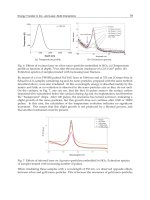
Fig. 3. Graphical illustration of the principle of the electronic cutting of the zone of interest
in the ITP stage of the ITP-CZE combination. L = leading ion, T = terminating ion, X =
matrix compound(s), Y, Z = analytes, R = resistance. Reprinted from ref. (Ölvecká et al.,
2001), with permission.
The principle of this hyphenated technique consists from well-defined preconcentration
(concentration LODs could be reduced by a factor of 10
3
when compared to conventional
single column CZE) and preseparation (up to 99% or even more interfering compounds
can be isolated (Danková et al., 1999)) of trace analytes in the first, wider, capillary
(isotachophoretic step) and subsequently a cut of important analytes accompanied with a
segment of the matrix, leading or terminator enters the second, narrower, capillary for the
final separation by CZE (Fig. 2, Fig.3). The presence of this segment results from the fact
that we do not want to lose a part of the analyzed zones and we must make a cut
generously. The zone of this segment survives for a certain time during the CZE stage and
this mean that ITP migration continues also in the second capillary for some time and it
influences strongly the results of the analysis, especially the detection times of analytes
used for identification of the analytes in CZE separations (Busnel et al., 2006; Gebauer et
al., 2007; Mikuš et al., 2006a). From this is clear that it is important in an ITP-CZE
combination to choose suitable electrolyte systems and find the optimum time to switch
the current from the preseparation capillary to the separation capillary (Křivánková et al.,
1995).
The ITP-CZE technique appeared to be very useful especially for the common universal
detectors producing relatively low concentration LODs (UV-VIS photometric detector). It
is because such method provides probably one of the most acceptable ratio simplicity-
cost: universality-concentration LOD in comparison to other column coupling methods
and detection systems. This suggestion is supported by many advanced applications of
the ITP-CZE-UV method in the pharmaceutical and biomedical field (Marák et al., 2007;
Mikuš et al., 2008a, 2008b, 2008c, 2009). Jumps in voltage (conductivity) between
neighboring zones result in permanently sharp boundaries between zones (Fig. 3) that is
extremely convenient for the conductivity detection in ITP. Although convenient to the
Column Coupling Electrophoresis in Biomedical Analysis
89
detection of the ITP zones, conductivity detection technique has a limited applicability in
the CZE separations (often measurements of small conductivity changes due to the zones
on a relatively high conductivity background of the carrier electrolyte) (Ölvecká et al.,
2001; Kaniansky et al., 2000).
3.1.1.2 ITP-ITP
The ITP-ITP combination represents the simplest possibility how to combine CE
techniques. For the general instrumental scheme valid also for ITP-ITP, see Fig. 1. In the
ITP–ITP mode both preseparation (wider) and analytical (narrower) capillaries are filled
with (i) the same leading electrolyte (one-dimensional ITP) or (ii) different electrolytes
(two-dimensional ITP) (Flottmann et al., 2006; Bexheti et al., 2006; Mikuš et al., 2006b;
Kubačák et al., 2006a, 2006b, 2007). The ITP separation in a concentration cascade,
introduced into conventional CE by Boček et al., (Boček et al., 1978) enhances the
detectabilities of the separated constituents from the response of the conductivity
detection due to well-known links between the concentration of the leading electrolyte
and the lengths (volumes) of the zones (Marák et al., 1990)
The first ITP stage of the ITP-ITP combination can apply all benefits as they are described
for the ITP stage of the ITP-CZE combination in section 3.1.1.1. On the other hand, the ITP-
ITP technique can take the highest advantage of the hyphenation with the MS detection
(Tomáš et al., 2010). It is because of an intrinsic feature of ITP to produce pure analyte zones,
i.e. those in which the analyte is accompanied only with counter ion, in the isotachophoretic
steady state. In this way, the maximum response of the MS detector can be obtained for the
analyte. Therefore, the ITP-ITP-MS hyphenation seems to be one of the most promissing
methods for the fully automatized biomedical analyses such as pharmacokinetic studies,
metabolomics, etc. An economic aspect of the ITP-ITP-MS method in comparison with the
HPLC-MS method for the ionic compounds is apparent.
3.1.1.3 ITP-CEC
Another approach in the column coupled electrophoresis is the use of ITP sample focusing
to improve the detection limits for the analysis of charged compounds in capillary
electrochromatography (CEC). Besides this, the on-line isotachophoretic stage can serve also
for a loadability enhancement (due to a large inner diameter of the ITP capillary). Both of
these effects are then responsible for a dramatic reduction of the sample concentration
detection limits through simultaneous acting of (i) large volume injection and (ii) analyte
stacking (Mazereeuw et al., 2000).
In the ITP-CEC combination (Fig. 4), the open ITP mode must be applied because of the
demands of the second stage (CEC) that is based on the EOF action. A coupled-column
set-up can be used, in which counterflow ITP focusing is performed, and the separation
capillaries are connected via a T-junction. For the schematic representation of the ITP–
CEC procedure see Fig. 5. From the application point of view, the first ITP stage is
advantageous especially for the injection of large volumes (tens of microliters) of diluted
samples. When a very large sample is introduced, however, the focusing time of the
sample often exceeds the migration time to the outlet of the ITP capillary. By applying a
hydrodynamic counterflow (applicable in the hydrodynamically open CE systems) the
ITP focusing will continue while extending the migration towards the outlet of the ITP
capillary. Although the hydrodynamically open CE systems have the advantage of
application of the supporting flows (counterflow, electroosmotic), it must be realized that
Biomedical Engineering – From Theory to Applications
90
the reproducibility of cutting and also overall analysis is generally lower than in the
hydrodynamically closed CE systems due to the fluctuations of the non selective flows in
the separation system.
Fig. 4. Schematic representation of the ITP–CEC set-up. Right scheme: Schematic
representation of the ITP–CEC–UV set-up with a (P) programmable capillary injection
system, (D) UV–VIS absorbance detector, (A) amperometer and (T) laboratory made
polyethylene T-piece. Untreated fused-silica capillaries of 220 m I.D. (1 and 2) and 75 m
(3) are used. Left scheme: Schematic representation of the entire ITP–CEC–MS set-up. The
electrospray needle with the sheath flow contains the CEC column, which is directly
connected with the electrospray. The spray is directed towards the inlet capillary of the
interface on the SSQ 710 mass spectrometer (MS). HV is the electrospray power supply.
Reprinted from ref. (Mazereeuw et al., 2000), with permission.
The first ITP stage of the ITP-CEC combination can apply all benefits as they are described
generally for the ITP stage of ITP-CZE combination in section 3.1.1.1. In ITP-CEC, the ITP
sample clean-up effect is extremely important for enhancing reproducibility of CEC
especially when injecting complex biological samples. The CEC stage of the ITP-CEC
technique can take a high advantage of the hyphenation with the UV-VIS or MS detection,
for the schemes of the experimental setups see Fig. 4. It is pronounced in the situations when
the selectors interfering with the detection must be used in the separation system in order to
establish the required selectivity. Immobilization of such selectors in the CEC column
prevents their entering into the detector cell resulting in the elimination of the detection
interferences. In this way, the maximum response of the UV-VIS or MS detector can be
obtained for the analyte. Hence, the ITP-CEC combination seems to be a powerful tool for
the on-line selective separation, sensitive determination and spectral identification of chiral
compounds and various other isomers and structurally related compounds (i.e.
“problematic” analytes) present in complex ionic matrices. The ITP-CEC-MS hyphenation
seems to be one of the most promissing methods for the fully automatized biomedical chiral
analyses such as enantioselective pharmacokinetic studies, metabolomics, etc. (Mazereeuw
et al., 2000).
Column Coupling Electrophoresis in Biomedical Analysis
91
Fig. 5. Schematic representation of the ITP–CEC procedure (with a supporting non selective
flow). The sample loading, ITP focusing step, sample zone transfer and CEC separation are
shown in step 1, 2, 3 and 4, respectively. The set-up contains a (D) UV–VIS absorbance or
MS detection, (T) terminator buffer and (L) leading buffer. Untreated fused-silica capillaries
of 220 m I.D. (1 and 2) and 75 m (3) are used. Reprinted from ref. (Mazereeuw et al.,
2000)., with permission.
3.1.1.4 CZE-CZE
CE separation system with tandem-coupled columns, i.e. CZE-CZE makes possible, within
certain limits, splitting a CZE run into a sequence of the separation and detection stages (for
the general instrumental scheme valid also for CZE-CZE, see Fig. 1). Therefore, the carrier
electrolyte employed in the first (separation) stage of the run could be optimized with
respect to the resolution of an analyte from complex (biological) matrix. In this way, a very
significant ‘‘in-column’’ clean-up of the analytes from complex ionic matrices can be reached
in the separation stage of the tandem by combining appropriate acid-basic (pH) and
complexing (selectors) conditions. Due to this, the detection (e.g. spectral) data could be
acquired in the detection stage of the tandem with almost no disturbances by matrix co-
migrants (Danková et al., 2003).
The carrier electrolyte employed in the second (detection) stage could be chosen to reach
favourable conditions in the acquisition of detection (e.g. spectral) data while maintaining
the resolution of the analyte from matrix constituents as achieved in the separation stage
Biomedical Engineering – From Theory to Applications
92
(Danková et al., 2003). Such two-dimensional systems reduce probability of component
overlap and improve peak identification capabilities since the exact position of a compound
in a two-dimensional electropherogram is dependent on two different separation
mechanisms (Sahlin, 2007).
The CZE-CZE combination can be set to achieve a remarkable selectivity. On the other hand,
it is considerably less sensitive than the ITP-CZE combination due to the absence of stacking
capability of the basic CZE technique. It can be overcome, fortunately, replacing a basic CZE
technique by an advanced one (e.g. stacking). The CZE-CZE technique is favorable for the
hyphenation with various detection techniques (e.g. spectral, electrochemical) because it
makes possible splitting of the CZE run into a sequence of the separation and detection
stages (Danková et al., 2003).
3.1.1.5 IEF-CZE, -CGE
Arduous proteomics tasks require techniques with high throughput and high efficiency in
order to screen a certain proteome expression and to monitor the effects of environmental
conditions and time on the expression. There seldom is, at present, a single separation mode
sufficient enough to deal with such complex samples. CE is a significant tool for the
separation of proteins and peptides (Dolnik &. Hutterer, 2001). To finish complicated
separation jobs, great efforts have been concentrated on the development of 2D CE (Yang et
al., 2003b). IEF, CGE and CZE are the most effective electrophoretic techniques for
zwitterionic compounds, therefore the on-line combination of these techniques is of the
highest importance for the protein analysis with perspectives of their automatization and
miniaturization (Kaniansky et al., 2000; Chen X. et al., 2002; Kvasnička et al., 2001).
When performing isoelectric focusing, one can fill the total volume of a capillary with
sample solution. It can be expected that the detection sensitivity of the hyphenated system
benefits from the concentration effect of the first dimension of IEF. This feature holds
advantage over other CE modes such as CZE, CGE, micellar electrokinetic capillary
chromatography (MECC), and capillary electrochromatography (CEC). Practically, IEF has a
power to concentrate analytes up to several hundred folds in a capillary (Shen et al., 2000).
Such a condensed and shortened analyte plug in a capillary is appropriate for sample
injection to other CE modes. Therefore IEF is a proper candidate for the first dimension in a
multi-dimensional CE system. Apparently, this will improve the sensitivity for mass
detection. It is advantageous over those systems in which IEF was utilized as the second
dimension. Nevertheless, the sensitivity of UV absorbance suffers from the necessity of the
CAs involved in IEF. Of course, isotachophoresis (ITP) as a pretreatment tool for CZE
separation also has a concentration effect (Kaniansky et al., 1999). ITP is carried out based on
the mobility differences of ions and, IEF, based on different pIs of ampholytic molecules.
Capillary isoelectric focusing (IEF) and capillary zone electrophoresis (CZE) can be on-line
hyphenated by a dialysis interface to achieve a 2D capillary electrophoresis (CE) system, i.e.
IEF-CZE (Fig. 6), as it was demonstrated by Yang et al. (Yang et al., 2003b). The system was
used with just one high-voltage power supply and three electrodes (one cathode shared by
the two dimensions). The focused and preseparated (according to differences in the
isoelectric points of the analytes) zones in the first dimension (i.e. the IEF) were driven to the
dialysis interface by electroosmotic flow (EOF), besides chemical mobilization from the first
anode to the shared cathode. Zero net charged analyte molecules focused in the first
dimension are recharged in the interface (I
2
in Fig. 6) according to the pH of the altered
buffer. The semi-permeable property of the interface ensures that macromolecules of
Column Coupling Electrophoresis in Biomedical Analysis
93
ampholytic analytes remain in the separation channel. In the second dimension (i.e. the
CZE), the preseparated zones were further separated (according to the ratios of charge and
mass, i.e. electrophoretic mobility) and driven by an inverted EOF, which originated from
the charged layer of a cationic surfactant adsorbed onto the inner wall of the capillary. It can
be concluded that the 2D IEF–CZE system possesses higher resolving power than each of
the single modes. This protocol of the 2D CE system endues the interface with durability
and makes for convenient performance. To reduce the dead volume, it is necessary to match
the inner diameter of the hollow fiber to that of the capillaries. The tangent surfaces of these
units should be made even and smooth.
Fig. 6. Construction of 2D IEF–CZE. Upper scheme: general overview. S: high-voltage power
supply; C1, C2: capillaries; I
1
, I
2
, I
3
: interfaces; D: detector. Lower scheme: detail of dialysis
interface. (1) capillaries; (2) buffer reservoir; (3) hollow fiber; (4) electrode; (5) buffer inlet; (6)
buffer outlet. Reprinted from ref. (Yang et al., 2003b), with permission.
A two-dimensional capillary isoelectric focusing–capillary gel electrophoresis (IEF–CGE)
system is another modification of the technique based on on-line combination of IEF with
zone electrophoresis (Yang et al., 2003a). It also can be accomplished just with one high-
voltage power supply and three electrodes. Chemical mobilization can be utilized to drive
the sample zones of the first dimension. To actualize 2D IEF–CGE performance, coated and
gel-filled capillaries are needed to eliminate the undesired EOF. In a gel-filled capillary the
emergence of bubbles is tedious. From this point of view, it is valuable to exploit a more
convenient and robust 2D CE system such as IEF-CZE (as illustrated above).
3.1.2 Hyphenation of electrophoretic and non electrophoretic techniques
Lately there were introduced into CE several hybrid on-line sample preparation techniques
that are still in development as there is a big effort (i) to simplify usually a very complex
Biomedical Engineering – From Theory to Applications
94
instrumental arrangement and simultaneously (ii) to ensure the enhancement of the
compatibility within and reproducibility of the procedure. The column coupled non
electrophoretic stages include (i) chromatography (Pálmarsdóttir & Edholm, 1995;
Pálmarsdóttir et al., 1996, 1997), (ii) SPE extraction (Puig et al., 2007), (iii) dialysis (Lada &
Kennedy, 1997), and (vi) flow injection analysis (FIA) (Mardones et al., 1999). A great
potential of the hybrid on-line sample preparation techniques is given by their
complementarity that enables to cumulate positive effects and/or overcome the weak points
of the individual sample preparation techniques. In addition, these techniques, likevise to
CE-CE, can be simultaneously combined also with stacking effects or chemical reaction in
order to enhance further overall analytical effect as it is demonstrated in the following
sections. From the practical point of view, the following sections are starting with the on-line
implementation of FIA because the flow injection principles and instrumental
procedures/arrangements are widely applied also for the effective integration of other non
electrophoretic techniques (SPE, LC, dialysis) with CE.
3.1.2.1 FI-CE
The concept of flow injection analysis (FIA) was introduced in the mid-seventies. It was
preceded by the success of segmented flow analysis, mainly in clinical and environmental
analysis. This advance, as well as the development of continuous monitors for process
control and environmental monitors, ensured the success of the FIA methodology
(Trojanowicz et al., 2009; Lü et al., 2009). A combination of CE with a flow injection (FI)
offers a great scale of sample preparation and the most frequently it is used for the on-line
implementation of chemical reactions. The technique of combined flow injection CE (FI-CE)
integrates the essential favorable merits of FI and CE. It utilizes the various excellent on-line
sample pretreatments and preconcentration (such as cloud point extraction, SPE,
ionexchange, DPJ and head-column FESS technique, analyte derivatization) of FI, which has
the advantages of high speed, accuracy, precision and avoiding manual handling of sample
and reagents. Therefore, the coupling of FI-CE is an attractive technique; it can significantly
expand the application of CE and has achieved many publications since its first appearance
(Mikuš & Maráková, 2010).
Fig. 7. Typical FI manifold used for the derivatization of the analytes and their on-line
introduction into the CE system. Reprinted from ref. (Mardones et al., 1999), with
permission.
A high potential of the FI-CE method in automatization of sample derivatization and
subsequent separation was demonstrated by Mardones et al. (Mardones et al., 1999). The
Column Coupling Electrophoresis in Biomedical Analysis
95
derivatization reaction for carnitine as the model analyte was carried out on a FI system
coupled with the CE equipment via a programmable arm (Valcárcel et al., 1998). The
arrangement is shown in Fig. 7. The derivatization reagent (FMOC-Cl) is introduced directly
into the loop of the injection valve (IV) when load position is selected, while the sample is
introduced into the system and it is mixed with the buffer (carbonate). Then, valve is
switched to the injection position allowing the mixing of sample–buffer and reagent
solution. In this position the flow is stopped for a defined time in the reactor loop (390 cm),
which is introduced into the thermostatic bath (50°C). Finally, the reaction mixture is
introduced via the mechanic arm into the CE system.
The third generation of flow-injection (laboratory-on valve, lab-on-valve or LOV) allows
scaling-down sample and reagent volumes to the 10–20 L range, while waste production is
typically 0.1–0.2 mL per assay (Solich et al., 2004). These facts make LOV an ideal tool for
on-line coupling with CE systems (Kulka et al., 2006).
3.1.2.2 SPE-CE
The new trends in the coupling between SPE–CE are focused on several strategies, one of
which involves developing new materials to increase the retention and selectivity of some
analytes. In this sense the increasing use of materials such as immunoaffinity sorbents has
been shown to overcome the problem of selectivity especially when complex samples are
analysed. The use of molecular imprinted polymers (MIP) could be also an attractive
alternative and further development is expected in this area in the near future. Carbon
nanostructures also seem to be very promising materials which are in the first stages of
development and so more research is expected in this field (Puig et al., 2007).
Fig. 8. Schematic diagram of the three types of interfaces for on-line SPE–CE coupling: (a)
vial interface; (b) valve interface; (c) T-split interface. Reproduced with permission from (a)
Stroink et al. (Stroink et al., 2003), (b) Tempels et al. (Tempels et al, 2007) and (c) Puig et al.
(Puig et al., 2007).
Extraction techniques now play a major role for sample preparation in CE. These techniques
can be used not only for reconstitution of the sample from small volumes but also for
sample purification in complex matrices and desalting for very saline samples that would
interfere with the electrophoretic process (e.g. FESS requires low conductivity sample).
Considerable progress has been made towards the coupling of solid phase extraction (SPE)
with a subsequent electrophoresis while coupling of liquid phase extraction (LLE) with
electrophoresis is less used. Before coupling the SPE and CE, the appropriate SPE conditions
for trapping and eluting the test compounds must be investigated. The breakthrough
Biomedical Engineering – From Theory to Applications
96
volumes, desorption efficiency and desorption volume must be studied too. Typical
approaches of the on-line coupling of SPE with CE, advantageous by a high flexibility and
variability of extraction volumes, are based on the use of a vial, valve or T-split interfaces.
Schematic diagram of these types of interfaces for on-line SPE–CE coupling are shown in
Fig.8.
An on-line SPE–CE approach based on a Tee-split interface was demonstrated by Puig et al.
(Puig et al., 2007). The Tee-split interface is required for the on-line coupling of SPE–CE and
to allow an injection volume that is suitable for CE analysis because the SPE elution volume
is considerably larger than the maximum volume that can be injected into the CE capillary.
Using this interface, a part of the SPE elution plug is injected while the rest of the sample is
flushed to waste. Depending on the matrix, however, the sample must be appropriately
pretreated prior to the injection into the first stage (i.e. SPE). As plasma is a relatively
complex sample, the introduction of a pretreatment step (protein precipitation) prior to
injection was necessary to prevent clogging of the SPE column.
For various specific purposes where chemical reaction and preconcentration must be
involved simultaneously (e.g. in case of peptide mapping), the on-line coupling of
microreactor (with an immobilized-enzyme), SPE preconcentrator and CE can be applied
(Bonneil & Waldron, 1999). The problems related to the preconcentrator, such as reversal of
EOF at low pH, can be eliminated by designing the on-line system in such a way that the
preconcentrator is not part of the separation capillary, unlike most configurations reported
in the SPE-CE literature. Consequently, the preconcentrator should not interfere with the
separation process. Benefits of the on-line microreactor-SPE-CE system include (i) sensitivity
(several hundred-fold preconcentration factor can be achieved) for the analyte products
isolated in very small quantities from complex (biological) samples, (ii) avoiding
conventional experimental steps that are quite long, labor intensive and require a lot of
sample handling. Such system can be reused for several samples with acceptable
reproducibility and relatively short analysis time. On the other hand, a loss of separation
efficiency can be observed that is induced by the multiple-valve design of the system and
dispersion of the desorption plug.
Another way of the integration of chemical reaction to the SPE-CE is the lab-on-valve (LOV)
interface. The automatic minicolumn SPE preconcentration in LOV module coupled on-line
with the CE equipment was proposed for the separation and quantification of mixtures of
target analytes in very diluted samples (Jiménez & de Castro, 2008). This method can be
applied with or without an on-line analyte derivatization depending on requirements. So that
the complex derivatization-SPE-CE method integrates several different working principles
such as (i) flow injection with chemical reaction, (ii) preseparation and preconcentration with
non electrophoretic (extraction) principles, (iii) final separation with electrophoretic principles
and detection of the separated zones. The usefulness of the LOV interface for the on-line
coupling with a CE instrument interfaced by the appropriate manifold was reflected in
excellent concentration LODs and linear dynamic ranges obtained.
Solid-phase microextraction (SPME) is interesting and alternative technique because it is
simple, can be used to extract analytes from very small samples and provides a rapid
extraction and transfer to the analytical instrument. Moreover, it can be easily combined
with other extraction and/or analytical procedures, improving to a large extent the
sensitivity and selectivity of the whole method (Lord & Pawliszyn, 2000; Ouyang &
Pawliszyn, 2006; Saito & Jinno, 2003; Fang et al., 2006a, 2006b). Even though SPME is
becoming an attractive alternative to using SPE, its use in combination with CE is still rather
Column Coupling Electrophoresis in Biomedical Analysis
97
limited. Such coupling has not been widely used because of its inherent drawbacks
regarding the low injection volumes typically required in CE (which are crucial to obtaining
good separation efficiency) and also because the different sizes of the separation capillaries
usually used for CE and the SPME fibers (Liu & Pawliszyn, 2006). Moreover, SPME suffers
from limited choice of selectivity in comparison with SPE since only few stationary phases
are avalaible (Puig et al., 2007).
3.1.2.3 LC-CE
When biological samples have to be analyzed, additional sample pretreatment prior to the
SPE step may be needed to remove compounds that jeopardize an effective analyte
concentration (or even block the SPE column) and the subsequent CE analysis. Sample
pretreatment prior to SPE can be achieved by carrying out a preceding separation.
Generally, sample analysis with on-line multidimensional separation systems can be
performed using a comprehensive or a heart-cut approach. The comprehensive approach
results in the analysis of the complete sample in all subsequent dimensions, whereas the
heart-cut approach analyzes only a small part of the pre-separated sample in the second
separation step. The comprehensive approach demands a slow preceding separation
compared to the subsequent separation in order to accomplish analysis of the complete
sample in all dimensions. Typical examples of such comprehensive systems are the on-line
size exclusion chromatography (SEC)–CE systems and reversed phase LC–CE systems
developed in the group of Jorgenson (Bushey & Jorgenson, 1990; Lemmo & Jorgenson, 1993;
Moore Jr. & Jorgenson, 1995; Hooker & Jorgenson, 1997), which are coupled by various
interfaces. These systems do not concentrate the chromatographic fractions prior to
introduction into the CE system, which reduces the sensitivities of the total systems. Efforts
to integrate such a focusing step would imply the need for an even slower preceding
separation step to create time for sample trapping in a SPE column, washing and desorption
of the concentrated fraction and sample introduction into the CE system. In practice, a
comprehensive multidimensional system with a focusing step seems almost impossible,
unless a number of columns are integrated into the system in a parallel fashion to enable
“parking” of the LC fractions. In that case, the LC fractions are stored in the focusing step on
various SPE columns and can be sent to the CE system at any convenient moment.
The heart-cut approach is less demanding and is best suitable for target analysis. In the case
of a heart-cut approach for an on-line system, it is also easier to integrate a concentration
step between the preceding and the final separation step because there are no time
constraints. Stroink et al. (Stroink et al., 2003) coupled SEC–SPE with CE through a vial-type
interface for the quantitative analysis of enkephalins in cerebrospinal fluid (CSF). The SEC
dimension separated the sample in a protein and a peptide-containing fraction. This
resulted in a relatively large volume of the peptide fraction (about 200L), requiring a
subsequent SPE step prior to CE analysis to obtain acceptable LODs.
Tempels et al. (Tempels et al., 2006) developed an on-line SEC–SPE–CE system with a Tee-
split interface (Fig. 9) for the isolation, concentration and separation of peptides or other
lower molecules in biological fluids (such as CSF). The SEC dimension served for the
fractionation of the sample so that a fraction having required molecular weight could be
easily selected (here, proteins were discarded). The small SPE column provided effective
sample preconcentration using small desorption volumes (425 nL). The Tee-split interface
enabled on-line injection of the concentrated analytes into the CE system without disturbing
separation efficiency.
Biomedical Engineering – From Theory to Applications
98
Fig. 9. Schematic diagram of the on-line SEC–SPE–CE system with the Tee-split interface.
The on-line SEC–SPE–CE system was built in three distinct parts: a SEC, a SPE and a CE
part. The SEC part consisted of a pump (pump 1), a valve (valve 1) for introduction of
sample, a SEC column, and a UV detector (detector 1). The SPE part comprised a pump
(pump 2), a micro valve (valve 3) for introduction of acetonitrile, and a SPE column. Valve 2
functioned as a selection valve to direct a fraction of solvent A towards the SPE column or to
detector 1. The CE part of the complete system is framed. Lengths of capillaries are shown in
italics (cm). The CE part consisted of a CE system with a build-in photodiode array detector.
The CE and SPE parts were connected by a micro Tee with a void volume of 29 nL. The SEC
part was filled with solvent A, whereas in the SPE and CE parts BGE was used. Reprinted
from ref. (Tempels et al., 2006), with permission.
Although LC-CE coupling is technically much more difficult than CE-CE, because it has to
be accompanied by collection, evaporation and reconstitution of fraction isolated by LC,
some of these actions can be eliminated implementing an advanced CE stage (with a
concentration capability) into LC-CE. Micro-column liquid chromatography (MLC) can be
used on-line with an advanced (stacking) CE for sample purification and concentration
allowing injection of microliter volumes into the electrophoresis capillary (Bushey &
Jorgenson, 1990; Pálmarsdóttir & Edholm, 1995). By using the double stacking procedure
with assistance of the backpressure almost complete filling of the electrophoresis capillary is
possible without significant loss of CZE separation performance. The combined system has
a much greater resolving power and peak capacity than either of the two systems used
independently of each other.
3.1.2.4 Dialysis-CE
Microdialysis is a widely accepted sampling and infusion technique frequently used to
sample small molecules from complex, often biological, matrices (Adell & Artigas, 1998;
Chaurasia, 1999). In the microdialysis, small molecules are able to diffuse across the dialysis
membrane into the probe, while large molecules, such as proteins and cell fragments, are
excluded. This is the sample cleanup provided by the microdialysis.
On-line microdialysis-CE assays for neurotransmitters to date have been most successful for
easily resolved analytes such as glutamate and aspartate (Thompson et al., 1999; Lada et al.,
Column Coupling Electrophoresis in Biomedical Analysis
99
1997, 1998). However, the efficiency and peak capacity of high-speed CE separations are
often not high enough to resolve complex mixtures. Recently, improvements in injection
technique and detection limits have improved separation efficiency (Bowser & Kennedy,
2001). In the microdialysis, the minimum volume required for analysis often determines the
rate at which the dialysate can be sampled. On-line microdialysis-derivatization-CE-LIF
assays as proposed by Lada et al. (Lada et al., 1997) (for the instrumental scheme see Fig. 10)
eliminate fraction collection. The separation capillary was coupled to the reactor capillary
via a flow-gated interface which allowed dialysate samples to be automatically injected onto
the separation capillary. This elimination of fraction collection, combined with the high
mass sensitivity of LIF or electrochemical detectors, makes sampling rates on the order of
seconds possible (Thompson et al., 1999; Lada et al., 1997, 1998). The microdialysis-CZE-LIF
system with on-line derivatization has the advantage of simultaneously obtained high
relative recoveries and good temporal resolution with (in-vivo) microdialysis sampling for
the real biological system (brain) (Lada et al., 1997).
Fig. 10. Diagram of the microdialysis:CZE-LIF system with on-line derivatization. Reprinted
from ref. (Lada et al., 1997), with permission.
3.2 Microchip format
Developments in the fields of microfluidics and microfabrication during the last 15 years
have given rise to microchips with broad ranges of functionality and versatility in the areas
of bioanalysis such as clinical applications (Li & Kricka, 2006) and chiral separations (Belder,
2006). Microfluidic devices such as microchips can provide several additional advantages
over electromigration techniques performed in capillary format. The heat dissipation is
much better in chip format compared with that in a capillary and therefore higher electric
fields can be applied across channels of microchip. This fact enables, along with a
considerably reduced length of channels, significant shortening of separation time
(millisecond analysis time is possible to achieve, see e.g. (Belder, 2006)). Sample and reagent
consumption is markedly reduced in microchannels. Hence, microchip capillary
electrophoresis (MCE) can provide a unique possibility of ultraspeed separations of
microscale sample amounts. Applicable are both electrophoretic (Gong & Hauser, 2006;
Belder, 2006) as well as electrochromatographic modes (Weng et al., 2006).
In practice, however, the resolution achievable in MCE devices is often lower compared to
that obtainable in classical CE utilizing considerably longer separation capillaries. In order
Biomedical Engineering – From Theory to Applications
100
to obtain sufficient resolution in MCE, different strategies have been used (Belder, 2006),
such as (i) enhancing the selectivity of the system as much as possible (changing type and
amount of selector, adding coselector, etc.), (ii) using of folded separation channels, the
column length can be extended without enlarging the compact footprint of the device, (iii)
using coated channels, internal coatings improve separation performance by the
suppression of both analyte wall interaction and electroosmosis. A use/combination of
above mentioned tools applicable in MCE gives a better chance for real-time process control
and for multidimensional separations and makes the MCE to be powerful tool in real
applications (pharmaceutical, biomedical, etc.). Sample pretreatment has been recognized as
another significant barrier to higher levels of integration. Other accompanied problems in
real applications of basic MCE are as follows: The detection volume of microfluidic devices
due to the channel dimension and the sampling amounts is rather small, which would
impact the detectable concentration. Fabricating a microchip with a large detection volume
can be easily performed, but the separation efficiency is usually insufficient (Hempel, 2000).
Another way is to inject a long sample band and then stack it into a narrow zone using on-
line preconcentration techniques prior to separation (Chien, 2003). In such case, not only the
preconcentration but also sample clean-up can be simultaneously carried out. Therefore,
further considerable enhancement of analytical capabilities can be achieved in the MCE
format using advanced single or multiple channel configurations.
Practically all of the advanced principles, effects and techniques described in previous
sections (2 and 3.1) are applicable also in microchip format. The most effective advanced
MCE approaches are briefly presented in this section.
3.2.1 Hyphenation of electrophoretic techniques
The column coupling (CC) configuration of the separation system is more compatible with
microfluidic devices than capillary electrophoresis (Bergmann et al., 1996), since the
manufacturing process is the same for simple and coupled channel chips (Huang et al., 2005).
The on-line coupling of sample pretreatment systems to MCE have a great interest because it
allows the automatization of the analytical process (from sample preparation to data
treatment), which is a current trend in analytical chemistry (Ma et al., 2006; Cho et al., 2004).
When we consider the sample amounts currently handled in conjuction with the separations
on MCE it is clear that direct couplings of the sample pretreatment procedures to the
separation stages of the analysis are almost inevitable (Kaniansky et al., 2003). For the above
mentioned purposes the electrophoretic pretreatment methods are mostly used and it is
important that they provide, mainly: (i) different separation mechanism in the pretreatment
and separation stages of the analysis; (ii) an electrophoretically driven removal of the matrix
constituents from the separation system on the pretreatment (to desalt the sample and reduce
the number of the sample constituents); (iii) processing an adequate amount of the sample (to
make the analyte detectable in the separation stage of the analysis); (iv) a nondispersive
transfer of the analyte after the preteatment to the separation stage.
3.2.1.1 ITP-ZE
ITP-ZE (ZE, zone electrophoresis) performed on microchip is the most frequently used
configuration similarly to the ITP-CZE in capillary format. It is because of the robustness
and application potential of the microchip ITP-ZE. ITP and ZE, as the basic electrophoretic
methods, differ in the sample loadabilities, spatial configurations of the separated
constituents, concentrating effects, and in part in applicabilities for particular categories of
Column Coupling Electrophoresis in Biomedical Analysis
101
the analytes they make tools that can be effectively on-line combined on the column
coupling chip in two general ways (Kaniansky et al., 2000; Wainright et al., 2002; Bodor et
al., 2002) (i) ITP, concentrating the sample constituents into a narrow pulse is intended,
mainly, as a sample injection technique for ZE; (ii) ITP, while concentrating the analyte and
some of the matrix constituents into a narrow pulse, serves mainly as a sample clean-up
technique and removes a major part of the sample matrix from the separation system before
the final ZE separation. For the separation mechanism of ITP-ZE in microchip format see
Fig. 2, that is principally the same for the capillary and microchip format. MCE provided
with the column-coupling (CC) configuration of the separation channels for the ITP-ZE
separations is illustrated in Fig. 11. Different volumes of the sample channels (S1, S2) serve
for a low or large volume injection depending on analyte and matrix concentration. At this
scheme, the contact conductivity detector is used, nevertheless, other common detectors
such as UV-VIS absorbance photometric detector, and especially LIF detector can be
successfully applied, see e.g. (Belder, 2006).
Fig. 11. MCE provided with the column-coupling (CC) configuration of the separation
channels. CC poly(methylmethacrylate) chip provided with the conductivity detection cells.
Details: C3 = terminating electrolyte channel; S1 and S2 = 9000 and 950 nL sample injection
channels, respectively; W = an outlet hole from the chip channels to a waste container; C1 =
first separation channel (3050 nL volume; 76x0.2x0.2 mm (length, width, depth)) with a
platinum conductivity sensor (D1); C2 = second separation channel (1680 nL volume;
42x0.2x0.2 mm) with a platinum conductivity sensor (D2). Reprinted from ref. (Kaniansky et
al., 2003), with permission.
3.2.1.2 ITP-GE
ITP-GE is proposed for the special category of separations where high molecular
compounds are separated from each other in presence or absence of matrix constituents
(Huang et al., 2005). A microchip for integrated ITP preconcentration with GE separation
enables to decrease the detectable concentration of biopolymers such as sodium dodecyl
sulfate (SDS)-proteins. Each channel of the chip is advantageously designed with a long
sample injection channel to increase the sample loading and allow stacking the sample into
a narrow zone using discontinuous ITP buffers. The preconcentrated sample is separated in
GE mode in sieving polymer solutions. All the analysis steps including injection,
preconcentration, and separation of the ITP-GE process are performed continuously,
controlled by a high-voltage power source with sequential voltage switching between the
analysis steps. Without deteriorating the peak resolution, the integrated ITP-GE system can
result in a decreased detectable concentration of tens-fold compared to the GE mode only.
The picture of the ITP-GE microchip and the protocol of the ITP-GE procedure on the
microfluidic device are illustrated in Fig. 12.
Biomedical Engineering – From Theory to Applications
102
Fig. 12. (a) Glass microchip developed for ITP-GE separation, consisting of three separation
elements; (b) protocol of the ITP-GE procedure on the microfluidic device. S: sample; SW:
sample waste; B: background electrolyte; L: leading electrolyte; T: terminating electrolyte. 1)
B and T loading; 2) S and L injection–S at ground, SW at high voltage; 3) stacking–T at
ground, B (well 6) at high voltage; 4) separation–B (well 5) at ground, B (well 6) at high
voltage. The electrodes not in use float. Channel lengths are expressed in mm. Reprinted
from ref. (Huang et al., 2005), with permission.
3.2.1.3 ITP-ITP
Undoubtedly, the use of MCE can be extended advantageously to 2-D ITP separations
(Ölvecká et al., 2001; Kaniansky et al., 2000). CE chip provided with the column-coupling
(CC) configuration of the separation channels and corresponding scheme of the equipment
for the ITP-ITP separations are the same as those ones for ITP-ZE illustrated in Fig.11. ITP-
ITP with the tandem-coupled separation channels makes possible a complete resolution of
various analytes, even the structurally related compounds (such as enantiomers). However,
this can lead only to a moderate extension of the concentration range within which such
analytes can be simultaneously quantified that is pronounced especially for the microfluidic
devices such as MCE. The best results in this respect can be achieved by using a
concentration cascade of the leading ions in the tandem coupled separation channels. Here,
a high production rate, favored in the first separation channel, is followed by the ITP
migration of the analytes in the second channel under the electrolyte conditions enhancing
their detectabilities. This enables to separate structurally related analytes with their higher
concentration ratios, and similarly, trace analyte besides higher concentration of matrix ions
(Ölvecká et al., 2001).
3.2.1.4 ZE-ZE
In a ZE-ZE on-line combination, different separation mechanisms are implemented via
appropriate compositions of the BGE solutions placed into the separation channels prior to
the ZE run. Column switching provides means that significantly enhance resolving power
attainable in the ZE separations performed on the CC chip. These, mainly include (i) on-
Column Coupling Electrophoresis in Biomedical Analysis
103
column sample purification of the multicomponent and/or high salinity samples and (ii)
different separation mechanism applicable in the coupled channels (2D features).
Undoubtedly, a very reproducible transfer process, a well defined and highly efficient
removal of the matrix constituents from the separation compartment and the use of different
separation mechanisms in the channels are features that makes column switching ZE on the
CC chip a very promising tool for a miniaturized analysis of multicomponenet samples. The
ZE-ZE based MCE operating with a hydrodynamically closed separation system, makes a
separation platform for highly reproducible migrations of separated constituents
(Kaniansky et al., 2004; Sahlin, 2007; Hanna et al., 2000). The same instrumentation and
channel arrangement as used for ITP-ZE, ITP-ITP or ITP-GE can be also applied for ZE-ZE
(of course, with appropriate electrolyte systems).
3.2.2 Hyphenation of electrophoretic and non electrophoretic techniques
3.2.2.1 Extraction techniques
An interesting focus of research is the emergent development of microchips in the field of
the chip-based SPE–CE. However, the research in this field is mainly centered in the
manufacturing process, so the application of such microdevices is rather limited. In the
coming years research in this field will focus on exploring the potential of chip-based SPE–
CE for its application in the analysis of real samples (Puig et al., 2007, 2008). SPE is the most
attractive way of coupling extraction with CE and, especially, MCE, particularly as it can
provide significant improvements in sensitivity without the use of electrokinetic injection
(Puig et al., 2007, 2008; Bertoncini & Hennion, 2004).
A potential of the affinity extraction in a chip format for a comprehensive proteomic
analysis was demonstrated by Slentz (Slentz et al., 2003). This paper reports a system for
three-dimensional chip electrochromatography (for the scheme of the chip, see Fig.13). The
steps involved include (i) chemical reaction (enzymatic digestion), (ii) affinity extraction
(selection of e.g. histidine-containing peptides), and (iii) CEC separation (reversed-phase
capillary electrochromatography of the selected peptides). Fluidic manipulations including
loading media, sample injection, and sample elution can be successfully performed by
voltage manipulation alone.
Fig. 13. Scheme of the column used and SEM of the microfabricated frit (frit B) and head of
the collocated monolithic support structure (COMOSS) column. Reprinted from ref. (Slentz
et al., 2003), with permission.