Báo cáo y học: "LMP-420, a small-molecule inhibitor of TNF-alpha, reduces replication of HIV-1 and Mycobacterium tuberculosis in human cell" pps
Bạn đang xem bản rút gọn của tài liệu. Xem và tải ngay bản đầy đủ của tài liệu tại đây (761.1 KB, 9 trang )
BioMed Central
Page 1 of 9
(page number not for citation purposes)
AIDS Research and Therapy
Open Access
Research
LMP-420, a small-molecule inhibitor of TNF-alpha, reduces
replication of HIV-1 and Mycobacterium tuberculosis in human cells
Soichi Haraguchi*
1
, Noorbibi K Day
1
, Wasu Kamchaisatian
1
,
Macarena Beigier-Pompadre
2
, Steffen Stenger
2
,
Nutthapong Tangsinmankong
1
, John W Sleasman
1
, Salvatore V Pizzo
3
and
George J Cianciolo
3
Address:
1
Department of Pediatrics, University of South Florida, 801 Sixth Street South, St. Petersburg, FL 33701, USA,
2
Institut für Klinische
Mikrobiologie, Immunologie und Hygiene der Friedrich-Alexander Universität Erlangen-Nürnberg, Erlangen, Germany and
3
Department of
Pathology, Duke University Medical Center, Durham, NC 27710, USA
Email: Soichi Haraguchi* - ; Noorbibi K Day - ; Wasu Kamchaisatian - ;
Macarena Beigier-Pompadre - ; Steffen Stenger - ;
Nutthapong Tangsinmankong - ; John W Sleasman - ; Salvatore V Pizzo - ;
George J Cianciolo -
* Corresponding author
Abstract
Background: Co-infections of human immunodeficiency virus (HIV) and Mycobacterium tuberculosis (M.
Tb) are steadily increasing and represent a major health crisis in many developing countries. Both
pathogens individually stimulate tumor necrosis factor-alpha (TNF) release from infected cells and TNF, in
turn, enhances the replication of each. A recent report on a Phase I clinical trial suggested that etanercept
(soluble TNF receptor) might be beneficial in treating HIV/M. Tb co-infected patients. We sought to
determine if a small molecule inhibitor of TNF synthesis and activity could block replication of either
organism and thus be a potential adjunct to existing drugs targeting these agents.
Results: LMP-420, a novel anti-inflammatory agent that inhibits TNF, was tested for HIV-1 inhibition both
alone and in combination with AZT (3' -azido-3-deoxythymidine). LMP-420 alone was tested against M.
Tb. HIV-1 infected human peripheral blood mononuclear cells (PBMC) or M. Tb-infected human alveolar
macrophages (AM) were treated with a single dose of LMP-420 and viral or bacterial replication
determined after 7 or 5 days respectively. Viral replication was determined from supernatant p24 levels
measured by ELISA. M. Tb replication was determined by bacterial culture of macrophage lysates. LMP-
420 alone inhibited HIV replication over 7 days with an IC
50
of ~300 nM. Combination of LMP-420 with
AZT doubled the level of HIV inhibition observed with AZT alone. LMP-420 alone inhibited the replication
of virulent M. Tb by >80%, more than that observed with anti-TNF antibody alone.
Conclusion: Inhibition of TNF with inexpensive, small-molecule, orally-active drugs may represent a
useful strategy for enhancing the activity of currently-available antiviral and anti-M. Tb agents, particularly
in those areas where co-infections with these pathogens act to synergistically enhance each other.
Published: 31 March 2006
AIDS Research and Therapy2006, 3:8 doi:10.1186/1742-6405-3-8
Received: 09 January 2006
Accepted: 31 March 2006
This article is available from: />© 2006Haraguchi et al; licensee BioMed Central Ltd.
This is an Open Access article distributed under the terms of the Creative Commons Attribution License ( />),
which permits unrestricted use, distribution, and reproduction in any medium, provided the original work is properly cited.
AIDS Research and Therapy 2006, 3:8 />Page 2 of 9
(page number not for citation purposes)
Background
AIDS and tuberculosis annually kill more than three mil-
lion people worldwide and the numbers are growing. Of
the >40 million adults and 5 million children infected
with HIV, 95 percent live in developing countries and
about one-third are co-infected with M. Tb. As many as
half of HIV-infected patients in Africa have M. Tb and up
to 80 percent of M. Tb-infected patients are infected with
HIV. People co-infected with both HIV and M. Tb have a
100-fold greater risk of developing active M. Tb disease
and becoming infectious, increasing the spread of disease
even further and faster. If active M. Tb goes untreated in
HIV+ patients, most will die within one year.
M. Tb is the most common opportunistic infection occur-
ring in HIV-infected individuals in resource poor coun-
tries and it accelerates HIV-associated morbidity and
mortality as well as viral replication [1]. Studies have
shown increased transcription of the HIV long terminal
repeat (LTR) in cultured monocytic cells exposed to either
live M. Tb or cell wall components [2]. In these same stud-
ies anti-TNF antibodies reduced the increased transcrip-
tion of the HIV LTR by >50% [2]. Kitaura et al. [3]
demonstrated that incubation of U1, a chronically HIV-
infected human promonocytic cell line, with various
strains of mycobacteria resulted in enhanced p24 antigen
release into the supernatant. The amount of TNF pro-
duced by U1 cells correlated with p24 antigen release and
antibody to TNF inhibited p24 release induced by myco-
bacteria. In a recent review Collins et al. [4] postulate that
higher viral loads, increased HIV diversity, and changes in
cytokine/chemokine levels in HIV-infected individuals
with M. Tb appear to be related to a localized immune
stimulation. They suggest that increased levels of TNF and
MCP-1, induced by M. Tb, may activate HIV replication in
lymphocytes, monocytes, and macrophages that are resi-
dent or have migrated to M. Tb-infected organs, such as
the pleura or lung. In addition, studies from the same
group demonstrated that the HIV present in blood, fol-
lowing a M. Tb-mediated burst in load and diversity, is
phylogenetically related to HIV clones that have evolved
independently in M. Tb-infected lung or pleural compart-
ments [5]. The potential of MCP-1 (CCL2) to upregulate
HIV replication was also confirmed by Fantuzzi et al. [6]
who reported that infection of monocyte-derived macro-
phages with laboratory-adapted HIV or primary viral iso-
lates in the continuous presence of anti-CCL2 antibody
resulted in significantly lower p24 release compared to
control cultures. Further, CCL2 neutralization resulted in
the intracellular accumulation of p24 antigen and they
suggested that CCL2 might represent an autocrine factor
important for enhancing virion production, most likely
by affecting the macrophage cytoskeleton.
Other organisms also enhance HIV replication through
increased TNF production. Zhao et al. [7] reported that the
protozoan parasite Leishmania enhances both HIV virus
transcription and production in human tonsillar tissue
infected ex vivo. Use of pentoxifylline and neutralizing
anti-TNF or anti-IL-1-alpha antibodies showed that this
Leishmania-mediated increase in HIV production was
linked to increased production of TNF and IL-1-alpha.
As noted above, TNF-mediated enhancement also applies
to the replication of M. Tb. Engele et al. [8] demonstrated
that infection of human alveolar macrophage (AM) with
virulent strains of mycobacteria induced the secretion of
significantly higher levels of TNF than attenuated strains
and that TNF levels correlated with the ability of the
mycobacteria to multiply intracellularly. Treatment of
infected macrophages with anti-TNF antibodies reduced,
while exogenously-added TNF enhanced, the growth rate
of intracellular bacteria.
Studies supporting the potential role of TNF in HIV repli-
cation and pathogenesis are those of De et al. [9], who
used HIV-transgenic mice (tg26) which appear normal at
birth but die within 3- 4 weeks. The skin of these trans-
genic mice shows diffuse scaling and expresses high levels
of both HIV mRNA and gp120. Sera of Tg26 mice have a
50-fold increase in TNF levels compared to those of non-
transgenic mice. Treatment with antibody to TNF reduced
serum TNF levels by ~75%, prevented death, resulted in
near normal growth, and produced a marked decrease in
skin lesions and a profound reduction in the expression of
HIV mRNA and gp120. Sha et al. [10] reported the results
of a clinical study in which etanercept (Enbrel
®
; dimerized
soluble TNF receptor) was used for as a single bolus to
treat HIV-infected subjects who had already received 12
weeks of HAART (highly active antiretroviral therapy) fol-
lowed by an additional 16 weeks of HAART with or with-
out recombinant human interleukin-2 (rhIL-2). Plasma
IL-6 and C-reactive protein levels increased after rhIL-2
treatment but etanercept pretreatment blunted these
increases and appeared to be well tolerated. Recently,
Wallis et al. [11] reported on a 28-day Phase I safety study
of etanercept in 16 patients co-infected with HIV-1 and M.
Tb. Etanercept (25 mg) was administered twice weekly,
beginning on day 4 of the M. Tb therapy, for 4 weeks.
Controls were 42 CD4-frequency-matched patients with
sputum smear-positive M. Tb and CD4 cell counts >200
cells/µl. In etanercept-treated subjects trends toward supe-
rior responses to M. Tb treatment were evident in body
mass, performance score, number of involved lung zones,
cavitary closure, and time to sputum culture conversion.
Etanercept treatment resulted in a 25% increase in CD4
cells by week 4 although none of these positive trends
were statistically significant because of the low number of
patients enrolled in this Phase I study.
AIDS Research and Therapy 2006, 3:8 />Page 3 of 9
(page number not for citation purposes)
Thus, several studies suggest that minimizing TNF levels
in both HIV and M. Tb-infected patients might decrease
both the replication of each pathogen as well as the patho-
genesis associated with co-infection of these two agents.
Furthermore, pilot clinical studies suggest that TNF levels
can be safely lowered in conjunction with existing antiret-
roviral or anti-M. Tb therapies. Currently-marketed TNF
antagonists are proteins requiring injection and with rela-
tively long half-lives, making it more difficult to cease TNF
antagonism once it has been initiated. In the present study
we sought to determine whether LMP-420, an anti-
inflammatory nucleoside analog that is a potent inhibitor
of TNF and MCP-1 synthesis, would block the replication
of either HIV or virulent M. Tb in human primary cell cul-
tures and whether LMP-420 might synergize with AZT to
inhibit HIV replication.
Results
Inhibition by LMP-420 of TNF secretion by LPS-stimulated
PBMC
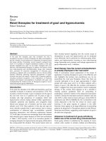
Figure 1A illustrates inhibition of TNF accumulation in
supernatants of human PBMC cultured for 20 h with 1 µg/
ml of LPS after a 2-h pretreatment with varying concentra-
tions of LMP-420. LMP-420 inhibits LPS-stimulated TNF
release in a dose-dependent manner with an IC
50
of
approximately 50 nM. At the highest concentration tested
(50 µM), LMP-420 inhibited 98.6% of TNF release com-
pared to a DMSO alone (0.5%) control. Since TNF
released from LPS-stimulated PBMC is derived mostly
from monocytes/macrophages, we wanted to determine if
LMP-420 could also inhibit TNF release from stimulated
lymphocytes. As shown in Figure 1B, LMP-420 also inhib-
ited TNF release from PBMC stimulated with either of two
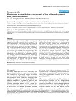
Inhibition of TNF by LMP-420Figure 1
Inhibition of TNF by LMP-420. A) Dose response of LMP-420 on human PBMC stimulated with LPS. PBMC were resuspended
to 3.75 × 10
6
total cells/ml in complete RPMI 1640 medium (containing 5% heat-inactivated human AB serum) and 0.4 ml of cell
suspension put into each well of a 48-well tissue culture plate. To each well was added 0.1 ml of media or media containing
LMP-420 (diluted from a stock solution of 338 mM in DMSO) to give the indicated final concentration. The cell cultures were
incubated for 2 h at 37°C in humidified 5% CO
2
and then 55 µl of media or LPS (S. typhosa, 1 µg/ml of media) was added to
each well. The cultures were incubated 20 h at 37°C, the contents of each well removed to a 5-ml polypropylene centrifuge
tube and centrifuged for 20 min at 400 g. The supernatants were removed to a fresh tube and frozen at -20°C until assayed by
solid phase ELISA (R & D Systems). B) Dose response of LMP-420 on human PBMC stimulated with anti-CD3 or SEB. PBMC
were resuspended to 3.75 × 10
6
total cells/ml in complete RPMI 1640 medium (containing 5% heat-inactivated human AB
serum) and 0.4 ml of cell suspension put into each well of a 48-well tissue culture plate. To each well was added 0.1 ml of
media or media containing LMP-420 (diluted from a stock solution of 338 mM in DMSO) to give the indicated final concentra-
tion. The cell cultures were incubated for 2 h at 37°C in humidified 5% CO
2
and then 55 µl of media or anti-CD3 (25 ng/ml
final concentration) or SEB (100 ng/ml final concentration) was added to each well. The cultures were incubated for 48 h at
37°C, the contents of each well removed to a 5-ml polypropylene centrifuge tube and centrifuged for 20 min at 400 g. The
supernatants were removed to a fresh tube and frozen at -20°C until assayed by solid phase ELISA (R & D Systems). C) RT-
PCR of samples prepared from LPS-stimulated human PBMC. PBMC (1 × 10
6
/well) were incubated for 2 h at 37°C with the
indicated concentration of LMP-420 and then stimulated for 3 h at 37°C with LPS (S. typhosa; 1 µg/ml). Cells were harvested,
total RNA extracted, cDNA prepared and RT-PCR performed for 30 cycles using primers for TNF-α and β-actin obtained
from R & D Systems.
0 0.01 0.1 1.0 10
C
BA
LMP-420 [
µ
M]
TNF-α
β
-actin
AIDS Research and Therapy 2006, 3:8 />Page 4 of 9
(page number not for citation purposes)
different lymphocyte-stimulating reagents, monoclonal
anti-CD3 antibody or SEB.
LMP-420 inhibits TNF at the transcriptional level
Inhibition of TNF release can occur at a number of differ-
ent points in stimulated cells. These might include initial
binding of the stimulating ligand, transcription of TNF
mRNA, translation of mRNA to protein, expression of
TNF on cell membranes, or the cleavage of TNF from the
cell membrane. Since LMP-420 inhibits TNF release from
PBMC stimulated with a variety of ligands other than LPS
(including IL-2, zymosan, Pansorbin; data not shown) we
assumed that inhibition was occurring at a post-stimulus
step. PBMC were treated for 2 h with different concentra-
tions of LMP-420, the cells stimulated an additional 3 h
with LPS, total mRNA isolated, cDNA prepared, and RT-
PCR performed using specific primers for human TNF. As
shown in Figure 1C, LMP-420 inhibits transcription of
mRNA for TNF in a dose-dependent manner, suggesting
that this is the mechanism by which it effects its inhibition
of TNF protein release.
LMP-420 is highly selective for TNF
In order to determine whether LMP-420 was for selective
for inhibition of TNF, PBMC were incubated with either
media or a single concentration of LMP-420 (1 µM; 20 ×
IC
50
for inhibition of TNF) for 2 h and then incubated
overnight with LPS (1 µg/ml). Supernatants were assayed
for released cytokines/chemokines using a Bio-Rad multi-
plex assay kit which measures 17 cytokines/chemokines
simultaneously. As shown in Table 1, LMP-420 potently
inhibits the release of TNF and MCP-1 (91 and 95%
respectively) with a lesser effect on the release of IL-1 and
IFN-γ (33 and 38% respectively). In contrast, there was no
significant effect on the levels of IL-6, IL-8, IL-10, G-CSF,
or MIP-1β. Although there were slight (~30% or less)
decreases in the levels of several other cytokines/chemok-
ines, the levels present were too low to ascribe significance
to these effects. Since these culture supernatants were
from overnight cultures we cannot at this time rule out the
possibility that the inhibition of MCP-1 is secondary to
the inhibition of TNF by LMP-420, since TNF can itself
stimulate release of MCP-1. This pattern of cytokine/
chemokine inhibition is, of course, only representative of
PBMC stimulated with a TLR4 ligand (LPS) which prefer-
entially stimulates monocytes/macrophages and a similar
pattern of cytokine/chemokine inhibition may not neces-
sarily be observed with other stimuli.
Inhibition of HIV-1 replication in human PBMC using LMP-
420
Because of LMP-420's ability to inhibit TNF release, both
from monocytes/macrophages and T lymphocytes, we
hypothesized that LMP-420 should inhibit the replication
of HIV-1 since HIV-1 can induce TNF and TNF can activate
HIV-1 viral LTR. Activated PBMC were incubated with var-
ying concentrations of LMP-420, ranging from 0- 50000
nM, at the same time that HIV was added and then incu-
bated for 7 days without further addition of LMP-420. As
shown in Figure 2, LMP-420 inhibits in a dose-dependent
manner the replication, as determined by HIV p24 anti-
gen release, of HIV in PBMC. The percent of p24 release,
compared to media controls, is 97%, 65%, 45%, 16% and
0.2% at LMP-420 concentrations of 5, 50, 500, 5000, and
Table 1: Selective inhibition of TNF-alpha by LMP-420
CYTOKINE LPS-Stimulated Release (pg/ml) Percent Inhibition
Media Alone + LMP-420 (1 µM)
IL-1β 15, 829 ± 75 10,670 ± 271 33
IL-2 24 ± 0 18 ± 1 25
IL-4 582 ± 54 419 ± 13 28
IL-5 7 ± 3 3 ± 1 57
IL-6 16,512 ± 190 17,249 ± 103 0
IL-7 26 ± 1 24 ± 1 8
IL-8 17,160 ± 221 17,575 ± 262 0
IL-10 1,963 ± 17 1,766 ± 62 10
IL-12 51 ± 3 76 ± 4 0
IL-13 44 ± 2 33 ± 0 25
IL-17 80 ± 1 56 ± 1 30
G-CSF 2,861 ± 98 4,794 ± 803 0
GM-CSF 93 ± 6 63 ± 4 32
MCP-1 2,714 ± 132 143 ± 0 95
MIP-1β 19,554 ± 63 19,127 ± 520 2
IFN-γ 424 ± 4 262 ± 1 38
TNF-α 7,628 ± 112 654 ± 7 91
AIDS Research and Therapy 2006, 3:8 />Page 5 of 9
(page number not for citation purposes)
50000 nM, respectively. Thus, at an LMP-420 concentra-
tion of 500 nM, HIV replication is inhibited by 55%. This
same concentration of LMP-420 would inhibit ~90% or
more of 24 h TNF release from LPS-stimulated PBMC (Fig-
ure 1A) and perhaps slightly less in cultures stimulated
with lymphocyte-specific stimuli (Figure 1B).
LMP-420 enhances the inhibitory effect of AZT on viral
replication
Our hypothesis was that the LMP-420 inhibition of HIV
replication shown in Figure 2 was due to the host cells'
inability to support that replication through TNF release
and subsequent activation of viral LTR in an autocrine or
paracrine fashion. Based on this postulated mechanism,
LMP-420 should synergize with antiviral agents, such as
AZT, which target the virus directly. As shown in Figure 3,
addition of suboptimal doses of 50 or 500 nM LMP-420
to a suboptimal dose of 10 nM AZT results in a further
decrease of HIV-1 p24 antigen release from 32% (AZT
alone) to 20% (AZT + 50 nM LMP-420) and 16% (AZT +
500 nM LMP-420) of controls. Addition of 50 or 500 nM
LMP-420 to 100 nM AZT resulted in a decrease of HIV-1
p24 production from 10% (AZT alone) to 6% (AZT + 50
nM LMP-420) and 4% (AZT + 500 nM LMP-420) of vehi-
cle control. Thus 500 nM LMP-420, a dose which by itself
results in a 55% inhibition of viral replication, used in
combination with AZT (10 nM or 100 nM) increases the
inhibition of viral replication by 50% and 60% respec-
tively when compared with AZT alone.
LMP-420 inhibits the replication of virulent M. Tb in
human AM
Since a compound which could inhibit the release of TNF
from both HIV and M. Tb would have added potential in
areas where both pathogens are endemic, we first exam-
ined the ability of LMP-420 to inhibit TNF release from M.
Tb-infected AM. AM derived from BAL were exposed to
varying concentrations of LMP-420 for 2 h prior to being
infected (MOI = 5) with M. Tb, the cells were cultured an
additional 24 h, and TNF levels in the supernatants deter-
mined. LMP-420 inhibited TNF release from M. Tb-
infected AM by 55, 68, and 90% at concentrations of 0.35,
3.5, and 35 µM respectively (data not shown). Since one
of us (S. Stenger) had previously shown that anti-TNF
antibody could block replication of virulent M. Tb [8], we
now sought to determine if LMP-420 could likewise
inhibit the replication of M. Tb in human AM. As shown
in Figure 4, addition of 10 µM LMP-420 to cultures of
human AM infected with a virulent strain of M. Tb
(H37Rv; MOI = 1; efficiency of infection = 36 ± 9%)
inhibits the replication of M. Tb by >80% over the subse-
quent 108 h of culture. The concentration of LMP-420
used (10 µM) would be expected to inhibit at least 70-
80% of the M. Tb-stimulated TNF release under the condi-
tions of these assays. Interestingly, the inhibition of repli-
cation by LMP-420 was even greater than that observed
using 10 µg/ml of an anti-TNF polyclonal antibody which
might suggest that LMP-420 inhibitory effects involve
more than just TNF inhibition.
LMP-420 is non-toxic to PBMC at the concentrations
tested
Inhibition of the replication of either HIV or M. Tb by
LMP-420 could arguably be the result of depletion of host
cells by LMP-420. We first tested LMP-420 for toxicity in a
MitoScan™ SMP assay looking at electron transfer with
NADH [12]. No toxicity was observed with LMP-420 at
concentrations up to 20 µM and the EC
50
for LMP-420-
mediated toxicity was 300 µM, a concentration that is
6000-fold greater than the EC
50
for inhibition of TNF (Fig-
ure 1A). In addition to examining LMP-420's effects on
mitochondrial function, three human lymphoid cell lines
(CEM, lymphoblastoid; THP-1, monocytic; K562, eryth-
roleukemic) were grown for 72 h in 50 µM LMP-420 with
no observed effects on cell proliferation, as measured by
[
3
H]-thymidine incorporation (data not shown). Further
confirmation of the fact that LMP-420 is non-toxic are
studies in which PBMC were stimulated for 72 h with 80
ng/ml of SEB in the presence or absence of either 2 µM or
0.4 µM LMP-420. Under these conditions LMP-420 inhib-
ited TNF release by ~80% and ~60% respectively but had
negligible effects on SEB-stimulated lymphocyte prolifer-
HIV-1
Ba-L
replication in PHA-stimulated human PBMC treated with indicated concentration of LMP-420Figure 2
HIV-1
Ba-L
replication in PHA-stimulated human PBMC treated
with indicated concentration of LMP-420. PBMC (10
6
/ml)
were infected with HIV-1
Ba-L
for 2 h, treated with LMP-420 at
the concentration indicated and incubated under the condi-
tions described in Methods. The supernatants were harvested
and stored at -80°C for HIV-1 p24 determinations as
described in Methods. Data represent the average ± SEM of
thee experiments using 3 different donors.
AIDS Research and Therapy 2006, 3:8 />Page 6 of 9
(page number not for citation purposes)
ation (~12% inhibition at 2 µM; 0% inhibition at 0.4 µM)
(data not shown). Cumulatively, these results suggest that
LMP-420's effects on either TNF release or the replication
of HIV-1 or M. Tb are not due to cellular toxicity.
Discussion
Co-infection with HIV and M. Tb is a major problem in
developing countries. In addition to difficulties associated
with simultaneous treatment of infections with two major
pathogens, data suggests that each of these pathogens can
enhance infection with the other. Numerous investiga-
tions have demonstrated that both HIV- and M. Tb-
infected cells produce TNF and that either autocrine or
paracrine produced TNF can enhance the replication of
both pathogens.
Although in vitro studies such as those presented here sug-
gest that targeting TNF to suppress replication of HIV and/
or M. Tb might be a logical approach to enhance existing
therapies, there are several reports of reactivation of latent
M. Tb in patients being treated with TNF antagonist ther-
apies [13,14]. The large majority of cases involving reacti-
vation of latent M. Tb in patients treated with TNF
antagonists appear to have occurred with infliximab
(Remicade
™
; humanized monoclonal antibody) with
~144 and ~35 cases of tuberculosis per 100,000 patients
occurring with infliximab and etanercept respectively
from January 1998- September 2002 [15]. In a recent
report of 12 cases of reactivation of latent M. Tb in
patients in California being treated with TNF antagonists,
11 of the patients were being treated with infliximab [13].
Since infliximab is capable of binding to the transmem-
brane form of TNF and can subsequently activate comple-
ment, it is possible that infliximab might injure or kill
TNF-expressing cells and thus function as a generalized
immunosuppressive. Indeed, infliximab treatment in
patients with Crohn's disease has been reported to induce
apoptosis of both monocytes and T lymphocytes [16,17].
Although reactivation of latent M. Tb has also occurred
with etanercept, it should be noted that a large proportion
of patients being treated with these TNF antagonist agents
are also being treated with other suppressive drugs such as
methotrexate or steroids. In the study noted above [13], 8
of the 12 patients experiencing reactivation of latent M. Tb
were being treated with prednisone, methotrexate or aza-
thioprine.
An advantage of a small-molecule inhibitor of TNF tran-
scription and release (such as LMP-420) over biologicals
such as infliximab or etanercept is that such a molecule
offers greater pharmacological control. The current TNF
antagonists are designed to neutralize circulating TNF and
are thus given at sufficiently high doses to maximize the
time between injections which is dictated by the pharma-
cological half-lives of the molecules. By their nature, these
antagonists neutralize essentially all of the circulating TNF
and are irreversible. Although a small molecule will likely
have a shorter half-life, this allows the intervention to be
stopped should the clinical situation warrant it. Further-
more, LMP-420, while a very potent inhibitor of TNF
release, inhibited only ~93% and 98% of TNF release
from LPS-stimulated PBMC at 0.5 and 5.0 µM respectively
(Figure 1A). The low level of TNF release which "escapes"
inhibition by LMP-420 may be sufficient to maintain
immune surveillance while LMP-420's inhibition of the
majority of released TNF may protect against TNF-related
pathogenesis. Nonetheless, since HIV+ patients are
already immunosuppressed by their disease, more exten-
sive testing, including clinical studies, will be necessary to
determine whether treatment with a TNF biosynthesis
inhibitor which is non-toxic and doesn't affect general cell
function, such as LMP-420, might also reactivate latent M.
Tb.
Our data confirms that LMP-420, an inhibitor of TNF
transcription and subsequent biosynthesis, is able to
inhibit the replication of both HIV-1 (Figure 2) and viru-
lent M. Tb (Figure 4) in primary cultures of human cells.
The doses at which LMP-420 inhibits replication of these
pathogens are doses which have not been found to be
toxic to either primary human leukocytes or human cell
lines. Furthermore, LMP-420 has also recently been dem-
onstrated to inhibit replication of M. Tb in human blood-
Effects of LMP-420 and/or zidovudine (AZT) on HIV-1 repli-cationFigure 3
Effects of LMP-420 and/or zidovudine (AZT) on HIV-1 repli-
cation. PBMC (10
6
/ml) were infected with HIV-1
Ba-L
for 2 h,
treated with LMP-420 and/or AZT at concentration indi-
cated, and incubated under condition described in Methods.
The supernatants were harvested and stored at -80°C for
HIV-1 p24 determinations as described in Methods. Data rep-
resent the average ± SEM of thee experiments using 3 differ-
ent donors.
AIDS Research and Therapy 2006, 3:8 />Page 7 of 9
(page number not for citation purposes)
derived dendritic cells [18]. Evidence doesn't suggest that
LMP-420 has any direct anti-viral (G. Cianciolo, unpub-
lished data) or anti-bacterial activity but rather, that it
inhibits replication of these two pathogens by inhibiting
the host cells' ability to support the pathogens' replica-
tion. In our studies we used an HIV strain (Ba-L) that is
known to be monocyte/macrophage tropic. Whether
LMP-420 would also affect T cell tropic strains of HIV
remains to be determined although HIV-infected T cells
also produce TNF [[19][20][21][22]] and LMP-420 inhib-
its TNF synthesis in T cells (Figure 1B). The dose at which
LMP-420 inhibits 50% of HIV replication in PBMC is at
least several-fold greater than the dose required to inhibit
50% of LPS-stimulated TNF release from PBMC. However,
HIV replication is measured after 7 days of culture and
LMP-420 was added only once, at the time of virus addi-
tion. Whether replenishing the LMP-420 during the 7 days
of culture would lower the effective concentration of LMP-
420 required for viral inhibition remains to be deter-
mined.
In 24-h cultures of LPS-stimulated PBMC (Table 1), 1.0
µM LMP-420 significantly inhibits the release of MCP-1
(95%) as well as TNF (91%). We have recently demon-
strated by RT-PCR (G. Cianciolo, data not shown) that
upregulation of mRNA for MCP-1 is completely blocked
in 1.0 µM LMP-420-treated PBMC after 3 h of LPS stimu-
lation. Nonetheless, we still cannot rule out the possibility
that the inhibition of MCP-1 is secondary to the inhibi-
tion of TNF since studies in human airway epithelial cells
have demonstrated strong TNF induction of MCP-1
mRNA within 1 h, the shortest time-period examined
[23]. However, LMP-420 has recently been shown to also
inhibit the upregulation of adhesion molecules (ICAM-1,
VCAM-1, CD40) on human brain-derived endothelial
cells activated by either TNF or lymphotoxin (LT; TNF-
beta) and to prevent the release of microparticles (a sign
of inflammation) from such activated cells [24]. Regard-
less of the mechanism of MCP-1 inhibition by LMP-420,
blockade of this chemokine is potentially advantageous
since MCP-1 has been demonstrated to enhance HIV rep-
lication [25]. Although the role of MCP-1 in the replica-
tion of M. Tb is less clear, a recent study demonstrated that
co-infection of macaques with simian immunodeficiency
virus (SIV) and Mycobacterium avium complex (MAC) sig-
nificantly increased levels of MCP-1 in both serum and tis-
sue samples [26].
Conclusion
The cell culture studies presented here confirm that strat-
egies designed to inhibit the release of pro-inflammatory
cytokines/chemokines, such as TNF and MCP-1, may be
beneficial in inhibiting the replication of HIV-1, M. Tb, or
both. The potential advantages of small-molecule, inex-
pensive, stable compounds over existing biologicals in
areas where both pathogens are endemic suggest that this
approach is deserving of further investigation.
Methods
Reagents
LMP-420, 2-NH
2
-6-Cl-9- [(5-dihydroxyboryl)-pentyl]
purine, a gift from LeukoMed, Inc. (Raleigh, NC), was
stored as a 10 mM stock solution in dimethylsulfoxide
(DMSO; Sigma-Aldrich, St. Louis, MO). Anti-CD3
(OKT3) antibody, staphylococcus enterotoxin B (SEB),
and lipopolysaccharide (LPS) were obtained from Ortho
Biotech (Bridgewater, NJ), Calbiochem (San Diego, CA),
and Sigma-Aldrich, respectively.
Separation and stimulation of PBMC
PBMC were separated from buffy-coated blood of healthy
donors by standard Ficoll-Hypaque gradient centrifuga-
tion procedures. PBMC were activated with 5 µg/ml of
phytohemagglutinin (PHA) plus 10 ng/ml of IL-2 in RPMI
1640 culture medium with L-glutamine (2 mM), supple-
mented with 20% fetal bovine serum and 100 U/ml pen-
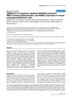
LMP-420 inhibits replication of M. Tb in human alveolar mac-rophages (AM)Figure 4
LMP-420 inhibits replication of M. Tb in human alveolar mac-
rophages (AM). AM were collected by bronchial-alveolar lav-
age from healthy volunteers and infected overnight in culture
with M. Tb (H37Rv; MOI = 1; efficiency of infection = 36 ±
9%). Cells (5 × 10
5
) were plated in 500 µl of medium in a 24-
well plate supplemented with nothing, rhuTNF (10 ng/ml),
anti-TNF (10 µg/ml) or LMP-420 (10 µM). The first time
point for plating to determine CFU was taken immediately
after the overnight infection. The second time point was
taken after 120 h (108 h after treatment was initiated). Data
represent the average ± SEM of thee experiments using 3 dif-
ferent donors.
AIDS Research and Therapy 2006, 3:8 />Page 8 of 9
(page number not for citation purposes)
icillin and 100 µg/ml streptomycin (growth medium) for
2- 3 days at 37°C in 5% CO
2
and 95% humidified air.
Infection of stimulated PBMC with HIV-1Ba-L
Activated PBMC were washed and infected with HIV-1
Ba-L
(NIH AIDS Research & Reference Reagent Program, Ger-
mantown, MD) at 500 × TCID
50
determined by previous
propagation in normal PBMC for 5- 7 days. HIV-1
Ba-L
-
infected PBMC were incubated at 37°C in 5% CO
2
and
95% humidified air for 2 h, mixed by gentle swirling every
20- 30 min, and centrifuged for 10 min at 200 g at room
temperature. Cell pellets were gently resuspended in
growth medium.
Treatment of infected cells with LMP-420 and/or AZT
HIV-1
Ba-L
infected PBMC (1 × 10
6
/ml/well) were treated
with or without drug (LMP-420 and/or AZT) with equiva-
lent amounts of DMSO in a 48-well plate at 37°C in 5%
CO
2
and 95% humidified air for 7 days. Compounds were
not cytotoxic at the concentrations used during the assays
and viability of infected and treated cells, as measured by
trypan blue, was >85%.
Titration of HIV-1 p24 antigen
Supernatants were collected at 7 days post-infection and
viral replication quantitated by measurement of the HIV-
1 specific core antigen, p24, by radioimmunoassay, using
the protocol provided by the supplier (Beckman Coulter,
Hialeah, FL).
Infection of AM with M. Tb
Human AM were obtained and infected with M. Tb as pre-
viously described [8]. Briefly, AM were obtained from the
discarded bronchoalveolar lavage (BAL) fluid of patients
who had undergone bronchoscopy for diagnostic pur-
poses after all identifiers had been removed. Purity
(>95%) of the cells was confirmed by α-naphthyl-acetate
esterase staining (Sigma-Aldrich) and flow cytometric
analysis (CD3 <1%, CD19, CD56, CD66, CD1 negative).
Viability was >96% as determined by trypan blue dye
exclusion.
AM were infected with single cell suspensions of M. Tb in
six-well culture plates at 1 × 10
6
cells/ml in a final volume
of 3 ml. After 4-h incubation at 37°C, extracellular bacte-
ria were removed by intensive rinsing with PBS. To quan-
titate mycobacterial growth, the adherent cells were
harvested by gentle scraping with a cell scraper and re-
plated at a concentration of 1 × 10
6
cells/ml in a 24-well
plate (final volume 500 µl) in complete medium without
antibiotics plus 10% human serum. Cell viability of
infected AM was determined by trypan blue exclusion and
was >99%.
For determination of CFU, infected cells were lysed with
0.3% saponin (Sigma-Aldrich) to release intracellular bac-
teria. At all time points an aliquot of un-lysed, infected
cells was harvested and counted, allowing an exact quan-
tification of cells as well as determination of cell viability.
Recovery of cells was >80% in all experiments, with cell
viability regularly >90%. CFUs of lysates were determined
as previously described [8].
Toxicity assays for LMP-420
General toxicity of LMP-420 was evaluated using the
MitoScan™ SMP assay (submitochondrial particle; Har-
vard Biosciences, Holliston, MA) per the manufacturer's
instructions. Verapamil-HCl (Sigma-Aldrich) was used as
an internal positive control (EC
50
~150 µM). Compounds
were prepared as a 338 mM stock (LMP-420) or a 285 mM
stock (verapamil-HCl) in DMSO and the highest concen-
tration of DMSO in the assays was 0.7%. MitoScan™ pro-
vides a rapid, homogeneous assay that correlates highly
with cell proliferation assays, cell viability assays and
other cytotoxicity or apoptosis endpoints such as MTT,
LDH and Alamar Blue assays. In addition to its effects on
mitochondrial activity, LMP-420 was tested for growth-
inhibitory effects on three human lymphoid cell lines
(CEM, THP-1, K-562; ATCC, Manassas, VA). To each of
duplicate sets of 6 wells of a 96-well cell culture plate was
added 5 × 10
3
of each cell type. One set was cultured for
72 h in media alone and the other was cultured in 50 µM
LMP-420. For the last 4 h of culture, 0.5 µCi of [
3
H]-thy-
midine (6.7 Ci/mmole; New England Nuclear-Perk-
inElmer Life and Analytical Sciences, Boston, MA) was
added to each well and the contents of each well harvested
onto glass fiber filters and incorporated radioactivity
determined by liquid scintillation spectrophotometry.
Abbreviations
AM, alveolar macrophage; BAL, bronchoalveolar lavage;
CFU, colony forming units; HIV, human immunodefi-
ciency virus type 1; LPS, lipopolysaccharides; LTR, long
terminal repeat; NO, nitric oxide; PBMC, peripheral blood
mononuclear cells; PHA, phytohemagglutinin; rhIL-2,
recombinant human interleukin-2; SEB, staphylococcus
enterotoxin B; TNF, tumor necrosis factor-alpha;
Competing interests
The author(s) declare that they have no competing inter-
ests.
Authors' contributions
SH, GJC, NKD and SS designed the experiments. SH, GJC,
WK and MBP performed the experiments. SH, GJC, WK,
NKD, NT, MBP and SS analyzed the data. SH, GJC, NKD,
WK, MBP, SS, NT, JWS and SP wrote, edited, and reviewed
the manuscript.
Publish with BioMed Central and every
scientist can read your work free of charge
"BioMed Central will be the most significant development for
disseminating the results of biomedical research in our lifetime."
Sir Paul Nurse, Cancer Research UK
Your research papers will be:
available free of charge to the entire biomedical community
peer reviewed and published immediately upon acceptance
cited in PubMed and archived on PubMed Central
yours — you keep the copyright
Submit your manuscript here:
/>BioMedcentral
AIDS Research and Therapy 2006, 3:8 />Page 9 of 9
(page number not for citation purposes)
Acknowledgements
The authors wish to acknowledge the assistance of Dr. Gregory Sem-
powski (Human Vaccine Institute) and Ms. Fang Wang (Department of
Pathology) of Duke University Medical Center in the performance of the
cytokine/chemokine multiplex assays and RT-PCR studies respectively, and
Mr. Elmer Dinglasan (Immunoparameters Laboratory) of All Children's
Hospital for his excellent technical assistance. This work was supported in
part by Eleanor Naylor Dana Charitable Trust. SS was funded by the Ger-
man Research Foundation (SFB 643), and MBP is an Alexander von Hum-
boldt fellow.
References
1. Toossi Z, Johnson JL, Kanost RA, Wu M, Luzze H, Peters P, Okwera
A, Joloba M, Mugyenyi P, Mugerwa RD, Aung H, Ellner JJ, Hirsch CS,
Uganda-Case Western Reserve Research Collaborations: Increased
replication of HIV-1 at sites of Mycobacterium tuberculosis
infection: potential mechanisms of viral activation. J Acquir
Immune Defic Syndr 2001, 28:1-8.
2. Zhang Y, Nakata K, Weiden M, Rom WN: Mycobacterium tubercu-
losis enhances human immunodeficiency virus-1 replication
by transcriptional activation at the long terminal repeat. J
Clin Invest 1995, 95:2324-2331.
3. Kitaura H, Ohara N, Kobayashi K, Yamada T: TNF-α-mediated
multiplication of human immunodeficiency virus in chroni-
cally infected monocytoid cells by mycobacterial infection.
APMIS 2001, 109:533-540.
4. Collins KR, Quiñones-Mateu ME, Toossi Z, Arts EJ: Impact of
tuberculosis on HIV-1 replication, diversity, and disease pro-
gression. AIDS Rev 2002, 4:165-176.
5. Collins KR, Quiñones-Mateu ME, Wu M, Luzze H, Johnson JL, Hirsch
C, Toossi Z, Arts EJ: Human immunodeficiency virus type I
(HIV-1) quasispecies at the sites of Mycobacterium tuberculo-
sis infection contribute to systemic HIV-1 heterogeneity. J
Virol 2002, 76:1697-1706.
6. Fantuzzi L, Spadaro F, Vallanti G, Canini I, Ramoni C, Vicenzi E,
Belardelli F, Poli G, Gessani S: Endogenous CCL2 (monocyte
chemotactic protein-1) modulates human immunodefi-
ciency virus type-1 replication and affects cytoskeleton
organization in human monocyte-derived macrophages.
Blood 2003, 102:2334-2337.
7. Zhao C, Papadopoulou B, Tremblay MJ: Leishmania infantum pro-
motes replication of HIV type 1 in human lymphoid tissue
cultured ex vivo by inducing secretion of the proinflamma-
tory cytokines TNF-α and IL-1α. J Immunol 2004,
172:3086-3093.
8. Engele M, Stößel E, Castiglione K, Schwerdtner N, Wagner M, Bölc-
skei P, Röllinghoff M, Stenger S: Induction of TNF in human alve-
olar macrophages as a potential evasion mechanism of
virulent Mycobacterium tuberculosis. J Immunol 2002,
168:1328-1337.
9. De SK, Devadas K, Notkins AL: Elevated levels of tumor necrosis
factor alpha (TNF-α) in human immunodeficiency virus type
1-transgenic mice: Prevention of death by antibody to TNF-
α. J Virol 2002, 76:11710-11714.
10. Sha BE, Valdez H, Gelman RS, Landay AL, Agosti J, Mitsuya R, Pollard
RB, Mildvan D, Namkung A, Ogata-Arakaki DM, Fox L, Estep S, Erice
A, Kilgo P, Walker RE, Bancroft L, Lederman MM: Effect of etaner-
cept (Enbrel) on interleukin 6, tumor necrosis factor alpha,
and markers of immune activation in HIV-infected subjects
receiving interleukin 2. AIDS Res Hum Retroviruses 2002,
18:661-665.
11. Wallis RS, Kyambadde P, Johnson JL, Horter L, Kittle R, Pohle M,
Ducar C, Millard M, Mayanja-Kizza H, Whalen C, Okwera A: A study
of the safety, immunology, virology, and microbiology of
adjunctive etanercept in HIV-1-associated tuberculosis. AIDS
2004, 18:257-264.
12. Knobeloch LM, Blondin GA, Harkin JM: Use of submitochondrial
particles for prediction of chemical toxicity in man. Bull Envi-
ron Contam Toxicol 1990, 44:661-668.
13. Centers for Disease Control and Prevention (CDC): Tuberculosis
associated with blocking agents against tumor necrosis fac-
tor-alpha ? California, 2002-2003. MMWR Morb Mortal Wkly Rep
2004, 53:683-686.
14. Keane J: TNF-blocking agents and tuberculosis: new drugs
illuminate an old topic. Rheumatology (Oxford) 2005, 44:714-720.
15. Wallis RS, Broder MS, Wong JY, Hanson ME, Beenhouwer DO:
Granulomatous infectious diseases associated with tumor
necrosis factor antagonists. Clin Infect Dis 2004, 38:1261-1265.
16. Lügering A, Schmidt M, Lügering N, Pauels HG, Domschke W, Kucha-
rzik T: Infliximab induces apoptosis in monocytes from
patients with chronic active Crohn's disease by using a cas-
pase-dependent pathway. Gastroenterology 2001, 121:1145-1157.
17. ten Hove T, van Montfrans C, Peppelenbosch MP, van Deventer SJH:
Infliximab treatment induces apoptosis of lamina propria T
lymphocytes in Crohn's disease. Gut 2002, 50:206-211.
18. Buettner M, Meinken C, Bastian M, Bhat R, Stössel E, Faller G, Cian-
ciolo G, Ficker J, Wagner M, Röllinghoff M, Stenger S: Inverse cor-
relation of maturity and antibacterial activity in human
dendritic cells. J Immunol 2005, 174:4203-4209.
19. Fujinaga K, Nakaya T, Ikuta K: Generation of endogenous tumor
necrosis factor-α in MOLT-4 cells during the acute replica-
tion phase of human immunodeficiency virus type 1 deter-
mines the subsequent latent infection. J Gen Virol 1998,
79:221-229.
20. Roux P, Alfieri C, Hrimech M, Cohen EA, Tanner JE: Activation of
transcription factors NF-κB and NF-IL-6 by human immuno-
deficiency virus type 1 protein R (Vpr) induces interleukin-8
expression. J Virol 2000, 74:4658-4665.
21. Lama J, Ware CF: Human immunodeficiency virus type 1 Nef
mediates sustained membrane expression of tumor necrosis
factor and the related cytokine LIGHT on activated T cells.
J Virol 2000, 74:9396-9402.
22. Jiménez JL, González-Nicolás J, Alvarez S, Fresno M, Muñoz-Fernán-
dez MA: Regulation of human immunodeficiency virus type 1
replication in human T lymphocytes by nitric oxide. J Virol
2001, 75:4655-4663.
23. Carpenter LR, Moy JN, Roebuck KA: Respiratory syncytial virus
and TNFalpha induction of chemokine gene expression
involves differential activation of Rel A and NF-kappaB I.
BMC Infect Dis 2002, 2:5.
24. Wassmer SC, Cianciolo GJ, Combes V, Grau GE: Inhibition of
endothelial activation: a new way to treat cerebral malaria?
PLoS Med 2005, 2:e245.
25. Vicenzi E, Alfano M, Ghezzi S, Gatti A, Veglia F, Lazzarin A, Sozzani S,
Mantovani A, Poli G: Divergent regulation of HIV-1 replication
in PBMC of infected individuals by CC chemokines: suppres-
sion by RANTES, MIP-1α, and MCP-3, and enhancement by
MCP-1. J Leukoc Biol 2000, 68:405-412.
26. Hendricks EE, Lin K-C, Boisvert K, Pauley D, Mansfield KG: Altera-
tions in expression of monocytes chemotactic protein-1 in
the simian immunodeficiency virus model of disseminated
Mycobacterium avium complex. J Infect Dis 2004, 189:1714-1720.