Báo cáo y học: "Psychogenic or neurogenic origin of agrammatism and foreign accent syndrome in a bipolar patient: a case report" pdf
Bạn đang xem bản rút gọn của tài liệu. Xem và tải ngay bản đầy đủ của tài liệu tại đây (552.21 KB, 7 trang )
BioMed Central
Page 1 of 7
(page number not for citation purposes)
Annals of General Psychiatry
Open Access
Case report
Psychogenic or neurogenic origin of agrammatism and foreign
accent syndrome in a bipolar patient: a case report
Stéphane Poulin
1
, Joël Macoir*
1,2
, Nancy Paquet
3
, Marion Fossard
1,2
and
Louis Gagnon
3
Address:
1
Centre de recherche Université Laval Robert-Giffard, 2601, rue de la Canardière Beauport (Qc), G1J 2G3, Canada,
2
Université Laval,
Faculté de médecine, Pavillon Ferdinand-Vandry, Québec, (Qc) G1K 7P4, Canada and
3
Service de médecine nucléaire, Hôtel-Dieu de Lévis, 143,
rue Wolfe, Lévis (Qc) G6V 3Z1, Canada
Email: Stéphane Poulin - ; Joël Macoir* - ;
Nancy Paquet - ; Marion Fossard - ; Louis Gagnon -
* Corresponding author
Abstract
Background: Foreign accent syndrome (FAS) is a rare speech disorder characterized by the
appearance of a new accent, different from the speaker's native language and perceived as foreign
by the speaker and the listener. In most of the reported cases, FAS follows stroke but has also been
found following traumatic brain injury, cerebral haemorrhage and multiple sclerosis. In very few
cases, FAS was reported in patients presenting with psychiatric disorders but the link between this
condition and FAS was confirmed in only one case.
Case presentation: In this report, we present the case of FG, a bipolar patient presenting with
language disorders characterized by a foreign accent and agrammatism, initially categorized as being
of psychogenic origin. The patient had an extensive neuropsychological and language evaluation as
well as brain imaging exams. In addition to FAS and agrammatism, FG also showed a working
memory deficit and executive dysfunction. Moreover, these clinical signs were related to altered
cerebral activity on an FDG-PET scan that showed diffuse hypometabolism in the frontal, parietal
and temporal lobes bilaterally as well as a focal deficit in the area of the anterior left temporal lobe.
When compared to the MRI, these deficits were related to asymmetric atrophy, which was
retrospectively seen in the left temporal and frontal opercular/insular region without a focal lesion.
Discussion: To our knowledge, FG is the first case of FAS imaged with an
18
F-FDG-PET scan. The
nature and type of neuropsychological and linguistic deficits, supported by neuroimaging data,
exclude a neurotoxic or neurodegenerative origin for this patient's clinical manifestations. For
similar reasons, a psychogenic etiology is also highly improbable.
Conclusion: To account for the FAS and agrammatism in FG, various explanations have been
ruled out. Because of the focal deficit seen on the brain imaging, involving the left insular and
anterior temporal cortex, two brain regions frequently involved in aphasic syndrome but also in
FAS, a cerebrovascular origin must be considered the best explanation to account for FG's language
deficits.
Published: 04 January 2007
Annals of General Psychiatry 2007, 6:1 doi:10.1186/1744-859X-6-1
Received: 06 October 2006
Accepted: 04 January 2007
This article is available from: />© 2007 Poulin et al; licensee BioMed Central Ltd.
This is an Open Access article distributed under the terms of the Creative Commons Attribution License ( />),
which permits unrestricted use, distribution, and reproduction in any medium, provided the original work is properly cited.
Annals of General Psychiatry 2007, 6:1 />Page 2 of 7
(page number not for citation purposes)
Background
Foreign accent syndrome (FAS) is a rare speech disorder
characterized by the appearance of a new accent, different
from the speaker's native language and perceived as for-
eign by the listener and, in most cases, by the speaker also.
Previous exposure to the new accent is not necessary for its
emergence. Different explanations of the functional origin
of FAS have been suggested, one of the more frequent
being impaired access to verbal-motor patterns or a mild
form of apraxia of speech. Clinical manifestations are het-
erogeneous among FAS patients but usually include seg-
mental (e.g., changes in vowel length and tenseness) and
prosodic (e.g., inappropriate word and sentence stress)
deficits. Very few cases (n = 40) have been reported since
the first descriptions of the syndrome by Pierre Marie in
1907 and Pick in 1919 [1]. It most often follows stroke
and then overlays the recovery phase of non-fluent apha-
sia though it could persist beyond this phase. FAS has also
been described following traumatic brain injury, cerebral
haemorrhage and multiple sclerosis [2-7]. In a recent
paper, Edwards, Patel and Pople [2] reviewed 35 case
studies of FAS and showed that in 26 of them, the syn-
drome resulted from cerebral infarct, while 9 were due to
head injury (6 cases), multiple sclerosis (2 cases) or epi-
sodes of psychosis (1 case). In 34% of these cases, FAS was
also associated with agrammatism. Agrammatism is a fre-
quent symptom of Broca's aphasia characterized by a def-
icit in sentence production. In spontaneous speech,
agrammatic patients speak non-fluently and produce tele-
graphic speech. They mainly use content words (nouns,
verbs, adjectives) and tend to omit or substitute function
words (prepositions, articles and auxiliaries) as well as
inflections or other grammatical morphemes. Among
reported FAS cases, few brain imaging studies have been
done and there is no consensus regarding the precise
region responsible for its occurrence. Neuroanatomically,
the vast majority of the lesions described were in the dom-
inant hemisphere and in most cases involved regions typ-
ically associated with Broca's aphasia. Subcortical
structures seem to be consistently affected [8].
Of all the reported FAS cases, very few (n = 3) cannot be
clearly related to a neurological event, revealed by clinical
exams and/or structural brain imaging studies [9-11]. For
two of these cases, a psychological origin was never sug-
gested although they were notable for psychiatric disor-
ders [9,10]. In the third case, given the normal functional
brain imaging results, conversion disorder was suggested
as an explanatory mechanism [11].
This paper reports the case of FG, a bipolar patient pre-
senting with language disorders characterized by a foreign
accent and agrammatism initially categorized as being of
psychogenic origin. Psychiatric patients do not commonly
manifest speech or language disorders except when
acutely psychotic. On formal language testing, schizo-
phrenic and bipolar patients may present semantic verbal
fluency and word finding difficulties when compared to
controls [12]. To our knowledge, there are no instances of
FAS and agrammatism previously reported in a bipolar
patient.
Case presentation
FG is a 74-year-old right-handed man. He has a grade
eleven education and worked as an auxiliary nurse. He
had suffered from a chronic bipolar disease since 1982,
with multiple episodes requiring many hospitalizations.
He came to our attention in July 2005 for acute exacerba-
tion of a bipolar disorder with suspected psychotic fea-
tures requiring inpatient treatment. At admission,
symptoms were compatible with manic exacerbation. Psy-
chotic features were not confirmed. Mental status exami-
nation revealed signs of his primary psychiatric disorder.
Moreover, a foreign accent, English-sounding, was noted.
FG had learned to deal with this long-lasting symptom so
he did not report it spontaneously. However, on explicit
questioning, he reported that this accent was socially
invalidating and completely impossible to control or
repress. FG is a native speaker of Quebec French but peo-
ple who met him thought he came from somewhere else,
most often Acadia (French-speaking areas of Eastern Can-
ada (New Brunswick, Prince Edward Island and Nova Sco-
tia) where the accent is markedly different from Quebec
French), France or an English-speaking foreign country.
Apart from this foreign accent, he also reported some Ger-
man- or Spanish-sounding words occasionally and spon-
taneously coming to his mind. No meaning is associated
with these words and the patient easily controls their
occurrence with no anxiety. Neurological examination
completed during the index hospitalization was unre-
markable except for an observed inability to turn back on
one foot (decomposition of the half-turn) when walking,
slight incoordination of the left arm on the cerebellar test-
ing, and slight micrographia. Snout and palmomental
primitive reflexes were also noted.
FG's past medical records reported the presence of this for-
eign accent in January 2003. It was first noticed at the psy-
chiatric outpatient clinic consultation, shortly after he was
discharged from the inpatient service, which was required
for manic exacerbation of his bipolar disorder in the fall
of 2002. The presence of agrammatism was also recorded
during the same period. Psychological factors were sus-
pected because of the patient's psychosocial background
(abuse by his father and emotional closeness to his
mother, who was English-speaking). Even though he was
exposed to English as a child, he never spoke or learned
this language. Without any other neurological symptoms,
his psychiatrist ascribed the foreign accent to a psycholog-
ical phenomenon operating at an unconscious level.
Annals of General Psychiatry 2007, 6:1 />Page 3 of 7
(page number not for citation purposes)
His neurological history is noteworthy for epilepsy
between the ages of 6 and 14 but without any other symp-
tomatic seizures thereafter. He also suffered from delirium
due to lithium intoxication 6 months before the onset of
the foreign accent. Finally he has been treated for an
essential tremor for many years and has neurosensory
hypoacusia. Otherwise, there was no prior history of
stroke, cranial trauma or encephalitis. When he developed
the language disorder, he was on stable doses of lithium,
valproate, quetiapine and perphenazine.
Although they appeared approximately 3 years earlier, the
functional origin of the FAS and agrammatism was
explored in FG through an extensive neuropsychological
and brain imaging study.
Neuropsychological evaluation
Neuropsychological testing showed no impairment in
tasks exploring orientation to time and space. FG's per-
formance was normal on the task exploring concentration
and selective attention [13]. He showed good face recog-
nition and presented no clinical signs of visual agnosia
[14]. There were no signs of unilateral neglect. Praxis abil-
ities were well preserved [15]. FG performed normally on
tasks exploring episodic memory. His performance was
within the normal range for the three recalls of the DMS-
48, a visual forced-choice recognition test [16], as well as
for the pictorial recognition memory test and the short
recognition memory test for faces [17]. The patient's short
term memory was normal in the visuospatial modality
(forward span = 5; backward span = 4) [18]as well as in
the verbal modality (forward digit span = 4; backward
digit span = 3; forward word span = 4). FG presented with
deficits on tests exploring working memory and executive
functions. He presented with a severe impairment on the
interference condition of the Brown-Peterson task [19], a
test that taps the ability to encode, maintain, and manip-
ulate information in working memory (see Table 1). His
performance on the Stroop Test [20], an instrument
designed to evaluate inhibition abilities (i.e. inhibition of
a habitual or more automatic response in favour of an
unusual one), was influenced by interference. He
obtained normal scores in the word reading and colour
naming but his performance was impaired in the colour-
word conditions. FG also showed abnormal performance
on the Trail Making [21], a test exploring mental flexibil-
ity (ability to manage more than one stimulus at a time
and to shift the course of an ongoing activity). He made
no mistakes but was slow on part A and his performance
was poor on Part B (alternated switching between num-
bers and letters). Finally, FG's performance was impaired
(2 SD below the normal range) on the D-Kefs Tower Test
[22], a complex task that measures the executive functions
of spatial planning, rule learning, and inhibition of
impulsive responding.
Language evaluation
With regard to language, speech output was fluent and
well articulated, with no signs of word-finding difficulties.
The patient however presented with mild agrammatism.
There were no phonemic or verbal paraphasias but speech
was sometimes telegraphic with omissions of function
Table 1: Performance of FG and norms (mean and standard deviation or range) on neuropsychological and language tests
Test FG's score Norm
Working memory and executive functions
- Brown-Peterson test
- no interference 100% 98.33% (4.47)
- mean of interference scores 42%* 97.22% (4.46)
- Stroop Test
- Color name reading 74 sec. 48.5 sec (25–86)
- Color naming 105 sec. 69.4 sec (46–123)
- Interference 249 sec.* 142.4 sec. (88–204)
- Trail making test
- Part A 61 sec.* 41.3 sec. (15)
- Part B 253 sec.* 111.4 sec. (72.2)
Language
- Picture naming (DO80) 72/80 74.9 (2.94)
- Letter fluency (PENO) 5* 45.46 (16.4)
- Category fluency (PENO) 14* 47.85 (9.8)
- Pyramids and Palm Trees Test 47/52 49.4 (1.74)
- Token test 29/36 29–36
- Spoken word/sentence-to-picture matching
(PENO)
44/47 44.6 (2.19)
- Written word/sentence-to-picture matching
(PENO)
12/12 10.81 (.81)
* Indicates a score below the norm or out of the normal range
Annals of General Psychiatry 2007, 6:1 />Page 4 of 7
(page number not for citation purposes)
words and grammatical bound morphemes as well as
impoverished syntactic structure. Auditory and visuo-ver-
bal input components were largely preserved. Compre-
hension abilities at the lexical-semantic level [23] as well
as at the syntactic-semantic level [15,24] were normal (see
Table 1). Reading and immediate and delayed repetition
were flawless for both words and nonwords. Written
spelling of nonwords was flawless but the patient's per-
formance on word writing to dictation was canonical of
surface agraphia with exclusive production of phonologi-
cal plausible errors and performance affected by ortho-
graphic regularity and lexical frequency. However, the
patient did not completely master the written language so
that these results cannot be interpreted as actual deficits.
FG's performance was normal in confrontation naming
[25] but he showed difficulties in letter and semantic cat-
egory fluency tasks [15] (see Table 1), a performance that
could be attributed to the deficit in executive functioning.
FG showed many characteristics usually reported for FAS.
There were no signs of dysarthria (no slow, slurred, grop-
ing or laboured articulation) or apraxia of speech (no dys-
fluency and no problems with phoneme sequencing) but
acoustic analysis performed on speech samples recorded
in Digital Audio Tape showed the presence of abnormali-
ties at the segmental and suprasegmental levels. Unfortu-
nately, we had no premorbid recording of the patient's
speech. However, FG himself as well as one of his close
friends, who has known him for over 30 years, confirmed
that he never had this particular strange accent before its
sudden appearance in January 2003.
Neuroimaging
Neuroimaging studies were performed while the patient
was in euthymic condition. A magnetic resonance imag-
ing (MRI) study including sagittal FLAIR and T2-weighted
sequences and axial FLAIR, proton density, T1 and T2-
weighted sequences was performed on December 8, 2005
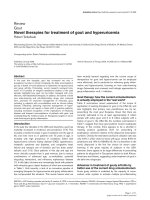
using the standard protocol. The first interpretation was
normal except for slight diffuse cerebral atrophy consid-
ered normal for his age (see Figure 1: serie 3 31/10 = axial
T2-weighted sequence showing diffuse cortical atrophy
predominating at the left sylvian fissure).
An
18
F-fluorodeoxyglucose brain positron emission tom-
ography was obtained with a dual-head coincidence cam-
era (Vertex MCD-AC, Phillips). After a 30-minute rest, 111
MBq
18
F-FDG were injected in a veinous catheter. There
was another 30-minute rest before starting the acquisition
(64 × 64 × 16 matrix, 64 steps, mean of 25 seconds/step
with decay correction). Measured attenuation and scatter
correction were applied to the iterative reconstruction
method.
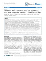
The reconstructed images showed diffuse hypometabo-
lism in the frontal, parietal and temporal lobes bilaterally
whereas the cerebellum, occipital lobe and subcortical
structures were spared. There was also a focal deficit in the
area of the anterior left temporal lobe with prominence of
the sylvian sulcus (see Figure 2). When compared to the
MRI, these deficits were related to asymmetric atrophy,
which was retrospectively seen in the left temporal and
frontal opercular/insular region without a focal lesion.
Discussion
We have reported the case of FG, a bipolar patient who
presented with a sudden onset of FAS and agrammatism.
He also showed a working memory deficit and executive
dysfunction. These clinical signs were related to altered
cerebral activity on the FDG-PET scan.
In FG, the FDG-PET scan is characterized by more diffuse
hypometabolism and by marked hypometabolism in the
area of the left insular and anterior temporal cortex. Func-
tional neuroimaging revealed focal deficit signs while the
MRI structural neuroimaging was initially considered a
normal variant for FG's age. The MRI scan showed slight
diffuse cerebral atrophy and an absence of indirect signs
of vascular pathology such as hyper intense signals on T2-
weighted images elsewhere in the brain. Retrospective
analysis of the MRI scan showed the same asymmetry as
noted on the PET scan, albeit less obviously. To our
Magnetic resonance imagingFigure 1
Magnetic resonance imaging. Axial T2-weighted
sequence showing diffuse cortical atrophy predominating at
the left sylvian fissure.
Annals of General Psychiatry 2007, 6:1 />Page 5 of 7
(page number not for citation purposes)
knowledge, FG is the first case of FAS imaged with an
18
F-
FDG-PET scan.
These structural and functional neuroimaging characteris-
tics differ substantially from what was previously reported
for bipolar disorder patients as a group. In fact, structural
neuroimaging studies do not typically show overall brain
volume loss but specific regional cerebral volume varia-
tions. Compared to controls, volume reductions in the
subgenual cortex and cerebellar vermis, associated with
enlargement in the striatum and amygdala, are usually
noted in bipolar patients [26]. Unlike FG, no previous
study showed insular cortex or anterior temporal cortex
reduction; on the contrary, one showed an increase in the
left insular/frontoparietal operculum cortex [27].
Despite variations in approaches (PET, SPECT, fMRI), par-
adigms used (at rest vs. while completing cognitive tasks),
mood states studied (depressive, manic, euthymic) and
treatment status (on mood stabilizers or not), converging
results have been reported on functional neuroimaging of
bipolar disorders [26]. Decreased metabolism and per-
fusion in the prefrontal cortex and particularly in the sub-
genual portion of the cingulated gyrus and striatum are
observed during depressive phases in bipolar patients.
Conversely, increased orbitofrontal cortex and cingulate
gyrus activity along with their related subcortical struc-
tures including the striatum and thalamus is reported in
manic states. Only one functional imaging study of
euthymic bipolar patients at rest is reported in the litera-
ture. In that study, a state-dependent activation of the
anterior part of the temporal lobe was observed for
depressive/dysphoric and manic states. While euthymic,
no altered temporal lobe activity was seen [28]. As a
whole, these results do not indicate that FG's bipolar sta-
tus may explain the altered functional imaging results. In
this patient, it is more likely that the language disorders
(FAS and agrammatism) are the external manifestation of
the marked hypometabolism of the left insular and ante-
rior temporal cortices.
The insula is frequently involved in major aphasic syn-
dromes and especially in Broca's aphasia. This type of
aphasia is caused by large lesions that damage the poste-
rior lateral frontal lobe, including the operculum, anterior
superior insula, anterior parietal lobe, and the white mat-
ter deep inside these structures. Lesions of the insula are
associated with impairments in speech production and
more specifically with articulatory planning deficits (i.e.,
apraxia of speech) [29]. This finding has received further
support from neuropsychological [29] and neuroimaging
studies [30]. The insula is also involved in sentence
processing (comprehension and production). Moreover,
patients with a lesion restricted to Broca's area usually did
not produce agrammatic speech [31]. Larger lesions of the
frontal and parietal opercula and the insula were required.
A previous case of FAS showed perfusion deficits on a
SPECT study in the regions of the left frontal motor cortex
extending to the insula and subcortical structures in addi-
tion to the left anterior temporal lobe [4].
Executive dysfunction could also represent a clinical man-
ifestation of the altered metabolism of the left insular cor-
tex. Executive functions represent several higher level
cognitive processes enabling adaptation to new or com-
plex situations. Traditionally considered abilities relying
on frontal lobes, the neural networks that underlie execu-
tive functions are now largely identified though not com-
pletely elucidated. They are probably specific, with each
recruiting various cortical areas of the brain, not only in
the frontal lobes but also in the parietal and temporal
lobes as well as the cerebellum [32,33]. Subcortical struc-
tures play a critical role in executive functions. Insular cor-
tex neural activity assessed by functional imaging was
correlated with deficits in executive functions in several
studies including normal [34-36] and clinical samples
[37,38]. FG is treated with a typical antipsychotic (per-
phenazine) and shows clinical signs of parkinsonism
(decomposition of the half-turn, micrographia) so that
executive dysfunction could be related to basal ganglia
impairment.
To account for FG's deficit, a possible neurotoxic origin
must be considered. With respect to the previous lithium
intoxication, delayed onset manifestation has never been
reported. Except for dysarthria, speech disorders are rare
in lithium neurotoxicity. Antipsychotics have never
shown consistent alteration of language and cognitive
functioning in clinical populations [39].
Because of the acute onset and stability of the symptoms
in FG, the presence of a neurodegenerative process is
highly improbable but should also be examined. Except
for cognitive function deficits, none of the DSM-IV-TR
[40] criteria for the diagnosis of dementia was met in FG.
He showed no episodic memory problems, no agnosia,
Brain positron emission tomographyFigure 2
Brain positron emission tomography.
18
F-FDG PET
showing focal deficit in the area of the anterior left temporal
lobe with proheminence of the sylvian sulcus.
Annals of General Psychiatry 2007, 6:1 />Page 6 of 7
(page number not for citation purposes)
no apraxia, and his language difficulties did not corre-
spond to what is usually encountered (i.e., word-finding
and comprehension problems) in the early phase of
major forms of dementia. Moreover, the patient's cogni-
tive impairment had no impact on his social participation
and activities of daily living. FG presented with abnormal-
ities in the left anterior temporal lobe, a cortical localiza-
tion compatible with frontotemporal dementia (FTD).
However, except for executive function deficits, the
patient's clinical profile did not meet the clinical diagno-
sis features of FTD [41]. Apart from episodes of decom-
pensation, he presented neither character change nor
disordered social conduct, the dominant features at the
onset of and throughout the course of FTD. With respect
to language, he did not show any of the supportive diag-
nosis features of FTD (aspontaneity, echolalia, persevera-
tion, etc). Finally, progressive nonfluent aphasia (PNFA)
is a clinical syndrome associated with FTD [41] in which
agrammatism is sometimes observed [42]. However, FG
did not present any of the PNFA core diagnostic features
(nonfluent spontaneous speech, phonemic paraphasias,
anomia). Moreover, FAS has never been reported in
PNFA, as in any other forms of dementia.
In FG's case, conversion disorder must be excluded as the
primary mechanism responsible for the foreign accent
and agrammatism. Speech disorders of conversion origin
typically present as dysarthria, mutism, aphonia or stut-
tering [43]. Foreign accent and agrammatism would be a
rather unusual presentation of conversion disorder. Fur-
thermore, FG had never heard of or known anyone suffer-
ing from this disorder before it appeared in 2003, making
unconscious mimicry almost impossible. Nevertheless,
conversion disorder may superimpose on complex neuro-
logical symptoms, giving them chronic course [44]. There-
fore, it cannot be totally ruled out that conversion
mechanisms contribute to the foreign accent and agram-
matism in FG. A typical chronic FAS has recently been
ascribed to conversion disorder [11]. For now, because of
the way the DSM-IV-TR criteria are formulated, there is no
way to convincingly exclude a conversion disorder contri-
bution to a neurological symptom of unknown origin
[40]. In fact, conversion disorder is the only DSM-IV-TR
diagnosis that includes in its definition criteria a putative
causative mechanism which, in any event, can never be
ruled out. Therefore, because of the absence of clear and
specific criteria, a diagnosis of conversion disorder is auto-
matically considered when there is no alternative hypoth-
esis. For the moment, functional brain imaging as well as
electrophysiological studies cannot help either. These
types of studies have shown alterations of specific brain
regions in neurological dysfunction of conversion origin
[45,46].
Conclusion
Initially attributed to a psychogenic phenomenon, the ori-
gin of FAS and agrammatism in FG is now clearer. Differ-
ent functional origins were considered and have been
largely ruled out. Neither FAS nor agrammatism have
been reported in bipolar disorder patients. Moreover,
neuroradiological correlates in these patients usually dif-
fer from what was observed in FG. The nature and type of
clinical manifestations also exclude a neurotoxic or neu-
rodegenerative origin for FG's cognitive symptoms. For
similar reasons, a conversion disorder also appears to be a
highly improbable etiology even though a possible contri-
bution cannot be totally excluded. Because of the focal
deficit seen on the brain imaging, involving the left insu-
lar and anterior temporal cortex, two brain regions fre-
quently involved in aphasic syndrome but also in FAS, a
cerebrovascular origin must be considered the best expla-
nation to account for FG's language deficits. We therefore
conclude that in this patient, as in few other reported
cases, the FAS is associated with agrammatism as a direct
consequence of a cerebral infarct.
Competing interests
The author(s) declare that they have no competing inter-
ests.
Authors' contributions
SP contributed to the patient's care and referred him to JM
for the clinical study. JM was the study coordinator. SP
and JM reviewed the existing literature and drafted the
manuscript. MF and NP reviewed the manuscript and con-
tributed to the writing. NP and LG conducted the brain
imaging exams and interpreted the data. All the authors
approved the final manuscript.
Acknowledgements
The authors gratefully acknowledge the cooperation of the patient
described in this case report, from whom written consent was obtained for
the publication of this study. JM would like to thank Dr Jean-Luc Nespou-
lous for his help in the literature review about FAS.
References
1. Pick A: Uber anerungen des sprach-characters als begleiter-
scheinung aphasischer strorungen. Zeitschrift fuer de gesamte
Neurun Psychologie 1919, 54:230-241.
2. Edwards RJ, Patel NK, Pople IK: Foreign accent following brain
injury: syndrome or epiphenomenon? Eur Neurol 2005,
53(2):87-91.
3. Munson PD, Heilman B: Foreign accent syndrome: anatomic,
pathophysiologic and psychosocial considerations. S D J Med
2005, 58(5):187-189.
4. Marien P, Verhoeven J, Engelborghs S, Rooker S, Pickut BA, De Deyn
PP: A role for the cerebellum in motor speech planning: Evi-
dence from foreign accent syndrome. Clin Neurol Neurosurg
2005, 105(5):518-522.
5. Fridriksson J, Ryalls J, Rorden C, Morgan PS, George MS, Baylis GC:
Brain damage and cortical compensation in foreign accent
syndrome. Neurocase 2005, 11(5):319-324.
6. Van Borsel J, Janssens L, Santens P: Foreign accent syndrome: an
organic disorder? J Commun Disord 2005, 38(6):421-429.
Publish with BioMed Central and every
scientist can read your work free of charge
"BioMed Central will be the most significant development for
disseminating the results of biomedical research in our lifetime."
Sir Paul Nurse, Cancer Research UK
Your research papers will be:
available free of charge to the entire biomedical community
peer reviewed and published immediately upon acceptance
cited in PubMed and archived on PubMed Central
yours — you keep the copyright
Submit your manuscript here:
/>BioMedcentral
Annals of General Psychiatry 2007, 6:1 />Page 7 of 7
(page number not for citation purposes)
7. Ryalls J, Whiteside J: An atypical case of Foreign Accent Syn-
drome. Clin Linguist Phon 2006, 20(2–3):157-162.
8. Carbary TJ, Patterson JP, Snyder PJ: Foreign Accent Syndrome
following a catastrophic second injury: MRI correlates, lin-
guistic and voice pattern analyses. Brain Cogn 2000, 43(1–
3):78-85.
9. Reeves RR, Norton JW: Foreign accent-like syndrome during
psychotic exacerbations. Neuropsychiatry Neuropsychol Behav Neu-
rol 2001, 14(2):135-138.
10. Coelho C, Robb M: Acoustic Analysis of Foreign Accent Syn-
drome: An Examination of Three Explanatory Models. Jour-
nal of Medical Speech-Language Pathology 2001, 9:227-242.
11. Verhoeven J, Marien P, Engelborghs S, D'Haenen H, De Deyn P: A
foreign speech accent in a case of conversion disorder. Behav
Neurol 2005, 16(4):225-232.
12. Dickerson F, Boronow JJ, Stallings C, Origoni AE, Cole SK, Yolken
RH: Cognitive functioning in schizophrenia and bipolar disor-
der: comparison of performance on the Repeatable Battery
for the Assessment of Neuropsychological Status. Psychiatry
Res 2004, 129(1):45-53.
13. Smith A: Symbol digit modalities test. Los Angeles: Western Psy-
chological Services; 1991.
14. Riddoch MJ, Humphreys GW: Birmingham Object Recognition
Battery. Hove: Lawrence Erlbaum Associates; 1993.
15. Joanette Y, Ska B, Belleville S, Lecours AR, Peretz I, Poissant A: Éval-
uation neuropsychologique dans la démence de type Alzhe-
imer: un compromis optimal. L'année gérontologique 1995,
3:69-83.
16. Barbeau E, Tramoni E, Joubert S, Mancini J, Ceccaldi M, Poncet M:
Evaluation de la mémoire de reconnaissance visuelle : nor-
malisation d'une nouvelle épreuve en choix forcé (DMS48)
et utilité en neuropsychologie clinique. In L'évaluation des trou-
bles de la mémoire Edited by: Van der Linden M. Marseille: Solal;
2004:85-101.
17. Warrington EK: The Camden Memory Test. Hove: Psychology
Press; 1996.
18. Milner B: Interhemispheric differences and psychological
processes. British Medical Bulletin 1971, 27:273-7.
19. Brown J: Some tests of the decay theory of immediate mem-
ory.
Q J Exp Psycho1 1958, 10:12-21.
20. Golden JC: Stroop color and word test. Chicago: Stoelting Co;
1978.
21. Reitan R, Wolfson D: The Halstead-Reitan Neuropsychological
Test Battery: Theory and clinical interpretation. Tucson:
Neuropsychology Press; 1993.
22. Delis DC, Kaplan E, Kramer JH: Delis-Kaplan executive function
system. London: The Psychological Corporation; 2001.
23. Howard D, Patterson K: The pyramids and palm trees test: A
test for semantic access from words and pictures. Bury St
Edmunds: Thames Valley Test Company; 1992.
24. De Renzi E, Faglioni P: Normative data and screening power of
a shortened version of the Token Test. Cortex 1978, 14:41-49.
25. Deloche G, Hannequin D: Test de dénomination orale
d'images-DO 80. Paris: Éditions du Centre de Psychologie Appli-
quée; 1997.
26. Strakowski SM, Delbello MP, Adler CM: The functional neuro-
anatomy of bipolar disorder: a review of neuroimaging find-
ings. Mol Psychiatry 2005, 10(1):105-116.
27. Lochhead RA, Parsey RV, Oquendo MA, Mann JJ: Regional brain
gray matter volume differences in patients with bipolar dis-
order as assessed by optimized voxel-based morphometry.
Biol Psychiatry 2004, 55(12):1154-1162.
28. Gyulai L, Alavi A, Broich K, Reilley J, Ball WB, Whybrow PC: I-123
iofetamine single-photon computed emission tomography in
rapid cycling bipolar disorder: a clinical study. Biol Psychiatry
1997, 41(2):152-161.
29. Dronkers NF: A new brain region for coordinating speech
articulation. Nature 1996, 384(6605):159-161.
30. Blank SC, Scott SK, Murphy K, Warburton E, Wise RJ: Speech pro-
duction: Wernicke, Broca and beyond. Brain 2002, 25(Pt
8):11829-1838.
31. Mohr JP, Pessin MS, Finkelstein S, Funkenstein HH, Duncan GW,
Davis KR: Broca aphasia: pathologic and clinical. Neurology
1978, 28(4):311-324.
32. Fassbender C, Murphy K, Foxe JJ, Wylie GR, Javitt DC, Robertson IH,
Garavan H: A topography of executive functions and their
interactions revealed by functional magnetic resonance
imaging. Brain Res Cogn Brain Res 2004, 20(2):
132-143.
33. Collette F, Hogge M, Salmon E, Van der Linden M: Exploration of
the neural substrates of executive functioning by functional
neuroimaging. Neuroscience 2006, 139(1):209-221.
34. Taylor SF, Kornblum S, Lauber EJ, Minoshima S, Koeppe RA: Isola-
tion of specific interference processing in the Stroop task:
PET activation studies. Neuroimage 1997, 6(2):81-92.
35. Dove A, Pollmann S, Schubert T, Wiggins CJ, von Cramon DY: Pre-
frontal cortex activation in task switching: an event-related
fMRI study. Brain Res Cogn Brain Res 2000, 9(1):103-109.
36. Menon V, Adleman NE, White CD, Glover GH, Reiss AL: Error-
related brain activation during a Go/NoGo response inhibi-
tion task. Hum Brain Mapp 2001, 12(3):131-143.
37. Peinemann A, Schuller S, Pohl C, Jahn T, Weindl A, Kassubek J: Exec-
utive dysfunction in early stages of Huntington's disease is
associated with striatal and insular atrophy: a neuropsycho-
logical and voxel-based morphometric study. J Neurol Sci 2005,
239(1):11-19.
38. Schmitz N, Rubia K, Daly E, Smith A, Williams S, Murphy DG: Neural
correlates of executive function in autistic spectrum disor-
ders. Biol Psychiatry 2006, 59(1):7-16.
39. Daban C, Amado I, Bourdel MC, Loo H, Olie JP, Poirier MF, Krebs
MO: Cognitive dysfunctions in medicated and unmedicated
patients with recent-onset schizophrenia. J Psychiatr Res 2005,
39(4):391-398.
40. American Psychiatric Association Task Force on DSM-IV: Diagnostic
and statistical manual of mental disorders : DSM-IV-TR Washington:
American Psychiatric Association; 2000.
41. Neary D, Snowden JS, Gustafson L, Passant U, Stuss D, Black S, Freed-
man M, Kertesz A, Robert PH, Albert M, Boone K, Miller BL, Cum-
mings J, Benson DF: Frontotemporal lobar degeneration: a
consensus on clinical diagnostic criteria. Neurology 1998,
51:1546-1554.
42. Grossman M, Ash S: Primary Progressive Aphasia: A Review.
Neurocase 2004, 10(1):3-18.
43. Lempert T, Dieterich M, Huppert D, Brandt T: Psychogenic disor-
ders in neurology: frequency and clinical spectrum. Acta Neu-
rol Scand 1990, 82(5):335-340.
44. Andrade C, Singh NM, Bhakta SG: Simultaneous true seizures
and pseudoseizures. J Clin Psychiatry 2006, 67(4):673.
45. Vuilleumier P: Hysterical conversion and brain function. Prog
Brain Res 2005, 150:309-329.
46. Black DN, Seritan AL, Taber KH, Hurley RA: Conversion hysteria:
lessons from functional imaging. J Neuropsychiatry Clin Neurosci
2004, 16(3):245-251.