Báo cáo y học: "Gender-specific effects of HIV protease inhibitors on body mass in mice" ppt
Bạn đang xem bản rút gọn của tài liệu. Xem và tải ngay bản đầy đủ của tài liệu tại đây (716.43 KB, 8 trang )
BioMed Central
Page 1 of 8
(page number not for citation purposes)
AIDS Research and Therapy
Open Access
Research
Gender-specific effects of HIV protease inhibitors on body mass in
mice
Melinda E Wilson*, Kimberly F Allred, Elizabeth M Kordik, Deana K Jasper,
Amanda N Rosewell and Anthony J Bisotti
Address: Department of Physiology, College of Medicine, University of Kentucky, Lexington, KY 40536, USA
Email: Melinda E Wilson* - ; Kimberly F Allred - ; Elizabeth M Kordik - ;
Deana K Jasper - ; Amanda N Rosewell - ; Anthony J Bisotti -
* Corresponding author
Abstract
Protease inhibitors, as part of highly active anti-retroviral therapy (HAART), have significantly
increased the lifespan of human immunodeficiency virus (HIV) infected patients. Several deleterious
side effects including dyslipidemia and lipodystrophy, however, have been observed with HAART.
Women are at a higher risk of developing adipose tissue alterations and these alterations have
different characteristics as compared to men. We have previously demonstrated that in mice the
HIV protease inhibitor, ritonavir, caused a reduction in weight gain in females, but had no effect on
male mice. In the present study, we examined the potential causes of this difference in weight gain.
Low-density lipoprotein receptor (LDL-R) null mice or wild-type C57BL/6 mice, were administered
15 μg/ml ritonavir or vehicle (0.01% ethanol) in the drinking water for 6 weeks. The percent of
total body weight gained during the treatment period was measured and confirmed that female
LDL-R gained significantly less weight with ritonavir treatment than males. In wild type mice,
however, there was no effect of ritonavir treatment in either sex. Despite the weight loss in LDL-
R null mice, ritonavir increased food intake, but no difference was observed in gonadal fat weight.
Serum leptin levels were significantly lower in females. Ritonavir further suppressed leptin levels in
(p < 0.05). Ritonavir did not alter serum adiponectin levels in either gender. To determine the
source of these differences, female mice were ovariectomized remove the gonadal sex hormones.
Ovariectomy prevented the weight loss induced by ritonavir (p < 0.05). Furthermore, leptin levels
were no longer suppressed by ritonavir (p < 0.05). This study demonstrates that gonadal factors in
females influence the hormonal control of weight gain changes induced by HIV protease inhibitors
in an environment of elevated cholesterol.
Background
The use of highly active anti-retroviral therapy (HAART)
has dramatically increased the lifespan of individuals
infected with the human immunodeficiency virus (HIV).
HAART often includes a cocktail of nucleoside reverse
transcriptase inhibitors and protease inhibitors that pre-
vent virus replication and assembly. While effective in
reducing the progression of AIDS, significant side effects
have been observed with long-term use of protease inhib-
itors [1-3]. HIV protease inhibitors have been associated
with an increase in atherosclerosis, dyslipidemia and lipo-
dystrophy. Adipose tissue alterations associated with pro-
Published: 1 May 2007
AIDS Research and Therapy 2007, 4:8 doi:10.1186/1742-6405-4-8
Received: 21 November 2006
Accepted: 1 May 2007
This article is available from: />© 2007 Wilson et al; licensee BioMed Central Ltd.
This is an Open Access article distributed under the terms of the Creative Commons Attribution License ( />),
which permits unrestricted use, distribution, and reproduction in any medium, provided the original work is properly cited.
AIDS Research and Therapy 2007, 4:8 />Page 2 of 8
(page number not for citation purposes)
tease inhibitor use include a loss of total body fat, with an
increase in fat deposition in the abdomen and in the dor-
socervical region leading to "buffalo humps" [4-6]. This
pattern of fat distribution is often associated with the
complex of symptoms including insulin resistance, hyper-
tension and dyslipidemia referred to as metabolic syn-
drome [7].
Gender differences have been observed in the incidence as
well as the severity of these adipose tissue alterations, with
women having a higher rate of reported disturbances [8].
The adipose tissue alterations observed were complex and
result in increased abdominal and breast accumulation
with reduced peripheral fat. These differences were not
due to age or the severity of the disease and are hypothe-
sized to be hormonal in nature.
Several adipose tissue-derived hormones play a role in
weight gain, obesity and are involved in the development
of metabolic syndrome [9-11]. Leptin plays a crucial role
for regulating weight gain by controlling fat mass. Leptin
levels are positively correlated with body mass index [12].
Additionally, leptin has been shown to reverse the dyslip-
idemia and lipodystrophy caused by HIV protease inhibi-
tors in mice [13]. Adiponectin is also produced from
adipose tissue and sensitizes skeletal muscle and liver to
the actions of insulin [14]. Adiponectin levels are nega-
tively correlated with body mass index [15].
Ritonavir induces atherosclerotic lesions in low-density
lipoprotein receptor knockout (LDL-R null) mice [16]. We
previously observed that females gained significantly less
weight than their male counterparts [17]. In the present
study, we have begun to investigate possible mechanisms
of this gender difference in male and female mice under-
going treatment with the HIV protease inhibitor, ritona-
vir.
Results
Ritonavir treatment reduced weight gain in female LDL-R
null mice
We had previously observed a decrease in weight gain in
female LDL-R null mice receiving ritonavir in the drinking
water as compared to males [17]. To begin to explore the
mechanisms of this effect in more detail we addressed the
effect of ritonavir treatment in male and female wild type
(C57BL/6) as well as LDL-R null mice. Both genotypes
were used to determine if the elevated cholesterol associ-
ated with LDL-R null mice [17], played a role in weight
gain. Beginning at six weeks of age male and female wild
type (C57BL/6) and LDL-R null mice were weighed and
randomly assigned to two treatment groups. One group
received vehicle (0.01% ethanol), while the other received
ritonavir (15 μg/day) in the drinking water as previously
described [16]. At the end of 6 weeks of treatment, the ani-
mals were weighed again. In wild type mice, both males
and females gained the same amount of weight expressed
as a percentage of total body weight (Figure 1). Ritonavir
had no effect on weight gain. LDL-R null mice gained
more weight overall. Ritonavir had no effect in males, but
suppressed weight gain in females (p < 0.05).
Ritonavir treatment does not alter serum levels of
cholesterol, insulin, glucose or 17
β
-estradiol
We have previously utilized this paradigm to investigate
the ability of ritonavir to induce atherosclerosis without
raising cholesterol levels further to isolate the direct effects
of ritonavir. LDL-R null mice had elevated cholesterol
compared to wild type mice, but ritonavir had no further
effect (Table 1). We confirmed that this dose of ritonavir
did not alter serum cholesterol levels. Ritonavir treatment
did not alter levels of insulin, blood glucose or 17β-estra-
diol in female mice.
Ritonavir does not alter adipose tissue amount in females
Since we observed no effect of ritonavir in wild type mice,
all future studies were performed in LDL-R null mice. Epi-
cardial and abdominal fat are important sources of adi-
pose tissue that play a role in the development of
metabolic syndrome and cardiovascular disease [18]. As
these two processes are associated with HAART, especially
ritonavir treatment, epicardial fat was removed and
weighed at the conclusion of the experiment. Epicardial
fat was dissected from the heart and weighed (Figure 2A).
Males had less epicardial fat, and ritonavir treatment
raised it to levels approaching those in females (p < 0.05).
Epicardial fat does not correlate with alterations in weight
gain. Additionally we isolated white adipose tissue from
the gonadal fat pad in the abdomen as previously
described [19](Figure 2B). Females had significantly less
abdominal fat adjusted for body weight as compared to
males (p < 0.05). Ritonavir treatment did not alter
abdominal fat in either sex.
Ritonavir increases food intake
Mice were monitored during the six weeks of ritonavir
treatment to assess their average daily water and food
intake (Table 2). Ritonavir increased water intake in males
(p < 0.05). No effect was seen in females. In both sexes,
ritonavir increased food intake (p < 0.01).
Leptin levels were suppressed by ritonavir
To begin to investigate potential hormonal mechanisms
by which ritonavir modulates weight gain we measured
the effect on two of the important adipose hormones
involved in weight gain; leptin and adiponectin. At the
end of the treatment period, leptin and adiponectin levels
were measured in the serum. Females had lower serum
levels of leptin as compared to males (p < 0.05) in either
treatment group (Figure 3A). Ritonavir treatment reduced
AIDS Research and Therapy 2007, 4:8 />Page 3 of 8
(page number not for citation purposes)
leptin levels in both males and females (p < 0.05). There
was no effect of ritonavir on adiponectin levels (Figure
3A). Additionally, we measured leptin protein levels in
white adipose tissue by western immunoblot analysis
(Figure 3B). The relative density of leptin expression in
adipose protein samples was quantified and expressed rel-
ative to vehicle treated males (Figure 3C). Blots were also
processed with an antibody to actin to ensure equal pro-
tein levels in each lane (data not shown). The protein lev-
els in the adipose tissue reflect the serum levels in terms of
gender. Ritonavir, however, did not significantly alter lep-
tin expression within white adipose tissue.
Ovariectomy reverses the effects of ritonavir in female
mice
To determine if hormonal factors from the ovary contrib-
ute to the gender-specific effects of ritonavir on weight
gain, the ovaries were surgically removed from female
mice at the beginning of the ritonavir treatment period.
Intact control animals lost weight as before (p < 0.05)
(Figure 4A). Ovariectomy prevented the weigh loss
induced by ritonavir. Ovariectomy also induced a small
but statistically insignificant gain in weight. No significant
differences were observed in the epicardial fat or the
gonadal fat pat in these animals (data not shown). Serum
leptin levels were also measured at the conclusion of the
ritonavir treatment (Figure 4B). Ritonavir no longer sup-
pressed leptin levels in ovariectomized mice. Leptin levels
were significantly increased (p < 0.05).
Discussion
In the present study, we have demonstrated that gender
influences one of the side effects of HIV protease inhibi-
tors by inducing different outcomes of weight gain in
male and female mice. Ritonavir treatment suppressed
body mass gain in female LDL-R null mice. Interestingly,
this effect only occurred in LDL-R null mice and not wild
type mice. Ritonavir also decreased leptin levels in serum
while increasing food intake. Ovariectomy prevented the
weight reduction and suppression of leptin in females,
indicating gonadal factors mediate this alteration in
weight gain in response to ritonavir.
We previously demonstrated that ritonavir treatment in
female LDL-R null mice caused a decrease in body weight
gain over the course of the six-week treatment period [17].
This dose of ritonavir does not have the same effect in
wild type mice. Other studies with higher doses, however,
have shown decreased weight gain in wild type mice [20]
suggesting that there is a continuum of effects based on
the dose of drug. The lower dose of ritonavir in this study
is relevant as low doses are often used to boost the bioa-
vailability of other components of HAART [21,22]. The
primary difference between LDL-R null and wild type
mice is elevated cholesterol and triglyceride levels. Ele-
vated serum triglyceride levels are one factor involved in
the metabolic consequences of HAART leading to cardio-
vascular disease and atherosclerosis [23]. The results of
the current study suggest that these differences also makes
females more susceptible to alterations in weight gain and
adipose tissue formation induced by ritonavir treatment.
The level of the molecular interaction of this effect
remains to be determined.
Baseline leptin levels were lower in females. This has pre-
viously been shown in wild type CD-1 mice, but to our
knowledge this is the first time it has been demonstrated
in LDL-R null mice [24]. Other studies with C57BL/6 mice
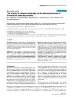
Weight gain in mice treated with ritonavirFigure 1
Weight gain in mice treated with ritonavir. At six
weeks of age, male and female wild type (C57BL/6) (top) or
LDL-R null mice (bottom) were administered ritonavir (15
μg/day) or vehicle (0.01% ethanol) through the drinking
water for six weeks. Mice were weighed at the beginning and
at the conclusion of the study. Bars represent the mean +/-
SEM, n = 6–8. * = significantly different from vehicle (p <
0.05).
AIDS Research and Therapy 2007, 4:8 />Page 4 of 8
(page number not for citation purposes)
have shown no difference or small increases in leptin in
females [25,26]. These differences are likely due to genetic
background or the age of the mice. This difference was
observed both in circulating levels of leptin in the serum
as well as at the level of leptin expression in abdominal
white adipose tissue.
In the present study, ritonavir suppressed serum leptin
levels, and increased food intake in both male and female
mice. This increase in food intake did not translate to
increased abdominal fat deposition or weight gain. Inter-
estingly, ritonavir increased epicardial fat in males. This
may correlate with the increased susceptibility of males to
the development of atherosclerosis in this model of HIV
protease inhibitor treatment [17]. Alternatively, in
females, weight gain is impaired. One possible explana-
tion is that ritonavir causes an alteration in energy
expenditure and metabolic activity of the liver or skeletal
muscle. Ritonavir has been shown to alter gene expression
in the liver that results in altered fatty acid metabolism
[20]. Even though in the present study, ritonavir does not
alter serum lipids, it can have subtle effects on liver and
adipose metabolism.
Ritonavir suppressed leptin levels in both male and
female mice. Leptin levels in females were lower than
males to begin with. It is possible that there is a threshold
level at which if leptin levels drop below, the ability to
maintain body weight in the face of a metabolic challenge
is lost. Previously, studies have shown that at high con-
centrations of ritonavir, leptin levels are reduced [13].
One possible explanation for the lack of correlation
between tissue expression and serum levels is an altera-
tion in leptin binding proteins by ritonavir. Ritonavir may
regulate serum binding proteins or the soluble leptin
receptor. These binding proteins affect the stability, deliv-
erance of leptin to targets or its clearance rate [27].
Women are more likely to develop adipose tissue altera-
tions induced by HAART [8]. Our data suggests that in
mice this is also the case. Clinical studies have demon-
strated that not all women develop adipose tissue altera-
tions and that they do not manifest themselves in the
same manner or have the same time of onset [8,28]. Ele-
vated triglycerides are, however, associated with the devel-
opment of adipose tissue alterations [28]. Our data add to
the growing body of evidence that females who have dys-
lipidemia would be more likely to develop adipose tissue
alterations. Additionally, it is also possible that gender
influences the metabolism or bioavailability of ritonavir.
This gender difference has been shown for the HIV pro-
tease inhibitor, indinavir [29].
Ovariectomy is well known to increase body weight in
rodents and humans [30]. The exact role of estrogen in
this process is not clear, but may involve regulation of lep-
tin or alterations in lipid metabolism in skeletal muscle
and adipose tissue [31,32]. We observed an increase in
weight gain following ovariectomy. Ritonavir did not
influence weight gain in the ovariectomized female mice,
suggesting an interaction at the site of action of ritonavir
and ovariectomy in the ability of the two to influence
body mass. Whether this interaction involves gonadal
hormones, such as estrogen, remains to be determined.
LDL-R null mice produce an environment of elevated
plasma cholesterol and triglycerides. It is in this environ-
ment that ritonavir alters weight gain. It remains possible
that the loss of LDL-R protein itself causes the effect. This
possibility has not been previously studied in detail in
these mice, but a lack of LDL-R in tissues other than the
liver may be important in regulating body weight. Addi-
tionally, LDL-R is expressed at a relatively high level in the
adrenal gland and could potentially play a role in regulat-
ing body weight by altering cortisol production [33].
Conclusion
In conclusion, this study reports the novel finding that
female mice are more likely to develop disturbances in
weight gain in response to ritonavir when they have a
background of elevated cholesterol. Ritonavir causes a
decline in serum leptin levels without altering adiponec-
tin levels. In concordance with the decline in leptin levels
there was increased food intake, however, no difference in
Table 1: Metabolic parameters of LDL-R null mice treated with ritonavir.
Male Female
Vehicle Ritonavir Vehicle Ritonavir
Total Cholesterol (mg/
ml)
1.32 +/- 0.06 1.23 +/- 0.11 0.96 +/- 0.06* 0.98 +/- 0.08*
Insulin (ng/ml) 0.80 +/- 0.01 0.73 +/- 0.01 0.80 +/- 0.08 0.72 +/- 0.02
Glucose (mg/dL) 180.17 +/- 17.6 176.67 +/- 14.5 155.17 +/- 6.4 149.00 +/- 8.2
Estradiol (pg/ml) ND ND 9.19 +/- 1.8 12.75 +/- 2.3
* = significantly different from male mice (p < 0.05)
ND = Not Determined
AIDS Research and Therapy 2007, 4:8 />Page 5 of 8
(page number not for citation purposes)
abdominal fat was observed. This suggests a secondary site
of action where ritonavir prevents adipose tissue forma-
tion or and increase in energy expenditure. Removing the
female sex hormones prevents the effects of ritonavir on
weight gain and serum leptin levels. This study begins to
investigate the mechanisms involved in the diverse
actions of HIV protease inhibitors and underscores the
complexity of the interactions between female hormones
and metabolism.
Methods
Animals
All animals were housed in the AAALAC certified animal
facilities at the University of Kentucky. Animals were
maintained on a 14:10 light/dark cycle at constant tem-
perature conditions with food (normal chow) and water
provided ad libitum. Wild type C57BL/6 mice were pur-
chased from Charles River (Wilmington, MA). The LDL-R
null and mice were supplied by The Jackson Laboratory
(Bar Harbor, ME). LDL-R null mice have been backcrossed
to a C57BL/6 background. At six weeks of age mice were
given vehicle control (0.01% ethanol) or ritonavir (15 μg/
day) in their drinking water for 6 weeks. A stock ritonavir
solution was made in ethanol and further diluted in the
drinking water. This regimen has previously been
described to induce atherosclerotic lesions in LDL-R null
mice without further altering plasma cholesterol levels
[16]. It produces significant physiological effects at rela-
tively low doses of ritonavir. At the time of tissue collec-
tion, animals were deeply anesthetized and blood was
collected by cardiac puncture. White adipose tissue was
dissected from the gonadal fat pad as previously described
[19] and frozen at -80°C until further use. Glucose meas-
urements were immediately made from whole blood
using a glucometer. Serum was then isolated and frozen at
-20°C until assayed as described below. A second set of
females was bilaterally ovariectomized prior to treatment
with ritonavir as described above. Briefly, a small incision
was made through the abdominal skin and muscle layer
of the animal around the area of the kidneys to expose the
ovary. The distal portion of each uterine horn was
clamped with a hemostat and the ovary was removed.
Blood assays
Serum was assayed for metabolic, lipid and hormonal
content using enzyme-linked immunoassays (ELISAs).
Leptin and insulin were measured using an ELISA from
ALPCO Diagnostics (Salem, NH). The intra-assay variance
and inter-assay variances for leptin and insulin were 7.8%,
Table 2: Food and water intake of LDL-R null mice treated with ritonavir.
Male Female
Vehicle Ritonavir Vehicle Ritonavir
Water intake (ml/day) 2.78 +/- 0.084 3.42 +/- 0.140 2.83 +/- 0.091* 2.96 +/- 0.107*
Food intake (g/day) 3.21 +/- 0.047 6.05 +/- 0.017# 2.87 +/- 0.035* 5.48 +/- 0.062*#
* = significantly different from male mice (p < 0.05)
# = significantly different from vehicle (p < 0.05)
Adipose tissue in mice treated with ritonavirFigure 2
Adipose tissue in mice treated with ritonavir. Male and
female LDL-R null mice were administered ritonavir (15 μg/
day) or vehicle (0.01% ethanol) through the drinking water
for six weeks. At the conclusion of the study, epicardial and
abdominal adipose tissue was removed and weighed. Data is
expressed as a percentage of final body weight. Bars repre-
sent the mean +/- SEM, n = 6–8. * = significantly different
from ritonavir (p < 0.05). # = significantly different from
males (p < 0.05).
AIDS Research and Therapy 2007, 4:8 />Page 6 of 8
(page number not for citation purposes)
Adipokine levels in mice treated with ritonavirFigure 3
Adipokine levels in mice treated with ritonavir. A) Serum leptin levels (left) and adiponectin levels (right) were meas-
ured by ELISA in male and female LDL-R null mice treated with ritonavir (15 μg/day) or vehicle (0.01% ethanol) for six weeks.
Bars represent the mean +/- SEM, n = 6–8. * = significantly different from vehicle (p < 0.05). # = significantly different from
males (p < 0.05). B) Leptin expression was identified by western immunoblot assay. Total protein was isolated from white adi-
pose tissue from male and female LDL-R null mice treated with ritonavir (15 μg/day) or vehicle (0.01% ethanol) for six weeks.
SDS-PAGE and immunoblot with a leptin antibody (#AFP6621299 obtained through the NHPP, NIDDK and Dr. A. F. Parlow)
was performed. The relative density of the bands was quantified. C) A representative immunoblot is shown. Recombinant
human leptin (rhLeptin) protein was included as a positive control.
AIDS Research and Therapy 2007, 4:8 />Page 7 of 8
(page number not for citation purposes)
10.5% and 8.7%, 8.5%, respectively. Adiponectin was
measured using an ELISA from Linco Research (St.
Charles, MO). The intra-assay variance and interassay var-
iances were 5.8% and 6.0%, respectively. Total cholesterol
was measured using an assay kit from Biovision (Moun-
tain View, CA). The intra-assay variance and inter-assay
variances were 2.8% and 2.6%, respectively. 17β-estradiol
was measured with a kit from Research Diagnostic Inc
(Concord, MA). The intra-assay variance and inter-assay
variances were 4.7% and 7.8%, respectively.
Western blot immunoassay
Adipose tissue protein was isolated as previously
described [34]. Briefly, proteins were isolated from frozen
white adipose tissue by homogenization in 1 mL isolation
media (250 mM Sucrose, 0.2 mM EDTA, 10 mM HEPES,
ddH
2
0, 1 tablet Roche
®
Complete Mini protease inhibitor
cocktail). The homogenate was then centrifuged at 7000 ×
g for 30 minutes at 4°C, and the fat pad discarded. After
removing the supernatant and pipetting it into a new 1.5
mL tube, the pellet containing the nuclear fraction was
brought up in 100–200 μL of sample buffer (40 mM Tris,
2% SDS, pH 8.0, ddH
2
0) and stored at -80°C. 600 μL 10%
TCA in acetone + 20 mM DTT was added to the superna-
tant containing the cytosolic fraction and placed at -20°C
for 1–2 hrs. The protein was precipitated by centrifugation
at 3500 × g for 30 minutes at 40°C, followed by two
washes in ice-cold 90% acetone between which the super-
natant was discarded and the pellet precipitated at 3500 ×
g for 3 minutes at 4°C. After the final wash, the pellet was
air dried for 5–10 minutes at room temperature to remove
residual acetone. Finally, the cytosolic protein was resus-
pended in 250–500 μL sample buffer and stored at -80°C.
15 μg of cytosolic protein was separated on a 12.5% SDS-
polyacrylamide gel. The separated proteins were then
transferred to nitrocellulose membranes. Each membrane
was blocked in 1:1 1 × PBS and Odyssey Blocking Buffer
(LI-COR) for 1 hour at room temperature. Primary anti-
bodies were diluted in 1:1 1 × PBS and Odyssey Blocking
Buffer + 0.2% Tween and incubated overnight at 4°C. The
concentrations of the primary antibodies used were: Anti-
actin (1:2000, Sigma-Aldrich) and anti-leptin (1:2000,
NHPP, NIDDK and Dr. A.J. Parlow). Recombinant leptin
was included as a positive control (NHPP, NIDDK, A.J.
Parlow). Fluorescently labeled secondary antibodies
(Rockland IRDye 800 or Molecular Probes AlexaFluor)
were diluted 1:5000 in 1 × PBS + Odyssey Blocking Buffer
+ 0.2% Tween + 0.01% SDS and incubated with the mem-
brane for 35 minutes in the dark at room temperature. The
membrane was then washed and the labeled proteins
were visualized on an Odyssey Infrared Imaging System
(LI-COR Biosciences, Lincoln, NE) as previously
described [35].
Statistics
Data were analyzed by two-way analysis of variance
(ANOVA), one-way ANOVA, and the Student Newman-
Keuls T-test was used for post-hoc comparisons, where
appropriate. Significance was considered at a p-value <
0.05. All experiments consisted of n = 6–8 animals per
experimental group.
Competing interests
The author(s) declare that they have no competing inter-
ests.
Ovariectomy reverses the effect of ritonavir in female miceFigure 4
Ovariectomy reverses the effect of ritonavir in
female mice. A) At six weeks of age, female LDL-R null
mice were bilaterally ovariectomized and administered riton-
avir (15 μg/day) or vehicle (0.01% ethanol) through the
drinking water for six weeks. An intact group was included as
a control. Mice were weighed at the beginning and at the
conclusion of the study. Bars represent the mean +/- SEM, n
= 6–8. B) Serum leptin levels were measured by ELISA in
ovariectomized female mice treated with ritonavir (15 μg/
day) or vehicle (0.01% ethanol) for six weeks. Bars represent
the mean +/- SEM, n = 6–8. * = significantly different from
vehicle (p < 0.05).
AIDS Research and Therapy 2007, 4:8 />Page 8 of 8
(page number not for citation purposes)
Authors' contributions
MW conceived the study and participated in its design and
coordination. MW wrote the manuscript. KA performed
the ELISAs and blood work. EK, DJ and AR treated and
monitored the animals and collected food and water data.
AB performed the western immunoblot analyses. All
authors read and approved the final manuscript.
Acknowledgements
The antibody to leptin (#AFP6621299) and recombinant human leptin
(#AFP496C) was obtained through the NHPP, NIDDK and Dr. A. F. Par-
low. This project was supported by NIH HL073693 (MEW) and grant P20
RR 15592 from the National Center for Research Resources (NCRR), a
component of the National Institutes of Health (NIH) and its contents are
sole the responsibility of the authors and do not necessarily represent the
official views of NCRR or NIH.
References
1. Carr A, Samaras K, Chisholm DJ, Cooper DA: Pathogenesis of
HIV-1-protease inhibitor-associated peripheral lipodystro-
phy, hyperlipidaemia, and insulin resistance. Lancet 1998,
351(9119):1881-1883.
2. Currier JS, Taylor A, Boyd F, Dezii CM, Kawabata H, Burtcel B, Maa
JF, Hodder S: Coronary heart disease in HIV-infected individu-
als. J Acquir Immune Defic Syndr 2003, 33(4):506-512.
3. Holmberg SD, Moorman AC, Williamson JM, Tong TC, Ward DJ,
Wood KC, Greenberg AE, Janssen RS: Protease inhibitors and
cardiovascular outcomes in patients with HIV-1. Lancet 2002,
360(9347):1747-1748.
4. Lo JC, Mulligan K, Tai VW, Algren H, Schambelan M: "Buffalo
hump" in men with HIV-1 infection. Lancet 1998,
351(9106):867-870.
5. Koutkia P, Grinspoon S: HIV-associated lipodystrophy: patho-
genesis, prognosis, treatment, and controversies. Annu Rev
Med 2004, 55:303-317.
6. Carr A, Samaras K, Chisholm DJ, Cooper DA: Abnormal fat distri-
bution and use of protease inhibitors. Lancet 1998,
351(9117):1736.
7. Sanchez-Torres RJ, Delgado-Osorio H: The metabolic syndrome
and its cardiovascular manifestations. Bol Asoc Med P R 2005,
97(4):271-280.
8. Galli M, Veglia F, Angarano G, Santambrogio S, Meneghini E, Gritti F,
Cargnel A, Mazzotta F, Lazzarin A: Gender differences in antiret-
roviral drug-related adipose tissue alterations. Women are
at higher risk than men and develop particular lipodystrophy
patterns. J Acquir Immune Defic Syndr 2003, 34(1):58-61.
9. Staiger H, Tschritter O, Machann J, Thamer C, Fritsche A, Maerker E,
Schick F, Haring HU, Stumvoll M: Relationship of serum adi-
ponectin and leptin concentrations with body fat distribution
in humans. Obes Res 2003, 11(3):368-372.
10. Lonnqvist F, Arner P, Nordfors L, Schalling M: Overexpression of
the obese (ob) gene in adipose tissue of human obese sub-
jects. Nat Med 1995, 1(9):950-953.
11. Maffei M, Halaas J, Ravussin E, Pratley RE, Lee GH, Zhang Y, Fei H,
Kim S, Lallone R, Ranganathan S, et al.: Leptin levels in human and
rodent: measurement of plasma leptin and ob RNA in obese
and weight-reduced subjects. Nat Med 1995, 1(11):1155-1161.
12. Caro JF, Sinha MK, Kolaczynski JW, Zhang PL, Considine RV: Leptin:
the tale of an obesity gene. Diabetes 1996, 45(11):1455-1462.
13. Riddle TM, Fichtenbaum CJ, Hui DY: Leptin replacement therapy
but not dietary polyunsaturated fatty acid alleviates HIV
protease inhibitor-induced dyslipidemia and lipodystrophy in
mice. J Acquir Immune Defic Syndr 2003, 33(5):564-570.
14. Yamauchi T, Kamon J, Waki H, Terauchi Y, Kubota N, Hara K, Mori
Y, Ide T, Murakami K, Tsuboyama-Kasaoka N, et al.: The fat-derived
hormone adiponectin reverses insulin resistance associated
with both lipoatrophy and obesity. Nat Med 2001, 7(8):941-946.
15. Arita Y, Kihara S, Ouchi N, Takahashi M, Maeda K, Miyagawa J, Hotta
K, Shimomura I, Nakamura T, Miyaoka K, et al.: Paradoxical
decrease of an adipose-specific protein, adiponectin, in obes-
ity. Biochem Biophys Res Commun 1999, 257(1):79-83.
16. Dressman J, Kincer J, Matveev SV, Guo L, Greenberg RN, Guerin T,
Meade D, Li XA, Zhu W, Uittenbogaard A, et al.: HIV protease
inhibitors promote atherosclerotic lesion formation inde-
pendent of dyslipidemia by increasing CD36-dependent
cholesteryl ester accumulation in macrophages. J Clin Invest
2003, 111(3):389-397.
17. Allred KF, Smart EJ, Wilson ME: Estrogen receptor-alpha medi-
ates gender differences in atherosclerosis induced by HIV
protease inhibitors. J Biol Chem 2006, 281(3):1419-1425.
18. Iacobellis G, Corradi D, Sharma AM: Epicardial adipose tissue:
anatomic, biomolecular and clinical relationships with the
heart. Nat Clin Pract Cardiovasc Med 2005, 2(10):536-543.
19. Johnson PR, Hirsch J: Cellularity of adipose depots in six strains
of genetically obese mice. J Lipid Res
1972, 13(1):2-11.
20. Goetzman ES, Tian L, Nagy TR, Gower BA, Schoeb TR, Elgavish A,
Acosta EP, Saag MS, Wood PA: HIV protease inhibitor ritonavir
induces lipoatrophy in male mice. AIDS Res Hum Retroviruses
2003, 19(12):1141-1150.
21. Lledo-Garcia R, Nacher A, Prats-Garcia L, Casabo VG, Merino-San-
juan M: Bioavailability and pharmacokinetic model for ritona-
vir in the rat. J Pharm Sci 2006.
22. Kappelhoff BS, Huitema AD, Crommentuyn KM, Mulder JW, Meen-
horst PL, van Gorp EC, Mairuhu AT, Beijnen JH: Development and
validation of a population pharmacokinetic model for riton-
avir used as a booster or as an antiviral agent in HIV-1-
infected patients. Br J Clin Pharmacol 2005, 59(2):174-182.
23. Mooser V: Atherosclerosis and HIV in the highly active
antiretroviral therapy era: towards an epidemic of cardio-
vascular disease? Aids 2003, 17(Suppl 1):S65-69.
24. Gui Y, Silha JV, Murphy LJ: Sexual dimorphism and regulation of
resistin, adiponectin, and leptin expression in the mouse.
Obes Res 2004, 12(9):1481-1491.
25. Ahren B: Diurnal variation in circulating leptin is dependent
on gender, food intake and circulating insulin in mice. Acta
Physiol Scand 2000, 169(4):325-331.
26. Lewitt MS, Brismar K: Gender difference in the leptin response
to feeding in peroxisome-proliferator-activated receptor-
alpha knockout mice. Int J Obes Relat Metab Disord 2002,
26(10):1296-1300.
27. Lammert A, Kiess W, Bottner A, Glasow A, Kratzsch J: Soluble lep-
tin receptor represents the main leptin binding activity in
human blood. Biochem Biophys Res Commun 2001, 283(4):982-988.
28. Jacobson DL, Knox T, Spiegelman D, Skinner S, Gorbach S, Wanke C:
Prevalence of, evolution of, and risk factors for fat atrophy
and fat deposition in a cohort of HIV-infected men and
women. Clin Infect Dis 2005, 40(12):1837-1845.
29. Kappelhoff BS, Huitema AD, Sankatsing SU, Meenhorst PL, Van Gorp
EC, Mulder JW, Prins JM, Beijnen JH:
Population pharmacokinet-
ics of indinavir alone and in combination with ritonavir in
HIV-1-infected patients. Br J Clin Pharmacol 2005, 60(3):276-286.
30. Wade GN, Gray JM, Bartness TJ: Gonadal influences on adipos-
ity. Int J Obes 1985, 9(Suppl 1):83-92.
31. Shimomura K, Shimizu H, Tsuchiya T, Abe Y, Uehara Y, Mori M: Is
leptin a key factor which develops obesity by ovariectomy?
Endocr J 2002, 49(4):417-423.
32. Kamei Y, Suzuki M, Miyazaki H, Tsuboyama-Kasaoka N, Wu J, Ishimi
Y, Ezaki O: Ovariectomy in mice decreases lipid metabolism-
related gene expression in adipose tissue and skeletal muscle
with increased body fat. J Nutr Sci Vitaminol (Tokyo) 2005,
51(2):110-117.
33. Hussain MM, Strickland DK, Bakillah A: The mammalian low-den-
sity lipoprotein receptor family. Annu Rev Nutr 1999,
19:141-172.
34. Gullicksen PS, Dean RG, Baile CA: Detection of DNA fragmenta-
tion and apoptotic proteins, and quantification of uncoupling
protein expression by real-time RT-PCR in adipose tissue. J
Biochem Biophys Methods 2004, 58(1):1-13.
35. Wilson ME, Allred KF, Bisotti AJ, Bruce-Keller A, Chuahan A, Nath A:
Estradiol negatively regulates HIV-LTR promoter activity in
glial cells. AIDS Res Hum Retroviruses 2006, 22(4):350-356.