Báo cáo y học: "Immune reconstitution inflammatory syndrome in association with HIV/AIDS and tuberculosis: Views over hidden possibilitie" pptx
Bạn đang xem bản rút gọn của tài liệu. Xem và tải ngay bản đầy đủ của tài liệu tại đây (293.7 KB, 7 trang )
BioMed Central
Page 1 of 7
(page number not for citation purposes)
AIDS Research and Therapy
Open Access
Hypothesis
Immune reconstitution inflammatory syndrome in association with
HIV/AIDS and tuberculosis: Views over hidden possibilities
Esaki Muthu Shankar, Ramachandran Vignesh, Kailapuri G Murugavel,
Pachamuthu Balakrishnan, Ramalingam Sekar, Charmaine AC Lloyd,
Suniti Solomon and Nagalingeswaran Kumarasamy*
Address: YRG Centre for AIDS Research and Education, VHS Hospital Campus, Rajiv Gandhi Salai – Information Technology Corridor, Taramani,
Chennai 600 113, India
Email: Esaki Muthu Shankar - ; Ramachandran Vignesh - ;
Kailapuri G Murugavel - ; Pachamuthu Balakrishnan - ; Ramalingam Sekar - ;
Charmaine AC Lloyd - ; Suniti Solomon - ;
Nagalingeswaran Kumarasamy* -
* Corresponding author
Abstract
Gut immune components are severely compromised among persons with AIDS, which allows
increased translocation of bacterial lipopolysaccharides (LPS) into the systemic circulation. These
microbial LPS are reportedly increased in chronically HIV-infected individuals and findings have
correlated convincingly with measures of immune activation. Immune reconstitution inflammatory
syndrome (IRIS) is an adverse consequence of the restoration of pathogen-specific immune
responses in a subset of HIV-infected subjects with underlying latent infections during the initial
months of highly active antiretroviral treatment (HAART). Whether IRIS is the result of a response
to a high antigen burden, an excessive response by the recovering immune system, exacerbated
production of pro-inflammatory cytokines or a lack of immune regulation due to inability to
produce regulatory cytokines remains to be determined. We theorize that those who develop IRIS
have a high burden of proinflammatory cytokines produced also in response to systemic bacterial
LPS that nonspecifically act on latent mycobacterial antigens. We also hypothesize that subjects that
do not develop IRIS could have developed either tolerance (anergy) to persistent LPS/tubercle
antigens or could have normal FOXP3+ gene and that those with defective FOXP3+ gene or those
with enormous plasma LPS could be vulnerable to IRIS. The measure of microbial LPS, anti-LPS
antibodies and nonspecific plasma cytokines in subjects on HAART shall predict the role of these
components in IRIS.
Background
Immune reconstitution inflammatory syndrome (IRIS): An
existing lacuna in HIV immunology?
IRIS is an adverse consequence of the restoration of path-
ogen-specific immune responses in HIV-infected patients
during the initial months of highly active antiretroviral
treatment (HAART) [1]. Even though IRIS is also closely
associated with certain other infectious (mycobacteria,
varicella zoster, herpesviruses, and cytomegalovirus) and
non-infectious (autoimmune) conditions [2-10], the
Published: 30 November 2007
AIDS Research and Therapy 2007, 4:29 doi:10.1186/1742-6405-4-29
Received: 12 September 2007
Accepted: 30 November 2007
This article is available from: />© 2007 Shankar et al; licensee BioMed Central Ltd.
This is an Open Access article distributed under the terms of the Creative Commons Attribution License ( />),
which permits unrestricted use, distribution, and reproduction in any medium, provided the original work is properly cited.
AIDS Research and Therapy
2007, 4:29 />Page 2 of 7
(page number not for citation purposes)
morbidity associated with HIV/tuberculosis (TB) is more
important [1,11] as the crisis seem to be alarming in third-
world nations, where the proportion of HIV/TB IRIS is
reportedly high, ranging from 11% to 43% [12-15]. This
could be due to differences in cohort characteristics, case
definitions and differences in the mean time interval
between TB diagnosis and antiretroviral therapy (ART)
initiation. Data from resource-limited countries on TB-
IRIS is scarce; a rate of 8% was reported from India [1].
Immunology of IRIS in HIV/TB is deficient and HIV-spe-
cific T lymphocyte responses have repeatedly shown to be
defective [16]. To understand the immunopathogenesis of
IRIS it will be crucial to elucidate the intrinsic dynamics of
immune cells after initiation of HAART [17]. Preliminary
investigations have shown that an acute exacerbation of
mycobacteria-specific Th1 response after HIV infection
control by HAART causes IRIS in HIV/TB [17,18].
Does CD4+ T-cell depletion lead to a breach in gut
immune cell integrity to initiate the proinflammatory
cytokine saga?
In the context of an HIV infected subject with latent pul-
monary TB, progressing to AIDS stage of HIV disease, the
acute stage of the infection is characterized by eventual
depletion in the number of CD4+ T-cells, the key orches-
trator of all immune mechanisms in the body. Recent
research has re-examined the rate of immunopathologic
events in HIV disease, where the first few weeks is charac-
terized by massive viremia and depletion of ~50% mem-
ory CD4+ T-cell (CCR5+) population especially in the gut
[19-26]. Since the gut associated lymphoid tissue (GALT)
comprises ~60% of entire lymphoid organ system, rich in
memory cells, its depletion has a strong consequence on
the entire CD4+ T-cell population. Memory CD4+ T-cells
in the lamina propria is depleted principally by Fas-FasL-
mediated cell death [26]. In addition, productive HIV
infection is favored by an inflammatory environment,
because Th1 cytokines (IL-2, IL-12, TNF-α) increase NFkB
activation in T-cells, which drives HIV transcription. Early
breach in the gut mucosal integrity and epithelial micro-
environment [19-21,27-30] leads to increased transloca-
tion of luminal microbial products [20] because the gut is
thought to be the principal source of microbial products
(especially LPS) and because it has a massive bacterial
load compared to other anatomical sites [31-33]. Translo-
cation results in chronic inflammation via Toll-like recep-
tor (TLR-4) stimulation, resulting in cytokine and
chemokine release driving persistent T-cell activation and
(tat mediated) apoptosis via activation-induced cell
death (AICD) [21]. However, due to lack of sufficient
CD4+ T-cells, complex inflammatory mechanisms might
not be expected due to anergy.
HAART, immunological restoration and the inflammatory
milieu: Who are the possible mediators?
Most of the subjects with HIV disease attend HIV testing
centers in India only after advanced clinical HIV disease
(AIDS) sets in and when their CD4+ T-cell counts are low
[1,11]. In spite of initiation of HAART, some experience a
'discordant response', whereby the HIV-1 RNA plasma
level is below the limit of detection but the CD4+ cell
count response is blunted. We propose that these individ-
uals with HIV/TB coinfection might not progress to clini-
cal IRIS owing to poor immune reconstitution despite
considerable virological recovery. As a consequence, a
substantial proportion of treated individuals show poor
CD4+ T-cell recovery [40]. This has also been correlated
with a lower nadir pretreatment CD4+ T-cell count, sug-
gestive of more extensive depletion of CD4+ T-cells in the
GALT during acute HIV infection, which may be refractory
to reconstitution with ART [19,41]. Initiation of HAART
allows 'partial' immune restoration [42], which however,
can result in the substantial proliferation and differentia-
tion of most of the immune components [43,44]. Due to
immune restoration, an inflammatory response against
infectious and non-infectious antigens (LPS) is mounted
leading to noticeable 'paradoxical worsening' [43-45],
with a shift toward a Th1 receptor profile, which increases
the levels of IFN-γ and IL-2 [46-51]. Therefore, persons
with latent TB or other systemic commensal antigens
(LPS) could lead to exaggerated inflammatory responses.
Studies also show that an inflammatory response is
required for the elimination of any gram-negative infec-
tion (i.e. LPS) [52]. HAART treatment (that enable 'partial'
immune reconstitution) considerably reduces circulating
LPS although total clearance may not be feasible for con-
siderable periods of time.
Bacterial LPS, the microbe-associated molecular patterns
(MAMP) of gram-negative bacteria are known potent acti-
vators of cells of inflammatory system. Plasma LPS levels
have been directly associated with the degree of intestinal
permeability following invasive gastrointestinal surgery
[34], inflammatory bowel disease (IBD) [35] and graft-
versus host disease (GVHD) [31,35-39]. Experimental SIV
infection of macaques resulted in raised circulating LPS
levels [21]. Recent studies have found significantly ele-
vated levels of plasma LPS in chronically HIV-infected
humans with progressive disease [21] and has correlated
convincingly with measures of innate and adaptive
immune activation. Besides, the study also has shown the
association between LPS and chronic in vivo stimulation
of monocytes, an association between raised plasma LPS;
and an association between reduction in plasma LPS and
CD4+ T-cell reconstitution with HAART [21]. Due to
abrupt increase in the numbers of CD4+ T-cells, the pat-
tern recognition receptors (PRR) induce signal transduc-
tion pathway molecules like NFkB, IL-1 receptor, TNF
AIDS Research and Therapy
2007, 4:29 />Page 3 of 7
(page number not for citation purposes)
receptor, MAP kinase receptor etc. [53]. Cytokines such as
IL-1 can also stimulate the NFkB binding molecule to acti-
vate NFkB [54-56], which induces the expression of
cyclooxygenase-2 (COX-2), which consequently leads to
tissue inflammation at the site where latent TB antigens
are located. Interestingly, the expression of the COX-2-
encoding gene, believed to be responsible for the massive
production of prostaglandins at inflammatory sites, is
transcriptionally regulated by NFkB [54]. NFkB resides in
the cytoplasm and is bound to its inhibitor. Furthermore,
injurious and inflammatory stimuli, such as free radicals
present in the plasma of the immune deteriorated host
leads to NFkB release that subsequently moves into the
nucleus to activate the genes responsible for COX-2
expression.
Alternatively, effector T-cells of the Th1 subset activates
macrophages by CD154 – CD4+0 interactions and by
secreting IFN-γ. Th1 subsets produce the proinflammatory
cytokines, IL-2, IFN-γ, and TNF-α, and Th2 cells, the anti-
inflammatory cytokines, IL-4, 5, 6, 10, and 13. In addi-
tion, macrophages that have phagocytosed TB bacilli pro-
duce IL-12 that stimulates the differentiation of naïve
CD4+ T-cells to the Th1 subset, which again produces
IFN-γ on encountering macrophage-associated microbial
antigens; IL-12 also increases the amount of IFN-γ pro-
duced by these T-cells. In different T-cell mediated dis-
eases, tissue injury is caused by a delayed-type
hypersensitivity response mediated by CD4+ T-cells or by
lysis of host cells by CD8+ CTLs. Some studies suggest that
circulating IL-6 levels prior to HAART may be associated
with IRIS [53]. CD4+ T-cells may react against cell or tis-
sue antigens and secrete cytokines that induce local
inflammation and activate macrophages. The actual tissue
injury is caused by the macrophages and other inflamma-
tory cells. CD8+ T-cells specific for antigens on autologous
cells may directly kill these cells. Increased LPS-binding
protein (LBP) may also increase the host response and
potentiate injury. We hypothesize that the excessive pres-
ence of LPS in HIV/TB coinfected subjects accounts for the
progression of IRIS and those that have LPS in limited
concentrations may not. In studies in which normal
human subjects were treated with LPS intravenously,
there was a shift toward a Th2 response with increased
expression of IL-10, [57-59] and the pretreatment of
healthy human volunteers with IL-10 reduced the LPS-
induced increases in chemokines [60,61]. Data from stud-
ies in normal human volunteers suggest that LPS increase
the production of circulating IL-10, which would then
blunt the proinflammatory response to a second bacterial
challenge [60,61]. The Th2 shift in sepsis suggests that an
excess of anti-inflammatory cytokines may result in
impaired lung host response. We therefore hypothesize
that this situation could also lead to extensive multiplica-
tion of TB bacilli. A brief overview of the concept is illus-
trated in figure 1.
The likelihood of 'normal' FOXP3+ gene and endotoxin-
tolerance among IRIS non-developers – Why?
This 'paradoxical worsening' could also be attributable to
additional presence of defective FOXP3+ gene among IRIS
developers. Presence of defective FOXP3+ gene in T-cells
has been reported to confer increased risk of inflamma-
tory conditions in human beings in contrast to a normal
FOXP3+ gene. After an initial exposure to LPS, monocytes
and macrophages become refractory to subsequent LPS
challenge (endotoxin-tolerance) (57 – 61). This initially
was believed to be protective against septic shock. How-
ever, recent evidence suggests that endotoxin-tolerance
impairs the host response to a second bacterial challenge
[62,63]. The prolonged presence of TB antigens (and a
normal FOXP3+ gene) could also lead to anergy and poor
immune responses to TB antigens despite HAART. Mono-
cytes obtained from septic patients have functional defects
that include profound defects in IL-1, 6, and TNF-α pro-
duction; loss of HLA class II antigen expression; and
impaired antigen presentation [64-69]. In patients with
sepsis, monocytes from survivors showed normal
cytokine response following LPS stimulation [64]. A
potential mechanism whereby endotoxin-tolerance devel-
ops is a down-regulation of LPS receptors such as mem-
brane CD14 on macrophages [70]. The exposure of
monocytes and macrophages to the anti-inflammatory
cytokines, IL-10 and TGF-β, is a second mechanism that
may be responsible for the monocyte deactivation that
resembles endotoxin-tolerance [71]. Studies performed
with human alveolar macrophages exposed to IL-10 in
vitro show increased intracellular bacterial replication of
Legionella pneumophila, [72] and decreased production of
proinflammatory cytokines [73]. These suggest that mac-
rophages and monocytes in septic patients may develop a
phenotype similar to that observed in endotoxin-toler-
ance, which could result in an impaired response to lung
pathogens. The development of tolerance was hypothe-
sized to be beneficial by diminishing the proinflamma-
tory response in patients with sepsis. However, some data
suggest that the development of tolerance may worsen
clinical outcomes because monocytes and macrophages
may not respond adequately to a bacterial challenge
[62,65,74]. The CD14/TLR complex and associated sign-
aling pathways are essential for the recognition of LPS by
macrophages, and several studies suggest that down-regu-
lation of CD14/TLR complexes on macrophages is
responsible for the development of tolerance
[63,70,75,76]. However, the development of tolerance
does not correlate with down-regulation of LPS-binding
sites [77], suggesting the possible role of other mecha-
nisms including the disruption of CD14/TLR
signaling pathways [78] and the macrophage exposure to
AIDS Research and Therapy
2007, 4:29 />Page 4 of 7
(page number not for citation purposes)
anti-inflammatory IL-10 [79]. Therefore, it is hypothe-
sized that subjects that do not progress to develop IRIS
(IRIS tolerant) despite HAART initiation could develop
tolerance (anergy) to persistent LPS/tubercle antigens.
Conclusion
It is hypothesized that proinflammatory cytokines pro-
duced excessively in response to systemic bacterial LPS
nonspecifically act on latent mycobacterial antigens lead-
ing to clinical deterioration and 'paradoxical worsening'
of inflammatory responses against both infectious (HIV/
TB) and non-infectious (LPS) microbial antigens. This
'paradoxical worsening' could also be attributable to addi-
tional presence of defective FOXP3+ gene among IRIS
developers. Subjects that do not progress to develop IRIS
(IRIS tolerant) despite HAART initiation could develop
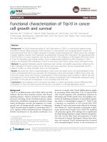
One possible mechanism that illustrates the immunology of IRIS in a subject with HIV/TB coinfectionFigure 1
One possible mechanism that illustrates the immunology of IRIS in a subject with HIV/TB coinfection. Compro-
mised gut immunity leads to increased translocation of luminal gram negative bacterial LPS into the systemic circulation. Initia-
tion of HAART in the subject leads to abrupt restoration of CD4+ T-cells and almost any pathogen-specific immune response.
IRIS developers have a high burden of LPS and proinflammatory cytokines produced against LPS could result in an exaggerated,
nonspecific attack on latent mycobacterial antigens that are presented in the local lymph nodes leading to localized inflamma-
tion. We also hypothesize that subjects that do not develop IRIS could have developed either tolerance (anergy) to persistent
LPS and tubercle antigens or could have normal FOXP3+ gene (not shown) and that those with defective FOXP3+ gene or
enormous plasma LPS could be vulnerable to IRIS (as demonstrated by researchers that defective FOXP3+ gene is associated
with increased risk for inflammatory conditions). (Bold lines indicate the availability of clinical/experimental evidence and
dashed lines indicate the possible mechanism).
AIDS Research and Therapy
2007, 4:29 />Page 5 of 7
(page number not for citation purposes)
either tolerance (anergy) to persistently existing LPS and
tubercle antigens. Thus far, no single treatment option
exists against IRIS and depends on the underlying infec-
tious agent and its clinical presentation. However, since
the pathogenesis is an inflammatory one, systemic corti-
costeroids or non-steroidal anti-inflammatory drugs
(NSAIDS) may assuage the symptoms. Therefore, studies
must be attempted to assess the role of immunological
correlates and possible markers of IRIS needs to be evalu-
ated to better understand the mechanisms behind IRIS in
HIV/TB or other opportunistic coinfections, which would
largely facilitate the timely management of IRIS in HIV/
AIDS.
Competing interests
The author(s) declare that they have no competing inter-
ests.
Authors' contributions
EMS, RV and NK conceived and proposed the hypothesis.
RV, KGM, PB, CAL, RS, SS, and NK provided additional
inputs to further develop the scientific concept; EMS, RV
and PB drafted the manuscript; SS and NK shared their
clinical expertise and critically revised the manuscript. All
authors read and approved the final manuscript. EMS, RV
and NK are the guarantors of the paper.
Acknowledgements
The authors are grateful to all the staff and patients of YRG CARE without
whose support and facilitation, this manuscript could not have been con-
ceived and drafted.
References
1. Kumarasamy N, Chaguturu S, Mayer KH, Solomon S, Yepthomi HT,
Balakrishnan P, Flanigan TP: Incidence of immune reconstitution
syndrome in HIV/tuberculosis-coinfected patients after initi-
ation of generic antiretroviral therapy in India. J Acquir Immune
Defic Syndr 2004, 37:1574-1576.
2. Keane NM, Price P, Lee S, Almeida CA, Stone SF, James I, French MA:
Restoration of CD4 T-cell responses to cytomegalovirus is
short-lived in severely immunodeficient HIV-infected
patients responding to highly active antiretroviral therapy.
HIV Med 2004, 5:407-414.
3. Stone SF, Price P, Brochier J, French MA: Plasma bioavailable
interleukin-6 is elevated in human immunodeficiency virus-
infected patients who experience herpes virus-associated
immune restoration disease after start of highly active
antiretroviral therapy. J Infect Dis 2001, 184:1073-1077.
4. Koval CE, Gigliotti F, Nevins D, Demeter LM: Immune reconstitu-
tion syndrome after successful treatment of Pneumocystis
carinii pneumonia in a man with human immunodeficiency
virus type 1 infection. Clin Infect Dis 2002, 35(4):491-493.
5. Bicanic T, Harrison T, Niepieklo A, Dyakopu N, Meintjes G: Symp-
tomatic relapse of HIV-associated cryptococcal meningitis
after initial fluconazole monotherapy: the role of fluconazole
resistance and immune reconstitution. Clin Infect Dis 2006,
43:1069-1073.
6. Lawn SD, Bicanic TA, Macallan DC: Pyomyositis and cutaneous
abscesses due to Mycobacterium avium : an immune reconsti-
tution manifestation in a patient with AIDS. Clin Infect Dis
2004, 38:461-463.
7. Sereti I, Sarlis NJ, Arioglu E, Turner ML, Mican JM: Alopecia univer-
salis and Graves' disease in the setting of immune restora-
tion after highly active antiretroviral therapy. AIDS 2001,
15:138-140.
8. Mirmirani P, Maurer TA, Herndier B, McGrath M, Weinstein MD,
Berger TG: Sarcoidosis in a patient with AIDS: a manifesta-
tion of immune restoration syndrome. J Am Acad Dermatol
1999, 41:285-286.
9. Lawn SD, Checkley A, Wansbrough-Jones MH: Acute bilateral
parotitis caused by Mycobacterium scrofulaceum : immune
reconstitution disease in a patient with AIDS. Sex Transm Infect
2005, 81:517-518.
10. Taylor CL, Subbarao V, Gayed S, Ustianowski AP: Immune recon-
stitution syndrome to Strongyloides stercoralis infection. AIDS
2007, 21:649-650.
11. Kumarasamy N, Vallabhaneni S, Flanigan TP, Mayer KH, Solomon S:
Clinical profile of HIV in India. Indian J Med Res 2005,
121:377-394.
12. Breton G, Duval X, Estellat C, Poaletti X, Bonnet D, Mvondo MD, et
al.: Determinants of immune reconstitution inflammatory
syndrome in HIV type 1-infected patients with tuberculosis
after initiation of antiretroviral therapy. Clin Infect Dis 2004,
39:1709-1712.
13. Wendel KA, Alwood KS, Gachuhi R, Chaisson RE, Bishai WR, Sterling
TR: Paradoxical worsening of tuberculosis in HIV infected
persons. Chest 2001, 120:193-197.
14. Shelburne SA, Visnegarwala F, Darcourt J, Graviss EA, Giordano TP,
White AC Jr, Hamill RJ: Incidence and risk factors for immune
reconstitution inflammatory syndrome during highly active
antiretroviral therapy. AIDS 2005, 19:399-406.
15. Shao H, Crump J, Ramadhani H, Uiso L, Sendui-Nguyaine , Kiwera R,
Ndosi1 E, Shao J, Bartlett J, Thielman N: A randomised trial of
early versus delayed fixed dose combination zidovudine/lam-
ivudine/abacavir in patients coinfected with HIV and tuber-
culosis: early findings of the tuberculosis and immune
reconstitution syndrome trial. Thirteenth Conference on Retrovi-
ruses and Opportunistic Infections. Denver, CO, February 2006 . [abstract
796]
16. Shelburne SA, Montes M, Hamill RJ: Immune reconstitution
inflammatory syndrome: more answers, more questions. J
Antimicrob Chemother 2006, 57:167-170.
17. Bourgarit A, Carcelain G, Martinez V, Lascoux C, Delcey V, Gicquel
B, Vicaut E, Lagrange PH, Sereni D, Autran B: Explosion of tuber-
culin-specific Th1-responses induces immune restoration
syndrome in tuberculosis and HIV co-infected patients. AIDS
2006, 20:F1-F7.
18. Race EM, Adelson-Mitty J, Kriegel GR, Barlam TF, Reimann KA, Letvin
NL, Japour AJ: Focal mycobacterial lymphadenitis following
initiation of protease-inhibitor therapy in patients with
advanced HIV-1 disease. Lancet 1998, 351:252-255.
19. Brenchley JM, Schacker TW, Ruff LE, Price DA, Taylor JH, Beilman GJ,
Nguyen PL, Khoruts A, Larson M, Haase AT, Douek DC: CD4+ T
cell depletion during all stages of HIV disease occurs pre-
dominantly in the gastrointestinal tract. J Exp Med 2004,
200:749-759.
20. Brenchley JM, Price DA, Douek DC: HIV disease: fallout from a
mucosal catastrophe? Nat Immunol 2006, 7:235-239.
21. Brenchley JM, Price DA, Schacker TW, Asher TE, Silvestri G, Rao S,
Kazzaz Z, Bornstein E, Lambotte O, Altmann D, Blazar BR, Rodriguez
B, Teixeira-Johnson L, Landay A, Martin JN, Hecht FM, Picker LJ, Led-
erman MM, Deeks SG, Douek DC: Microbial translocation is a
cause of systemic immune activation in chronic HIV infec-
tion. Nat Med 2006, 12:1365-1371.
22. Mattapallil JJ, Douek DC, Hill B, Nishimura Y, Martin M, Roederer M:
Massive infection and loss of memory CD4+ T cells in multi-
ple tissues during acute SIV infection. Nature 2005,
434:1093-1097.
23. Veazey RS, DeMaria M, Chalifoux LV, Shvetz DE, Pauley DR, Knight
HL, Rosenzweig M, Johnson RP, Desrosiers RC, Lackner AA: Gas-
trointestinal tract as a major site of CD4+ T cell depletion
and viral replication in SIV infection. Science 1998,
280:427-431.
24. Guadalupe M, Reay E, Sankaran S, Prindiville T, Flamm J, McNeil A,
Dandekar S: Severe CD4+ T-cell depletion in gut lymphoid tis-
sue during primary human immunodeficiency virus type 1
infection and substantial delay in restoration following highly
active antiretroviral therapy. J Virol 2003, 77:11708-11717.
AIDS Research and Therapy
2007, 4:29 />Page 6 of 7
(page number not for citation purposes)
25. Picker LJ, Hagen SI, Lum R, Reed-Inderbitzin EF, Daly LM, Sylwester
AW, Walker JM, Siess DC, Piatak M Jr, Wang C, Allison DB, Maino
VC, Lifson JD, Kodama T, Axthelm MK: Insufficient production
and tissue delivery of CD4+ memory T cells in rapidly pro-
gressive simian immunodeficiency virus infection. J Exp Med
2004, 200:1299-1314.
26. Li Q, Duan L, Estes JD, Ma ZM, Rourke T, Wang Y, Reilly C, Carlis J,
Miller CJ, Haase AT: Peak SIV replication in resting memory
CD4+ T cells depletes gut lamina propria CD4+ T cells.
Nature 2005, 434:1148-1152.
27. Mehandru S, Poles MA, Tenner-Racz K, Horowitz A, Hurley A, Hogan
C, Boden D, Racz P, Markowitz M: Primary HIV-1 infection is
associated with preferential depletion of CD4+ T lym-
phocytes from effector sites in the gastrointestinal tract. J
Exp Med 2004, 200:761-770.
28. George MD, Reay E, Sankaran S, Dandekar S: Early antiretroviral
therapy for simian immunodeficiency virus infection leads to
mucosal CD4+ T-cell restoration and enhanced gene expres-
sion regulating mucosal repair and regeneration. J Virol 2005,
79:2709-2719.
29. Kotler DP: HIV infection and the gastrointestinal tract. AIDS
2005, 19:107-117.
30. Sharpstone D, Neild P, Crane R, Taylor C, Hodgson C, Sherwood R,
Gazzard B, Bjarnason I: Small intestinal transit, absorption, and
permeability in patients with AIDS with and without diar-
rhoea. Gut 1999, 45:70-76.
31. Cooke KR, Olkiewicz K, Erickson N, Ferrara JL: The role of endo-
toxin and the innate immune response in the pathophysiol-
ogy of acute graft versus host disease. J Endotoxin Res 2002,
8:441-448.
32. Macpherson AJ, Harris NL: Interactions between commensal
intestinal bacteria and the immune system. Nat Rev Immunol
2004, 4:478-485.
33. Takeda K, Kaisho T, Akira S: Toll-like receptors. Annu Rev Immunol
2003, 21:335-376.
34. Schietroma M, Carlei F, Cappelli S, Amicucci G: Intestinal permea-
bility and systemic endotoxemia after laparotomic or lapar-
oscopic cholecystectomy. Ann Surg 2006, 243:359-363.
35. Caradonna L, Amati L, Magrone T, Pellegrino NM, Jirillo E, Caccavo
D: Enteric bacteria, lipopolysaccharides and related
cytokines in inflammatory bowel disease: biological and clin-
ical significance. J Endotoxin Res 2000, 6:205-214.
36. Hill GR, Teshima T, Gerbitz A, Pan L, Cooke KR, Brinson YS, Craw-
ford JM, Ferrara JL: Differential roles of IL-1 and TNF-α on
graft-versus-host disease and graft versus leukemia.
J Clin
Invest 1999, 104:459-467.
37. Schietroma M, Cappelli S, Carlei F, Di Giuro G, Agnifili A, Recchia CL,
Antonellis M, Amicucci G: Intestinal and systemic endotoxae-
mia after laparotomic or laparoscopic cholecystectomy. Chir
Ital 2006, 58:171-177.
38. Wellmann W, Fink PC, Benner F, Schmidt FW: Endotoxaemia in
active Crohn's disease. Treatment with whole gut irrigation
and 5-aminosalicylic acid. Gut 1986, 27:814-820.
39. Wyatt J, Vogelsang H, Hubl W, Waldhoer T, Lochs H: Intestinal
permeability and the prediction of relapse in Crohn's dis-
ease. Lancet 1993, 341:1437-1439.
40. Nicastri E, Chiesi A, Angeletti C, Sarmati L, Palmisano L, Geraci A,
Andreoni M, Vella S, Italian Antiretroviral Treatment Group (IATG):
Clinical outcome after 4 years follow-up of HIV-seropositive
subjects with incomplete virologic or immunologic response
to HAART. J Med Virol 2005, 76:153-160.
41. Douek DC, Picker LJ, Koup RA: T cell dynamics in HIV-1 infec-
tion. Annu Rev Immunol 2003, 21:265-304.
42. Bower M, Fox P, Fife K, Gill J, Nelson M, Gazzard B: Highly active
antiretroviral therapy (HAART) prolongs time to treatment
failure in Kaposi's sarcoma. AIDS 1999, 13:2105-2111.
43. Chien J, Johnson H: Paradoxical reactions in HIV and pulmo-
nary TB. Chest 1998, 114:933-936.
44. Kunimoto DY, Chui L, Nobert E, Houston S: Immune mediated
"HAART" attack during treatment for tuberculosis. Int J
Tuberc Lung Dis 1999, 3:944-947.
45. Shelburne SA 3rd, Hamill RJ, Rodriguez-Barradas MC, Greenberg SB,
Atmar RL, Musher DW, Gathe JC Jr, Visnegarwala F, Trautner BW:
Immune reconstitution inflammatory syndrome: emer-
gence of a unique syndrome during highly active antiretrovi-
ral therapy. Medicine (Baltimore) 2002, 81:213-227.
46. Mitsuyasu R: HIV protease inhibitors: immunological insights.
AIDS 1999, 13(Suppl 1):S19-S27.
47. Narita M, Ashkin D, Hollender ES, Pitchenik AE: Paradoxical wors-
ening of tuberculosis following antiretroviral therapy in
patients with AIDS. Am J Respir Crit Care Med 1998, 158:157-161.
48. John M, French MAH: Exacerbation of the inflammatory
response to Mycobacterium tuberculosis after antiretroviral
therapy.
Med J Aust 1998, 169:473-474.
49. Fishman JE, Saraf-Lavi E, Narita M, Hollender ES, Ramsinghani R,
Ashkin D: Pulmonary tuberculosis in AIDS patients: transient
chest radiographic worsening after initiation of antiretrovi-
ral therapy. AJR Am J Roentgenol 2000, 174:43-49.
50. Furrer H, Malinverni R: Systemic inflammatory reaction after
starting highly active antiretroviral therapy in AIDS patients
treated for extrapulmonary tuberculosis. Am J Med 1999,
106:371-372.
51. Stone SF, Price P, Brochier J, French MA: Plasma bioavailable
interleukin-6 is elevated in human immunodeficiency virus-
infected patients who experience herpesvirus-associated
immune restoration disease after start of highly active
antiretroviral therapy. J infect Dis 2001, 184:1073-1077.
52. Shahin RD, Engberg I, Hagberg L, Eden CS: Neutrophil recruit-
ment and bacterial clearance correlated with LPS respon-
siveness in local gram-negative infection. J Immunol 1987,
138:3475-3480.
53. Dadgostar H, Zarnegar B, Hoffmann A, Qin XF, Truong U, Rao G,
Baltimore D, Cheng G: Cooperation of multiple signaling path-
ways in CD4+0-regulated gene expression in B lymphocytes.
Proc Natl Acad Sci USA 2002, 5:1497-1502.
54. D'Acquisto F, May MJ, Ghosh S: Inhibition of nuclear factor
kappa B (NFkB): an emerging theme in anti-inflammatory
therapies. Molec Interv 2002, 2:22-35.
55. Seaman DR: Joint complex dysfunction, a novel term to
replace subluxation/subluxation complex: etiological and
treatment considerations. J Manipulative Physiol Ther 1997,
20:634-644.
56. Seaman DR: The diet-induced proinflammatory state: a cause
of chronic pain and other degenerative diseases? J Manipulative
Physiol Ther 2002, 25:168-179.
57. van der Poll T, de Wall Malefyt R, Coyle SM, Lowry SF: Antiinflam-
matory cytokine responses during clinical sepsis and experi-
mental endotoxemia: sequential measurements of plasma
soluble interleukin (IL)-1 receptor type II, IL-10, and IL-13. J
Infect Dis 1997, 175:118-122.
58. Zimmer S, Pollard V, Marshall GD: Effects of endotoxin on the
Th1/Th2 response in humans. J Burn Care Rehabil 1996,
17:491-496.
59. Lauw FN, Lauw FN, ten Hove T, Dekkers PE, de Jonge E, van
Deventer SJ, van Der Poll T: Reduced Th1, but not Th2, cytokine
production by lymphocytes after in vivo exposure of healthy
subjects to endotoxin. Infect Immun 2000, 68:
1014-1018.
60. Olszyna DP, Pajkrt D, van Deventer SJ, van der Poll T: Effect of
interleukin 10 on the release of the CXC chemokines growth
related oncogene GRO-alpha and epithelial cell-derived neu-
trophil activating peptide (ENA)-78 during human endotox-
emia. Immunol Lett 2001, 78:41-44.
61. Olszyna DP, Pajkrt D, Lauw FN, van Deventer SJ, van Der Poll T:
Interleukin 10 inhibits the release of CC chemokines during
human endotoxemia. J Infect Dis 2000, 181:613-620.
62. West MA, Heagy W: Endotoxin tolerance: a review. Crit Care
Med 2002, 30:S64-S73.
63. Dobrovolskaia MA, Vogel SN: Toll receptors, CD14, and macro-
phage activation and deactivation by LPS. Microbes Infect 2002,
4:903-914.
64. Munoz C, Carlet J, Fitting C, Misset B, Blériot JP, Cavaillon JM: Dys-
regulation of in vitro cytokine production by monocytes dur-
ing sepsis. J Clin Invest 1991, 88:1747-1754.
65. Volk HD, Thieme M, Heym S, Döcke WD, Ruppe U, Tausch W, Man-
ger D, Zuckermann S, Golosubow A, Nieter B, et al.: Alterations in
function and phenotype of monocytes from patients with
septic disease: predictive value and new therapeutic strate-
gies. Behring Inst Mitt 1991, 88:208-215.
66. Wilson CS, Seatter SC, Rodriguez JL, Bellingham J, Clair L, West MA:
In vivo endotoxin tolerance: impaired LPS-stimulated TNF
release of monocytes from patients with sepsis, but not
SIRS. J Surg Res 1997, 69:101-106.
AIDS Research and Therapy
2007, 4:29 />Page 7 of 7
(page number not for citation purposes)
67. Döcke WD, Randow F, Syrbe U, Krausch D, Asadullah K, Reinke P,
Volk HD, Kox W: Monocyte deactivation in septic patients:
restoration by IFN-g treatment. Nat Med 1997, 3:678-681.
68. Wolk K, Docke WD, von Baehr V, Volk HD, Sabat R: Impaired
antigen presentation by human monocytes during endotoxin
tolerance. Blood 2000, 96:218-223.
69. Adib-Conquy M, Adrie C, Moine P, Asehnoune K, Fitting C, Pinsky
MR, Dhainaut JF, Cavaillon JM: NF-kappaB expression in mono-
nuclear cells of patients with sepsis resembles that observed
in lipopolysaccharide tolerance. Am J Respir Crit Care Med 2000,
162:1877-1883.
70. Sugawara S, Nemoto E, Tada H, Miyake K, Imamura T, Takada H:
Proteolysis of human monocyte CD14 by cysteine protein-
ases (gingipains) from Porphyromonas gingivalis leading to
lipopolysaccharide hyporesponsiveness. J Immunol 2000,
165:411-418.
71. Randow F, Syrbe U, Meisel C, Krausch D, Zuckermann H, Platzer C,
Volk HD: Mechanism of endotoxin desensitization: involve-
ment of interleukin 10 and transforming growth factor beta.
J Exp Med 1995, 181:1887-1892.
72. Park DR, Skerrett SJ: IL-10 enhances the growth of Legionella
pneumophila in human mononuclear phagocytes and
reverses the protective effect of IFN-gamma: differential
responses of blood monocytes and alveolar macrophages. J
Immunol 1996, 157:2528-2538.
73. Raychaudhuri B, Fisher CJ, Farver CF, Malur A, Drazba J, Kavuru MS,
et al.: Interleukin 10 (IL-10)-mediated inhibition of inflamma-
tory cytokine production by human alveolar macrophages.
Cytokine 2000, 12:1348-1355.
74. Heagy W, Hansen C, Nieman K, Cohen M, Richardson C, Rodriguez
JL, Thomassen MJ: Impaired ex vivo lipopolysaccharide stimu-
lated whole blood tumor necrosis factor production may
identify "septic' intensive care unit patients. Shock 2000,
14:271-276.
75. Nomura F, Akashi S, Sakao Y, Sato S, Kawai T, Matsumoto M, Nakan-
ishi K, Kimoto M, Miyake K, Takeda K, Akira S: Endotoxin toler-
ance in mouse peritoneal macrophages correlates with
down-regulation of surface toll-like receptor 4 expression. J
Immunol 2000, 164:3476-3479.
76. Sato S, Nomura F, Kawai T, Takeuchi O, Mühlradt PF, Takeda K, Akira
S: Synergy and crosstolerance between toll-like receptor
(TLR) 2- and TLR-4- mediated signaling pathways. J Immunol
2000, 165:7096-7101.
77. Fahmi H, Chaby R: Desensitization of macrophages to endo-
toxin effects is not correlated with a down-regulation of
lipopolysaccharide-binding sites.
Cell Immunol 1993,
150:219-229.
78. Jacinto R, Hartung T, McCall C, Li L: Lipopolysaccharideand
lipoteichoic acid induced tolerance and cross-tolerance: dis-
tinct alterations in il-1 receptor-associated kinase. J Immunol
2002, 168:6136-6141.
79. Reddy RC, Chen GH, Tekchandani PK, Standiford TJ: Sepsis-
induced immunosuppression: from bad to worse. Immunol Res
2001, 24:273-287.
Publish with BioMed Central and every
scientist can read your work free of charge
"BioMed Central will be the most significant development for
disseminating the results of biomedical research in our lifetime."
Sir Paul Nurse, Cancer Research UK
Your research papers will be:
available free of charge to the entire biomedical community
peer reviewed and published immediately upon acceptance
cited in PubMed and archived on PubMed Central
yours — you keep the copyright
Submit your manuscript here:
/>BioMedcentral