Báo cáo y học: "ertheim cung cấp cho các bạn kiến thức về ngành y đề tài: The prognostic significance of facial lymphoedema in HIV-seropositive subjects with Kaposi sarcoma" pps
Bạn đang xem bản rút gọn của tài liệu. Xem và tải ngay bản đầy đủ của tài liệu tại đây (1.88 MB, 6 trang )
BioMed Central
Page 1 of 6
(page number not for citation purposes)
AIDS Research and Therapy
Open Access
Case report
The prognostic significance of facial lymphoedema in
HIV-seropositive subjects with Kaposi sarcoma
L Feller*
1
, JN Masipa
2
, NH Wood
1
, EJ Raubenheimer
3
and J Lemmer
1
Address:
1
Department of Periodontology and Oral Medicine, University of Limpopo School of Dentistry, Pretoria, South Africa,
2
Department of
Maxillofacial and Oral Surgery, University of Limpopo School of Dentistry, Pretoria, South Africa and
3
Department of Oral Pathology, University
of Limpopo School of Dentistry, Pretoria, South Africa
Email: L Feller* - ; JN Masipa - ; NH Wood - ;
EJ Raubenheimer - ; J Lemmer -
* Corresponding author
Abstract
Background: Kaposi Sarcoma (KS) is a multifocal angioproliferative neoplasm characterized by
inflammation, oedema, neoangiogenesis and spindle cell proliferation. The pathogenesis of human
immunodeficiency virus (HIV)-associated KS (HIV-KS) is multifactorial. HHV-8 is an essential factor
but not in itself sufficient to cause HIV-KS, the development of which is influenced by HIV, by
increased production of cytokines and by growth factors. Whether HIV-KS is a true malignancy or
a reactive hyperplastic inflammatory condition is debatable.
Results and Conclusion: Oedema of the face, legs and hands is a prominent feature of HIV-KS
and is probably caused by lymphoedema related to the HIV-KS lesions. The cases of two HIV-
seropositive subjects with KS-associated facial lymphoedema are reported. Extensive oral HIV-KS
in association with facial oedema in the absence of anti-retroviral treatment appears to be an
indication of a poor prognosis.
Introduction
Kaposi sarcoma (KS) is an endothelial tumour of lym-
phatic endothelial origin that affects mucocutanous sites,
but may also involve internal organs [1]. Human immun-
odeficiency virus (HIV)-associated KS (HIV-KS) is the
most common tumour in HIV infection and it may occur
at any level of CD4+ T cell count during HIV infection, but
usually affects HIV-seropositive subjects with CD4+ T cell
count below 200 cells/μl [2].
The natural course of HIV-KS is unpredictable. It may be
either a mild or a life-threatening disease but the overall
prognosis without treatment is poor. Aggressive HIV-KS is
associated with extensive intraoral exophytic lesions,
sometimes with oedema and sometimes as in the case of
aggressive HIV-KS at any site, with pulmonary involve-
ment [3-5].
Lymphoedema associated with HIV-KS commonly affects
the face, the neck, the external genitalia and the lower
extremities. It may be present before the appearance of
clinically overt KS lesions and the lymphoedematous site
is predisposed to secondary bacterial infections [6].
Oral HIV-KS lesions are single or multifocal, initially
present as macules that may progress to papulo-nodular
lesions and eventually become confluent to form large
exophytic masses [7]. The lesions are bluish-purple or red,
may be indolent or may be locally aggressive [8,9]. Oral
HIV-KS most frequently affects the hard palate, the gingi-
Published: 29 January 2008
AIDS Research and Therapy 2008, 5:2 doi:10.1186/1742-6405-5-2
Received: 6 November 2007
Accepted: 29 January 2008
This article is available from: />© 2008 Feller et al; licensee BioMed Central Ltd.
This is an Open Access article distributed under the terms of the Creative Commons Attribution License ( />),
which permits unrestricted use, distribution, and reproduction in any medium, provided the original work is properly cited.
AIDS Research and Therapy 2008, 5:2 />Page 2 of 6
(page number not for citation purposes)
vae and the dorsum of the tongue, and in advanced cases
the tumour may progress into the underlying bone [10].
In 71% of HIV-seropositive subjects with KS the oral cav-
ity will be affected at some time during their KS disease,
and subjects with oral HIV-KS have a higher mortality rate
than subjects with exclusively cutaneous manifestations
[3,11].
The aim of this paper is to report that the onset of facial
lymphoedema in two subjects with extensive HIV-KS of
the mouth of some duration who had not received antiret-
roviral treatment was rapidly followed by death. We sug-
gest that the onset of facial lymphoedema under
circumstances comparable to those mentioned above,
may have serious prognosticatory significance. As far as
we are aware this observation has not previously been
reported.
Case presentation
Case 1
A 28-year-old black male with an unremarkable medical
history presented with numerous nodules on the face (Fig
1), and with multifocal, purple-red, maculo-papular
lesions on the gingivae (Fig 2), and on the hard palate (Fig
3). The patient reported that the facial and intra-oral
lesions had appeared concurrently three months prior to
our examination.
KS was provisionally diagnosed. Serological tests con-
firmed HIV infection, and microscopic examination of a
biopsy specimen from the palate showed pronounced vas-
cular and spindle cell proliferation, slit-like vascular
spaces with extravasated red blood cells. These findings
provided confirmation of the provisional diagnosis of KS.
The patient refused anti-retroviral treatment and systemic
cytotoxic chemotherapy, and was temporarily lost to fol-
low-up but returned to our clinic three months later. He
now presented with severe oedema of the face, an increase
in number of the facial KS lesions (Fig 4), and extensive
enlargement of the oral KS lesions affecting the maxillary
and mandibular gingivae and the hard palate. (Fig 5). The
patient was in severe pain, could not eat, and had diffi-
culty speaking. He again refused any investigations and
treatment, returned home and died two weeks later, the
cause of death being undetermined.
Case 2
A 55-year old HIV-seropositive man with a CD4+ T cell
count of 12 × 10
6
/l and CD4+ T cell and a percentage of
total lymphocytes of 2.11%, had extensive cutaneous and
oral lesions as well as pronounced oedema of the face (Fig
6), and the lower extremities.
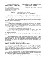
Multifocal purple-red maculo-papular KS lesions on the pal-ateFigure 3
Multifocal purple-red maculo-papular KS lesions on the pal-
ate. Note the pseudomembranous candidiasis (arrow).
KS nodules on the face at initial presentationFigure 1
KS nodules on the face at initial presentation.
Multifocal maculo-papular KS lesions on the gingivaeFigure 2
Multifocal maculo-papular KS lesions on the gingivae.
AIDS Research and Therapy 2008, 5:2 />Page 3 of 6
(page number not for citation purposes)
The patient was wasted, had been treated for tuberculosis
but had chronic obstructive lung disease, pulmonary
hypertension with right ventricular enlargement, and
intractable gastroenteritis. He had developed several cuta-
neous Kaposi sarcomata two months prior to our exami-
nation. The patient reported that the facial oedema and
the oral lesions had been rapidly getting worse over the
last three weeks.
The oral lesions were exophytic, red lobulated masses
affecting the palate, maxillary gingivae and the dorsum of
the tongue (Fig 7). Microscopic examination of a biopsy
specimen from the tongue showed features consistent
with those of KS.
The patient died 7 days after our examination, while sys-
temic investigation was in progress, and before cytotoxic
chemotherapy could be initiated.
Discussion
Lymphoedema
Lymphoedema is characterized by an abnormal accumu-
lation of protein-rich interstitial fluid in the presence of
normal capillary filtration, resulting in swelling of the
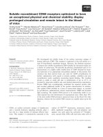
Oedema of the faceFigure 6
Oedema of the face. Note the pronounced periorbital
oedema.
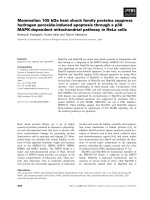
Photograph of the face three months after the initial presen-tation (Fig 1)Figure 4
Photograph of the face three months after the initial presen-
tation (Fig 1). Note the oedema of the face, in particular of
the periorbital tissues.
Photograph showing the worsening of the palatal lesions (three months after the initial examination (Figs 2 & 3)Figure 5
Photograph showing the worsening of the palatal lesions
(three months after the initial examination (Figs 2 & 3).
AIDS Research and Therapy 2008, 5:2 />Page 4 of 6
(page number not for citation purposes)
affected soft tissue. This is in contrast to oedema that
develops when capillary filtration rate exceeds lymph
drainage [12].
Lymphoedema can be categorized as primary and second-
ary. Primary lymphoedema is rare and is associated with
agenesis, hypoplasia or ectasia of lymphatic channels. Sec-
ondary lymphoedema usually occurs due to damage to, or
obstruction of normal lymphatic channels interfering
with lymphatic flow. Common causes of secondary lym-
phoedema include infections that lead to obliteration of
lymphatic lumina, extensive surgical excision of lymph
nodes, post surgical scarring, malignancies that start in, or
spread to lymph nodes, and radiation treatment that
causes fibrosis, blocking the lymphatics. Lymphoedema
affects mainly the extremities, which have limited collat-
eral lymphatics which cannot readily drain excess fluid
[12,13].
Chronic damage to, or blockade of lymphatic channels
may result in low-output failure of lymph circulation.
Long-standing, high-protein lymphoedema has the
potential to stimulate angiogenesis and neovasculariza-
tion both of lymphatics and of blood vessels, lipid depo-
sition, subcutaneous fibrosis and hyperkeratosis. In
addition, stasis of lymph implies failure of transport of
fluid, crystalloids, macro-molecules and lymphoid cells
through the lymphatics, the lymph nodes, and from the
tissues back to the blood stream. There will also be local
immune impairment and depletion of nutritional factors
in the affected lymphoedemateous regions [12,14]. The
development of lymphoedema may be attributed to the
high interstitial protein concentration with increased
osmotic pressure that causes retention of fluid in the con-
nective tissue [15], and to release of growth factors, in par-
ticular vascular endothelial growth factor and basic
fibroblast growth factor, that may bring about subcutane-
ous fibrosis and hyperkeratosis [15,16].
The local environment in chronic lymphoedema
Lymphoedema is associated with local immune impair-
ment. Migration of immunoregulatory cells such as den-
dritic cells, T lymphocytes and macrophages from
peripheral tissue sites to regional lymph nodes via lym-
phatic vessels is compromised in the setting of lym-
phoedema. This results in ineffective clearance of antigens
and in impaired local adaptive cellular immune responses
as well as in immune surveillance. In addition, in lym-
phoedemateous areas there are alterations in the profiles
of the local cytokines, growth factors and adhesion mole-
cules and there is dysregulation of immunoinflammatory
reactions leading to progressive endothelial cell prolifera-
tion with the development of abnormalities in the pattern
of the lymphatic vessels. These alterations in the local
microenvironment and in the immune competence of the
lymphoedemateous areas may play roles of cofactors in
the initiation and promotion of KS [12,17].
Lymphoedema associated with HIV-KS
Lymphoedema may precede the development of HIV-KS;
may be present at the time of diagnosis of HIV-KS; or may
develop later in parallel with the progression of HIV-KS
disease [17].
The clinical oedema observed in some subjects with KS
may be pronounced, as in the presented cases, and affects
mainly the lower extremities and the periorbital areas
[6,15]. Lymphangioscintigraphy in subjects with KS
shows a variety of abnormal patterns of the lymphatic
channels, corresponding to the distribution of the cutane-
ous KS lesions [18], and to the clinical lymphoedema. At
times the oedema with its associated subcutaneous fibro-
sis and with hyperkeratosis may camouflage the cutane-
ous KS lesions and make the diagnosis of KS difficult
[15,19].
In addition to the factors previously, mentioned HHV-8
infection of lymphatic endothelial cells and of cells resi-
dent in lymph nodes may cause both damage to lym-
phatic channels and enlargement of lymph nodes, with
impeded lymphatic drainage and consequent lym-
phoedema [12,15,18]. HHV-8 is a necessary factor but not
in its own sufficient to cause KS: but chronic lym-
phoedema together with HHV-8 may initiate the develop-
ment of in-situ KS that with time progresses to become
clinically manifest KS [20].
Our first patient refused hospitalization or any further
investigations or treatment at all, therefore we can only
Intraoral view showing extensive KS lesions on the hard pal-ate, the soft palate and the dorsum of the tongueFigure 7
Intraoral view showing extensive KS lesions on the hard pal-
ate, the soft palate and the dorsum of the tongue.
AIDS Research and Therapy 2008, 5:2 />Page 5 of 6
(page number not for citation purposes)
state that the facial oedema was present but not whether
it may have had a non-HIV/KS related cause. The second
patient was in hospital in the care of physicians who
recorded none of the other possible causes of facial lym-
phoedema than HIV-KS.
Lymphoedema that precedes KS may in the presence of
HHV-8 infection be a predisposing factor to the develop-
ment of KS from lymphatic endothelial cells [16,20]. On
the other hand, it has been shown [19,20], that fibroma-
like nodules in the submucosa, of uncertain etiology, in
the presence of lymphoedema and HHV-8 may evolve to
in-situ KS.
The simultaneous development of lymphoedema and KS,
or the development of lymphoedema in parallel with the
progression of established KS may be attributed to the
HHV-8-induced exuberant proliferation of endothelial
cells that may lead to the occlusion of lymphatic vascular
lumens. When this is widespread and involves multiple
lymphatic channels, it has the potential to cause lym-
phoedema. The obliteration of the lymphatic channels
may be supplemented by their compression by the enlarg-
ing KS [18,19]. This process is most probably responsible
for the severe clinical oedema associated with rapidly pro-
gressive HIV-KS disease.
Treatment of HIV-KS associated lymphoedema
Treatment of lymphoedema associated with HIV-KS
focuses on treatment of both the HIV-KS and the lym-
phoedema. Highly active antiretroviral therapy (HAART)
together with cytotoxic chemotherapy is the treatment of
choice for the management of HIV-KS. At times this may
also improve the associated lymphoedema. The lym-
phoedema should be treated in parallel to the treatment
of HIV-KS with diuretic agents, and in cases where the
lower extremities are involved, by elevation of the limbs
and compressive dressings. The preservation of skin integ-
rity is important to prevent secondary bacterial and fungal
infections. Application of emollients to the affected lym-
phoedematous skin may prevent cutaneous breakdown. If
breakdown does occur, application of topical antimicro-
bial agents is necessary [21].
Comments
According to the AIDS clinical trial group staging classifi-
cation for AIDS-associated Kaposi sarcoma, lym-
phoedema and exophytic HIV-KS oral lesions are
independently associated with poor prognosis [3,4]. The
two cases reported here demonstrate that in HIV-seropos-
itive subjects with established nodular oral KS lesions, the
development of facial lymphoedema rapidly leads to
death. The pathogenic mechanisms that bring about the
rapid development of facial lymphoedema and that lead
to death are obscure.
In sub-Saharan Africa, the natural course of HIV-KS in the
absence of HAART is characterized by rapid disease pro-
gression associated with high HHV-8 burden and short
life expectancy. In this geographic region, HIV-KS has
reached epidemic proportions [22,23]. HIV-KS in the
mouth is common, and subjects with extensive exophytic
oral lesions and tumour-associated oedema have higher
death rates than HIV-seropositive subjects having exclu-
sively cutaneous lesions [3].
Since there is no cure for HIV-KS, conventionally its treat-
ment focused on control of tumour growth and pallia-
tion. HAART should always be instituted in HAART-naïve
HIV-seropositive subjects with KS, since it promotes
regression of KS lesions. However, in about 6,5% of these
subjects HIV-KS may flare up shortly after the introduc-
tion of HAART as an immune reconstitution inflamma-
tory syndrome (IRIS) [24].
In light of the cases presented here and our personal expe-
rience, it might be advisable to treat oral HIV-KS with
cytotoxic chemotherapy at early maculo-papular stages
when the lesions are still asymptomatic. Such a treatment
protocol might have several advantages. Firstly, systemic
cytotoxic chemotherapy might prevent or at least delay
the development of extensive exophytic oral lesions that
have a poor prognosis; secondly it might prevent the
development of lymphoedema associated with late stages
of HIV-KS; and thirdly, in the early stages of oral HIV-KS,
limited cytotoxic chemotherapy is sufficient to control
tumour growth compared to more extensive chemother-
apy regimen needed to treat advanced oral HIV-KS.
Indeed, systemic chemotherapy given to HIV-seropositive
subjects has the risk of exaggerating the existing state of
immuno-suppression, thus further increasing the suscep-
tibility to opportunistic infections and neoplasms, how-
ever, limited doses of cytotoxic chemotherapy is not
usually associated with such adverse effects.
Acknowledgements
Informed consent was obtained from the relatives of these patients for pub-
lication of this case report and any accompanying images. A copy of the
written consent is available for review by the Editor-in-Chief of this journal.
References
1. Wang HW, Trotter MW, Lagos D, Bourboulia D, Henderson S, Mäk-
inen T, Elliman S, Flanagan AM, Alitalo K, Boshoff C: Kaposi sar-
coma herpesvirus-induced cellular programming
contributes to the lymphatic endothelial gene expression in
Kaposi sarcoma. Nat Genet 2004, 36:687-693.
2. Schwartz AA: Kaposi's sarcoma: an update. J Surg Oncol 2004,
87:146-151.
3. Fauci AS, Lane HC: Human immunodeficiency virus disease:
AIDS and related disorders. In Harrison's principles of internal med-
icine 16th edition. Edited by: Kasper DL, Braunwald E, Fauci AS,
Hauser SL, Longo DL, Jameson JL. New York: McGraw-Hill;
2005:1076-1139.
4. Krown SE, Testa MA, Huang J: AIDS-related Kaposi's sarcoma:
Prospective validation of the AIDS clinical trials group stag-
ing classification. J Clin Oncol 1997, 15:3085-3092.
Publish with BioMed Central and every
scientist can read your work free of charge
"BioMed Central will be the most significant development for
disseminating the results of biomedical research in our lifetime."
Sir Paul Nurse, Cancer Research UK
Your research papers will be:
available free of charge to the entire biomedical community
peer reviewed and published immediately upon acceptance
cited in PubMed and archived on PubMed Central
yours — you keep the copyright
Submit your manuscript here:
/>BioMedcentral
AIDS Research and Therapy 2008, 5:2 />Page 6 of 6
(page number not for citation purposes)
5. Krown SE, Metroka C, Wernz JC: Kaposi's sarcoma in Acquired
immune Deficiency Syndrome: A proposal for uniform eval-
uation, response, and staging criteria. J Clin Oncol 1989,
7:1201-1207.
6. Bossuyt L, Van den Oord JJ, Degreef H: Lymphangioma-like vari-
ant of AIDS-associated Kaposi's sarcoma with pronounced
edema formation. Dermatology 1995, 190:324-326.
7. Feller L, Lemmer J, Wood NH, Jadwat Y, Raubenheimer EJ: HIV-
associated oral Kaposi sarcoma and HHV-8: a review. J Int
Acad Periodontol 2007, 9:129-136.
8. Feller L, Lemmer J, Wood NH, Raubenheimer EJ: Necrotizing gin-
givitis of Kaposi sarcoma affected gingivae. SADJ 2006,
61:314-317.
9. Feller L, Wood NH, Lemmer J: HIV-associated Kaposi Sarcoma:
Pathogenic mechanisms. Oral Surg Oral Med Oral Pathol Oral Radiol
Endod 2006, 104:521-529.
10. Lager I, Altini M, Coleman H, Ali H: Oral Kaposi's sarcoma: a clin-
ico-pathologic study from South Africa. Oral Surg Oral Med Oral
Pathol Oral Radiol Endod 2003, 96:701-710.
11. Epstein JB, Cabay RJ, Glick M: Oral malignancies in HIV disease:
changes in disease presentation, increasing understanding of
molecular pathogenesis, and current management. Oral Surg
Oral Med Oral Pathol Oral Radiol Endod 2005, 100:571-578.
12. Ruocco V, Schwaartz RA, Ruocco E: Lymphedema: an immuno-
logically vulnerable site for development of neoplasms. J Am
Acad Dermatol 2002, 47:124-127.
13. Creager MA, Dzau VS: Vascular diseases of the extremities. In
Harrison's Principles of Internal Medicine 16th edition. McGraw-Hill;
2005:1486-1494.
14. Witte MH, Witte CL, Way DL: Medical ignorance, AIDS-
Kaposi's sarcoma complex and the lymphatic system. West J
Med 1990, 153:17-23.
15. Hengge UR, Stocks K, Goos M: Acquired immune deficiency syn-
drome-related hyperkeratotic Kaposi's sarcoma with severe
lymphoedema: report of five cases. Br J Dermatol 2000,
142:501-505.
16. Schwartz RA, Cohen JB, Watson RA, Gascón P, Ahkami RN, Ruszczak
Z, Halpern J, Lambert W: Penile Kaposi's sarcoma preceded by
chronic penile lymphoedema. Br J Dermatol 2000, 142:153-156.
17. Smith KJ, Skelton H: Kaposi's sarcoma-like angiosarcomas may
reflect a common lymphatic endothelium differentiation
pattern as Kaposi's sarcoma in association with chronic
lymphedema. Int J Dermatol 2006, 45:623-626.
18. Witte MH, Fiala M, McNeill GC, Witte CL, Williams WH, Szabo J:
Lymphangioscintigraphy in AIDS-associated Kaposi sar-
coma. AJR 1990, 155:311-315.
19. Ramdial PK, Chetty R, Singh B, Singh R, Aboobaker J: Lymphedema-
tous HIV-associated Kaposi's sarcoma. J Cutan Pathol 2006,
33:474-481.
20. Konstantinopoulos PA, Dezube BJ, Pantanowitz L: Morphologic
and immunophenotypic evidence of in-situ Kaposi's sar-
coma. BMC Clinical Pathology 2006, 6:7. doi:10.1186/1472-6890-6-7.
21. Allen PJ, Gillespie DL, Redfield RR, Gomez ER: Lower extremity
lymphedema caused by acquired immune deficiency syn-
drome-related Kaposi's sarcoma: Case report and review of
the literature. J Vasc Surg 1995, 22:178-181.
22. Di Lorenzo G, Konstantinopoulos PA, Pantanowitz L, Di Trolio R, De
Placido S, Dezube BJ: Management of AIDS-related Kaposi's
sarcoma. Lancet Oncol 2007, 8:167-176.
23. Krown SE: AIDS-associated Kaposi's sarcoma: is there still a
role for interferon alpha? Cytokines and Growth Factor Reviews
2007, 18:395-402.
24. Bower M, Nelson M, Young AM, Thirlwell C, Newsom-Davis T, Man-
dalia S, Dhillon T, Holmes P, Gazzard BG, Stebbing J: Immune
reconstitution inflammatory syndrome with Kasposi's sar-
coma. J Clin Oncol 2005, 23:5224-5228.