Extracellular signal-regulated kinase 1/2 plays a pro-life role in experimental brain stem death via MAPK signal-interacting kinase at rostral ventrolateral medulla pps
Bạn đang xem bản rút gọn của tài liệu. Xem và tải ngay bản đầy đủ của tài liệu tại đây (608.15 KB, 9 trang )
RESEA R C H Open Access
Extracellular signal-regulated kinase 1/2 plays a
pro-life role in experimental brain stem death via
MAPK signal-interacting kinase at rostral
ventrolateral medulla
Samuel HH Chan, Enya YH Sun, Alice YW Chang
*
Abstract
Background: As the origin of a life-and-death signal detected from systemic arterial pressure, which sequentially
increases (pro-life) and decreases (pro-death) to reflect progressive dysfunction of central cardiovasc ular regulation
during the advancement towards brain stem death in critically ill patients, the rostral ventrolateral medulla (RVLM)
is a suitable neural substrate for mechanistic delineation of this fatal phenomenon. The present study assessed the
hypothesis that extracellular signal-regulated kinase 1/2 (ERK1/2), a member of the mitogen-activated protein
kinases (MAPKs) that is important for cell survival and is activated specifically by MAPK kinase 1/2 (MEK1/2), plays a
pro-life role in RVLM during brain stem death. We further delineated the participation of MAPK signal-interacting
kinase (MNK), a novel substrate of ERK in this process.
Methods: An experimental model of brain stem death that employed micro injection of the organophosphate
insecticide mevinphos (Mev; 10 nmol) bilaterally into RVLM of Sprague-Dawley rats was used, in conjunction with
cardiovascular, pharmacological and biochemical evaluations.
Results: Results from ELISA showed that whereas the total ERK1/2 was not affected, augmented phosphorylation
of ERK1/2 at Thr202 and Tyr204 in RVLM occurred preferentially during the pro-life phase of experimental brain
stem death. Furthermore, pretreatment by microinjection into the bilateral RVLM of a specific ERK2 inhibitor, ERK
activation inhibitor peptide II (1 nmol); a specific MEK1/2 inhibitor, U0126 (5 pmol); or a specific MNK1/2 inhibitor,
CGP57380 (5 pmol) exacerbated the hypotension and blunted the augmented life-and-death signals exhibited
during the pro-life phase . Those pretreatments also blocked the upregulated nitric oxide synthase I (NOS I)/protein
kinase G (PKG) signaling, the pro-life cascade that sustains central cardiovascular regulatory functions during
experimental brain stem death.
Conclusions: Our results demonstrated that activation of MEK1/2, ERK1/2 and MNK1/2 in RVLM plays a preferential
pro-life role by sustaining the central cardiovascular regulatory machinery during brain stem death via upregulation
of NOS I/PKG signaling cascade in RVLM.
Background
Although brain stem death is currently the legal defini-
tion of death in Taiwan and many countries [1,2], the
detailed cellular an d molecular mechanisms underlying
this phenomenon of paramount medical importance are
still unclear. The invariable prognosis that asystole
occurs within hours or days after the diagnosis of brain
stem death [3] strongly suggests that permanent impair-
ment of the brain stem cardiovascular regulatory
machinery precedes death. Better understanding of the
mechanistic aspects of the dysfunction of central cardio-
vascular regulation during brain stem death should
therefore enrich the dearth of information currently
available on this fatal phenomenon.
* Correspondence:
Center for Translational Research in Biomedical Sciences, Chang Gung
Memorial Hospital-Kaohsiung Medical Center, Kaohsiung County 83301,
Taiwan
Chan et al. Journal of Biomedical Science 2010, 17:17
/>© 2010 Chan et al; licensee BioMed Central Ltd. This is an Open Access article distribut ed under the terms of the Creative Commons
Attribution License (http://crea tivecommons.org/li censes/by/2 .0), which permits unrestricted use, distribution, and reproduction in
any medium, provided the original work is properly cited.
One suitable experimental animal model for mechanistic
evaluation of brain stem death uses the organophosphate
poison mevinphos (3-(dimethoxyphosphinyloxyl)-2-bute-
noic acid methyl ester (Mev), a US Environmental Protec-
tion Agency Toxicity Category I pesticide, as the
experimental insult [4]. At the same time, as the origin of
a life-and-death signal [5] that reflects failure of the central
cardiovascular regulatory machinery during brain stem
death [6-8] and a brain stem site via which Mev acts to eli-
cit cardiovascular toxicity [9], the rostral ventrolateral
medulla (RVLM) is a suitable neural substrate for mechan-
istic evaluation of this fatal phenomenon [4]. Of interest is
that the waxing and waning of the life-and-death signal,
which mirrors the fluctuation of neuronal functionality in
RVLM, presents itself as the low-frequency (LF) compo-
nent in the systemic arterial pressure (SAP) spectrum of
intensive-care unit patients [6-8]. More importantly, the
distinct phases of augmentation followed by reduction of
the LF p ower exhibited during Mev intoxication [10-13]
can be designated the pro-life and pro-death phase of cen-
tral cardiovascular regulation in this model of brain stem
death [4]. Based on this model, our laboratory has pre-
viously demonstrated that nitric oxide (NO) generated by
NO synthase I (NOS I) in RVLM, followed by activation
of the soluble guanylyl cyclase/cyclic GMP/protein kinase
G (PKG) cascade, is responsible for the pro-life phase; per-
oxynitrite formed by a reaction between NOS II-derived
NO and superoxide anion underlies the pro-death phase
[10-13].
As death represents the end of existence for an indivi-
dual, we proposed previously [4] that multiple pro-life
and pro-death programs must be activated in RVLM
during the progression toward brain stem death. There-
fore, one meaningful direction in our search for the cel-
lular and molecular mechanisms of brain stem death is
to identify these regulatory programs. In this re gard, the
extracellular signal-regulated kinases (ERKs) present
themselves as another reasonable candidate for the pro-
life program. As a member of the mitogen-activated
protein kinases (MAPKs), ERK1/2 pathway is activated
specifically by MAPK kinase 1/2 (MEK1/2), and is an
important signal for cell survival [14-19]. Sustained inhi-
bition of ERK expression leads to the induction of apop-
tosis in rat neuronal PC12 cells [15]. On the other hand,
ERK is strongly and persistently activated during cell
survival [16,17]. Wit h particular relevance to this study,
ERK1/2 activation leads to NOS I induction in rat PC12
cells [18] and in rat aortic smooth muscle cells [19].
Based on our Mev intoxication model [4], the present
study evaluated the hypothesis that MEK1/2 and ERK1/
2 in RVLM play a pro-life role during brain stem death
by activating the NOS I/PKG cascade. We further deli-
neated the participation of MAPK signal-interacting
kinase (MNK), a n ovel substrate of ERK [20,21] in this
process. Our results demonstrated that activation of
MEK1/2, ERK1/2 and MNK1/2 in RVLM plays a prefer-
ential pro-life role by sustaining central cardiovascular
regulatory functions d uring brain stem death via upre-
gulation of the NOS I/PKG signaling cascade in RVLM.
Methods
Adult male Sprague-Dawley rats (285-345 g, n = 164)
purchased from the Experimental An imal Center of the
National Science Council, Taiwan, Republic of China
were used. All experimental procedures carried out in
this study have been approved by the Laboratory Animal
Committee of the Chang G ung Memorial Hospital-
Kaohsiung Medical Center, and were in compliance
with the guidelines for animal care set forth by this
Committee.
General preparation
Preparatory surgery was carried out under an induction
dose of pentobarbital sodium (50 mg/kg, i.p.), and
included cannulation of a femoral artery and a femoral
vein, together with tracheal intubation. During the
recording session, which routinely commenced 60 min
after the administration of pentobarbital sodium,
anesthesia was maintained by intravenous infusion of
propofol (Zeneca, Macclesfield, UK) at 20-25 mg/kg/h.
We have demonstrated previously [22] that this scheme
provided satisfactory anesthetic maintenance while pre-
servi ng the capacity of central cardiovascular regulation.
Body temperature of the animals was maintained at 37°
C w ith a heating pad, and rats were allowed to breathe
spontaneously with room air during the entire recording
session.
Animal model of brain stem death
The Mev intoxication model of brain stem death [4] was
used. Since Mev induces comparable cardiovascular
responses on given systemically or directly to RVLM [9],
we routinely microinjected Mev bilaterally into RVLM
to elicit site-speci fic effects [9-13]. SAP signals recorded
from the femoral artery were simultaneously subjected
to on-line power spectral analysis [9-13,23]. We were
particularly interested in the low-frequency (LF; 0.25-0.8
Hz) component in the SAP spectrum because its power
density mirrors the prevalence of baroreceptor reflex
(BRR)-mediated sympathetic neurogenic vasomotor dis-
charges that emanate from this brain stem site [23].
More importantly, our laboratory demonstrated pre-
viously [10-13] that the power density of this spectral
signal exhibits biphasic changes that reflect the pro-life
and pro-death phases seen during the progression
towards brain stem death in patients who succumbed to
organophosphate poisoning [8]. Heart rate (HR) was
derived instantaneously from SAP signals. Temporal
Chan et al. Journal of Biomedical Science 2010, 17:17
/>Page 2 of 9
changes in the power density of the LF component, pul-
satile SAP, mean SAP (MSAP) and HR were routinely
followed for 180 min after Mev administration in an on-
line and real-time manner.
Microinjection of test agents
Microinjection bilaterally of test agents into RVLM,
each at a volume of 50 nl, wa s carried out st ereotaxi-
cally and sequentially [9-13] via a glass micropipette
connected to a 0.5-μl Hamilton (Reno, NV, USA)
microsyringe. The coordinates used were: 4.5-5 mm
posterior to lambda, 1.8-2.1 mm lateral to midline,
and 8.1-8.4 mm below the dorsal surface of cerebel-
lum. These coordinates were selected to cover the
ventrolateral medulla at which functionally identified
sympathetic premotor neurons reside [24]. Test
agents used included Mev (kindly provided by Huik-
wang Corporation, Tainan , Taiwan), a specific ERK2
inhibitor [25], ERK activation inhibitor peptide II
(Calbiochem, San Diego, CA, USA); a specific MEK1/
2 inhibitor [26,27], U0126 (Calbiochem); or a specific
MNK1/2 inhibitor [28,29], CGP57380 (Tocris, Ellis-
ville, MO, USA). All test agents used for pretreat-
ments were given 30 min before the administration of
Mev. The doses were adopted fro m previous reports
[25-29] that used those test agents for the same pur-
pose as in this study. Application of the same amount
of artificial cerebrospinal fluid (aCSF) controlled for
possible volume effect of microinjection of Mev, and
0.2% DMSO serving as the vehicle control for ERK
activation inhibitor peptide II, U0126 or CGP57380.
The composition of aCSF was (mM): NaCl 117,
NaHCO
3
25, glucose 11, KCl 4.7, CaCl
2
2.5, MgCl
2
1.2 and NaH
2
PO
4
1.2. To avoid the confounding
effects of drug interactions, each animal received only
one pharmacological treatment.
Collection of tissue samples from ventrolateral medulla
We routinely collected tissue sa mples for subsequent
biochemical evaluations [10-13] during the peak of the
pro-life and pro-death phase (Mev group), or 30 or 180
min after microinjection of aCSF or 0.2% DMSO into
RVLM (vehicle cont rol group). Animals were killed with
an overdose of pentobarbital sodium and tissues from
both sides of the ventrolateral medulla, at the level of
RVLM (0.5-1.5 mm rostral to the obex), were collected
by micropunches made with a 1 mm (i.d.) stainless-steel
bore to cover the anatomical bounda ries of RVLM.
Medullary tissues collected from anesthetized animals
but without treatment served as the sham-controls. The
conc entr ation of total proteins extracted from those tis-
sue samples was determined by the BCA protein assay
(Pierce, Rockford, IL, USA).
ELISA for ERK or phosphorylated ERK
Cell lysate from ventrolateral medulla was subjected to
enzyme-linked immunosorbent assay (ELISA) according
to the manufacturer’s protocol of a commercial kit (Cell
Signaling, Danvers, MA, USA) to detect the levels of
ERK1/2 or phosph orylated ERK1/2 at Thr202/Tyr204.
The final absorbance of reaction solution at 450 nm was
determined by spectrophotometry using an ELISA
microtiter plate reader (Anthros Labtec, Salzburg, Aus-
tria),andexpressedasfoldchangesagainstsham-
controls.
Western blot analysis
Western blot analy sis [10-13,23] was carried out using
a rabbit polyclonal antiserum against NOS I, NOS II
(Santa Cruz, Santa Cruz, CA, USA) or PKG (Calbio-
chem); or a mouse monoclonal antiserum against
nitrotyrosine (Upstate, Lake Placid, NY, USA) or b-
actin (Chemicon, Temecula, CA, USA). This was fol-
lowed by incubation with horseradish peroxidase-con-
jugated donkey anti-rabbit IgG (Amersham
Biosciences, Little Cha lfont, Bucks, UK) for NOS I,
NOS II or PKG, or sheep anti-mouse IgG (Amersham
Biosciences) for nitrotyrosine or b-actin. Specific anti-
body-antigen comple x was detected by an enha nced
chemiluminescence Western blot detection system
(NEN, Boston, MA, USA). The amount of protein pro-
duct was quantified by the ImageMaster Video Docu-
mentation System (Amersham Pharmacia Biotech), and
wasexpressedastheratiotob-actin protein.
Histology
In some animals that were not used for biochemical
analysis, the brain stem was removed at the end of the
physiological experiment and fixed in 3 0% sucrose in
10% formaldehyde-saline solut ion for at leas t 72 h. Fro-
zen 25-μm sections of the medulla oblongata stained
with neural red were used for histological verification of
the microinjection sites.
Statistical analysis
All va lues are expressed as mean ± SEM. The averaged
value of MSAP or HR calculated every 20 min after
administration of test agents or vehicle, the sum total o f
power density for the LF component in the SAP spec-
trum over 20 min, or the protein expression level in
RVLM during each phase of experimental b rain stem
death, were used for statistical analysis. One-way or
two-way ANOVA with repeated measures was used, as
appropriate, to assess group means. This was followed
by the Scheffé multiple-range test for post hoc assess -
ment of individual means. P < 0.05 was considered to
be statistically significant.
Chan et al. Journal of Biomedical Science 2010, 17:17
/>Page 3 of 9
Results
Mev intoxication model of brain stem death
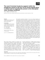
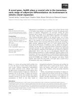
Figure 1 shows that co-microinjection bilaterally of Mev
(10 nmo l) and vehicle into RVLM elicited a progressive
hypotension that became signif icant 100 min after appli-
cation, accompanied by indiscernible alterations in HR.
Concurrent changes in power density of the LF compo-
nent of SAP signals revealed two distinct phases [10-13].
The pro-life Phase I entailed a significantly augmented
LF power that endured 80-10 0 min to reflect sustained
brain stem cardiovascular regulatory functions. The pro-
death Phase II, which lasted the remainder of our 180-
min observation period, exhibited further and significant
reduction in the power density of this spectral compo-
nent to below baseline, which signifies failure of central
cardiovascular regulation that precedes brain stem death
[4].
Preferential activation of ERK1/2 in RVLM during the pro-
life phase
We first evaluated the fundamental premise that ERK1/2
in RVLM is activated during experimental brain stem
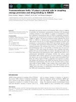
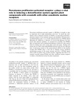
death. Quantification by ELISA revealed that the total
ERK1/2 in ventrolateral medulla was not affected by
microinjection of Mev into the bilateral RVLM (fig. 2).
Interestingly, phosphorylated ERK1/2 (pERK1/2) at
Thr202 and Tyr204 in RVLM was significantly and pre-
ferentially augmented during the pro-life phase (fig. 2),
of which returned to baseline during the pro-death
phase. The level of both ERK1/ 2 and pERK1/2 in ven-
trolateral medulla of vehicle groups was comparab le to
sham-controls.
Activation of MEK1/2, ERK1/2 or MNK1/2 in RVLM
sustains central cardiovascular regulation associated with
experimental brain stem death
Based on the stipulation that the magnitude and dura-
tion of the LF component of SAP signals during experi-
mental brain stem death ref lect the pre valence of the
life-and-death signal [4], we next employed pharmacolo-
gical blockade to evaluate whether a causal relationship
exists between activation of ERK1/2 in RVLM and cen-
tral cardiovascular regulation during brain stem death.
Pretreatment with microinjection into the bilateral
RVLM of ERK activation inhibitor peptide II (1 nmol),
which binds specifically to ERK2 to prevent its interac-
tion with MEK [25], exacerbated significantly the hypo-
tension and blunted the augmented power density of
the LF component of SAP signals during the pro-life
phase (fig. 1), withou t affecting HR. Similar results were
obtained o n local application bilaterally into RVLM of
U0126(5pmol),aspecificinhibitorofMEK1and
MEK2 [26,27] (fig. 1). Those pretreatments also
significantly shortened the pro-life phase to 25-3 5 min
by shifting the prevailing phase of the 180-min observa-
tion period toward the pro-death phase (fig. 1). Intrigu-
ingly, comparable results were also obtained on
pretreatment with microinjection bilaterally into RVLM
of CGP57380 (5 pmol), a specific cell-permeable
MNK1/2 inhibitor [28,29] (fig. 1); although a dose of 1
pmolwasineffectiveagainstthecardiovascular
responses during the pro-life phase (maximal MSAP:
112.5 ± 5.2 versus 113.0 ± 5.8 mmHg; maxi mal HR:
356.8 ± 20.1 versus 354.6 ± 16.6 bpm; maximal LF
power: 73.6 ± 6.7 versus 75.2 ± 7.5 mmHg
2
when com-
pared to 0.2% DMSO pretreatment; mean ± SEM, n = 4
animals). On the other hand, ERK activation inhibitor
peptide II, U0126 or CGP57380 did not significantly
affect the hypotension and decrease in LF power already
exhibited during the pro-death phase. Furthermore, pre-
treatment with vehicles exerted minimal effects on the
phasic cardiovascular responses.
Activation of MEK1/2 or ERK1/2 underlies the
augmentation of NOS I or PKG in RVLM during the pro-
life phase
We previously demonstrated [10-13] that NOS I/PK G
signaling in RVLM is responsible for sustaining central
cardiovascular regulation during the pro-life phase in
our Mev intoxication modelofbrainstemdeath.Itis
therefore conceivable that MEK1/2 or ERK1/2 in RVLM
may confer its pro-life actions via the NOS I/PKG cas-
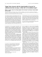
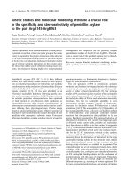
cade. As r eported previously [10-13], Western blot ana-
lysis revealed a significant augmentation of NOS I or
PKG expression in ventrolateral medulla during the pro-
life phase, followed by a return to baseline during the
pro-death phase (fig. 3). Pharmacological blockade was
again used to ascertain that these temporally correlated
biochemical changes are causally linked to MEK1/2 or
ERK1/2 activation in RVLM during experimental brain
stem death. Pretreating animals by microinjection into
the bilateral RVLM of ERK activation inhibitor peptide
II (1 nmol) or U0126 (5 pmol) significantly blunted the
augmented NOS I or PKG protein expression at ventro-
lateral medulla during the pro-life phase (fig. 3). On the
other hand, the protein levels of NOS I and PKG during
the pro-death phase were not affected by these
pretreatments.
Activation of MEK1/2 or ERK1/2 is not responsible for the
augmentation of NOS II or peroxynitrite in RVLM during
the pro-death phase
We also demonstrated in our previous studies [10-13]
that a progressive augmentation of NOS II and nitrotyr-
osine (an experimental index for peroxynitrite) expres-
sion in RVLM underlies the failure of central
Chan et al. Journal of Biomedical Science 2010, 17:17
/>Page 4 of 9
Figure 1 Activation of MEK1/2, ERK1/2 or MNK1/2 in RVLM sustained central cardiovas cular regulation associated with experi mental
brain stem death. Temporal changes in mean systemic arterial pressure (MSAP), hear rate (HR) or power density of low-frequency (LF)
component of SAP signals in rats that received pretreatment by microinjection bilaterally into RVLM of vehicle (Veh; aCSF or 0.2% DMSO), ERK
activation inhibitor peptide II (Peptide II; ERK2 inhibitor), U0126 (MEK1/2 inhibitor) or CGP57380 (MNK1/2 inhibitor), 30 min before local
application (at arrow) of aCSF or Mev (10 nmol) to the bilateral RVLM. Values are mean ± SEM, n = 5-7 animals per experimental group. *P <
0.05 versus Veh+aCSF group, and
+
P < 0.05 versus Veh+Mev group at corresponding time-points in the Scheffé multiple-range test.
Chan et al. Journal of Biomedical Science 2010, 17:17
/>Page 5 of 9
car diovascular regulatory functions during experimental
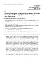
brain stem death. As such, MEK1/2 or ERK1/2 activa-
tion in RVLM may also lead to an antagonism o f this
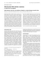
augmentation. However, pretreatment with ERK activa-
tion inhibitor peptide II (1 nmol) or U0126 (5 pmol),
similar to the vehicle controls, exerted no influence
against the increase of NOS II and nitrotyrosine protein
expression in ventrolateral medulla during both phases
of experimental brain stem death (fig. 4).
Activation of MNK1/2 leads to upregulation of NOS I or
PKG in RVLM during the pro-life phase
Our final series of experiments investigated whether
activation of MNK1/2 is upstream to the augmented
NOS I or PKG expression in RVLM during the pro-life
phase. Western blot analysis again revealed that micro-
injection bilaterally into RVLM of an effective dose of
CGP57380 (5 pmol) significantly blunted the elevat ed
NOS I or PKG p rotein level at ventrolateral medulla
during the pro-life phase of experimental brain stem
death (fig. 3), without af fecting the increase of NOS II
or nitrotyrosine protein expression (fig. 4).
Discussion
Based on a clinically relevant experimental model [4],
the present study provided novel demonstrations that
MEK1/2 or ERK1 /2 activation in RVLM sustains central
cardiovascular regulation during the progression towards
brain stem death. We further showed that mechanisti-
cally, this pro-life role was executed v ia upregulation of
the pro-life NOS I/PKG signaling cascade by MNK1/2.
It is generally contended that of the three MAPKs
characterized in mammals, ERK1/2 plays a crucial role
in survival responses [30-33]. On the other hand, Jun N-
terminus kinase (JNK) and p38 MAPK primarily med-
iate inflammatory response [34,35] and promote cell
death [36-38]. In rats that received transient middle cer-
ebral artery occlusion, pERK is strongly expressed at the
ischemic penumbra in cerebral cortex and is essentially
involved in cell survival [33]. The MEK/ERK survival
pathway mediates neuroprotection of striatal neurons
[32] or hippocampal CA1/CA3 neurons [31] against glu-
tamatergic neuronal cell death. It also protects s ympa-
thetic neurons against apoptosis induced by the
antimitotic nucleoside cytosine arabinoside [30]. The
Figure 2 Activation of ERK1/2 in RVLM during the pro-life
phase of experimental brain stem death. Changes in total ERK or
phosphorylated ERK at Thr202 and Tyr204 in folds relative to sham-
control (SC), detected in ventrolateral medulla during the pro-life
phase I (MI) or pro-death phase II (MII) of experimental brain stem
death or during comparable time points after treatment with aCSF
(AI or AII). Values are presented as mean ± SEM of triplicate analyses
on tissue samples pooled from 5-7 animals in each experimental
group. *P < 0.05 versus corresponding aCSF group (AI or AII) in the
Scheffé multiple-range analysis.
Figure 3 Activation of MEK1/2, ERK1/2 or MNK1/2 leads to
phasic upregulation of NOS I/PKG cascade in RVLM during
experimental brain stem death. Illustrative gels or summary of
fold changes against aCSF controls in ratio of nitric oxide synthase I
(NOS I) or protein kinase G (PKG) relative to b-actin protein
detected in ventrolateral medulla of rats that received ERK
activation inhibitor peptide II, U0126 or CGP57380 into bilateral
RVLM, 30 min before induction of experimental brain stem death.
Values are mean ± SEM of triplicate analyses on samples pooled
from 5-7 animals per experimental group. *P < 0.05 versus aCSF
group and
+
P < 0.05 versus Mev group in the Scheffé multiple-
range test.
Chan et al. Journal of Biomedical Science 2010, 17:17
/>Page 6 of 9
present study further identified a novel survival function
for MEK1/2 or ERK1/2 at RVLM in the form of a pro-
life role during experimental brain stem death.
We previously demonstrated a pro-life role for NOS I/
PKG cascade at RVLM in experimental brain stem
death [10-13]. The present study further revealed that
this pro-life signaling cascade is downstream to activa-
tion of MEK/ERK in RVLM. This demonstration is
echoed by the observation [19] that gene transfer or
gene kn ockdown of MEK increases or decreases NOS I
expression in culture d rat aortic smooth muscle cells.
Moreover, the MEK/ERK pathway, but not JNK or p38
MAPK, is required for NOS I mRNA or protein expres-
sion and activity in PC12 cells [18]. There are two possi-
ble, though not necessarily mutually exclusive,
mechanisms for MEK/ERK to elicit NOS I induction.
One possibility is for ERK1/2 to transcriptionally upre-
gulate NOS I. The promoter region of NOS I gene con-
tains putative cis-elements of binding sites for cAMP
response element (CREB), C/EBP and c-Myc [39,40],
which are c andidates for ERK nuclear targeting i n the
mediation of gene transcription. Another possibility is
for ERK to exert posttranslational modificat ion by phos-
phorylation of NOS I protein. ERK1/2 is tightly bound
to its physiologically relevant substrates, such as MNKs
and p90RSK1 for subsequent physiological responses.
Whereas p90RSK1 is a well-known substrate for ERK1/
2, MNK1 and MNK2 are novel serine/threonine protein
kinases that could be phosphorylated by ERK1/2 [20].
MNK1 activation is inhibit ed by MEK inhibitor
PD98 059 [20,21], suggesting that it is an important reg-
ulator for MNK activation. It follows that MNK1/2 may
enhance phosphorylation of NOS I on activation by
MEK/ERK. Whether the implied augmentation of NOS
I or PKG expression in RVLM by MNK1/2 observed
during experimental brain stem death in the present
study entails transcriptional upregulation remains to be
investigated.
Our results also showed that the pro-life role of MEK,
ERKandMNKinRVLMduringexperimentalbrain
stem death is manifested by sustaining central cardio-
vascular regulation. In this regard, it is of interest to
note that these cellular signals may be linked to angio-
tensin II (Ang II), a well-known peptide that is crucial
to the elevation of SAP in RVLM. Activation of the
ERK/CREB/c-fos cascade mediates the l ong-term
pressor effect of Ang II in RVLM [26]. MEK and ERK1/
2 also participate in Ang II-induced vascular smooth
muscle cell contraction [41]. Furthermore, MNK med-
iates Ang II-induced protein synthesis in vascular
smooth muscle cells [29]. Whether Ang II in RVLM
plays a pro-life role during brain stem death via activa-
tion of the MEK/ERK/MNK cascade, however, awaits
documentation.
Conclusion
In conclusion, the present study revealed that the MEK/
ERK/MNK cascade in RVLM plays a pro-life role during
experimental brain stem death by sustaining the central
cardiovascular regulatory machinery via NOS I/PKG
signaling.
Acknowledgements
Supported by research grants NSC97-2320-B-182A-007-MY3, NSC97-2321-B-
182A-004 and NSC98-2321-B-182A-005 (SHHC) and NSC96-2320-B-182A-016-
Figure 4 Activation of MEK1/2, ERK1/2 or MNK1/2 did not
affect NOS II/peroxynitrite signaling in RVLM during
experimental brain stem death. Illustrative gels or summary of
fold changes against aCSF controls in ratio of NOS II or nitrotyrosine
(NT; marker for peroxynitrite) relative to b-actin protein detected in
ventrolateral medulla of rats that received ERK activation inhibitor
peptide II, U0126 or CGP57380 into bilateral RVLM, 30 min before
induction of brain stem death. Note that NT is presented as %
relative to b-actin because it is below detection limit (ND) in aCSF
controls. Values are mean ± SEM of triplicate analyses on samples
pooled from 5-7 animals per experimental group. *P < 0.05 versus
aCSF group and
+
P < 0.05 versus Mev group in the Scheffé multiple-
range test.
Chan et al. Journal of Biomedical Science 2010, 17:17
/>Page 7 of 9
MY3, NSC97-2321-B-182A-006 and NSC98-2321-B-182A-003 (AYWC) from the
National Science Council, Taiwan, Republic of China.
Authors’ contributions
EYHS performed the physiological experiments and carried out the ELISA.
SHHC and AYWC conceived the study, participated in experimental design,
and drafted and revised the manuscript. All authors have read and approved
the final manuscript.
Competing interests
The authors declare that they have no competing interests.
Received: 19 February 2010 Accepted: 15 March 2010
Published: 15 March 2010
References
1. Anonymous: Report of the Medical Consultants on the Diagnosis of
Death to the President’s Commission for the Study of Ethical Problems
in Medicine and Biomedical and Behavioral Research. J Am Med Assoc
1981, 246:2184-2186.
2. Hung TP, Chen ST: Prognosis of deeply comatose patients on ventilators.
J Neurol Neurosurg Psychiatry 1995, 58:75-80.
3. Pallis C: ABC of Brain Stem Death London: British Medical Journal Press 1983.
4. Chan JYH, Chang AYW, Chan SHH: New insights on brain stem death:
From bedside to bench. Prog Neurobiol 2005, 77:396-425.
5. Kuo TBJ, Yang CCH, Chan SHH: Selective activation of vasomotor
component of SAP spectrum by nucleus reticularis ventrolateralis in rats.
Am J Physiol 1997, 272:H485-H492.
6. Kuo TBJ, Yien HW, Hseu SS, Yang CCH, Lin YY, Lee LC, Chan SHH:
Diminished vasomotor component of systemic arterial pressure signals
and baroreflex in brain death. Am J Physiol 1997, 273:H1291-H1298.
7. Yien HW, Hseu SS, Lee LC, Kuo TBJ, Lee TY, Chan SHH: Spectral analysis of
systemic arterial pressure and heart rate signals as a prognostic tool for
the prediction of patient outcome in intensive care unit. Crit Care Med
1997, 25:258-266.
8. Yen DHT, Yien HW, Wang LM, Lee CH, Chan SHH: Spectral analysis of
systemic arterial pressure and heart rate signals of patients with acute
respiratory failure induced by severe organophosphate poisoning. Crit
Care Med 2000, 28:2805-2811.
9. Yen DHT, Yen JC, Len WB, Wang LM, Lee CH, Chan SHH: Spectral changes
in systemic arterial pressure signals during acute mevinphos intoxication
in the rat. Shock 2001, 15:35-41.
10. Chan JYH, Chan SHH, Chang AYW: Differential contributions of NOS
isoforms in the rostral ventrolateral medulla to cardiovascular responses
associated with mevinphos intoxication in the rat. Neuropharmacology
2004, 46:1184-1194.
11. Chan JYH, Chan SHH, Li FCH, Cheng HL, Chang AYW: Phasic cardiovascular
responses to mevinphos are mediated through differential activation of
cGMP/PKG cascade and peroxynitrite via nitric oxide generated in the
rat rostral ventrolateral medulla by NOS I and II isoforms.
Neuropharmacology 2005, 48:161-172.
12. Chan JYH, Cheng HL, Chou JLJ, Li FCH, Dai KY, Chan SHH, Chang AYW:
Heat shock protein 60 or 70 activates NOS 1- and inhibits NOS II-
associated signaling, and depresses mitochondrial apoptotic cascade
during brain stem death. J Biol Chem 2007, 282:4585-4600.
13. Chan JYH, Wu CHY, Tsai CY, Cheng HL, Dai KY, Chan SHH, Chang AYW:
Transcriptional up-regulation of nitric oxide synthase II by nuclear
factor-B at rostral ventrolateral medulla in a rat mevinphos intoxication
model of brain stem death. J Physiol 2007, 581:1293-1307.
14. Klesse LJ, Meyers KA, Marshall CJ, Parada LF: Nerve growth factor induces
survival and differentiation through two distinct signaling cascades in
PC12 cells. Oncogene 1999, 18:2055-2068.
15. Xia Z, Dickens M, Raingeaud J, Davis RJ, Greenberg ME: Opposing effects
of ERK and JNK-p38 MAP kinases on apoptosis. Science 1995,
270:1326-1331.
16. Terasawa K, Ichimura A, Sato F, Shimizu K, Tsujimoto G: Sustained
activation of ERK1/2 by NGF induces microRNA-221 and 222 in PC12
cells. FEBS J 2009, 276:3269-3276.
17. Xue L, Murray JH, Tolkovsky AM: The Ras/phosphatidylinositol 3-kinase
and Ras/ERK pathways function as independent survival modules each
of which inhibits a distinct apoptotic signaling pathway in sympathetic
neurons. J Biol Chem 2000, 275:8817-8824.
18. Schonhoff CM, Bulseco DA, Brancho DM, Parada LF, Ross AH: The Ras-ERK
pathway is required for the induction of neuronal nitric oxide synthase
in differentiating PC12 cells. J Neurochem 2001, 78:631-639.
19. Nakata S, Tsutsui M, Shimokawa H, Tamura M, Tasaki H, Morishita T, Suda O,
Ueno S, Toyohira Y, Nakashima Y, Yanagihara N: Vascular neuronal NO
synthase is selectively upregulated by platelet-derived growth factor:
involvement of the MEK/ERK pathway. Arterioscler Thromb Vasc Biol 2005,
25:2502-2508.
20. Fukunaga R, Hunter T: MNK1, a new MAP kinase-activated protein kinase,
isolated by a novel expression screening method for identifying protein
kinase substrates. EMBO J 1997, 16:1921-1933.
21. Waskiewicz AJ, Flynn A, Proud CG, Cooper JA: Mitogen-activated protein
kinases activate the serine/threonine kinases Mnk1 and Mnk2. EMBO J
1997, 16:1909-1920.
22. Yang CH, Shyr MH, Kuo TBJ, Tan PPC, Chan SHH: Effects of propofol on
nociceptive response and power spectra of electroencephalographic
and systemic arterial pressure signals in the rat: correlation with plasma
concentration. J Pharmacol Exp Ther 1995, 275:1568-1574.
23. Li PL, Chao YM, Chan SHH, Chan JYH: Potentiation of baroreceptor reflex
response by heat shock protein 70 in nucleus tractus solitarii confers
cardiovascular protection during heatstroke. Circulation 2001,
103:2114-2119.
24. Ross CA, Ruggiero DA, Park DH, Joh TH, Sved AF, Fernandez-Pardal J,
Saavedra JM, Reis DJ: Tonic vasomotor control by the rostral ventrolateral
medulla: effect of electrical or chemical stimulation of the area
containing C1 adrenaline neurons on arterial pressure, heart rate, and
plasma catecholamines and vasopressin. J Neurosci 1984, 4:474-494.
25. Kelemen BR, Hsiao K, Goueli SA: Selective in vivo inhibition of mitogen-
activated protein kinase activation using cell-permeable peptides. J Biol
Chem 2002, 277:8741-8748.
26. Chan SHH, Wang LL, Tseng HL, Chan JYH: Upregulation of AT1 receptor
gene on activation of protein kinase Cb/nicotinamide adenine
dinucleotide diphosphate oxidase/ERK1/2/c-fos signaling cascade
mediates long-term pressor effect of angiotensin II in rostral
ventrolateral medulla. J Hypertens 2007, 25:1845-1861.
27. Namura S, Iihara K, Takami S, Nagata I, Kikuchi H, Matsushita K,
Moskowitz MA, Bonventre JV, Alessandrini A: Intravenous administration of
MEK inhibitor U0126 affords brain protection against forebrain ischemia
and focal cerebral ischemia. Proc Natl Acad Sci USA 2001,
98:11569-11574.
28. Chrestensen CA, Eschenroeder A, Ross WG, Ueda T, Watanabe-Fukunaga R,
Fukunaga R, Sturgill TW: Loss of MNK function sensitizes fibroblasts to
serum-withdrawal induced apoptosis. Genes Cells 2007, 12:1133-1140.
29. Ishida M, Ishida T, Nakashima H, Miho N, Miyagawa K, Chayama K,
Oshima T, Kambe M, Yoshizumi M: Mnk1 Is required for angiotensin II-
induced protein synthesis in vascular smooth muscle cells. Circ Res 2003,
93:1218-1224.
30. Anderson CNG, Tolkovsky AM: A Role for MAPK/ERK in sympathetic
neuron survival: Protection against a p53-dependent, JNK-independent
induction of apoptosis by cytosine arabinoside. J Neurosci 1999,
19:664-673.
31. Boscia F, Esposito CL, Di Crisci A, de Franciscis V, Annunziato L, Cerchia L:
GDNF selectively induces microglial activation and neuronal survival in
CA1/CA3 hippocampal regions exposed to NMDA insult through Ret/
ERK signalling. PLoS One 2009, 4:e6486.
32. Laing JM, Golembewski EK, Wales SQ, Liu J, Jafri MS, Yarowsky PJ,
Aurelian L: Growth-compromised HSV-2 vector Delta RR protects from N-
methyl-D-aspartate-induced neuronal degeneration through redundant
activation of the MEK/ERK and PI3-K/Akt survival pathways, either one
of which overrides apoptotic cascades. J Neurosci Res 2008, 86:378-391.
33. Li F, Omori N, Sato K, Jin G, Nagano I, Manabe Y, Shoji M, Abe K:
Coordinate expression of survival p-ERK and proapoptotic cytochrome c
signals in rat brain neurons after transient MCAO. Brain Res 2002,
958:83-88.
34. Adhikary G, Sun Y, Pearlman E: C-Jun NH
2
terminal kinase (JNK) is an
essential mediator of Toll-like receptor 2-induced corneal inflammation.
J Leukoc Biol 2008, 83:991-997.
35. Ruano D, Revilla E, Gavilan MP, Vizuete ML, Pintado C, Vitorica J, Castano A:
Role of p38 and inducible nitric oxide synthase in the in vivo
Chan et al. Journal of Biomedical Science 2010, 17:17
/>Page 8 of 9
dopaminergic cells’ degeneration induced by inflammatory processes
after lipopolysaccharide injection. Neuroscience 2006, 140:1157-1168.
36. Harper SJ, LoGrasso P: Signalling for survival and death in neurones: the
role of stress-activated kinases, JNK and p38. Cell Signal 2001, 13:299-310.
37. Zawada WM, Meintzer MK, Rao P, Marotti J, Wang X, Esplen JE, Clarkson ED,
Freed CR, Heidenreich KA: Inhibitors of p38 MAP kinase increase the
survival of transplanted dopamine neurons. Brain Res 2001, 891:185-196.
38. Rawal N, P arish C, Castelo-Branco G, Arenas E: Inhibition of JNK increases
survival of transplanted dopamine neurons in Parkinsonian rats. Cell
Death Differ 2007, 14:381-383.
39. Bros M, Boissel JP, Godtel-Armbrust U, Forstermann U: The untranslated
region of exon 2 of the human neuronal nitric oxide synthase (NOS1)
gene exerts regulatory activity. Gene 2007, 405:36-46.
40. Jeong Y, Won J, Kim C, Yim J: 5’-Flanking sequence and promoter activity
of the rabbit neuronal nitric oxide synthase (nNOS) gene. Mol Cells 2000,
10:566-574.
41. Touyz RM, He G, Deng LY, Schiffrin EL: Role of extracellular signal-
regulated kinases in angiotensin II-stimulated contraction of smooth
muscle cells from human resistance arteries. Circulation 1999, 99:392-399.
doi:10.1186/1423-0127-17-17
Cite this article as: Chan et al.: Extracellular signal-r egulated kinase 1/2
plays a pro-life role in experimental brain stem death via MAPK signal-
interacting kinase at rostral ventrolateral medulla. Journal of Biomedical
Science 2010 17:17.
Submit your next manuscript to BioMed Central
and take full advantage of:
• Convenient online submission
• Thorough peer review
• No space constraints or color figure charges
• Immediate publication on acceptance
• Inclusion in PubMed, CAS, Scopus and Google Scholar
• Research which is freely available for redistribution
Submit your manuscript at
www.biomedcentral.com/submit
Chan et al. Journal of Biomedical Science 2010, 17:17
/>Page 9 of 9