Platelet function and Isoprostane biology. Should Isoprostanes be the newest member of the Orphan-ligand family? potx
Bạn đang xem bản rút gọn của tài liệu. Xem và tải ngay bản đầy đủ của tài liệu tại đây (1.73 MB, 13 trang )
Ting and Khasawneh Journal of Biomedical Science 2010, 17:24
/>The cost of publication in Journal of Biomedical Science
is bourne by the National Science Council, Taiwan.
Open Access
REVIEW
© 2010 Ting and Khasawneh; licensee BioMed Central Ltd. This is an Open Access article distributed under the terms of the Creative
Commons Attribution License ( which permits unrestricted use, distribution, and repro-
duction in any medium, provided the original work is properly cited.
Review
Platelet function and Isoprostane biology. Should
Isoprostanes be the newest member of the
Orphan-ligand family?
Harold J Ting and Fadi T Khasawneh*
Abstract
While there have been many reports investigating the biological activity and signaling mechanisms of isoprostanes,
their role in biology, particularly in platelets, appears to still be underestimated. Moreover, whether these lipids have
their own receptors is still debated, despite multiple reports that discrete receptors for isporpstanes do exist on
platelets, vascular tissues, amongst others. This paper provides a review of the important literature of isoprostanes and
provides reasoning that isoprostanes should be classified as orphan ligands until their receptor(s) is/are identified.
Review
Maintaining proper function of platelets is vital as their
primary task is to stop bleeding from an injured vessel, a
process known as hemostasis [1,2]. The hemostatic plug
that forms in order to halt blood loss must be capable of
rapid dissolution upon wound healing [3]. Nonetheless,
blood flow must remain unimpeded in all other instances
to ensure effective nutrient and waste exchange. Thus,
platelets are, necessarily, firmly regulated blood elements
that must be highly and quickly responsive to activating
stimuli but otherwise are "completely" quiescent. Mal-
functions in either of these behaviors leads to a host of
disorders [3,4]. Furthermore, various deficiencies in acti-
vation result in bleeding diseases which are associated
with morbidity and mortality and may require lifetime
treatment (e.g., von Willebrand disease) [4,5]. Conversely,
improper activation, or recruitment of platelets to sites
where hemostasis is not needed are hallmarks of myocar-
dial infarction, ischemic stroke, peripheral artery disease
and other thrombotic ailments that together represent a
major source of mortality [6]. Thus, the mechanism of
platelet regulation and more specifically, their activation
is of great interest as understanding these signaling path-
ways will allow for the development of specific and ratio-
nally developed therapeutic intervention strategies.
Platelets are the second most abundant cells of the
blood numbering hundreds of millions per milliliter of
whole blood [7]. Yet, this still only comprises a very small
fraction of blood volume, as they are individually minus-
cule. This derives from the fact that platelets are not
themselves "true" cells but are merely cellular fragments
[8]. Thus, they lack nuclei; which makes certain modifica-
tions to their signaling or effector molecules irreversible
(e.g. nonspecific cyclooxygenase inhibition when plate-
lets are exposed to aspirin) [9]. Platelet function returns
only upon replacement with newly synthesized cells. To
this end, platelets are produced in the bone marrow and
are derived from very large cells called megakaryocytes
[10]. As megakaryocytes develop, they undergo a bud-
ding process that results in the release of several thou-
sand platelets per megakaryocyte allowing for rapid
replenishment in the absence of faults in platelet regula-
tion [8,10].
Platelet Activation
While a platelet lacks several organelles that are present
in other cell systems, it possesses complex structures that
are essential for its central role in hemostasis; which can
be inappropriately marshaled in thrombosis-based
events. Platelets are normally smooth and discoid in
shape, hence their name [11]. If platelets are stimulated
by one of a group of agonists (thrombin, thromboxane A
2
(TXA
2
), ADP, etc) they initiate and undergo a sequence of
physiological and anatomical changes [1,11-15]. The first
* Correspondence:
1
Department of Pharmaceutical Sciences, College of Pharmacy, Western
University of Health Sciences, Pomona, California 91766, USA
Ting and Khasawneh Journal of Biomedical Science 2010, 17:24
/>Page 2 of 13
discernible sign of platelet activation is shape change (i.e.,
platelets become spherical), and is associated with the
extension of long pseudopodia [16]. This is due to an ele-
vation in actin and myosin to levels that are only
exceeded by muscle cells and is initiated by increases in
cytosolic calcium (Ca
2+
) that results in phosphorylation
of myosin light chain by a Ca
2+
-calmodulin-dependent
kinase, which in turn enhances myosin binding of actin
[1,17]. In fact, experimentally induced activation can be
achieved through exposure to Ca
2+
ionophores in addi-
tion to physiological agonists and/or their derivatives
[18].
Platelets also express adhesive proteins on their surface
that allows them to adhere to the exposed subendothe-
lium in a injured blood vessel, as well as to surface pro-
teins of nearby platelets [2,11]. Therefore, the next phase
of activation is characterized by adhesion and aggrega-
tion of platelets as they bind to the damaged tissue as well
as each other, thereby preventing further blood loss from
a wound. In addition, platelets contain several types of
intercellular granules (i.e., alpha and dense granules) [19].
Alpha granules contain growth factors (such as platelet-
derived growth factor, insulin-like growth factor-1, tissue
growth factor-β, and platelet factor-4), the adhesion mol-
ecule, P-selectin, and clotting proteins (such as thrombo-
spondin, fibronectin, and von Willebrand factor) [20].
Dense granules contain platelet agonists such as adenine
nucleotides (ADP), ionized Ca
2+
, and signaling molecules
(such as histamine, serotonin, and epinephrine) [21,22].
Secretion is considered the next stage of platelet activa-
tion, as these chemicals play an essential role in the
hemostatic process as they serve to amplify platelet
response [13]. Due to this exponential activation, many of
these steps overlap among a population of platelets.
Hence, aggregation is reinforced by the secreted fibrino-
gen and thrombospondin, further binding the platelets
together, as well as by the dense granule-secreted agonists
which can signal further secretion (thus providing a
strong positive feedback loop). These substances are
thought to potentiate each others' effects. Finally, actin
and myosin mediate platelet retraction as activated plate-
lets condense the loose clot formed previously to seal a
vascular wound into a hard, dense mass capable of resist-
ing dispersion until wound healing is complete [23].
Platelet Signaling
Central to platelet activation is the mobilization of Ca
2+
from stores within the platelet that then signals additional
Ca
2+
entry into the cell from the extracellular environ-
ment. In this connection, the Ca
2+
ionophore A23187
mediates platelet shape change, aggregation, and secre-
tion, essentially acting identically to other platelet ago-
nists [18]. The particular temporal arrangement of
platelet activation is believed to be a result of increasing
concentrations of Ca
2+
and possibly other intracellular
signaling transmitters. The responses appear to be chron-
ological, but this is not due to any prerequisites of a previ-
ous stage but because of the order of their dependence on
Ca
2+
concentration [1,24]. Thus, since shape change
requires the least Ca
2+
concentrations to trigger, it's the
most difficult to inhibit. On the other hand, secretion and
aggregation require greater Ca
2+
concentrations, and,
consequently, are more readily inhibited. The signaling
pathways controlling the initiation or the amplification of
intracellular Ca
2+
entry are thus of major interest in plate-
let biology. While there are a host of additional effectors,
comprised of G-proteins, MAP Kinases, and other mole-
cules, these all integrate at the level of activating the
GPIIb/IIIa on platelet surface [25]. When platelets are
activated, this adhesive molecule undergoes a conforma-
tional change so that it can recognize fibrinogen mole-
cules, which allows for the formation of platelet
aggregates [16,25].
Platelets are activated through several signaling modal-
ities. Aggregation initiates within seconds upon exposure
to ADP, thrombin, serotonin, and epinephrine. Thrombin
is considered the most potent physiologic agonist and
thus has been widely used to study secretion along with
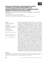
arachidonic acid (AA), endoperoxides, or TXA
2
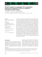
(Figure 1)
as they can induces platelet shape change, aggregation,
and secretion [26]. In contrast, platelet stimulation by
epinephrine is not associated with change in platelet
shape [27]. Additionally, the effects of "low" concentra-
tion of collagen are thought to be dependent on arachido-
nate metabolism. Aggregation is usually required for
secretion as the dense packing and resultant decrease in
Figure 1 Structure of arachidonic acid (the precursor for all pros-
taglandins), various TPR ligands, PGF
2α
, and the most abundant
isoprostane 8-iso-PGF
2α
.
Ting and Khasawneh Journal of Biomedical Science 2010, 17:24
/>Page 3 of 13
interstitial spaces serves to concentrate otherwise low
levels of released AA metabolites [13,28]. One exception
to this requirement is thrombin as it can induce secretion
in nonaggregated suspensions [1]. Due to the presence of
numerous, biologically active metabolites, one critical
activation arm of platelets is dependent on AA. AA,
which is the most abundant, is a 20-carbon unsaturated
fatty acid [29]. The release of AA from the membrane by
phospholipases, and subsequent metabolic modifications
leads to the formation of well-characterized prostaglan-
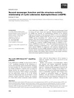
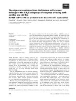
dins and thromboxanes (Figure 2). Of primary impor-
tance to platelet function is the formation of TXA
2
, which
is generated from arachidonic acid in reaction catalyzed
by the platelet cyclooxygenase-1 enzyme [30]. Generated
TXA
2
then binds to its G-protein coupled receptor
(GPCR) known as TXA
2
receptor (abbreviated as TPR).
There are two splice variants for TPR with distinct tissue
expression, i.e., the placental α-isoform and the endothe-
lial β-isoform [31]. Interestingly, using isoform-specific
TPR antibodies, TPR-α but not TPR-β was immunopre-
cipitated from platelets [32]. Furthermore, consistent
with this finding, platelets were found to express high lev-
els of mRNA for the α-isoform and low levels of β-iso-
forms. Taken together, these data suggest a limited role, if
any, for the β-isoforms in platelet function.
Interaction of TXA
2
, or other agonists to their cognate
receptors, leads to transduction of activating signals into
secondary messengers. One major pathway for this
response is the GPCRs [29,33-35]. G-proteins, which
consist of three different subunits, α, β and γ, can be
divided into four major families, G
q
, G
12
, G
i
and G
s
, of
which platelets have been found to express several dis-
tinct members [34,36]. More specifically, a host of in vitro
approaches involving reconstitution studies, affinity
copurification experiments or cross-linking studies with
photoactivated GTP analogs demonstrated that platelets
express G
q
, G
16
(G
q
family), G
12
, G
13
(G
12
family), G
s
, as
well as G
o
, G
i
and G
z
(G
i
family) [33,35,37-41]. These
studies have specifically revealed that TPR couples to the
G
q
and G
13
isoforms. Additionally, U46619, a stable TXA
2
mimetic, induces a rapid, transient rise in intracellular
Ca
2+
in platelets and in HEK293 cells cotransfected with
G
αq
or G
α11
and the α-isoform of TPR [42]. Further evi-
dence also indicates that the TPRα isoform can function-
ally couple to G
q
or to G
11
in vivo.
The G-protein, Gα
q
, signaling pathway starts by the
activation of phospholipase C (PLC) which in turn
metabolizes phosphatidylinositol 4,5-bisphosphate (PIP
2
)
into inositol 1,4,5-trisphosphate (IP
3
) and diacylglycerol
(DAG) [43,44]. IP
3
then binds to its receptor and raises
cytosolic Ca
2+
concentrations by inducing Ca
2+
release
from vesicles into the cytoplasm [45,46]. DAG serves to
stimulate protein kinase C (PKC) which in turn activates
phospholipase A
2
(PLA
2
) [47]. It is thought that both the
increase in cytoplasmic Ca
2+
and the production of DAG
are necessary for full platelet activation, and lead to the
activation of the glycoprotein GPIIb/IIIa[48,49]. This GP
is a heterodimeric complex of two GPs on the platelet
surface that serves as the fibrinogen receptor [16,25].
Fibrinogen is a dimeric molecule that serves as a molecu-
lar bridge which crosslinks platelets, thereby enabling
platelet aggregation and formation of a primary hemo-
static plug [50]. On this basis, activation of GPIIb/IIIa is
absolutely critical for platelet function. Under in vitro set-
tings, the conformational change required for the forma-
tion of "active" GPIIb/IIIa requires calcium [48,49,51].
Taken together, it's believed that increases in intracellular
Ca
2+
are the ultimate mediator of activation in platelets.
Arachidonic acid metabolites such as TXA
2
, have been
shown to trigger platelet responses dependent on stimu-
lation of G
12/13
-/G
q
-coupled receptors [37,38,41,52]. Sig-
naling through these receptors has been shown to
enhance phosphorylation of several tyrosine kinase fami-
lies (Src, Syk and FAK) [53]. Consistent with the role of
G
12/13
-coupled receptors, low doses of U46619 was found
to trigger tyrosine phosphorylation of FAK, Syk and Src
[54]. Secretion of TXA
2
(or other AA metabolites that act
though TPRs such as isoprostanes) from activated plate-
lets and other sources may then mediate further activa-
tion through this tyrosine-kinase-dependent signaling
pathway [55]. Additionally, thrombin has been reported
to induce phosphorylation of FAK in both platelets and
HEK293 cells, and binding of GPIIb/IIIa to fibrinogen ini-
tiates a second sustained wave of tyrosine phosphoryla-
tion [56,57]. In fact, GPCR-mediated activation of
tyrosine kinases is well characterized during integrin-
mediated assembly of cytoskeletal and signaling proteins
to focal adhesion sites [58]. Interestingly, U46619 medi-
ated activation was found to be independent of GPIIb/
IIIa binding to fibrinogen or the interaction of secreted
ADP with its platelet receptors (i.e., P2Y
1
and/or P2Y
12
)
[54]. Signaling through this modality alone was insuffi-
cient to stimulate full platelet activation, but synergized
with the G
z
-linked adrenaline receptor (epinephrine) to
mediate platelet aggregation [29,59,60]. In fact, it has
been reported that combined signaling via G
12/13
and G
i
is
required for full platelet activation [61,62]. Furthermore,
signaling through both the G
12/13
-dependent Rho-kinase,
and the tyrosine-kinase-dependent pathways was found
to be required for the synergistic activation of GPIIb/IIIa
[63]. Thus, these signals converge with additional signals
ensuing from the engagement of G
z
-coupled receptors
[33,36]. Together, this data reveals that a combination of
agonists at subthreshold levels or with low potency can
Ting and Khasawneh Journal of Biomedical Science 2010, 17:24
/>Page 4 of 13
serve to activate platelets in the absence of more potent
and perhaps more intentional activation.
Collectively, platelet TPRs are known to couple to the
four major families of G-proteins, which in turn activate
numerous downstream effectors, including second mes-
senger systems such as IP
3
/DAG, cAMP, small G proteins
(Ras, Rho, and Rac, effectors such as p160 ROCK, as well
as the Ca
2+
/calmodulin system) [33,34,36,64-67], phos-
phoinositide-3(PI3) kinase, activation of Syk, Src, and
FAK tyrosine kinase and mitogen-activated protein
kinase (MAPK, specifically p38 and p42) as well as pro-
tein kinase A and C (PKA and PKC) [54,65,68]. Addition-
ally, the action of many platelet agonists (ADP, thrombin,
low dose collagen) serves to mediate synthesis and subse-
Figure 2 A schematic representation of the arachidonic acid metabolism pathway. After its liberation by phospholipases, ((i.e., phospholipase
A
2
(PLA
2
) or phospholipase C (PLC)), the free arachidonic acid may undergo enzymatic metabolism by the lipoxygenases which produce HPETEs and
leukotrienes, and the cyclooxygenases (COX-1, COX-2) which generate prostaglandins and thromboxanes. The specific repertoire of the arachidonic
acid metabolites produced may vary according to the expression profile of these enzymes in different cell types. In platelets, for example, arachidonic
acid is metabolized by COX-1 into the prostaglandin endoperoxides, PGG
2
and PGH
2
. Next, thromboxane synthetase further metabolizes PGH
2
into
TXA
2
, which is a potent activator of platelet aggregation, with a half-life of 20-30 seconds. Thromboxane A
2
is then hydrolyzed to the inactive form
TXB
2
(not shown). On the other hand, if PGH
2
is metabolized by prostacyclin synthetase, then PGI
2
would be produced (e.g., in endothelial cells). Fur-
thermore, if PGH
2
is acted upon by PGD or PGE isomerase, then PGD
2
, and PGE
2
are produced, respectively (e.g., in renal cells). Finally, if the PG re-
ductase metabolizes PGH
2
, then PGF
2α
is produced (e.g., pulmonary vessels). Thus, the biological functions of arachidonic acid are exerted indirectly
after its metabolism into prostaglandin and thromboxane metabolites.
Ting and Khasawneh Journal of Biomedical Science 2010, 17:24
/>Page 5 of 13
quent secretion of TXA
2
[1,49,63]. Thus, TXA
2
is not only
a potent direct activator of platelet function, it is also a
key effector in other agonist mediated pathways. Fortu-
nately, TXA
2
is also highly unstable (a half life of around
30 seconds) and functions primarily as an autocrine or
local paracrine signal allowing for tight spatial regulation
of platelet activation [69]. The discovery of this central
role for AA metabolite pharmacological activity has
motivated the design of drugs with TPR antagonistic
activity.
Isoprostanes
While research on arachidonic acid metabolites have
focused on the traditional enzyme mediated pathway,
there is another potential route for arachidonic acid mod-
ification, i.e., a free radical mediated pathway [70,71].
This metabolic cascade has led to the investigation of a
class of "naturally" occurring prostaglandin-like products
known as isoprostanes. These are produced by the free
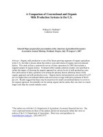
radical mediated oxidation of unsaturated fatty acids (Fig-
ure 3) in membrane phospholipids as opposed to the
enzymatically catalyzed oxidation found with the classi-
cal AA derivatives such as TXA
2
[70,72]. As the forma-
tion of isoprostanes is not enzymatically-directed, but
random chemical degradation, there is a larger variety of
molecules produced in vivo (Figure 3). Whereas the
endoperoxide prostaglandin G
2
(PGG
2
) is specifically
formed by the cyclooxygenase enzymes (COX-1 and
COX-2), four classes of isoprostanes are produced as a
result of the free-radical oxidation of AA (Figure 3), with
each class containing 16 subtypes of isoprostanes result-
ing in 64 individual isoprostane molecules [73].
Due to their interesting chemical properties and large
number of distinct members, isoprostanes are of clinical
interest for two main reasons: 1. they are ligands for pros-
taglandin receptors, and thus may exhibit biological
activity like TXA
2
and other AA metabolites [70,74]; and
2. they have been found to associate with the oxidative
status of an organism [75,76]. Moreover, there is evidence
that their levels serve as a predictor of the onset and
severity of inflammatory diseases such as atherosclerosis
and Alzheimer's disease [75,77]. Indeed, isoprostanes are
thought to participate in the pathogenesis of Alzheimer's
disease. Evaluation of the blood and urinary levels of cer-
tain isoprostanes' and their metabolites, respectively, has
been demonstrated to be a reliable approach to the
assessment of lipid peroxidation, and therefore of oxida-
tive stress in vivo [78]. More specifically, evidence points
to the possibility that isoprostanes may be involved in the
genesis of certain disease states. For example, in vitro
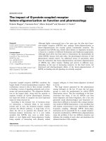
Figure 3 A schematic representation of the metabolic cascade for the non-enzymatic generation of isoprostanes. This is a proposed scheme
in which four series of regioisomers of PGG
2
are formed, before they are reduced to PGF
2α
isomers. As shown, isoprostanes can be formed from arachi-
donic acid in situ in phospholipids, from which they are presumably cleaved by phospholipases A
2
. PGG
2
spontaneously rearranges to PGD
2
and PGE
2
thereby generating isoprostanes of the D and E series. The initial step in the formation of an isoprostane from arachidonic acid (I) is the generation of
a lipid free radical by the abstraction of a hydrogen atom from one of the three methylene-interrupted carbon atoms, C7, C10, or C13, as shown here,
by a free radical (FR•) which may be a hydroxyl radical (HO•), a superoxide radical (O
2
-
•) or other free radical, and results in (II). Radical attack at C-10 is
shown, abstraction at the other positions determines the relative proportion of the isomers formed. The lipid free radical is converted to a peroxy rad-
ical by reaction with molecular oxygen. The peroxy radical cyclizes in an intramolecular reaction that yields an endoperoxide (III). The free radical chain
reaction will continue to propagate until quenched by an antioxidant.
Ting and Khasawneh Journal of Biomedical Science 2010, 17:24
/>Page 6 of 13
studies revealed that isoprostanes can induce oligoden-
drocyte progenitor cell death and induce vasoconstric-
tion and mitogenesis, as well as inflame endothelial cells
to bind monocytes, a critical initiating event in athero-
genesis [79-81]. An in vivo mouse model suggested that
isoprostanes are involved in the development of thrombi
at sites of vascular injury [82]. Furthermore, LDLR- and
ApoE-deficient mouse models demonstrated that these
oxidation products accelerate the development of athero-
sclerosis independent of de novo TXA
2
synthesis or
changes in plasma lipid levels [83]. In patients with ath-
erosclerosis and acute myocardial infarction, levels of iso-
prostanes were also found to be elevated and their
reduction coincided with decreased atherogenesis, sug-
gesting a role for this oxidized lipid in the development of
this disease state [76,84].
Most of the studies examining the biological activity of
isoprostanes have been conducted with a specific form,
8-iso-PGF
2α
(Figure 1), as it is one of the most abundantly
produced in vivo [85]. Much work has been done with
this compound as it is commercially available, having
been previously synthesized for unrelated reasons and
was therefore readily available for a host of studies (i.e.,
infusion, bioassay, receptor binding/affinity studies, etc).
Additionally, it exhibits chemical stability that signifi-
cantly exceeds that of TXA
2
, suggesting it's potential for
long-term signaling capacity that may lead to systemic
priming of platelets [83]. To this end, 8-iso-PGF
2α
has
been reported to exhibit significant biological activity.
Specifically, it has been found to be a mitogen in 3T3 cells
and in vascular smooth muscle cells and evidence sug-
gests it may play a role in pulmonary oxygen toxicity
[86,87]. This biological activity may be a result of modifi-
cation of the integrity and fluidity of membranes, a char-
acteristic consequence of oxidative damage [88]. This
occurs as a result of the distorted shape of isoprostanes
relative to the normal fatty acids present in membrane
phospholipids and could be critical in modifying the
hemodynamic properties in vascular tissues into a more
dysfunctional microenvironment conducive to initiating
chronic disease states.
Isoprostane Signaling Pathways
Given the plethora of reports that suggest 8-iso-PGF
2α
exerts biological actions on platelets, elucidating the con-
centrations necessary to elicit these effects and reconcil-
ing these with the levels reported to circulate in vivo is of
relevance to investigating its underlying mechanism of
action. In pursuit of this goal, it was found that there is a
minimum threshold concentration of 8-iso-PGF
2α
at
which it has the capacity to induce platelet shape change
and above which it can alter the formation of thrombox-
ane or irreversible aggregation in response to platelet
agonists [89,90]. Additionally, 8-iso-PGF
2α
synergistically
mediates aggregation upon exposure to subthreshold
concentrations of platelet agonists [74]. Such a modality
is supported by findings that when epinephrine and AA
were added to platelet rich plasma (PRP) in subthreshold
concentrations, they acted in a synergistic manner to pro-
duce platelet aggregation[29]. This synergistic platelet
activation in response to dual exposure to 8-iso-PGF
2α
and other agonists would be most likely in settings where
platelet activation and enhanced free radical formation
(and thus isoprostane formation) coincide, a characteris-
tic microenvironment of atherosclerosis. This synergism
was found to be abrogated by calcium channel inhibitors,
an α
2
-receptor antagonist and inhibitors of PLC, MAP
kinase, and COX pathways [29]. Since increased cytosolic
Ca
2+
is essential to platelet activation, the proposed
mechanism for potentiation between platelet agonists is
the activation of the Ca
2+
signaling cascade. Thus, a rise
in cytosolic Ca
2+
levels induced by the first agonist primes
platelets for an enhanced functional response to a second
agonist. In accord with this possible mechanism, increas-
ing concentrations of 8-iso-PGF
2α
resulted in dose-
dependent, irreversible platelet aggregation in the pres-
ence of subthreshold concentrations of collagen, ADP,
AA, and analogs of TXA
2
(i.e., I-BOP, U46619)[74]. This
phenomenon was not evident when platelets were pre-
treated with either COX inhibitors or TPR antagonists,
indicating a clear dependence of aggregation on the sec-
ondary formation of TXA
2
. Interestingly, 8-iso PGF
2α
failed to desensitize the calcium or inositol phosphate
responses to platelet stimulation by these agonists. Fur-
thermore, 8-iso-PGF
3α
a related chemical to 8-iso-PGF
2α
failed to initiate platelet shape change or aggregation nor
did it raise intracellular calcium or inositol phosphates,
suggesting a structural requirement for engaging the
receptor's ligand binding domain(s).
In the course of characterizing the properties of iso-
prostanes, it was discovered that they exert their biologi-
cal activity on a host of cell types: platelets, kidney, and
others, presumably via the activation of TPR [80,91,92]. It
has previously been shown that 8-iso-PGF
2α
induces
intracellular Ca
2+
mobilization in cells co-transfected
with TPR
α
and G
αq
or G
α11
[42]. More specifically, co-
transfection of G
α11
produced greater mobilization of
intracellular Ca
2+
than that stimulated by G
αq
. Surpris-
ingly, in human platelets, 8-iso PGF
2α
failed to cause a
dose-dependent increase in TPR
α
phosphorylation, in
spite of stimulating inositol phosphate formation [32]. It
is possible that the capacity of 8-iso-PGF
2α
for in vivo
platelet activation manifests only if it's delivered through
an especially concentrated mechanism, such as from
microvesicles shed by activated cells, or through selective
Ting and Khasawneh Journal of Biomedical Science 2010, 17:24
/>Page 7 of 13
reincorporation of secreted isoprostanes into the mem-
brane[93]. Nevertheless, this explanation is only partially
satisfactory since the TXA
2
mimetic U46619, but not 8-
iso-PGF
2α
, reduced glomerular insulin space and
increased inositol 1,4,5-trisphosphate production in rat
glomeruli and mesangial cells in a an apparently TPR-
dependent fashion (i.e., blocked by the TPR antagonist
SQ29,548)[91]. Conversely, rat aortic smooth muscle cells
were found to possess specific binding sites for both
TXA
2
and 8-iso-PGF
2α
and displayed functional
responses to both agonists, such as time- and dose-
dependent activation of MAP kinases [74,91]. Interest-
ingly, the addition of 8-iso-PGF
2α
and U46619 together
did not potentiate or antagonize the maximal level of
Ca
2+
mobilized in either platelets or transfected HEK293
cells, which suggests that 8-iso-PGF
2α
and U46619 are
acting through the same pathway (TPR) [42]. In line with
this notion, SQ29,548 was found to be equally potent in
abolishing the Ca
2+
response in both platelets and trans-
fected HEK293 cells upon stimulation with either U46619
or 8-iso-PGF
2α
. Pretreatment of platelets or transfected
cells with thrombin, on the other hand, did not desensi-
tize the rise in intracellular Ca
2+
upon subsequent stimu-
lation with either U46619 or 8-iso-PGF
2α
, which provides
further evidence that these lipids share a common signal-
ing pathway, though previous work showing abrogation
of effect by 8-iso-PGF
2α
in the presence of COX inhibitors
suggests that formation of TXA
2
is the potential link at
the TPR modality [74].
Studies have also revealed that 8-iso-PGF
2α
stimulates
platelet shape change and reversible aggregation through
a TPR-mediated process [74]. In support of this, 8-iso-
PGF
2α
was found to be a potent vasoconstrictor in the rat
lung and kidney, which was specific through TPRs[81,92].
Furthermore, a TPR antagonist was shown to block 8-iso-
PGF
2α
-induced vasoconstriction of renal glomeruli,
carotid arteries, and vascular smooth muscle cells
[92,94,95]. Additionally, it was found that the proathero-
genic effect of 8-iso-PGF
2α
is mediated via TPR activation
and is secondary to the induction of specific inflamma-
tory mediators, such as sICAM-1 and MCP-1 but not ET-
1, at the site of lesion development [83]. On the other
hand, several reports disputed the notion that the stimu-
latory effects of 8-iso-PGF
2α
are primarily mediated
through TPRs, adding more complexity to this issue. The
primary alternative signaling mechanism predicts the
existence of unidentified discrete isoprostane receptors in
human platelets and smooth muscle cells, the basis for
which is found in studies detailing differences between
the potencies of 8-iso-PGF
2α
and TPR agonists in induc-
ing DNA synthesis and MAP-kinase activation
[74,83,91,96,97]. Further complicating matters, this alter-
native proposal has also been recently disputed with sev-
eral possible explanations for the noted discrepancies
such as variations in the experimental conditions/cellular
preparations, or inherent differences in the potency of
the ligands employed [94]. In summary, there are clear
ambiguities concerning the mechanisms by which iso-
prostanes modulate cellular function.
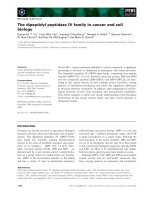
As a distinct and further confounding layer of complex-
ity it has been recently reported that 8-iso-PGF
2α
signals
through both stimulatory and inhibitory pathways in
platelets and that this inhibition by 8-iso-PGF
2α
operates
through a cAMP-dependent mechanism (Figure 4) [70].
Additionally, reduction of isoprostane formation by vita-
min E in combination with the suppression of TXB
2
bio-
synthesis (a metabolic marker of TXA
2
) was shown to be
more effective than the two approaches alone in experi-
mental atherosclerosis [98]. In this connection, by block-
ing TXA
2
synthesis, aspirin (ASA) appears to facilitate
increased isoprostane production from AA, which in
turn, may amplify the anti-thrombotic effects of ASA
itself through a secondary inhibitory process. Taken
together, it might be predicted that a therapeutic regimen
combining ASA along with a TPR antagonist would be
more beneficial than therapy with ASA alone. Specifi-
cally, under these conditions, the isoprostane stimulatory
effects would be blocked by TPR antagonism, while its
inhibitory effects would be promoted by elevating the
levels of circulating isoprostane. Thus, specific isopros-
tane-receptor interactions may mediate agonist activa-
tion of one effector pathway, yet act as an antagonist for
an alternate pathway.
Alternative Isoprostane Signaling Pathways
Despite this body of evidence associating elevated iso-
prostane with oxidative stress and vascular disease
pathology, as well as supporting a potential role for iso-
prostanes in mediating a host of disease processes such as
apoptosis, brain cell damage, and thrombosis, their bio-
logical activity and signaling mechanisms remain poorly
understood. A major hindrance to teasing out the mecha-
nism(s) is that specific inhibition of isoprostanes is not
universally reported. Aside from prostaglandin H
2
-TXA
2
and isoprostanes, the TPR receptors share other endoge-
nous ligands such as HETE. Moreover, other AA deriva-
tives (free radical-dependent or otherwise) may be
biologically relevant and signal through TPR, thus further
obfuscating the activity of isoprostanes on platelet biol-
ogy [99]. One of the most promising avenues for research
is thus isolating the contributions of signaling through
the TPR which is known to competently bind to isopros-
tanes. Studies report ligation of both existing membrane
and nuclear prostaglandin receptors by isoprostanes
[100,101]. However, the possibility of signaling through
Ting and Khasawneh Journal of Biomedical Science 2010, 17:24
/>Page 8 of 13
other isoprostane receptors is raised by studies reporting
an apparent inability of isoprostanes to ligate or signal
efficiently through either TPR isoform in vitro, despite
evidence that their in vivo actions are mediated by TPR
[91,94].
One potential alternative signaling mechanism posits a
contribution by the phenomenon of GPCR heterodi-
merization, which is a result of a specific receptor having
multiple isoforms, or non-isoform receptors that can
freely dimerize with each other. Heterodimerization has
been reported to alter receptor properties such as regula-
tion and ligand binding affinity [102]. In addition, studies
indicate that GPCR heterodimers may mediate changes
in the signaling preferences/characteristics of the individ-
ual receptors [100,102-104]. An example is found in the
dimerization of the β1 and β2 adrenergic receptors,
which enhances cAMP formation in response to isoprot-
ernol and has also been implicated in regulating cardiac
contractility [105]. Similarly, dimerization of the alpha
and beta isoforms of the TPR has been shown to mediate
Figure 4 Schematic representation of a model describing the inhibitory and stimulatory signaling pathways for TPR-dependent modula-
tion of platelet activation by 8-iso-PGF
2α
.
Ting and Khasawneh Journal of Biomedical Science 2010, 17:24
/>Page 9 of 13
alterations in both receptor regulation and signaling
[103,104]. Consistent with previous reports, 8-iso-PGF
2α
stimulated TPR-mediated IP
3
generation less potently
than IBOP and U46619 in cells expressing TPR
α
or TPR
β
individually. In contrast, while cells stably expressing
both TPR
α
and TPR
β
, exhibited significantly enhanced IP
3
generation following treatment with 8-iso-PGF
2α
, this
was not the case with IBOP or U46619. This finding was
not due to preferential binding to an isoform or in combi-
nation as there were no differences in the capacity for 8-
iso-PGF
2α
to displace the TPR antagonist SQ29,548 in
membranes generated from TPR
α
, TPR
β
or TPR
α
/TPR
β
co-expressing HEK cells despite signaling more efficiently
through a TPR
α
/TPR
β
heterodimer. However, it has been
reported that SQ29,548 does not fully occupy the binding
site for 8-iso-PGF
2α
in the TPR
α
/TPR
β
heterodimer.
These data together indicate that heterodimerization
does not modify the well characterized TPR binding site,
but instead may create an alternative isoprostane binding
site. Additionally, the possibility exists that downstream
G protein coupling is modified with GPCR heterodi-
merization. For example, if the TPR
α
/TPR
β
heterodimer
were more efficiently coupled to Gq in co-transfected
cells it might be expected that IP
3
and calcium signals
would be elevated. However, the absence of a similarly
enhanced signaling response with IBOP or U46619
stands in contradiction to this hypothesis. Finally, it's dif-
ficult to infer/interpret the biological relevance of the
impact of TPR
α
/TPR
β
heterodimer formation on isopros-
tane biology in platelets given that platelets do not
express TPRTPR
β
.
Yet another potential mechanism for isoprostane medi-
ated signaling is found at signal transduction, whereby
the response following activation of GPCR's is altered;
this is a particularly enticing avenue for future investiga-
tion since chronic disease states such as atherosclerosis
are characterized by persistent, subacute levels of dysreg-
ulation. In this connection, following their activation, dis-
sociated Gα subunits may not bind to their originally
coupled GPCR receptors. Instead, the final equilibrium of
the reassociation process for liberated Gα is determined
by the relative expression and affinity of the various acti-
vated GPCR's[106]. To illustrate, following PAR1 receptor
activation, both the level of PAR1 presentation and its Gα
affinity would decrease as PAR1 is internalized following
activation along with receptor alterations due to PAR1/
ligand interactions. Together, these effects would pro-
mote increased Gα coupling to TPRs and thus a conse-
quent shift to a higher ligand affinity state for this
receptor. Expression/affinity-mediated TPR/G-protein
coupling raises the possibility of competition for G-pro-
teins between TPRs and other GPCRs, and helping to
define the predominant signaling pathways through
which TPRs signal under different experimental condi-
tions and in different cell types. In support of this hypoth-
esis, it was found that activation of Gα
i
-coupled receptors
increased the potency and the efficacy of inositol phos-
phate production induced by bradykinin or UTP activa-
tion [106]. In addition, other studies demonstrated
synergistic interactions between U46619 and ADP as well
as U46619 and epinephrine [59,60,107,108].
Isoprostane Binding
Due to these sometimes confounding reports on isopros-
tane signaling, attempts have been made to elucidate the
specific segment(s) that define the receptor ligand-bind-
ing pocket of isoprostanes to TPR's, which will also
address the question of whether isoprostanes can physi-
cally interact with TPRs or not. We, recently reported
that 8-iso-PGF
2α
coordinates with specific residues on
platelet TPR's and that Phe
196
(Figure 4) specifically
serves as a unique TPR binding site for this ligand [70].
Furthermore, it was revealed that TPRs exhibit ligand
specificity, in both G-protein and TPR cotransfected
HEK293 cells as well as in platelets. Consistent with pre-
vious reports regarding the relative potency, the maximal
Ca
2+
response observed in platelets was 3- to 4-fold
greater after stimulation with U46619 than with 8-iso-
PGF
2α
[42]. This is critical as the signaling in platelet acti-
vation appears to integrate at the level of elevating intrac-
ellular Ca
2+
. Previously it was noted that 8-iso-PGF
2α
signals through both stimulatory and inhibitory pathways
in platelets and that the inhibitory effects of 8-iso-PGF
2α
operated through a cAMP dependent mechanism (Figure
4). This is supported by reports that 8-iso-PGF
2α
interacts
with platelets at two separate binding sites [70,74,91].
One of these sites was found to mediate a small rise in
intracellular Ca
2+
, a concomitant increase in inositol
phosphates and protein kinase C activation as well as
supporting irreversible platelet aggregation, when stimu-
lated by TXA
2
/PGH
2
analogs. The other site mediates the
majority of the calcium released from intracellular stores
and platelet shape change [109,110]. Additionally, as
mentioned elsewhere, the rapid, agonist-induced phos-
phorylation of TPR
α
appears to involve signaling through
low affinity binding sites. This was verified in studies
using platelets pretreated with GR32191 (which blocks
the low affinity TPR sites) where it was found that neither
low concentrations of I-BOP, nor high concentrations of
agonist resulted in TPR
β
phosphorylation[109].
Isoprostane in vivo Levels
In discussing isoprostanes it is important to note that iso-
prostanes can be produced in vivo at levels several orders
of magnitude higher than classical prostaglandins/throm-
Ting and Khasawneh Journal of Biomedical Science 2010, 17:24
/>Page 10 of 13
boxanes, and that they remain largely stable in circulation
in comparison to ligands such as TXA
2
itself [69,71].
Consequently, the biological effects of these signaling
modalities could, in theory, have a substantial systemic
impact on cellular functions along a broad temporal
range, characteristic of chronic disease states. Further-
more, it is known that the in vivo levels of isoprostanes
can be enhanced by the presence of vascular disease, thus
further associating this oxidative marker to the chronic
dysfunction characterized by oxidative stress [76,77,84].
However, one obfuscating complication remains in
deducing the role of isoprostanes in mediating platelet
activation; this derives in part from the fact that the
reported EC
50
concentrations of isoprostanes required to
elicit functional responses in platelets are much higher
than their measured concentrations in the circulation,
even in syndromes of oxidant stress [74]. The highest
plasma levels recorded in patients remain outside the
range of concentration necessary to evoke biological
responses in platelets or in other cell types. Thus, 8-iso-
PGF
2α
does not likely function as a conventional, circulat-
ing hormone in vivo, and even potential autocoidal func-
tions may necessitate highly concentrated forms of
delivery to local receptors. Nonetheless, it's possible that
these lipids do achieve such concentrations locally (com-
partmentalization), and hence modulate platelet function
at punctuate microenvironenments conducive for their
effect. Another possible explanation to this potential con-
flict is that incidental activation of TPR receptors by 8-iso
PGF
2α
may contribute at subthreshold levels to the
adverse effects of oxidant stress in vivo as would be the
case with some of the alternative signaling modalities
described previously.
Conclusion
An alternative to the classical COX-mediated AA modifi-
cation pathway has more recently been identified, that of
chemical degradation. More specifically, free radical-
induced oxidative modification of AA, which results in
the production of a group of chemicals called isopros-
tanes [71,81]. Furthermore, isoprostanes can circulate in
vivo at concentrations orders of magnitude higher than
other AA metabolites such as TXA
2
and remain much
more chemically stable (Table 1) [111-115]. This family of
lipid-mediators, particularly 8-iso-PGF
2α
, has been
strongly correlated with the oxidative microenviron-
ments found in various disease states. Many reports sug-
gest that isoprostanes produce their biological activity by
directly interacting with TPRs (e.g., on platelets), and a
plethora of reports indicate they are associated with
increased risk of several vascular diseases. This associa-
tion manifests in a broad range of cell types but almost all
appeared dependent on mediating TPR activation, and
secondarily, several G-proteins. Further complicating the
task of elucidating its underlying mechanism of effect,
reports have revealed that 8-iso-PGF
2α
signals through
both stimulatory and inhibitory pathways in platelets.
While the identity of the receptor that mediates its inhib-
itory effects remains unknown, evidence indicates that
it's coupled to Gs. And this is indicative of the continued
need for further research in this field as there are often
conflicting reports on the activity and signaling pathways
of this class of chemicals; possibly due to the subtle
nature of their contribution to platelet activation. Taken
together, this suggests the possibility that in chronic and
sustained dysregulated states as found in vascular dis-
ease, isoprostanes could possess a significant systemic
impact on cellular functions without initiating an acute
thrombotic event in the absence of other agonists and as
such remains an intriguing area of further research.
Abbreviations
TXA
2
: thromboxane A
2
; TPR: thromboxane A
2
receptor; AA: arachidonic acid;
GPCR: G-protein coupled receptor; Ca
2+
: calcium
Competing interests
The authors declare that they have no competing interests.
Table 1: A comparison between certain biological properties of TXA
2
and 8-iso-PGF
2α
Lipid Half life
(T
1/2
)
Plasma
Concentration
(endogenous)
Method of synthesis Receptors
TXA
2
20-30
seconds
111
TXB
2
(1-66 pg/ml)
113
Enzymatic
26
TPR
α
& TPR
β
31
8-iso-PGF
2α
1-10
minutes
112
351-1831 pg/ml
(dinordihydro
metabolite)
114
Non-emzymatic &
enzymatic
73,115
TPR
α
80
& TPR
β
74,91
and
ISR
70,74,97
Ting and Khasawneh Journal of Biomedical Science 2010, 17:24
/>Page 11 of 13
Authors' contributions
FTK: Prepared the manuscript and figures; HJT: Manuscript preparation, and
reference formatting.
Acknowledgements
The authors would like to thank Dr. Wallace J Murray for his help with structure
drawings. This work was supported by Intramural Funding Support from the
College of Pharmacy at Western University of Health Sciences (to F.T.K).
Author Details
Department of Pharmaceutical Sciences, College of Pharmacy, Western
University of Health Sciences, Pomona, California 91766, USA
References
1. Zucker MB, Nachmias VT: Platelet activation. Arteriosclerosis 1985, 5:2-18.
2. Buller HR, Ten Cate T: Coagulation and platelet activation pathways. A
review of the key components and the way in which these can be
manipulated. Eur Heart J 1995, 16(Suppl L):8-10.
3. Yardumian DA, Mackie IJ, Machin SJ: Laboratory investigation of platelet
function: a review of methodology. J Clin Pathol 1986, 39:701-712.
4. Bick RL: Platelet function defects: a clinical review. Semin Thromb
Hemost 1992, 18:167-185.
5. Dubois C, Panicot-Dubois L, Gainor JF, Furie BC, Furie B: Thrombin-
initiated platelet activation in vivo is vWF independent during
thrombus formation in a laser injury model. J Clin Invest 2007,
117:953-960.
6. Willerson JT: Conversion from chronic to acute coronary heart disease
syndromes. Role of platelets and platelet products. Tex Heart Inst J 1995,
22:13-19.
7. Sahud MA: Clinical and biochemical characteristics of a primary platelet
disorder. Acta Univ Carol Med Monogr 1972, 53:317-324.
8. Shaw T: The role of blood platelets in nucleoside metabolism:
regulation of megakaryocyte development and platelet production.
Mutat Res 1988, 200:67-97.
9. Patrono C: Aspirin as an antiplatelet drug. N Engl J Med 1994,
330:1287-1294.
10. Avraham H: Regulation of megakaryocytopoiesis. Stem Cells 1993,
11:499-510.
11. Jackson SP, Nesbitt WS, Westein E: Dynamics of platelet thrombus
formation. J Thromb Haemost 2009, 7(Suppl 1):17-20.
12. Born GV: Aggregation of blood platelets by adenosine diphosphate
and its reversal. Nature 1962, 194:927-929.
13. Charo IF, Feinman RD, Detwiler TC: Interrelations of platelet aggregation
and secretion. J Clin Invest 1977, 60:866-873.
14. Kattelman EJ, Venton DL, Le Breton GC: Characterization of U46619
binding in unactivated, intact human platelets and determination of
binding site affinities of four TXA2/PGH2 receptor antagonists (13-APA,
BM 13.177, ONO 3708 and SQ 29,548). Thromb Res 1986, 41:471-481.
15. Parise LV, Venton DL, Le Breton GC: Thromboxane A2/prostaglandin H2
directly stimulates platelet shape change independent of secreted
ADP. J Pharmacol Exp Ther 1982, 222:276-281.
16. Jirouskova M, Jaiswal JK, Coller BS: Ligand density dramatically affects
integrin alpha IIb beta 3-mediated platelet signaling and spreading.
Blood 2007, 109:5260-5269.
17. FitzGerald GA, Healy C, Daugherty J: Thromboxane A2 biosynthesis in
human disease. Fed Proc 1987, 46:154-158.
18. Gerrard JM, White JG, Rao GH: Effects of the lonophore A23187 on the
blood platelets II. Influence on ultrastructure. Am J Pathol 1974,
77:151-166.
19. Kaplan KL, Broekman MJ, Chernoff A, Lesznik GR, Drillings M: Platelet
alpha-granule proteins: studies on release and subcellular localization.
Blood 1979, 53:604-618.
20. Harrison P, Cramer EM: Platelet alpha-granules. Blood Rev 1993, 7:52-62.
21. Meyers KM, Holmsen H, Seachord CL: Comparative study of platelet
dense granule constituents. Am J Physiol 1982, 243:R454-461.
22. McNicol A, Israels SJ: Platelet dense granules: structure, function and
implications for haemostasis. Thromb Res 1999, 95:1-18.
23. Jennings LK, Fox JE, Edwards HH, Phillips DR: Changes in the cytoskeletal
structure of human platelets following thrombin activation. J Biol
Chem 1981, 256:6927-6932.
24. Rink TJ: Cytosolic calcium in platelet activation. Experientia 1988,
44:97-100.
25. Shattil SJ: Signaling through platelet integrin alpha IIb beta 3: inside-
out, outside-in, and sideways. Thromb Haemost 1999, 82:318-325.
26. Arthur JF, Jackson SP: Is thrombin the problem or (dis)solution? Blood
2009, 113:6046-6047.
27. Yun-Choi HS, Park KM, Pyo MK: Epinephrine induced platelet
aggregation in rat platelet-rich plasma. Thromb Res 2000, 100:511-518.
28. Zucker MB, Peterson J: Inhibition of adenosine diphosphate-induced
secondary aggregation and other platelet functions by acetylsalicylic
acid ingestion. Proc Soc Exp Biol Med 1968, 127:547-551.
29. Saeed SA, Rasheed H, Fecto FA, Achakzai MI, Ali R, Connor JD, Gilani AU:
Signaling mechanisms mediated by G-protein coupled receptors in
human platelets. Acta Pharmacol Sin 2004, 25:887-892.
30. Charo IF, Feinman RD, Detwiler TC, Smith JB, Ingerman CM, Silver MJ:
Prostaglandin endoperoxides and thromboxane A2 can induce
platelet aggregation in the absence of secretion. Nature 1977,
269:66-69.
31. Habib A, Vezza R, Creminon C, Maclouf J, FitzGerald GA: Rapid, agonist-
dependent phosphorylation in vivo of human thromboxane receptor
isoforms. Minimal involvement of protein kinase C. J Biol Chem 1997,
272:7191-7200.
32. Habib A, FitzGerald GA, Maclouf J: Phosphorylation of the thromboxane
receptor alpha, the predominant isoform expressed in human
platelets. J Biol Chem 1999, 274:2645-2651.
33. Brass LF, Hoxie JA, Kieber-Emmons T, Manning DR, Poncz M, Woolkalis M:
Agonist receptors and G proteins as mediators of platelet activation.
Adv Exp Med Biol 1993, 344:17-36.
34. Gilman AG: G proteins: transducers of receptor-generated signals.
Annu Rev Biochem 1987, 56:615-649.
35. Shenker A, Goldsmith P, Unson CG, Spiegel AM: The G protein coupled to
the thromboxane A2 receptor in human platelets is a member of the
novel Gq family. J Biol Chem 1991, 266:9309-9313.
36. Hepler JR, Gilman AG: G proteins. Trends Biochem Sci 1992, 17:383-387.
37. Offermanns S, Hu YH, Simon MI: Galpha12 and galpha13 are
phosphorylated during platelet activation. J Biol Chem 1996,
271:26044-26048.
38. Knezevic I, Borg C, Le Breton GC: Identification of Gq as one of the G-
proteins which copurify with human platelet thromboxane A2/
prostaglandin H2 receptors. J Biol Chem 1993, 268:26011-26017.
39. Giesberts AN, van Ginneken M, Gorter G, Lapetina EG, Akkerman JW, van
Willigen G: Subcellular localization of alpha-subunits of trimeric G-
proteins in human platelets. Biochem Biophys Res Commun 1997,
234:439-444.
40. Nakahata N, Miyamoto A, Ohkubo S, Ishimoto H, Sakai K, Nakanishi H,
Ohshika H, Ohizumi Y: Gq/11 communicates with thromboxane A2
receptors in human astrocytoma cells, rabbit astrocytes and human
platelets. Res Commun Mol Pathol Pharmacol 1995, 87:243-251.
41. Djellas Y, Manganello JM, Antonakis K, Le Breton GC: Identification of
Galpha13 as one of the G-proteins that couple to human platelet
thromboxane A2 receptors. J Biol Chem 1999, 274:14325-14330.
42. Kinsella BT, O'Mahony DJ, Fitzgerald GA: The human thromboxane A2
receptor alpha isoform (TP alpha) functionally couples to the G
proteins Gq and G11 in vivo and is activated by the isoprostane 8-epi
prostaglandin F2 alpha. J Pharmacol Exp Ther 1997, 281:957-964.
43. Rittenhouse-Simmons S: Production of diglyceride from
phosphatidylinositol in activated human platelets. J Clin Invest 1979,
63:580-587.
44. Lee SB, Rhee SG: Significance of PIP2 hydrolysis and regulation of
phospholipase C isozymes. Curr Opin Cell Biol 1995, 7:183-189.
45. Streb H, Irvine RF, Berridge MJ, Schulz I: Release of Ca2+ from a
nonmitochondrial intracellular store in pancreatic acinar cells by
inositol-1,4,5-trisphosphate. Nature 1983, 306:67-69.
46. Joseph SK, Thomas AP, Williams RJ, Irvine RF, Williamson JR: myo-Inositol
1,4,5-trisphosphate. A second messenger for the hormonal
mobilization of intracellular Ca2+ in liver. J Biol Chem 1984,
259:3077-3081.
47. Sano K, Takai Y, Yamanishi J, Nishizuka Y: A role of calcium-activated
phospholipid-dependent protein kinase in human platelet activation.
Received: 5 January 2010 Accepted: 6 April 2010
Published: 6 April 2010
This article is available from: 2010 Ting and Khasawneh; licensee BioMed Central Ltd. This is an Open Access article distributed under the terms of the Creative Commons Attribution License ( ), which permits unrestricted use, distribution, and reproduction in any medium, provided the original work is properly cited.Journal of Biomedical Science 2010, 17:24
Ting and Khasawneh Journal of Biomedical Science 2010, 17:24
/>Page 12 of 13
Comparison of thrombin and collagen actions. J Biol Chem 1983,
258:2010-2013.
48. Levy-Toledano S: Platelet signal transduction pathways: could we
organize them into a 'hierarchy'? Haemostasis 1999, 29:4-15.
49. McNicol A, Israels SJ: Platelets and anti-platelet therapy. J Pharmacol Sci
2003, 93:381-396.
50. Muszbek L, Bagoly Z, Bereczky Z, Katona E: The involvement of blood
coagulation factor XIII in fibrinolysis and thrombosis. Cardiovasc
Hematol Agents Med Chem 2008, 6:190-205.
51. Jennings LK, Phillips DR: Purification of glycoproteins IIb and III from
human platelet plasma membranes and characterization of a calcium-
dependent glycoprotein IIb-III complex. J Biol Chem 1982,
257:10458-10466.
52. Offermanns S, Laugwitz KL, Spicher K, Schultz G: G proteins of the G12
family are activated via thromboxane A2 and thrombin receptors in
human platelets. Proc Natl Acad Sci USA 1994, 91:504-508.
53. Dorsam RT, Kim S, Murugappan S, Rachoor S, Shankar H, Jin J, Kunapuli SP:
Differential requirements for calcium and Src family kinases in platelet
GPIIb/IIIa activation and thromboxane generation downstream of
different G-protein pathways. Blood 2005, 105:2749-2756.
54. Minuz P, Fumagalli L, Gaino S, Tommasoli RM, Degan M, Cavallini C, Lecchi
A, Cattaneo M, Lechi Santonastaso C, Berton G: Rapid stimulation of
tyrosine phosphorylation signals downstream of G-protein-coupled
receptors for thromboxane A2 in human platelets. Biochem J 2006,
400:127-134.
55. Egan KM, Wang M, Fries S, Lucitt MB, Zukas AM, Pure E, Lawson JA,
FitzGerald GA: Cyclooxygenases, thromboxane, and atherosclerosis:
plaque destabilization by cyclooxygenase-2 inhibition combined with
thromboxane receptor antagonism. Circulation 2005, 111:334-342.
56. Needham LK, Rozengurt E: Galpha12 and Galpha13 stimulate Rho-
dependent tyrosine phosphorylation of focal adhesion kinase, paxillin,
and p130 Crk-associated substrate. J Biol Chem 1998, 273:14626-14632.
57. Shattil SJ, Kashiwagi H, Pampori N: Integrin signaling: the platelet
paradigm. Blood 1998, 91:2645-2657.
58. Natarajan K, Berk BC: Crosstalk coregulation mechanisms of G protein-
coupled receptors and receptor tyrosine kinases. Methods Mol Biol
2006, 332:51-77.
59. Pulcinelli FM, Daniel JL, Riondino S, Gazzaniga PP, Salganicoff L:
Fibrinogen binding is independent of an increase in intracellular
calcium concentration in thrombin degranulated platelets. Thromb
Haemost 1995, 73:304-308.
60. Banga HS, Simons ER, Brass LF, Rittenhouse SE: Activation of
phospholipases A and C in human platelets exposed to epinephrine:
role of glycoproteins IIb/IIIa and dual role of epinephrine. Proc Natl
Acad Sci USA 1986, 83:9197-9201.
61. Nieswandt B, Schulte V, Zywietz A, Gratacap MP, Offermanns S:
Costimulation of Gi- and G12/G13-mediated signaling pathways
induces integrin alpha IIbbeta 3 activation in platelets. J Biol Chem
2002, 277:39493-39498.
62. Dorsam RT, Kim S, Jin J, Kunapuli SP: Coordinated signaling through
both G12/13 and G(i) pathways is sufficient to activate GPIIb/IIIa in
human platelets. J Biol Chem 2002, 277:47588-47595.
63. Offermanns S: Activation of platelet function through G protein-
coupled receptors. Circ Res 2006, 99:1293-1304.
64. Shock DD, He K, Wencel-Drake JD, Parise LV: Ras activation in platelets
after stimulation of the thrombin receptor, thromboxane A2 receptor
or protein kinase C. Biochem J 1997, 321(Pt 2):525-530.
65. Klages B, Brandt U, Simon MI, Schultz G, Offermanns S: Activation of G12/
G13 results in shape change and Rho/Rho-kinase-mediated myosin
light chain phosphorylation in mouse platelets. J Cell Biol 1999,
144:745-754.
66. Gratacap MP, Payrastre B, Nieswandt B, Offermanns S: Differential
regulation of Rho and Rac through heterotrimeric G-proteins and
cyclic nucleotides. J Biol Chem 2001, 276:47906-47913.
67. Paul BZ, Daniel JL, Kunapuli SP: Platelet shape change is mediated by
both calcium-dependent and -independent signaling pathways. Role
of p160 Rho-associated coiled-coil-containing protein kinase in
platelet shape change. J Biol Chem 1999, 274:28293-28300.
68. Nakahata N: Thromboxane A2: physiology/pathophysiology, cellular
signal transduction and pharmacology. Pharmacol Ther 2008,
118:18-35.
69. Bhagwat SS, Hamann PR, Still WC, Bunting S, Fitzpatrick FA: Synthesis and
structure of the platelet aggregation factor thromboxane A2. Nature
1985, 315:511-513.
70. Khasawneh FT, Huang JS, Mir F, Srinivasan S, Tiruppathi C, Le Breton GC:
Characterization of isoprostane signaling: evidence for a unique
coordination profile of 8-iso-PGF(2alpha) with the thromboxane A(2)
receptor, and activation of a separate cAMP-dependent inhibitory
pathway in human platelets. Biochem Pharmacol 2008, 75:2301-2315.
71. Morrow JD, Hill KE, Burk RF, Nammour TM, Badr KF, Roberts LJ: A series of
prostaglandin F2-like compounds are produced in vivo in humans by a
non-cyclooxygenase, free radical-catalyzed mechanism. Proc Natl Acad
Sci USA 1990, 87:9383-9387.
72. Morrow JD, Roberts LJ: The isoprostanes. Current knowledge and
directions for future research. Biochem Pharmacol 1996, 51:1-9.
73. Janssen LJ: Isoprostanes: an overview and putative roles in pulmonary
pathophysiology. Am J Physiol Lung Cell Mol Physiol 2001,
280:L1067-1082.
74. Pratico D, Smyth EM, Violi F, FitzGerald GA: Local amplification of platelet
function by 8-Epi prostaglandin F2alpha is not mediated by
thromboxane receptor isoforms. J Biol Chem 1996, 271:14916-14924.
75. Pratico D, Tangirala RK, Rader DJ, Rokach J, FitzGerald GA: Vitamin E
suppresses isoprostane generation in vivo and reduces atherosclerosis
in ApoE-deficient mice. Nat Med 1998, 4:1189-1192.
76. Pratico D: F(2)-isoprostanes: sensitive and specific non-invasive indices
of lipid peroxidation in vivo. Atherosclerosis 1999, 147:1-10.
77. Pratico D, V MYL, Trojanowski JQ, Rokach J, Fitzgerald GA: Increased F2-
isoprostanes in Alzheimer's disease: evidence for enhanced lipid
peroxidation in vivo. FASEB J 1998, 12:1777-1783.
78. Morrow JD, Frei B, Longmire AW, Gaziano JM, Lynch SM, Shyr Y, Strauss
WE, Oates JA, Roberts LJ: Increase in circulating products of lipid
peroxidation (F2-isoprostanes) in smokers. Smoking as a cause of
oxidative damage. N Engl J Med 1995, 332:1198-1203.
79. Brault S, Martinez-Bermudez AK, Roberts J, Cui QL, Fragoso G, Hemdan S,
Liu HN, Gobeil F Jr, Quiniou C, Kermorvant-Duchemin E, Lachance C,
Almazan G, Varma DR, Chemtob S: Cytotoxicity of the E(2)-isoprostane
15-E(2t)-IsoP on oligodendrocyte progenitors. Free Radic Biol Med 2004,
37:358-366.
80. Banerjee M, Kang KH, Morrow JD, Roberts LJ, Newman JH: Effects of a
novel prostaglandin, 8-epi-PGF2 alpha, in rabbit lung in situ. Am J
Physiol 1992, 263:H660-663.
81. Cracowski JL: The putative role of isoprostanes in human
cardiovascular physiology and disease: following the fingerprints.
Heart 2003, 89:821-822.
82. Cayatte AJ, Du Y, Oliver-Krasinski J, Lavielle G, Verbeuren TJ, Cohen RA: The
thromboxane receptor antagonist S18886 but not aspirin inhibits
atherogenesis in apo E-deficient mice: evidence that eicosanoids other
than thromboxane contribute to atherosclerosis. Arterioscler Thromb
Vasc Biol 2000, 20:1724-1728.
83. Tang M, Cyrus T, Yao Y, Vocun L, Pratico D: Involvement of thromboxane
receptor in the proatherogenic effect of isoprostane F2alpha-III:
evidence from apolipoprotein E- and LDL receptor-deficient mice.
Circulation 2005, 112:2867-2874.
84. Delanty N, Reilly MP, Pratico D, Lawson JA, McCarthy JF, Wood AE, Ohnishi
ST, Fitzgerald DJ, FitzGerald GA: 8-epi PGF2 alpha generation during
coronary reperfusion. A potential quantitative marker of oxidant stress
in vivo. Circulation 1997, 95:2492-2499.
85. Morrow JD, Minton TA, Badr KF, Roberts LJ: Evidence that the F2-
isoprostane, 8-epi-prostaglandin F2 alpha, is formed in vivo. Biochim
Biophys Acta 1994, 1210:244-248.
86. Hanasaki K, Nakano T, Arita H: Receptor-mediated mitogenic effect of
thromboxane A2 in vascular smooth muscle cells. Biochem Pharmacol
1990, 40:2535-2542.
87. Vacchiano CA, Tempel GE: Role of nonenzymatically generated
prostanoid, 8-iso-PGF2 alpha, in pulmonary oxygen toxicity. J Appl
Physiol 1994, 77:2912-2917.
88. Morrow JD, Awad JA, Boss HJ, Blair IA, Roberts LJ: Non-cyclooxygenase-
derived prostanoids (F2-isoprostanes) are formed in situ on
phospholipids. Proc Natl Acad Sci USA 1992, 89:10721-10725.
89. Morrow JD, Minton TA, Roberts LJ: The F2-isoprostane, 8-epi-
prostaglandin F2 alpha, a potent agonist of the vascular thromboxane/
endoperoxide receptor, is a platelet thromboxane/endoperoxide
receptor antagonist. Prostaglandins 1992, 44:155-163.
Ting and Khasawneh Journal of Biomedical Science 2010, 17:24
/>Page 13 of 13
90. Yin K, Halushka PV, Yan YT, Wong PY: Antiaggregatory activity of 8-epi-
prostaglandin F2 alpha and other F-series prostanoids and their
binding to thromboxane A2/prostaglandin H2 receptors in human
platelets. J Pharmacol Exp Ther 1994, 270:1192-1196.
91. Fukunaga M, Yura T, Grygorczyk R, Badr KF: Evidence for the distinct
nature of F2-isoprostane receptors from those of thromboxane A2. Am
J Physiol 1997, 272:F477-483.
92. Takahashi K, Nammour TM, Fukunaga M, Ebert J, Morrow JD, Roberts LJ,
Hoover RL, Badr KF: Glomerular actions of a free radical-generated
novel prostaglandin, 8-epi-prostaglandin F2 alpha, in the rat. Evidence
for interaction with thromboxane A2 receptors. J Clin Invest 1992,
90:136-141.
93. Barry OP, Pratico D, Lawson JA, FitzGerald GA: Transcellular activation of
platelets and endothelial cells by bioactive lipids in platelet
microparticles. J Clin Invest 1997, 99:2118-2127.
94. Audoly LP, Rocca B, Fabre JE, Koller BH, Thomas D, Loeb AL, Coffman TM,
FitzGerald GA: Cardiovascular responses to the isoprostanes
iPF(2alpha)-III and iPE(2)-III are mediated via the thromboxane A(2)
receptor in vivo. Circulation 2000, 101:2833-2840.
95. Hou X, Gobeil F Jr, Peri K, Speranza G, Marrache AM, Lachapelle P, Roberts
J, Varma DR, Chemtob S, Ellis EF: Augmented vasoconstriction and
thromboxane formation by 15-F(2t)-isoprostane (8-iso-prostaglandin
F(2alpha)) in immature pig periventricular brain microvessels. Stroke
2000, 31:516-524.
96. Fukunaga M, Makita N, Roberts LJ, Morrow JD, Takahashi K, Badr KF:
Evidence for the existence of F2-isoprostane receptors on rat vascular
smooth muscle cells. Am J Physiol 1993, 264:C1619-1624.
97. Longmire AW, Roberts LJ, Morrow JD: Actions of the E2-isoprostane, 8-
ISO-PGE2, on the platelet thromboxane/endoperoxide receptor in
humans and rats: additional evidence for the existence of a unique
isoprostane receptor. Prostaglandins 1994, 48:247-256.
98. Cyrus T, Tang LX, Rokach J, FitzGerald GA, Pratico D: Lipid peroxidation
and platelet activation in murine atherosclerosis. Circulation 2001,
104:1940-1945.
99. Belhassen L, Pelle G, Dubois-Rande JL, Adnot S: Improved endothelial
function by the thromboxane A2 receptor antagonist S 18886 in
patients with coronary artery disease treated with aspirin. J Am Coll
Cardiol 2003, 41:1198-1204.
100. Wilson SJ, Roche AM, Kostetskaia E, Smyth EM: Dimerization of the
human receptors for prostacyclin and thromboxane facilitates
thromboxane receptor-mediated cAMP generation. J Biol Chem 2004,
279:53036-53047.
101. Kunapuli P, Lawson JA, Rokach JA, Meinkoth JL, FitzGerald GA:
Prostaglandin F2alpha (PGF2alpha) and the isoprostane, 8, 12-iso-
isoprostane F2alpha-III, induce cardiomyocyte hypertrophy.
Differential activation of downstream signaling pathways. J Biol Chem
1998, 273:22442-22452.
102. Devi LA: Heterodimerization of G-protein-coupled receptors:
pharmacology, signaling and trafficking. Trends Pharmacol Sci 2001,
22:532-537.
103. Sasaki M, Miyosawa K, Ohkubo S, Nakahata N: Physiological significance
of thromboxane A(2) receptor dimerization. J Pharmacol Sci 2006,
100:263-270.
104. Laroche G, Lepine MC, Theriault C, Giguere P, Giguere V, Gallant MA, de
Brum-Fernandes A, Parent JL: Oligomerization of the alpha and beta
isoforms of the thromboxane A2 receptor: relevance to receptor
signaling and endocytosis. Cell Signal 2005, 17:1373-1383.
105. Zhu WZ, Chakir K, Zhang S, Yang D, Lavoie C, Bouvier M, Hebert TE, Lakatta
EG, Cheng H, Xiao RP: Heterodimerization of beta1- and beta2-
adrenergic receptor subtypes optimizes beta-adrenergic modulation
of cardiac contractility. Circ Res 2005, 97:244-251.
106. Quitterer U, Lohse MJ: Crosstalk between Galpha(i)- and Galpha(q)-
coupled receptors is mediated by Gbetagamma exchange. Proc Natl
Acad Sci USA 1999, 96:10626-10631.
107. Soslau G, Arabe L, Parker J, Pelleg A: Aggregation of human and canine
platelets: modulation by purine nucleotides. Thromb Res 1993,
72:127-137.
108. Pulcinelli FM, Pesciotti M, Pignatelli P, Riondino S, Gazzaniga PP:
Concomitant activation of Gi and Gq protein-coupled receptors does
not require an increase in cytosolic calcium for platelet aggregation.
FEBS Lett 1998, 435:115-118.
109. Takahara K, Murray R, FitzGerald GA, Fitzgerald DJ: The response to
thromboxane A2 analogues in human platelets. Discrimination of two
binding sites linked to distinct effector systems. J Biol Chem 1990,
265:6836-6844.
110. Furci L, Fitzgerald DJ, Fitzgerald GA: Heterogeneity of prostaglandin H2/
thromboxane A2 receptors: distinct subtypes mediate vascular
smooth muscle contraction and platelet aggregation. J Pharmacol Exp
Ther 1991, 258:74-81.
111. Hamberg M, Svensson J, Hedqvist P, Strandberg K, Samuelsson B:
Involvement of endoperoxides and thromboxanes in anaphylactic
reactions. Adv Prostaglandin Thromboxane Res 1976, 1:495-501.
112. Basu S: Metabolism of 8-iso-prostaglandin F2alpha. FEBS Lett 1998,
428:32-36.
113. Catella F, Healy D, Lawson JA, FitzGerald GA: 11-Dehydrothromboxane
B2: a quantitative index of thromboxane A2 formation in the human
circulation. Proc Natl Acad Sci USA 1986, 83:5861-5865.
114. Burke A, Lawson JA, Meagher EA, Rokach J, FitzGerald GA: Specific
analysis in plasma and urine of 2,3-dinor-5, 6-dihydro-isoprostane
F(2alpha)-III, a metabolite of isoprostane F(2alpha)-III and an oxidation
product of gamma-linolenic acid. J Biol Chem 2000, 275:2499-2504.
115. Jourdan KB, Mitchell JA, Evans TW: Release of isoprostanes by human
pulmonary artery in organ. Biochem Biophys Res Commun 1997,
233:668-72.
doi: 10.1186/1423-0127-17-24
Cite this article as: Ting and Khasawneh, Platelet function and Isoprostane
biology. Should Isoprostanes be the newest member of the Orphan-ligand
family? Journal of Biomedical Science 2010, 17:24