Damaged DNA-binding protein 2 (DDB2) protects against UV irradiation in human cells and Drosophila ppt
Bạn đang xem bản rút gọn của tài liệu. Xem và tải ngay bản đầy đủ của tài liệu tại đây (1.42 MB, 14 trang )
Sun et al. Journal of Biomedical Science 2010, 17:27
/>Open Access
RESEARCH
BioMed Central
© 2010 Sun et al; licensee BioMed Central Ltd. This is an Open Access article distributed under the terms of the Creative Commons At-
tribution License ( which permits unrestricted use, distribution, and reproduction in any
medium, provided the original work is properly cited.
Research
Damaged DNA-binding protein 2 (DDB2) protects
against UV irradiation in human cells and
Drosophila
Nian-Kang Sun
†1,2
, Chun-Ling Sun
†1
, Chia-Hua Lin
1
, Li-Mai Pai
1
and Chuck CK Chao*
1
Abstract
Background: We observed previously that cisplatin-resistant HeLa cells were cross-resistant to UV light due to
accumulation of DDB2, a protein implicated in DNA repair. More recently, we found that cFLIP, which represents an
anti-apoptotic protein whose level is induced by DDB2, was implicated in preventing apoptosis induced by death-
receptor signaling. In the present study, we investigated whether DDB2 has a protective role against UV irradiation and
whether cFLIP is also involved in this process.
Methods: We explored the role of DDB2 in mediating UV resistance in both human cells and Drosophila. To do so,
DDB2 was overexpressed by using a full-length open reading frame cDNA. Conversely, DDB2 and cFLIP were
suppressed by using antisense oligonucleotides. Cell survival was measured using a colony forming assay. Apoptosis
was monitored by examination of nuclear morphology, as well as by flow cytometry and Western blot analyses. A
transcription reporter assay was also used to assess transcription of cFLIP.
Results: We first observed that the cFLIP protein was upregulated in UV-resistant HeLa cells. In addition, the cFLIP
protein could be induced by stable expression of DDB2 in these cells. Notably, the anti-apoptotic effect of DDB2
against UV irradiation was largely attenuated by knockdown of cFLIP with antisense oligonucleotides in HeLa cells.
Moreover, overexpression of DDB2 did not protect against UV in VA13 and XP-A cell lines which both lack cFLIP.
Interestingly, ectopic expression of human DDB2 in Drosophila dramatically inhibited UV-induced fly death compared
to control GFP expression. On the other hand, expression of DDB2 failed to rescue a different type of apoptosis induced
by the genes Reaper or eiger.
Conclusion: Our results show that DDB2 protects against UV stress in a cFLIP-dependent manner. In addition, the
protective role of DDB2 against UV irradiation was found to be conserved in divergent living organisms such as human
and Drosophila. In addition, UV irradiation may activate a cFLIP-regulated apoptotic pathway in certain cells.
Background
Apoptosis plays an important role during the develop-
ment and the lifespan of multicellular organisms by elimi-
nating various cells via processes mediated mainly by
caspase enzymes [1,2]. DNA-damaging agents such as
ultraviolet (UV) light or chemotherapeutic agents can
cause apoptosis via pathways that involve mitochondria
[3,4]. Several intracellular signals are known to regulate
the apoptosis process. For instance, Bcl-2 and Bcl-xL
inhibit apoptosis by preventing the mitochondrial
changes that lead to activation of the Apaf-1/caspase-9-
apoptosome [5]. In addition, the X-linked inhibitor of
apoptosis proteins (XIAP) prevents apoptosis by directly
inhibiting the action of caspases [6]. On the other hand,
apoptosis can be activated by death receptors, which are
part of the superfamily of tumor necrosis factor (TNF)
receptor, such as Fas, which recruits caspase-8 via the
FADD/MORT1 adaptor [7,8].
The Fas signaling pathway is a complex set of events
that can be regulated by both cellular and viral proteins,
including cellular-FLICE inhibitory proteins (cFLIP) [9].
A recent study showed that UV irradiation could cause
* Correspondence:
1
Department of Biochemistry and Molecular Biology, Chang Gung University,
Gueishan, Taoyuan 333, Taiwan
†
Contributed equally
Full list of author information is available at the end of the article
Sun et al. Journal of Biomedical Science 2010, 17:27
/>Page 2 of 14
apoptosis by activating the Fas signaling pathway in vari-
ous cells [10]. The cFLIP associates with the signaling
complex which is downstream of death receptors. Three
cFLIP splicing variants have been identified: cFLIPL,
cFLIPS, and cFLIPR, and all three have been shown to act
as inhibitors of apoptosis. cFLIP is a catalytically inactive
homologue of pro-caspase-8/10 that negatively interferes
with pro-apoptotic signaling. The importance of cFLIP in
humans was shown by the finding that dysregulation of
cFLIP expression is observed in numerous autoimmune
diseases and cancers [11].
UV light is known to induce DNA repair in irradiated
cells. Damaged DNA-binding (DDB) proteins, which
mediate a key process in nucleotide excision repair after
UV damage, represent a complex consisting of the two
subunits DDB1 (127 kDa) and DDB2 (48 kDa) [12,13].
The human DDB2 cDNA has been characterized [14] and
the DDB2 protein has been demonstrated to rely on
DDB1 to recognize UV-damaged DNA [15]. We and oth-
ers have previously found that overexpression of DDB2
enhances nuclear excision repair in both hamster [16,17]
and human cells [18,19]. Notably, cisplatin-resistant
HeLa cells are cross-resistant to UV, and exhibit both
stronger DNA repair processes and increased DDB activ-
ity compared to parental cells [20,21]. We also found that
the cellular level of DDB2 protein may regulate sensitivity
to UV irradiation [22]. However, the complete mecha-
nism underlying resistance to UV irradiation still remains
unclear. Recently, we demonstrated that overexpression
of DDB2 induced cFLIP, and partially inhibited TNF-
induced apoptosis [23], an observation which may be
related to UV resistance.
In the present study, DDB2 was found to increase resis-
tance to UV in HeLa cells in a cFLIP-dependent manner.
Notably, ectopic expression of human DDB2 in Droso-
phila also protected this organism against UV irradiation.
These results support the notion that DDB2-mediated
DNA repair may be required in UV resistance. In addi-
tion, UV irradiation may activate a cFLIP-regulated apop-
totic pathway in certain cells.
Methods
Cell lines and culture
Human HeLa (S3), VA13, XP-A, and HEK293 cells
(obtained from the American Type Tissue Collection),
and cisplatin-resistant HeLa cell lines (HR3 and HR18)
[21,22] were prepared and maintained as described previ-
ously [22]. HR18, a DDB2 knockdown cell line, was
obtained by transfecting DDB2 antisense cDNA in HR3
cells [22].
Western blot analysis
Cell extracts and Western blot analysis were done as pre-
viously described [22]. Fifty μg of proteins were detected
with antibodies reactive against DDB2 [22], caspase-8,
caspase-9 (Cell Signaling Technology, Beverly, MA), cas-
pase-3, PARP, DFF, cFLIP, Bcl-2, Bcl-xL, or β-actin (Santa
Cruz Biotechnology, Santa Cruz, CA). The antigen-anti-
body complexes were visualized using enhanced chemilu-
minescence reaction according to the instructions
provided by the manufacturer (Pierce, Rockford, IL).
Cell clonogenicity and apoptosis
Sensitivity to UV irradiation (UVC, 0.5 J/m
2
/s) was deter-
mined by clonogenicity and apoptosis as described previ-
ously [22]. In some cases, HR18 cells were either infected
with β-Gal or DDB2 recombinant virus for 36 hrs prior to
exposure to UV. For assessment of clonogenicity, a colony
with at least 50 cells was used. For assessment of apop-
totic cells [23], at least 500 nuclei were examined for each
sample 24 hrs after UV irradiation as described previ-
ously [22]. Apoptotic cells were also determined from the
distribution of sub-G1 cells by using flow cytometry [24].
Stained nuclei were then analyzed using a Becton Dickin-
son FACScan (San Jose, CA) with 10,000 events per
determination. LYSYS II software was used to assess cell
cycle distribution.
Construction and production of recombinant DDB2
adenovirus
Replication-deficient recombinant adenoviruses contain-
ing either DDB2 or β-Gal were generated as described
earlier [22,25]. Cells were infected with adenoviruses at a
multiplicity of infection (MOI) of 3,000 for 36 hrs unless
indicated otherwise before UV irradiation.
Real-Time polymerase chain reaction (RT-PCR)
Real-time PCR was performed using an ABI Prism 7700
(Applied Biosystems, Foster City, CA) as described before
[26]. Total RNA (10 μg) was converted into cDNA using
oligo-dT primers with the SuperScript first-strand syn-
thesis system (Invitrogen, Carlsbad, CA). PCR amplifica-
tions of 10 ng of the cDNA were performed in triplicates
using Taq-Man Master Mix (Applied Biosystems). Quan-
tification and fold change of RNA abundance was calcu-
lated using the standard curve method. GenBank
sequence numbers (U97074
, U18300, NM_000996) were
used to design primers for cFLIP, DDB2, and ribosomal
protein L35a, respectively [26].
Inhibition assay with antisense oligonucleotides (ASO)
For antisense experiments, phosphothioated cFLIP anti-
sense oligonucleotides (ASO) (ACTTGTCCCTGCTC-
CTTGAA) or control phosphothioated oligonucleotides
(GGATGGTCCCCCCTCCACCAGGAGA), which were
synthesized by PAN Facility, Stanford University, were
delivered into cells by lipofection (Invitrogen) at a final
concentration of 600 nM, as described earlier [26]. After
4 hrs, the medium was removed, and was replaced with
Sun et al. Journal of Biomedical Science 2010, 17:27
/>Page 3 of 14
the appropriate cell growth medium containing the oligo-
nucleotides for 24 hrs. For the experiments requiring
overexpression of DDB2 or β-Gal, cells were also replaced
with growth medium containing the respective viruses
(Ad-DDB2 or Ad-β-Gal) for 36 hrs, and then the cells
were stimulated with UV and incubated for an additional
24 hrs.
Isolation and expression of cFLIP cDNA
Human cFLIP cDNA (The GenBank sequence number
U97074
) containing full-length open reading frame was
isolated by PCR amplification of total RNA extract (2 μg)
from HeLa cells. The following PCR primer sequences
were used: 5' GGTACCGACCCTTGTGAGCTTC-
CCTAGTCTAAG 3' (forward primer) and 5'CTCGAG-
GGTGTGAGCCACTACGCCCAG (reverse primer),
with both containing extra sequences for the restriction
sites Kpn I and Xho I (bold), respectively. The PCR prod-
ucts were ligated into the pGEM-Teasy vector (Promega,
Madison, WI), and then subcloned into the expression
vector pcDNA3 (Invitrogen) by using the enzymes Kpn I
and Xho I, to obtained pcDNA3-cFLIP. The isolated
cDNA sequence was confirmed by automatic sequencing,
and expressed in HEK293 cells by transfection with lipo-
fectamine (Invitrogen).
cFLIP promoter reporter assay
Human cFLIP promoter fused to luciferase reporter gene
was applied to study the transactivation by DDB2. Sub-
confluent growing cells were co-transfected with a total
of 3 μg of plasmid DNA containing 1 μg of pFP-1, with a
potential cFLIP promoter region (flanking from -920 to
+43 of cFLIP exon 1 start site; GenBank accession num-
ber AF238465
, a kind gift from Dr. B. M. Evers, The Uni-
versity of Texas Medical Branch at Galveston, Galveston,
TX), or its deletion variants, together with the indicated
amount of pcDNA3, pcDNA3-DDB2, or pcDNA3-
HMG1. The deletion variants of cFLIP promoter were
generated by manipulating pFP-1 with the appropriate
restriction endonucleases and ligases. After incubation
for the time indicated, the cells were lysed, and the
luciferase activity of the lysates was measured (Promega)
with a β-scintillation counter (PerkinElmer, Waltham,
MA).
Overexpression of DDB2 in Drosophila and measurement
of toxicity
The enzymes BglII/NotI were used to release GFP and
hDDB2 from pEGFP-N1 and pEGFP-N1-hDDB2. The
resulting fragments were subcloned into the expression
vector pUAST. The constructs were named pUG and
pUG-hDDB2, respectively. Ubiquitous expression of GFP
and GFP-hDDB2 were driven by hsGal4. After 2 hrs of
heat shock, dechorinated embryos or dissected larvae
were homogenized in 2× sample buffer.
Survival assay in Drosophila
The third instar larvae were heat-shocked at 37°C for 2
hrs to induce GFP or GFP-hDDB2 expression, followed
by exposure to UV at 0-80 J/m
2
. Fifty larvae were exposed
to each dose. Two independent insertion lines of each
construct and four independent experiments were car-
ried out. After exposure to UV, the larvae were incubated
at 25°C until the adult flies eclosed.
Apoptosis assay in fruit flies
The following transgenic fly strains were used for the
genetic analysis: GMR>Reaper (or GMR>Rpr), UAST-
DDB2, UAST-p35, GMR-Gal4, UAST-eiger were used. As
a control, p35 (baculovirus cell death inhibitor) was used
to block reaper-induced apoptosis in Drosophila. All
genetic crosses were performed at either 25°C or 29°C.
The strain GMR>Reaper features a rough and reduced
eye phenotype. The degree of apoptosis was determined
by eye phenotype.
Results
Resistance to UV in cisplatin-resistant HeLa cells is
associated with increased levels of DDB2
We first assessed the level of DDB2 protein in the cispla-
tin-resistant HeLa cells HR3 and HR18. While HR3 cells
were obtained by treating HeLa cells with repeated cycles
of cisplatin, HR18 were derived from HR3 cells following
expression of antisense cDNA to knockdown DDB2 [22].
By western blot analysis, we observed that the amount of
DDB2 protein in HR3 cells was around two times that
seen in control HeLa cells (Fig. 1A). On the other hand,
DDB2 in HR18 cells was lower than in control HeLa cells
(Fig. 1A). When the viability of these cells upon UV irra-
diation was monitored, we noted that HR3 cells were
more resistant to UV than HR18 or control HeLa cells
(Fig. 1A). The level of DDB1 in HR3 and HR18 was simi-
lar to control cells (Fig. 1A), an observation which sug-
gested that resistance to UV in these cells may be
associated mostly with DDB2. Next, we measured the
level of apoptosis in the UV-irradiated cells by assessing
nuclear morphology (Fig. 1B) or by flow cytometry (Fig.
1C). We confirmed that HR3 cells were more resistant to
UV than HR18 or control HeLa cells in both assays (Fig.
1B and 1C). Overexpression of DDB2 in HR18 cells was
shown to increase resistance to UV in these cells com-
pared to overexpression of β-Gal or to control HR18 cells
(Fig. 1D). In addition, HR18 cells overexpressing DDB2
showed lower apoptosis in response to UV compared to
control cells (Fig. 1E and 1F). These results indicate that
resistance to UV is associated with an increased level of
DDB2.
Sun et al. Journal of Biomedical Science 2010, 17:27
/>Page 4 of 14
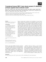
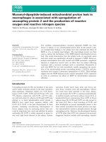
Figure 1 Protection against UV-induced cytotoxicity by forced expression of DDB2. (A, B, C) Sensitivity to UV is presented for stable cell lines
indicated or for (D, E, F) HR18 cells which express DDB2. The protein levels of DDB2, DDB1, and β-actin are shown in the insets. The plotted values
represent means ± S.D. of experiments performed in triplicates. IC
50
values were also indicated in A and D.
Sun et al. Journal of Biomedical Science 2010, 17:27
/>Page 5 of 14
DDB2 protects against UV-induced apoptosis in a caspase-
8 and/or caspase-9 dependent manner
We further investigated the levels of apoptotic markers in
these cells. While UV irradiation was found to induce the
cleavage and activation of caspases-8, 9, and 3 in both
control HeLa and HR3 cells, the cleavage/activation was
considerably reduced in HR3 cells (Fig. 2A). Similarly,
cleavage of both PARP and DNA fragmentation factor
(DFF45) substrates was also decreased in UV-irradiated
HR3 cells compared to control cells (Fig. 1A). In UV-irra-
diated HR18 cells, overexpression of DDB2 was found to
decrease the cleavage/activation of caspases-8, 9, and 3
compared to control cells (Fig. 2B). In addition, DFF45
protein level and PARP cleavage was also decreased in
UV-irradiated HR18 cells (Fig. 2B). We also observed that
cisplatin-resistant HeLa cells HR6, which expressed a low
level of DDB2 [22], also showed increase UV resistance
following overexpression of DDB2 (data not shown).
These results support the notion that DDB2 protects
against UV-induced apoptosis in a caspase-8 and/or cas-
pase-9-dependent manner.
Overexpression of DDB2 increases cFLIP level and
resistance to UV
The protective effect of DDB2 against UV irradiation may
be associated with various regulators of apoptosis. To
assess this possibility, we examined the level of cFLIP,
Bcl-2, and Bcl-xL in UV-resistant cells. The level of cFLIP
protein was increased in HR3 cells compared to control
HeLa cells (Fig. 3A). On the other hand, HR18 cells,
which express a low level of DDB2, showed low cFLIP
compared to control cells (Fig. 3A). Notably, we found
that overexpression of DDB2 using an adenovirus system
increased the level of cFLIP in HR18 cells (Fig. 3B). In this
case, increased expression of DDB2 was noticed 24 hrs
following virus infection, whereas the level of cFLIP
increased only 36 hrs following virus infection (Fig. 3B).
Overexpression of control Gal did not influence the level
of DDB2 or cFLIP compared to mock-treated cells (Fig.
3B). The stimulation of cFLIP following overexpression of
DDB2 was also detected in HeLa cells (data not shown).
To verify whether DDB2 could enhance resistance to UV,
we overexpressed DDB2 in HR18 cells and monitored
apoptosis following UV irradiation. Overexpression of
DDB2 was shown to protect against UV irradiation in a
time-dependent manner (Fig. 3C). UV resistance corre-
lated with the level of cFLIP protein in this case since UV
resistance was maximum at 36 and 72 hours following
virus infection (Figs. 3B and 3C). These results indicate
that UV resistance may be associated with increased lev-
els of DDB2 and cFLIP.
We also monitored the mRNA levels of DDB2 and
cFLIP in these cells by using quantitative PCR (Table 1).
The relative level of endogenous DDB2 mRNA in HeLa,
HR3, and HR18 cells were 1, 1.3, and 0.9, respectively. On
the other hand, the level of cFLIP mRNA in HeLa, HR3,
and HR18 was respectively 1, 5, and 2.6. Following over-
expression of DDB2, HR18 cells displayed a 22-fold
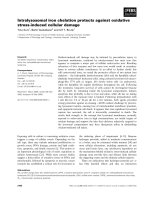
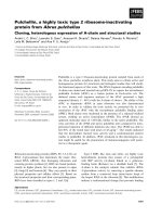
Figure 2 Overexpression of DDB2 is associated with reduced caspase activation following UV treatment. (A) Reduced caspase activation in
cisplatin-resistant cells following UV irradiation. Cell extracts were immunoblotted with the antibodies indicated. (B) Reduced caspase activation in
DDB2-expressing cells following UV irradiation.
Sun et al. Journal of Biomedical Science 2010, 17:27
/>Page 6 of 14
increase of DDB2 mRNA and a 3.4-fold increase of cFLIP.
In comparison, HR18 cells that overexpressed β-Gal
showed a more modest increase of DDB2 mRNA (1.78
fold) and cFLIP (1.9 fold), indicating that virus infection
had a low effect on these cells. From these results, we can
see that the level of DDB2 and cFLIP is increased in cispl-
atin-resistant cells, and that the level of these two pro-
teins appears to correlate with resistance to UV.
Low upregulation of cFLIP promoter activity by
overexpression of DDB2
To examine whether DDB2 may upregulate cFLIP gene
by activating its transcriptional, a cFLIP promoter (-920/
+43, setting the transcription initiation site as +1), which
had been fused to a luciferase cDNA as a reporter gene,
was co-transfected with a plasmid expressing DDB2
(pcDNA3-DDB2) in HEK293 cells. The cFLIP promoter
contains several potential cis-acting elements for transac-
tivators, including E2F (Fig. 4, bottom panel. A series of
deletion mutants were constructed as indicated (Fig. 4,
bottom panel). Transient expression analysis in HEK293
cells indicated the presence of cFLIP core promoters
located 920 bp upstream of the putative transcription ini-
tiation sites. Deletion of these elements reduced basal
promoter activity (Fig. 4, top panel, open bars). The core
promoters contain multiple active E2F sites, followed by a
site at -488 to -258, which represents a critical determi-
nant of negative regulation for this promoter activity. In
addition, Sp1 and AP1 sites located at -158 to -67 may be
essential transcription elements. Overexpression of
DDB2 induced nearly a two-fold increase of FLIP pro-
moter activity in HEK293 cells (Fig. 4, top panel). Nota-
bly, all the 5'-deletion mutants displayed similar promoter
activities as the full-length promoters (Fig. 4, -920/+43).
The promoter activity was undetected in the 3'-deletion
mutants (-920/-487 and -920/-258) where sequences
spanning the transcriptional initiation site were deleted.
These results suggest that DDB2 slightly enhances cFLIP
promoter activity, and that the trans-activation effect
may involve multiple transcription factors.
Knockdown of cFLIP using antisense oligonucleotides
decreases the anti-apoptotic effects of DDB2 against UV
irradiation
The data presented above suggest that cFLIP mediates
the protective effect of DDB2 against UV-induced apop-
tosis. To test this possibility more directly, we used cFLIP
antisense oligonucleotides (ASO) to decrease the level of
this protein in HR18 cells. The level of cFLIP was effi-
ciently decreased by treatment with 600 nM of cFLIP
antisense ASO, whereas control ASO ("CO ASO") did not
affect this protein (Fig. 5A, insert). As shown in Figure
5A, cFLIP antisense markedly sensitized HR18 cells to
apoptosis following UV irradiation. Notably, overexpres-
sion of DDB2 was shown to partially protect HR18 cells
against apoptosis induced by UV (Fig. 5A). In contrast,
forced expression of control β-Gal did not exhibit any
protective effect (Fig. 5A). cFLIP ASO attenuated the
protective effects of DDB2 overexpression against UV-
induced apoptosis (Fig. 5A).
Overexpression of DDB2 also decreased UV-induced
cleavage of both DFF and PARP in HR18 cells (Fig. 5B). In
contrast, forced expression of control β-Gal did not
exhibit any protection effect on the cleavage of either
DFF or PARP (Fig. 5B). cFLIP ASO decreased the protec-
tive effect of DDB2 overexpression against UV-induced
cleavage of DFF and PARP (Fig. 5B). However, the apop-
tosis level of HR18 cells that overexpressed DDB2, and
which were treated with cFLIP ASO, was still lower than
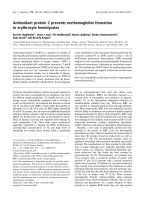
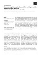
Figure 3 Stimulation of cFLIP expression and attenuation of UV
sensitivity by forced expression of DDB2. (A) Overexpression of
cFLIP in HR3 cells. The plotted values represent means ± S.D. of three
experiments (right panel). (B) Stimulation of cFLIP expression by forced
expression of DDB2. Whole cell extracts of HR18 cells, treated as indi-
cated, were subjected to immunoblot analysis with specific antibodies.
(C) Cell sensitivity to UV after Ad-DDB2 virus infection. The plotted val-
ues represent means ± S.D. of three experiments. ** Significant differ-
ence against control (p < 0.05).
Sun et al. Journal of Biomedical Science 2010, 17:27
/>Page 7 of 14
that of control cFLIP-suppressed cells (Fig. 5B). In this
case, the level of apoptosis was more significant in DDB2-
expressing cells (p < 0.01) compared to control cells (p <
0.05) (Fig. 5). This observation suggests that the activa-
tion of cFLIP by DDB2 may play a more protective role
against UV than endogenous cFLIP. These results indi-
cate that DDB2 protection against UV-induced apoptosis
may proceed via a pathway regulated by cFLIP.
Overexpression of DDB2 in cells lacking cFLIP does not
offer a protective effect against UV irradiation
We also used human VA13 and XP-A cells which express
low level of cFLIP. Surprisingly, these cells did not display
upregulation of cFLIP following DDB2 overexpression
(Fig. 6A). Notably, sensitivity to UV was not affected by
overexpression of DDB2 in VA13 and XP-A cells (Fig.
6B). Cell extracts of cFLIP cDNA transfected cells are
included as cFLIP protein marker. Besides, we observed
that overexpression of cFLIP substantially enhanced cell
viability in both VA13 and XP-A cells (Fig. 6B). as well as
in HEK293 cells (data not shown).
Protection against UV toxicity by DDB2 in Drosophila
Earlier, we and others found that overexpression of DDB2
enhances nuclear excision repair in both hamster [16,17]
and human cells [18,19]. To explore whether the protec-
tive role of DDB2 is conserved in other living organisms,
we expressed human DDB2 cDNA in the fruit fly Droso-
phila, and exposed the resulting flies to UV irradiation.
DDB2-GFP fusion construct and GFP control construct
were expressed in Drosophila with nuclear fluorescence
signals in the salivary gland of the third instar larvae (data
not shown). To detect protein expression, we isolated
proteins from embryos or third instar larvae after a heat-
shock induction of 2 hrs. The DDB2-GFP protein were
detected by Western blot using mouse anti-GFP anti-
body. In this case, the molecular weight of DDB2-GFP is
75 kDa and that of GFP is 27 kDa. UV-induced toxicity
was considerably suppressed in flies that overexpressed
DDB2 compared to control flies that overexpressed GFP
(Fig. 7B).
We also verified whether DDB2 could prevent apopto-
sis induced by the pro-apoptotic genes Rpr or eiger in
fruit flies. We first observed that flies that overexpressed
either Rpr or eiger showed apoptosis in the eyes as shown
by the reduced eye size compared to control flies (Fig. 7C,
panels a and e vs. panels b and f, respectively). On the
other hand, overexpression of DDB2 failed to rescue this
apoptotic effect (Fig. 7, panels c and g). In control experi-
ments, apoptosis induced by activated Rpr could be
mostly rescued by overexpression of p35 (Fig. 7C, panel b
vs. panel d). Even severe apoptosis in the eye was resulted
by activated eiger (Fig. 7C, compare panel e and panel f).
These results suggest that overexpression of DDB2 may
protect against UV toxicity, but that DDB2 is unable to
rescue activated apoptosis in Drosophila.
Discussion
In the present study, we demonstrated that DDB2
increased resistance to UV irradiation in a cFLIP-depen-
dent manner in cisplatin-selected HeLa cells. The marked
decrease of apoptosis following overexpression of DDB2
may partially explain why cisplatin-selected cells are
cross-resistant to UV [21,22] and TNF treatments
[26,27]. Although cisplatin-induced apoptosis can be
mediated by the Fas signaling pathway [27,28], this path-
way involves the mitochondria and the action of caspase-
9 [29]. However, attenuation of intracellular DDB2 levels
in HR3 cells did not affect apoptosis induced by either
cisplatin or mitomycin C, which potentially stimulate
mitochondrial death signals [22]. These observations sug-
gest that DDB2 may not be involved in regulating mito-
chondrial death pathway. Therefore, the increase of
DDB2 and cFLIP expression in cisplatin-resistant cells
Table 1: Induction of endogenous cFLIP mRNA levels in cells following Ad-DDB2 infection.
Fold increase of mRNAa
Cell/Adv infection DDB2 cFLIP
HeLa 1.006 ± 0.181 1.006 ± 0.167
HR3 1.302 ± 0.025 5.087 ± 0.298
HR18 0.909 ± 0.088 2.612 ± 0.010
HR18/Ad-β-Gal 1.780 ± 0.314 1.906 ± 0.220
HR18/Ad-DDB2 22.851 ± 0.115 3.446 ± 0.123
b
a
The numbers indicate mean ± standard deviation of three experiments.
Those samples were examined 60 h after virus infection.
b
Significant difference to HR18/Ad-β-Gal, p < 0.01.
Sun et al. Journal of Biomedical Science 2010, 17:27
/>Page 8 of 14
Figure 4 Upregulation of cFLIP promoter activity by forced expression of DDB2. HEK293 cells were co-transfected with cFLIP reporter together
with either control vector (pcDNA3) or DDB2-expressing vector (pcDNA3-DDB2). After 24 hrs, the luciferase activity was measured. The plotted values
represent means ± S.D. from three independent transfections. The schematic presentation of full-length cFLIP promoter (-920/+43) and its deletion
mutants are indicated below. Putative cis-elements are also indicated at positions relative to the transcription initiation site (+1). The construct number
at the top indicates the length of the tested promoter region upstream of the putative transcription initiation site (designated by the bent arrow at
+1). Luciferase activity is shown relative to the full-length cFLIP promoter (-920/+43). Significant difference to the control for each promoter is indicat-
ed.
Sun et al. Journal of Biomedical Science 2010, 17:27
/>Page 9 of 14
Figure 5 Resistance to UV in HR18 cells depends on DDB2 and cFLIP. (A) Attenuation of DDB2 protection against UV-induced apoptosis by knock-
down of cFLIP using antisense oligonucleotides. HR18 cells were treated as described in the Materials and methods. The plotted values represent
means ± S.D. of experiments performed in triplicates. Inset: immunoblot with specific antibodies. (B) Attenuation of UV-induced caspase activity by
forced expression of DDB2, and resensitization by cFLIP antisense oligonucleotides.
Sun et al. Journal of Biomedical Science 2010, 17:27
/>Page 10 of 14
may explain the cross-resistance to UV in these cells, but
not resistance to cisplatin.
Overexpression of DDB2 has also been shown to pro-
mote global genomic repair in hamster cells [16,17].
Notably, forced expression of mutant DDB2 (DDB2-
82TO), which is defective in DDB1 interaction and dam-
age recognition [30], also protects cells against UV-
induced apoptosis in HeLa cells [22]. These results
strongly suggest that the regulation of UV-induced apop-
tosis by DDB2 may be independent of DNA repair in
Figure 6 Lack of attenuation of UV sensitivity by forced expression of DDB2 in cFLIP-lacking cells. (A) Lack of stimulation of cFLIP expression
by forced expression of DDB2 in human VA13 and XP-A cells. Whole cell extracts of infected cells were compared. Cell extracts of cFLIP-transfected
cells are also shown as cFLIP protein indicator. (B): Cell sensitivity to UV treatment after Ad-DDB2 virus infection in human VA13 and XP-A cells. The
plotted values represent means ± S.D. of three experiments.
Sun et al. Journal of Biomedical Science 2010, 17:27
/>Page 11 of 14
Figure 7 Protection against UV toxicity by DDB2 in Drosophila. (A) Expression of hDDB2-GFP in Drosophila. Protein extracts from GFP and GFP-
hDDB2 expressing embryos were immunoblotted with anti-GFP antibodies. (B) Protection effect of hDDB2 against UV irradiation in fly larvae. GFP or
hDDB2 overexpressing larvae were collected and then irradiated with UV (0-100 J/m
2
). Four days later, surviving adults were counted and the survival
rate was calculated. (n>4000). (C) Lack of inhibition of Reaper- or Eiger-induced apoptosis by hDDB2 over-expression. Eye morphology of wild-type
and Reaper/eiger-overexpressing flies were examined. To analyze the effect of hDDB2, GMRGal4 was used to drive the expression of Reaper (GMR>Rpr)
or Eiger (GMR>eiger) simultaneously with hDDB2 (GMR>Rpr, UAS-hDDB2 and GMR>eiger, UAS-hDDB2, respectively). Panel a-d: Reaper-induced apop-
tosis, resulting in small eyes, was not rescued by DDB2 overexpression. Panel a, Eye morphology of wild-type OreR fly; Panel b, Eye morphology of Rpr
overexpressing fly; Panel c, Eye morphology of Rpr and DDB2 overexpressing fly; Panel d, Eye morphology of Rpr and P35 overexpressing fly. Panel
e~g: Eiger-induced apoptosis not rescued by DDB2 overexpression. Panel e, Eye morphology of wild-type OreR fly; Panel f, Eye morphology of eiger
overexpressing fly; Panel g, Eye morphology of eiger and DDB2 overexpressing fly.
Sun et al. Journal of Biomedical Science 2010, 17:27
/>Page 12 of 14
these cells. Thus, DDB2 as a DNA repair protein also has
a role in regulating cell response to agents that activate
cell surface death signals such as UV and TNF. Impor-
tantly, the protection effect of DDB2 against UV was only
detected in cells whose cFLIP levels are accumulated at
high levels (Fig. 2). This protection effect was decreased
when cFLIP levels became low or were attenuated by
ASO (see Figs. 3 and 5). However, overexpression of
DDB2 in hamster cells may or may not exhibit protection
against UV [16,17]. These divergent observations may be
due to different treatments during which cellular level of
cFLIP likely has a critical role in UV resistance. In addi-
tion, forced expression of DDB2 in cFLIP-lacking cells
(VA13 and XP-A) did not induce cFLIP accumulation, or
protection against UV (Fig. 6), indicating that the DDB2-
cFLIP pathway responsible for the protective effect
against UV-induced apoptosis is probably not evolution-
arily conserved.
DDB2 transcription can be stimulated by E2F1, which
does not require p53, but can be potentiated by this pro-
tein in primary mouse hepatocytes [31]. Moreover,
microarray analysis has demonstrated that FLIP is one of
the E2F1-regulated genes in Saos-2 cells [32]. These find-
ings suggest that E2F1 may also play a role in upregulat-
ing cFLIP, thereby exerting an apoptotic resistance by
inhibiting DISC formation [33] in the acquisition of UV
resistance. Consistent with this idea, we observed that
E2F1 accumulated in UV-resistant HeLa cells. However,
E2F1 has also been implicated in causing apoptosis [34].
Additional overexpression of E2F1 does not increase
endogenous cFLIP expression more than overexpression
of DDB2 alone (data not shown). Thus, the increased
E2F1 level in the resistant cells is not enough to support
apoptotic resistance mediated by DDB2-cFLIP. Although
induction of cFLIP by DDB2 is required for protecting
cells from UV-induced apoptosis, at least in HeLa cells,
we could not exclude the possible involvement of other
genes expression for DDB2-induced cross-resistance.
The expression of DDB2 is also transcriptionally regu-
lated by p53 in cell-type dependent manner [35]. Since
HeLa cells express lower levels of p53 due to infection
with human papillomavirus, continuous exposure of cells
to cisplatin during selection for resistance may activate
p53 and increase DDB2 [35], thereby upregulating cFLIP
levels and providing an opportunity for the cells to escape
UV-induced apoptosis. An apoptotic threshold signifi-
cantly regulated by p53-Bcl2 connection has been pro-
posed [36]. In this model, p53-dependent signals, like the
induction of Bax and direct inhibition of Bcl2, may syner-
gize with p53-independent signals including the induc-
tion of Bim to antagonize Bcl2 function and promote
apoptosis. This model may explain the chemotherapeutic
response of cancer cells as most of the DNA modifying
anti-cancer drugs induce mitochondria death pathway.
Our findings herein, together with others, suggest that
DDB2-cFLIP or p53-dependent DDB2-cFLIP expression,
accounts for an additional route in cell resistance for
agents that preferentially evoke cell surface death path-
way in specific cell type.
We have previously demonstrated that overexpression
of DDB2 could potentiate DNA repair and protect against
UV toxicity in human HeLa and hamster V79 cell lines
[16,22]. Similarly, overexpression of DDB2 protects
against UV toxicity in Drosophila, suggesting that DDB2
may exert its protective activity both in vitro and in vivo.
Other authors have also shown that DDB2 could enhance
global genomic repair and suppress UV-induced muta-
genesis in rodent cells [17]. The effects of DDB2 on DNA
repair was further supported by recent studies. For exam-
ple, overexpression of DDB2 potentiated nuclear excision
repair in mouse embryonic fibroblasts that were irradi-
ated with low doses of UV as shown by accumulation of
DDB1 in the nucleus, degradation of p53, and low level of
p21
Waf1/Cip1
, which is believed to be an inhibitor of repair
synthesis [37]. In addition, knockdown of DDB2 in MCF-
7 cells caused a decrease of cancer cell growth and colony
formation. Inversely, introduction of the DDB2 gene into
MDA-MB231 (low DDB2) cells stimulated growth and
colony formation [38]. DDB2 may play a role in potentia-
tion of MCF-7 cell growth by exerting a negative regula-
tion of the sod2 gene [39]. Hence, DDB2 also plays an
important role in the positive regulation of cell growth. In
most cases, DDB2 overexpression only partially reverses
induced apoptosis, suggesting that severe damage in cells
may override the protective function of DDB2. Surpris-
ingly, however, overexpression of DDB2 is unable to res-
cue activated apoptosis (induced by rpr or eiger) in
Drosophila. As such, Drosophila with less potent apopto-
sis design is needed to re-examine the effect of DDB2 in
regulation of apoptosis in flies. Recently, the potentiation
or lacking effects of DDB2 on DNA damage-induced
apoptosis has also been reported in different cell types
[40-42]. It is concluded that different genetic make-up
between cell types may play an important role in the reg-
ulation of DDB2-mediated cell response to UV stress.
Conclusion
Our results show that DDB2 protects cells against UV
irradiation via the action of cFLIP, which mediates an
anti-apoptosis response following irradiation. Ectopic
expression of human DDB2 in the fruit fly Drosophila
also inhibited UV-induced fly death but it failed to rescue
apoptosis activated by either Reaper or eiger gene. The
protective role of DDB2 against UV stress may be con-
served in various living organisms, whereas cFLIP expres-
sion may be one of the many mechanisms in mediating
protective DDB2 during the acquisition of apoptotic
resistance.
Sun et al. Journal of Biomedical Science 2010, 17:27
/>Page 13 of 14
Abbreviations
ASO: antisense oligonucleotides; CDDP: cisplatin; cFLIP: FLICE inhibitory pro-
teins; DDB2: UV-DNA damage binding protein 2; DFF: DNA fragmentation fac-
tor; β-Gal: β-Galactosidase; GAPDH: glyceraldehyde 3-phosphate
dehydrogenase; GFP: green fluorescence protein; MTT: 3-(4,5-dimethylthiazol-
2-yl)-2,5-diphenyltetrazolium bromide; PARP: Poly-ADP ribose polymerase; PCR:
polymerase chain reaction; SDS-PAGE: sodium dodecyl sulfate-polyacrylamide
gel electrophoresis; UV: ultraviolet radiation. XP-A: xeroderma pigmentosum
group A.
Competing interests
The authors declare that they have no competing interests.
Authors' contributions
NKS, CLS, and CCKC conceived and designed the experiments. NKS, CLS, and
CHL performed the experiments. NKS, CLS, LMP, and CCKC analyzed the data.
CCKC wrote the paper. All authors have read and approved the final manu-
script.
Acknowledgements
The authors would like to thank Dr. Burt Vogelstein (Johns Hopkins University)
for providing vectors used in this study (pAdTrackCMV, pShuttleCMV, and
pAdEasy1); Dr. Sue Lin-Chao (Institute of Molecular Biology, Academia Sinica,
Taipei) and PAN Facility (Stanford University) for providing antisense oligonu-
cleotides; and Dr. B. M. Evers (Department of Surgery, The University of Texas
Medical Branch at Galveston, Galveston, TX) for providing the cFLIP promoter.
The authors also thank Mr. T C. Wu and Mr. K Y. Peng for sharing unpublished
data. In addition, the authors thank Mr. Jan Martel for help during preparation
of the manuscript. This work was supported by intramural funds from Chang
Gung University (CMRPD32024, 33003, 140271) and by grants from the
National Science Council, R.O.C. (NSC 92-2320-B-182-054, NSC95-2320-B-182-
005).
Author Details
1
Department of Biochemistry and Molecular Biology, Chang Gung University,
Gueishan, Taoyuan 333, Taiwan and
2
Division of Biomedical Sciences, Chang
Gung Institute of Technology, Gueishan, Taoyuan 333, Taiwan
References
1. Hengartner MO: The biochemistry of apoptosis. Nature 2000,
407:770-776.
2. Kumar S: Caspase function in programmed cell death. Cell Death Differ
2007, 14:32-43.
3. Kim CN, Wang X, Huang Y, Ibrado AM, Liu L, Fang G, Bhalla K:
Overexpression of Bcl-X(L) inhibits Ara-C-induced mitochondrial loss
of cytochrome c and other perturbations that activate the molecular
cascade of apoptosis. Cancer Res 1997, 57:3115-3120.
4. Kluck RM, Bossy-Wetzel E, Green DR, Newmeyer DD: The release of
cytochrome c from mitochondria: a primary site for Bcl-2 regulation of
apoptosis. Science 1997, 275:1132-1136.
5. Li P, Nijhawan D, Budihardjo I, Srinivasula SM, Ahmad M, Alnemri ES, Wang
X: Cytochrome c and dATP-dependent formation of Apaf-1/caspase-9
complex initiates an apoptotic protease cascade. Cell 1997, 91:479-489.
6. Deveraux QL, Reed JC: IAP family proteins suppressors of apoptosis.
Genes Dev 1999, 13:239-252.
7. Ashkenazi A, Dixit VM: Death receptors: signaling and modulation.
Science 1998, 281:1305-1308.
8. Nagata S: Apoptosis by death factor. Cell 1997, 88:355-365.
9. Irmler M, Thome M, Hahne M, Schneider P, Hofmann K, Steiner V, Bodmer
JL, Schröter M, Burns K, Mattmann C, Rimoldi D, French LE, Tschopp J:
Inhibition of death receptor signals by cellular FLIP. Nature 1997,
388:190-195.
10. Rehemtulla A, Hamilton CA, Chinnaiyan AM, Dixit VM: Ultraviolet
radiation-induced apoptosis is mediated by activation of CD-95 (Fas/
APO-1). J Biol Chem 1997, 272:25783-25786.
11. Bagnoli M, Canevari S, Mezzanzanica D: Cellular FLICE-inhibitory protein
(c-FLIP) signalling: A key regulator of receptor-mediated apoptosis in
physiologic context and in cancer. Int J Biochem Cell Biol 2010,
42:210-213.
12. Abramic M, Levine AS, Protic M: Purification of an ultraviolet-inducible,
damage-specific DNA-binding protein from primate cells. J Biol Chem
1991, 266:22493-22500.
13. Keeney S, Chang GJ, Linn S: Characterization of a human DNA damage
binding protein implicated in xeroderma pigmentosum E. J Biol Chem
1993, 268:21293-21300.
14. Dualan R, Brody T, Keeney S, Nichols AF, Admon A, Linn S: Chromosomal
localization and cDNA cloning of the genes (DDB1 and DDB2) for the
p127 and p48 subunits of a human damage-specific DNA binding
protein. Genomics 1995, 29:62-69.
15. Hwang BJ, Toering S, Francke U, Chu G: p48 Activates a UV-damaged-
DNA binding factor and is defective in xeroderma pigmentosum group
E cells that lack binding activity. Mol Cell Biol 1998, 18:4391-4399.
16. Sun NK, Lu HP, Chao CC: Overexpression of damaged-DNA-binding
protein 2 (DDB2) potentiates UV resistance in hamster V79 cells. Chang
Gung Med J 2002, 25:723-733.
17. Tang JY, Hwang BJ, Ford JM, Hanawalt PC, Chu G: Xeroderma
pigmentosum p48 gene enhances global genomic repair and
suppresses UV-induced mutagenesis. Mol Cell 2000, 5:737-744.
18. Fitch ME, Cross IV, Turner SJ, Adimoolam S, Lin CX, Williams KG, Ford JM:
The DDB2 nucleotide excision repair gene product p48 enhances
global genomic repair in p53 deficient human fibroblasts. DNA Repair
2003, 2:819-826.
19. Wakasugi M, Kawashima A, Morioka H, Linn S, Sancar A, Mori T, Nikaido O,
Matsunaga T: DDB accumulates at DNA damage sites immediately after
UV irradiation and directly stimulates nucleotide excision repair. J Biol
Chem 2002, 277:1637-1640.
20. Chu G, Chang E: Cisplatin-resistant cells express increased levels of a
factor that recognizes damaged DNA. Proc Natl Acad Sci USA 1990,
87:3324-3327.
21. Chao CC, Huang SL, Huang HM, Lin-Chao S: Cross-resistance to UV
radiation of a cisplatin-resistant human cell line: overexpression of
cellular factors that recognize UV-modified DNA. Mol Cell Biol 1991,
11:2075-2080.
22. Sun NK, Kamarajan P, Huang H, Chao CC: Restoration of UV sensitivity in
UV-resistant HeLa cells by antisense-mediated depletion of damaged
DNA-binding protein 2 (DDB2). FEBS Lett 2002, 512:168-172.
23. Wyllie AH, Morris RG, Smith AL, Dunlop D: Chromatin cleavage in
apoptosis: association with condensed chromatin morphology and
dependence on macromolecular synthesis. J Pathol 1984, 142:67-77.
24. Darzynkiewicz Z, Li X: Measurements of cell death by flow cytometry
Portland Press; 1996.
25. He TC, Zhou S, da Costa LT, Yu J, Kinzler KW, Vogelstein B: A simplified
system for generating recombinant adenoviruses. Proc Natl Acad Sci
USA 1998, 95:2509-2514.
26. Sun CL, Chao CC: Cross-resistance to death ligand-induced apoptosis in
cisplatin-selected HeLa cells associated with overexpression of DDB2
and subsequent induction of cFLIP. Mol Pharmacol 2005, 67:1307-1314.
27. Kamarajan P, Sun NK, Chao CC: Up-regulation of FLIP in cisplatin-
selected HeLa cells causes cross-resistance to CD95/Fas death
signalling. Biochem J 2003, 376:253-260.
28. Kamarajan P, Sun NK, Sun CL, Chao CC: Apaf-1 overexpression partially
overcomes apoptotic resistance in a cisplatin-selected HeLa cell line.
FEBS Lett 2001, 505:206-212.
29. Mansouri A, Ridgway LD, Korapati AL, Zhang Q, Tian L, Wang Y, Siddik ZH,
Mills GB, Claret FX: Sustained activation of JNK/p38 MAPK pathways in
response to cisplatin leads to Fas ligand induction and cell death in
ovarian carcinoma cells. J Biol Chem 2003, 278:19245-19256.
30. Wittschieben BB, Wood RD: DDB complexities. DNA Repair (Amst) 2003,
2:1065-1069.
31. Prost S, Lu P, Caldwell H, Harrison D: E2F regulates DDB2: consequences
for DNA repair in Rb-deficient cells. Oncogene 2007, 26:3572-3581.
32. Stanelle J, Stiewe T, Theseling CC, Peter M, Putzer BM: Gene expression
changes in response to E2F1 activation. Nucleic Acids Res 2002,
30:1859-1867.
33. Scaffidi C, Schmitz I, Krammer PH, Peter ME: The role of c-FLIP in
modulation of CD95-induced apoptosis. J Biol Chem 1999,
274:1541-1548.
Received: 5 February 2010 Accepted: 17 April 2010
Published: 17 April 2010
This article is available from: 2010 Sun et al; licensee BioMed Central L td. This is an Open Access article distributed under the terms of the Creative Commons Attribution License ( ), which permits unrestricted use, distribution, and reproduction in any medium, provided the original work is properly cited.Journal of Biomedical Science 2010, 17:27
Sun et al. Journal of Biomedical Science 2010, 17:27
/>Page 14 of 14
34. Phillips AC, Ernst MK, Bates S, Rice NR, Vousden KH: E2F-1 potentiates cell
death by blocking antiapoptotic signaling pathways. Mol Cell 1999,
4:771-781.
35. Tan T, Chu G: p53 Binds and activates the xeroderma pigmentosum
DDB2 gene in humans but not mice. Mol Cell Biol 2002, 22:3247-3254.
36. Hemann MT, Lowe SW: The p53-Bcl-2 connection. Cell Death Differ 2006,
13:1256-1259.
37. Stoyanova T, Yoon T, Kopanja D, Mokyr MB, Raychaudhuri P: The
xeroderma pigmentosum group E gene product DDB2 activates
nucleotide excision repair by regulating the level of p21
Waf1/Cip1
. Mol
Cell Biol 2008, 28:177-187.
38. Kattan Z, Marchal S, Brunner E, Ramacci C, Leroux A, Merlin JL, Domenjoud
L, Dauca M, Becuwe P: Damaged DNA binding protein 2 plays a role in
breast cancer cell growth. PLoS One 2008, 3:e2002.
39. Minig V, Kattan Z, van Beeumen J, Brunner E, Becuwe P: Identification of
damaged DNA binding 2 protein as a transcriptional regulator of the
constitutive sod2 gene expression in human breast cancer cells. J Biol
Chem 2009, 284:14165-14176.
40. Barakat BM, Wang Q-E, Han C, Milum K, Yin D-T, Zhao Q, Wani G, Arafa E-
SA, El-Mahdy MA, Wani AA: Overexpression of DDB2 enhances the
sensitivity of human ovarian cancer cells to cisplatin by augmenting
cellular apoptosis. Int J Cancer in press.
41. Stoyanova T, Roy N, Kopanja D, Bagchi S, Raychaudhuri P: DDB2 decides
cell fate following DNA damage. Proc Natl Acad Sci USA 2009,
106:10690-10695.
42. Stubbert LJ, Smith JM, Hamill JD, Arcand TL, McKay BC: The anti-
apoptotic role for p53 following exposure to ultraviolet light does not
involve DDB2. Mutat Res 2009, 663:69-76.
doi: 10.1186/1423-0127-17-27
Cite this article as: Sun et al., Damaged DNA-binding protein 2 (DDB2) pro-
tects against UV irradiation in human cells and Drosophila Journal of Biomed-
ical Science 2010, 17:27