The signals of FGFs on the neurogenesis of embryonic stem cells ppsx
Bạn đang xem bản rút gọn của tài liệu. Xem và tải ngay bản đầy đủ của tài liệu tại đây (1.97 MB, 11 trang )
Chen et al. Journal of Biomedical Science 2010, 17:33
/>Open Access
RESEARCH
BioMed Central
© 2010 Chen et al; licensee BioMed Central Ltd. This is an Open Access article distributed under the terms of the Creative Commons
Attribution License ( which permits unrestricted use, distribution, and reproduction in
any medium, provided the original work is properly cited.
Research
The signals of FGFs on the neurogenesis of
embryonic stem cells
Ching-Wen Chen
1
, Chin-San Liu
2
, Ing-Ming Chiu
3
, Shih-Cheng Shen
1
, Hung-Chuan Pan
4
, Kun-Hsiung Lee
5
, Shinn-
Zong Lin
6
and Hong-Lin Su*
1,7
Abstract
Background: Neural induction is a complex process and the detailed mechanism of FGF-induced neurogenesis
remains unclear.
Methods: By using a serum-free neural induction method, we showed that FGF1 dose-dependently promoted the
induction of Sox1/N-cadherin/nestin triple positive cells, which represent primitive neuroblasts, from mouse
embryonic stem (ES) cells.
Results: We demonstrated that FGF1, FGF2, and FGF4, but not FGF8b, enhanced this neurogenesis. Especially, FGF-
enhanced neurogenesis is not mediated through the rescue of the apoptosis or the enhancement of the proliferation
of Sox1
+
cells. We further indicated that the inactivation of c-Jun N-terminal kinase-1 (JNK-1) and extracellular signal-
related kinase-2 (ERK-2), but not p38 mitogen-activated protein kinase (MAPK), inhibited the neural formation through
the inhibition of ES differentiation, but not through the formation of endomesodermal cells.
Conclusions: These lines of evidence delineated the roles of FGF downstream signals in the early neural differentiation
of ES cells.
Background
In the early gastrula of the chicken, temporary treatment
of the primitive ectoderm with Hensen's node for 5 hours
steers the ectoderm to become the neural fate [1,2]. FGF
was shown to be responsible for this instructive ability of
node and for the maintenance of later neural instructive
signals [3,4]. FGF first activates ERNI during early gastru-
lation and consequently triggers the zinc-finger tran-
scriptional activator, Churchill, and its downstream target
Sip1 in late gastrulation [4]. In Xenopus, the study of neu-
ral induction has revealed the essential role of Ras/MAPK
activation for neurogenesis in uncommitted ectoderm
and in dissociated animal cap cells, suggesting that the
requirement of FGF signals in neural induction is con-
served in chordates [5].
ES cells, which resemble epiblast cells in the blastocyst,
provide an alternative approach to the study of early
development in mammals [6,7]. Several one-step neural
induction models have been established. Trans-retinoic
acid (RA), a pro-neural inducer, enriches the neural pop-
ulation in a serum-containing embryoid bodies (EBs) sys-
tem [8,9]. However, RA treatment has several drawbacks,
including the caudalization of the neural fate, blockage of
forebrain induction, and the disruption of normal
embryogenesis [9-11]. Co-culture of ES cells with mouse
skull-derived stromal cells, such as PA6 cells, or bone
marrow-derived cells, such as MS5 cells, efficiently
induces the ES cells to become neuron lineages [8,12].
However, the factors contributing to this stromal-derived
inducing activity are still uncharacterized. ES cells cul-
tured in serum-free Neurobasal medium with N2B27
supplement efficiently differentiate into Sox1
+
neural pre-
cursors, which represent the earliest committed neuro-
blast cells in the developing embryo [13,14]. Specific
neuronal subtypes, such as dopaminergic and serotonin-
ergic neurons, are derived from the Sox1 neuroblasts by
the addition of defined patterning factors. Although the
Neurobasal/N2B27 model provides a simple monocul-
ture differentiation system for ES cells, these cells often
undergo apoptosis on days 3 to 5. Recently, an efficient
neural-induction monoculture system with a high sur-
* Correspondence:
1
Department of Life Sciences, National Chung-Hsing University, Taichung,
T
aiwan
Full list of author information is available at the end of the article
Chen et al. Journal of Biomedical Science 2010, 17:33
/>Page 2 of 11
vival rate for differentiating ES cells was developed and
termed as serum-free embryoid bodies formation (SFEB)
method [15]. This simple and reproducible system con-
sists of defined components and is suitable for the explo-
ration of downstream FGF signals in the early
neurogenesis of mammals.
Methods
Cell culture and differentiation
Sox1-GFP knock-in ES cells (46C), from Dr. Austin Smith
(University of Cambridge, UK), and ESC 26 cells, were
both well-characterized and germline transmissible
[14,16]. The culture condition of both cells [14,16] and
the SFEB method [15] has been described previously in
detail.
Reagents
Human recombinant FGF2, FGF4 and FGF8b were all
from R&D Systems. Recombinant human FGF1 was pre-
pared from Prof. Chiu in Institute of Cell and Systems
Medicine, the National Health Research Institutes, Tai-
wan [17]. Synthetic inhibitors of FGF signaling, including
SU5402, LY294002, SB203580, and SP600125, were from
Calbiochem; U0126 was purchased from Tocris.
Stable cell establishment
The plasmid Flag-DsRedT4-NLS was a gift from Tim
Shroeder at Helmholtz Center Munich, Institute of Stem
Cell Research, Germany. The genes of JNK dominant
negative mutants, Flag-JNK1a1apf and Flag-JNK2a2apf
[18,19], were obtained from Addgene http://
www.addgene.org and fused with a IRES-DsRed as a
reporter. The plasmids were transfected into ES cells with
lipofectamine 2000 (Invitrogen). After selection with 0.4
mg/ml G418 for two weeks, stable clones with red fluo-
rescence were picked up and maintained with 0.2 mg/ml
G418. The selected ES cells showed normal ES cell mor-
phology and pluripotent gene expression (data not
shown).
Immunocytochemistry
Cells were fixed in 4% cold paraformaldehyde and perme-
abilized with 0.3% Triton-X 100. Immunocytochemistry
was performed with the following primary antibodies:
OCT3/4 (1:500, Santa Cruz), Nanog (1:100, Cosmo Bio,
Japan), Sox2 (1:4000, Chemicon), N-cadherin (1:100,
DSHB, Iowa), FGF receptor 1 (FGFR1) and FGFR3 (both
1:100, Santa Cruz), FGFR2 (1:500, Abcam) and GFP
(1:1000, Aves Labs). Images of immunostaining were cap-
tured usinga fluorescent microscope (Nikon ECLIPSE
80I) or confocal microscope (LSM510 Meta, Zeiss).
Flow cytometry
Sox1-GFP ES cells were fully dissociated and analyzed
with flow cytometry (FC500, Beckman Coulter). Apopto-
sis was measured by staining for Annexin V (AbD Sero-
tec) at room temperature for 10 min in the dark.
RT-PCR analysis
Total RNA was isolated from ES cells using REzol™ C&T
reagent (Protech technology, Taiwan). Primers were
applied to detect the expression of FGFR1 (5'-CAC ACT
GCC TTC TCC TCC TC-3', 5'-CTC TGC CTC CCT
GTC TTC TG-3'), FGFR2 (5'-GGG GAT GTG GAG TTT
GTC TG-3', 5'-GCT TCT TGG TCG TGG TCT TC-3'),
FGFR3 (5'-CGG CTA CCT GTG AAG TGG AT-3', 5'-
GCT TGG TCT GTG GGA CTG TT-3'), FGFR4 (5'-AGG
AAA TGT GGC TGC TCT TG-3', 5'-GGT GTG TCC
AGT AGG GTG CT-3'), Sox1 (5'-CCT CGG ATC TCT
GGT CAA GT-3', 5'-TAC AGA GCC GGC AGT CAT
AC-3'), and G3PDH (5'-GTG AAG GTC GGT GTG AAC
G-3', 5'-GGT GAA GAC ACC AGT AGA CAC TC-3').
Western blot analysis
ES cells were lysed in RIPA buffer (50 mM Tris pH7.5,
150 mM NaCl, 10 mM EDTA, 1% NP-40, 0.1% SDS) plus
a cocktail of proteinase inhibitors (Sigma-Aldrich). Dena-
tured proteins were separated by 10% SDS-PAGE and
then transferred to PVDF membranes. Samples were
detected with antibodies to ERK1/2, phosphoERK1/2
(pERK1/2), p38 and pp38, JNKs and pJNKs, AKT and
pAKT. All MAPK-related antibodies were from Cell Sig-
nals and diluted 1:1000 for immunoblotting. Chemilumi-
nescence of immunoreactive bands was detected using
secondary horseradish peroxidase-conjugated antibodies
(Jackson ImmunoResearch) and ECL reagents (Amer-
sham).
Results
FGF1 enhanced the generation of Sox1
+
cells from ES cells
Two germline-transmissible mouse ES cell lines, ESC 26
and Sox1-GFP knock-in cells (46C), were used in this
study and the ESC 26 cell was characterized with the
expression of pluripotent makers (Fig. 1B to 1D). After
dissociation, ES cells were cultured at 2 × 10
6
cells/10 ml
in a defined, serum-free, neural differentiation medium
(SFEB method) (Fig. 1A), which is an efficient neural
induction method with rare mesendoderm formation
[15]. We showed that ES-derived Sox1-GFP
+
cell was
coexpressed several neural markers, such as nestin, pax6,
N-cadherin and Zic1 (Fig. 1E to 1H). In addition, GFAP
was not detected in differentiating 46C cells on day 6 (Fig.
1I), indicating that the Sox1
+
cells under the SFEB culture
represented primitive neuroblast cells [15]. Exogenous
FGF1, applied from day 1 through day 3, dramatically
enhanced the neural induction of ESC26 and 46C cells in
a dose-dependent manner, as revealed by the counting of
N-cadherin
+
colonies (Fig. 1J) and FACS analysis on day
6, respectively (Fig. 2A). These results suggest that FGF
Chen et al. Journal of Biomedical Science 2010, 17:33
/>Page 3 of 11
was sufficient to promote the formation of neuroblast
cells derived from ES cells.
We next tested the effects of different FGFs on neural
formation of ES cells. FGF1, FGF2, and FGF4 all showed
significantly elevated neural induction in 46C cells (Fig.
2A). However, FGF8b, even at the high concentration of
80 ng/ml, failed to enhance the neural induction of ES
cells (Fig. 2A). We further investigated the expression of
FGFRs in ES cells during neural induction and found that
the expression of FGFR4 gradually declined (Fig. 2B),
which is in agreement with the finding that FGFR4 is
excluded from the neuroectoderm of mouse embryos
[20]. In contrast, FGFR1, FGFR2, and FGFR3 expressions
were significantly increased during the conversion of ES
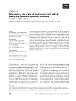
Figure 1 The characteristics of the ES cells and their neural derivatives. (A) Schematic procedure of SFEB for neural induction of ES cells. Undif-
ferentiated ESC 26 cells were characterized by pluripotent markers such as Oct4 (B), Nanog (C) and Sox2 (D). The 46C ES-derived GFP
+
cells were co-
expressed with neural markers, such as nestin (E), pax6 (F), N-cadherin (G), Zic1 (H), but not GFAP (I) on day 6. Nuclei of ES cells were stained with DAPI
in blue (B-I). ESC 26 cells were treated with 20, 40, and 80 ng/ml FGF1 from day 1 through day 3 and the N-cadherin
+
colonies were estimated under
fluorescent microscope (J) on day 6 from three independent experiments. A cell cluster with over 50 μm was counted as a colony and a colony was
N-cadherin positive if over half of the cells in the colony expressed N-cadherin. Scale bar, 10 μm in B.
Chen et al. Journal of Biomedical Science 2010, 17:33
/>Page 4 of 11
into neuroblast cells. Immunocytostaining revealed that
both FGFR1 and FGFR3 were detected in cytosol and
nuclei in neural derivatives (Fig. 2C). On day 6, GFP
+
sig-
nals were colocalized with FGFR1- and FGFR3-express-
ing cells, suggesting that both signals may be involved in
neurogenesis (Fig. 2C). RT-PCR and immunostaining,
shown in Figs. 2B and 2C, indicated that the expression of
FGFR2 in differentiating ES cells was robustly induced
and was localized on the cell membrane and cytosol,
rather than in the nucleus. We also found that FGFR2 was
not completely coexpressed with the GFP in 46C cells on
day 6 (Fig. 2C), suggesting that FGFR2 is involved in the
formation of subtypes of neurons. Taken together, these
results suggest that FGFR1 and FGFR3 are generally
required for neural induction and FGF8b is incompetent
on the enhancement of neurogenesis of ES cells.
Neural induction enhanced by FGF was not mediated
through the anti-apoptosis or cell proliferation on Sox1
+
cells
We treated 46C ES cells with or without FGF1 from day 1
through day 3 and detect the Sox1-GFP
+
cells from day 1
to day 8 (Fig. 3A). The number of Sox1
+
cells became 20%
of total cells on day 3 and reached the plateau, 50% of
total cells, on day 7. Treatment of FGF1 consistently and
dose-dependently enhanced the neurogenesis on day 3
through day 7. We also found that FGF treatment can
promote but cannot shorten the time of the neural induc-
tion from ES cells. The Sox1-GFP
+
cells did not appear on
differentiation day 2, regardless of the FGF1 treatment.
The increase of Sox1
+
cells in the FGF1-treated condi-
tion may result from enhanced proliferation and/or
reduced apoptosis of neuroblast cells. To test these possi-
bilities, FGF1 was incubated with the 46C cells, and the
apoptosis and proliferation of Sox1
+
cells were analyzed
by staining of activated caspase-3 and Ki67, respectively.
Double staining of cleaved caspase-3 and GFP revealed
that less than 5% double positive cells were detected (Fig.
3B). Similar results were obtained in FGF1-treated Sox1
+
cells (data not shown). The percentages of Ki67
+
cells in
Sox1
+
population were 24.75% (196/792) and 25.48%
(362/1421) in SFEB- and SFEB/FGF1-treated cells respec-
tively (Fig. 3C and 3D), demonstrating that FGF-triggered
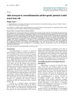
Figure 2 The FGF effects on the neurogenesis of ES cells and the FGFR expressions in ES cells. (A) After treatment with FGF1, FGF2, FGF4, and
FGF8b from day 1 to day 3 using the SFEB method, the numbers of 46C ES-derived Sox1-GFP
+
cells were estimated by flow cytometry on day 6 (n =
3 for each panel). (B) On indicated days, FGFRs in 46C ES cells were analyzed by RT-PCR. (C) Expression of FGFRs and the GFP
+
ES cells was analyzed by
immunostaining on day 6 or day 2. Single GFP positive cells were indicated by arrow. Nuclei of all cells are revealed by DAPI staining in blue. Scale bar,
10 μm in C. *, p < 0.01, Anova test.
Chen et al. Journal of Biomedical Science 2010, 17:33
/>Page 5 of 11
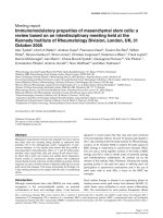
Figure 3 The apoptosis and the proliferation on committed neuroblast cells. (A) The induction of Sox1-GFP
+
cells from 46C cells were detected
by flow cytometry under the SFEB and SFEB/FGF1 condition. (B) The differentiating ES cells were labeled with cleaved caspase-3 (red), which detects
the cleaved fragment of caspase-3 (17/19 kDa), in Sox1/GFP
+
cells on differentiating day 4. (C, D) Proliferating GFP
+
cells were marked with the nuclear
staining of ki67 on day 4. (E) Total apoptotic cells, characterized with Annexin-V labeling, were estimated by flow cytometry after FGF and/or z-VAD-
fmk, a membrane-permeable pan-caspase inhibitor, from day 1 to day 4. Culture media were changed every day. (F) Total cell numbers were counted
in triplicate using trypan blue exclusion at indicated times.
Chen et al. Journal of Biomedical Science 2010, 17:33
/>Page 6 of 11
neurogenesis may not mediated through the enhance-
ment of Sox1 cell proliferation.
We also found that on day 1 through day 4, the total
number of apoptotic cells was not reduced after treat-
ment with 40 ng/ml FGF1, or with 5 μM of a pan-caspase
inhibitor, z-VAD-fmk. Even after the addition of both
FGF1 and z-VAD-fmk, the rescue of apoptotic cells was
not significant (Fig. 3E). The total ES cell population was
also counted on differentiation days 1 to 4. No statistical
significance in number was seen after treatment with
FGF1 and/or z-VAD-fmk (Fig. 3F). In sum, these results
suggest that the FGF-steering neurogenesis mainly
depends on the enforcing differentiation of ES cells,
rather than on anti-apoptosis or cell proliferation.
Neural induction of ES cells was mediated through the
activation of MAPK pathways
Given that phosphorylated intracellular domains of
FGFRs activate downstream phosphoinositide-3 kinase
(PI3K)/AKT and three major serine/threonine MAPKs,
including ERK 1/2, JNKs, and p38 kinases, we further
investigated which MAPK pathways were responsible for
the FGF-dependent neural induction. We found that sin-
gle suspended ES cells continued to initiate phosphory-
lated JNK during differentiation (Fig. 4A). Significant
enhancement of ERK activation was observed in 20 ng/ml
FGF1-treated ES cells, providing the linkage of biochemi-
cal evidences of FGF signal with its pro-neural function.
FGF1 promoted the AKT phosphorylation and the activi-
ties of all three MAPKs in differentiating ES cells at 12 hr
differentiation (Fig. 4B). Immunoblotting showed that the
total amount of AKT, JNK, p38 MAPK, and ERK1/2 pro-
tein expression was not altered between control and
SFEB conditions. Especially, JNK1 and ERK2 were the
major phosphorylated isoforms of JNKs and ERKs in the
differentiating ES cells, respectively.
Specific pharmacological inhibitors of MAPKs, shown
affecting their respective kinase targets in Fig. 4B, were
administrated to delineate the kinases involved in neuro-
genesis. We found that a PI3K/AKT inhibitor, LY294002,
significantly reduced the formation of Sox1-GFP
+
cells
under SFEB and SFEB/FGF1 conditions (Fig. 4C and 4D).
Intriguingly, a JNK inhibitor and an ERK inhibitor,
SP600125 and U0126, respectively, dramatically blocked
the neural formation of ES cells and abolished the FGF-
mediated neurogenesis (Fig. 4C and 4D). Nevertheless,
there was no significant reduction of Sox1-GFP
+
cells
after treatment with p38 kinase inhibitor, in both exoge-
nous FGF present or absent condition (Fig. 4C and 4D).
In addition, to verify the role of JNK isotypes in neural
differentiation of ES cells, stable clones expressing the
JNK1 and JNK2 dominant negative mutants (JNK1a1apf
and JNK2a2apf) were established (Fig. 5A and 5B). We
found that specific inhibition of JNK1, but not JNK2, sig-
nificantly reduced the formation of Sox1
+
and N-cad-
herin
+
cells (Fig. 5C, 5D and 5E), indicating that JNK1 is
essential for the neural induction of ES cells.
Response-time windows for the FGF-mediated
neurogenesis
To verify the FGF response windows during ES differenti-
ation, 40 ng/ml FGF1 was incubated with 46C cells for 24
hr on individual day 1 to 4 (Fig. 6A). ES-derived neural
cells were analyzed on day 6 by FACS. FGF1 treatment in
the first 24 hr window was sufficient to promote Sox1 cell
induction (Fig. 6B, the lane D1). Neurogenic effects were
also observed when the ES cells were incubated with
FGF1 on day 2 or 3 (Fig. 6B, the lane D2 and D3). This
result argues that transient FGF activation is sufficient to
enforce early cell-fate commitment and neural induction
of ES cells. In contrast, JNK and ERK inhibitors caused
only a short-term reduction of neurogenesis and a delay
in commitment. As shown in Figs. 6C and 6D, neural
inhibition was observed on day 6 when MAPK signals
were constantly depressed throughout days 1 to 3 (Fig.
6D; the lane D1-3). Transient treatments of both inhibi-
tors on individual days did not show the suppression of
neural induction (Fig. 6D; the lane D1, D2 and D3). Inter-
estingly, we also found that GFP
+
cell population with the
treatment of MAPK inhibitors throughout days 1 to 3
gradually increased from 26 ± 5.5% on day 6 to 55 ± 6.7%
of total cells on day 9 (data not shown), suggesting that
inhibition of JNK and ERK retards the ES cell commit-
ment, rather than promotes non-neural lineages.
Cell lineages of the ES cells treated with MAPK inhibitors
Reduction of the neural induction by the JNK and ERK
inhibitors could be caused by the increased undifferen-
tiating ES cells or non-neural lineages. In this study, we
demonstrated that inactivation of both JNK and ERK
enhanced the expression of pluripotent markers Oct4
and Nanog in differentiating ES cells on day 6 (Figs. 7A
and 7B), indicating that both phosphorylated JNK and
ERK are negative regulators of self-renewal of ES cells. It
is recently documented that ERK2 null ES cells fail to
commit into neural and mesodermal cells [21-24]. Simi-
larly, rare brachyury (T) expressed cells were found in
SP600125- and U0126-treated ES cells, compared to 5.2
± 0.2% brachyury-positive cells in the total population
under SFEB (Fig. 7C and 7E). The Sox17
+
cells, repre-
senting endoderm of differentiating ES cells, only
showed less 5% of total ES cells on day 6 under the SFEB
condition (Fig. 7D). No significant elevation of Sox17
+
cells was observed in JNK/ERK inhibitors treated ES
cells (Fig. 7F). In addition, we also did not find the
appearance of cytokeratin 14 (K14) positive cells, repre-
senting the epidermal precursor cells, in the SFEB-dif-
ferentiating ES cells even after the treatment of MAPK
Chen et al. Journal of Biomedical Science 2010, 17:33
/>Page 7 of 11
inhibitors. These results demonstrated that the reduc-
tion of neural formation by the inactivation of MAPK
was caused by the blockage of ES differentiation, rather
than by the enhancement of formation of mesoendoder-
mal nor epidermal lineages.
Discussion
Neural induction requires sequential signals to direct
uncommitted ectoderm into the definitive neural plate
[25]. Cumulative evidence supports the fact that FGF is
an essential factor for neurogenesis [26,27]. Interestingly,
activation of the Ras/MAPK pathway, rather than the
Figure 4 Effects of MAPK inhibitors on neural induction of ES cells. (A) Total cell lysates were collected from differentiating ES cells at indicated
times under SFEB condition. Kinetic JNKs activation was analyzed by western blot. FGF1 dose-effect on differentiating ES cells was revealed by ERK
phosphorylation at 30 min differentiation. (B) Downstream FGF signals were further detected with individual specific antibodies at 12 hr post-treat-
ment of 40 ng/ml FGF1 (lane 3), or with inhibitors (lane 4) of PI3K/AKT (LY 294002, 10 μM), JNK1/2 (SP 600125, 10 μM), p38 MAPK (SB 203580, 20 μM),
and ERK1/2 (U0126, 5 μM). After treatment with the inhibitors (C) or FGF1 (40 ng/ml) plus the inhibitors (D) from day 1 to day 3, the derived cells were
collected for FACS analysis on day 6. The same concentrations of reagents were applied in these experiments. Representative results were shown from
experiments done at least in triplicate.
Chen et al. Journal of Biomedical Science 2010, 17:33
/>Page 8 of 11
Figure 5 Genetic inhibition of JNKs in differentiating ES cells. (A) Flag-tagged dominant-negative mutants of JNK1 and JNK2 (JNK1a1-apf and
JNK2a2-apf) were conjugated with IRES-DsRed for the tracing of the consistently expressing cells. (B) The expression of flag, phosphorylated JNKs,
phosphorylated c-Jun (pc-Jun) and total amount of JNK1 and JNK2 were revealed by western blot. (C) Their efficiencies of neural formation were es-
timated by FACS analyses. The expressions of neural markers are also examined, such as Sox1 (D), nestin (D) and N-cadherin (N-cad) (E).
Figure 6 Response windows of FGF and MAPK inhibitors in differentiating ES cells. (A) FGF1 at 40 ng/ml was applied to 46C ES cells on individual
days (D1, D2, D3, D4) or from day 1 through 4 (D1-4). (B) Derived GFP
+
cells were analyzed by FACS on day 6. Independent experiments done in trip-
licate are illustrated. (C) As the indicated experimental conditions, the induction of Sox1-GFP
+
cells on day 6 was shown in (D) after FACS analysis.
SP600125 and U0126, 10 μM and 5 μM, respectively.
Chen et al. Journal of Biomedical Science 2010, 17:33
/>Page 9 of 11
Figure 7 Both inhibitors of JNK and ERK retarded ES differentiation. After treatment with 10 μM SP600125, 2 or 10 μM U0126 from days 1-3, ES
cells were plated on 0.1% matrigel-coated glasses and stained with anti-Oct4 (A) and anti-Nanog antibodies (B) on day 6. The ratio of undifferentiated
pluripotent ES cells to total DAPI
+
cells (n>500 cells) was estimated from experiments done in triplicate. Brachyury (T) (C), Sox17 (D) and cytokeratin
14 (E) expressions, representing mesodermal, endodermal and surface ectodermal cell lineages respectively, were examined in ES cells on day 6 with
SFEB treatment. Nuclei of all cells are seen by DAPI staining in blue. The statistic results of the cell numbers in panel C and D were also estimated,
respectively (E, F).
Chen et al. Journal of Biomedical Science 2010, 17:33
/>Page 10 of 11
diluted BMP ligands, has been shown to be responsible
for the neural cell fate of the fully dissociated animal cap
cells, arguing against the simplistic neural default model
[5]. The primitive streak- or organizer-derived BMP
inhibitors are not the only signals required for neurogen-
esis. FGF and the other developmental cues, such as Wnt
and Notch, also participate in neural induction in a
sophisticated manner [25].
It is noteworthy to emphasize that the activation of
MAPK during ES differentiation may not solely depend
on FGFR signals and other neural instructing factors
could also contribute to the neural induction through
JNK or ERK activation, such as insulin-like growth factor
(IGF) [28]. Treatment of JNK and ERK inhibitors should
simultaneously abolish the endogenous receptor tyrosine
kinase signals of differentiating ES cells. Here we showed
that neural induction of ES cells was accompanied with
the elevated expression of FGFRs and the activation of
MAPK pathway (Figs. 2B, 4A and 4B). Pharmacological
evidences (Fig. 4C) further supported that differentiation
into primitive neuroepithelial cells relied on the activa-
tion of both JNK and ERK pathways, but not the p38
MAPK pathway (Fig. 4C). Exogenous FGF-triggered neu-
rogenesis was completely reduced by the JNK and ERK
inhibitors (Fig. 4D). Taken together, these data highlights
the importance of FGFR activation and of individual
MAPK signals in the ES-neuron conversion.
Both pharmacological and genetic evidences support
the important role of JNK1 for the neural induction of ES
cells (Fig. 4C, D and 5). These results are consistent with
the previous finding that JNK1
-/-
ES cell has a significant
reduction in RA-triggered neurogenesis and that JNK/
Stress-associated activated protein 1 (JSAP1) is involved
in early embryonic neurogenesis [29,30]. While a neural
tube defect is only observed in JNK1/JNK2 double-
knockout mice and a JNK1 and JNK2 single-null embryo
is normal [31]. It is important to further explore the rea-
son of discrepancy between in vitro and in vivo data and
the JNK regulatory networks which participate in neural
fate decision and the development of primitive neuroec-
toderm.
Genetic manipulation has shown that ERK1-null mice
are healthy after birth, whereas disruption of the ERK2
gene results in abnormal trophectodermal and mesoder-
mal development [32,33]. In vitro ES differentiation has
also revealed that inhibition of ERK2 completely blocks
neural and mesodermal formation, suggesting that ERK2
is essential for the initiation of cell fate commitment of
epiblast cells [21,24]. In this study, we showed that inhibi-
tion of MAPK signals sustained the undifferentiated sta-
tus and the expression of pluripotent markers under the
SFEB condition. In future studies, it will be important to
understand how the regulatory networks of MAPKs are
affected after deprivation of LIF and how they initiate
somatic cell induction in ES cells.
Conclusions
Based on a simple and efficient neural induction method,
we demonstrate that FGF-triggered neurogenesis of ES
cells is not involved in cell proliferation or inhibition of
apoptosis. Activation of the ERK2 and JNK1 pathways,
rather than p38 MAP kinase, is mainly responsible for the
neural induction of ES cells. Release of pharmacological
inhibition re-initiated the ES differentiation and neuro-
genesis, indicating that the FGF pathway participates in
the initiation of ES commitment into embryonic cell lin-
eages.
List of abbreviations
ESC: embryonic stem cell; FGF: fibroblast growth factor;
MAPK: mitogen-activated protein kinase; SFEB: serum-
free embryoid body-like formation.
Competing interests
The authors declare that they have no competing interests.
Authors' contributions
CWC, SCS, HCP and HLS carried out the neural differentiation and drafted the
manuscript. KHL provided the mES cells and participated in the design of the
study. CSL, IMC SZL and HLS participated in the design of the study and per-
formed the statistical analysis. All authors read and approved the final manu-
script.
Acknowledgements
This work was supported by the Changhua Christian Hospital (C.S.L.), National
Health Research Institutes (H.L.S.) as well as the National Science Council
(H.L.S.) of Taiwan. This work was also granted from the Taichung Veterans Gen-
eral Hospital and National Chung Hsing University (TCVGH-NCHU-9776614
and -977602; to H.L.S and H.C.P.), Taichung, Taiwan. We also thank for the sup-
port from the core laboratory of tissue engineering and stem cells center in
NCHU.
Author Details
1
Department of Life Sciences, National Chung-Hsing University, Taichung,
Taiwan,
2
Department of Medical Research, Changhua Christian Hospital,
Changhua, Taiwan,
3
Institute of Cellular and Systems Medicine, National Health
Research Institutes; Miaoli, Taiwan,
4
Department of Neurosurgery, Taichung
Veterans General Hospital; Taichung, Taiwan,
5
Animal Technology Institute
Taiwan; Miaoli, Taiwan,
6
Center for Neuropsychiatry, China Medical University
and Hospital, Taichung, Taiwan; China Medical University Beigang Hospital,
Yunlin, Taiwan; Department of Immunology, China Medical University,
Taichung, Taiwan and
7
Department of Physical Therapy, China Medical
University, Taichung, Taiwan
References
1. Streit A, Berliner AJ, Papanayotou C, Sirulnik A, Stern CD: Initiation of
neural induction by FGF signalling before gastrulation. Nature 2000,
406:74-78.
2. Streit A, Stern CD: Establishment and maintenance of the border of the
neural plate in the chick: involvement of FGF and BMP activity. Mech
Dev 1999, 82:51-66.
3. Linker C, Stern CD: Neural induction requires BMP inhibition only as a
late step, and involves signals other than FGF and Wnt antagonists.
Development 2004, 131:5671-5681.
Received: 28 December 2009 Accepted: 29 April 2010
Published: 29 April 2010
This article is available from: 2010 Chen et al; licensee BioMed Central Ltd. This is an Open Access article distributed under the terms of the Creative Commons Attribution License ( which permits unrestricted use, distribution, and reproduction in any medium, provided the original work is properly cited.Journa l of Biome dical Scie nce 2010, 17:33
Chen et al. Journal of Biomedical Science 2010, 17:33
/>Page 11 of 11
4. Sheng G, dos Reis M, Stern CD: Churchill, a zinc finger transcriptional
activator, regulates the transition between gastrulation and
neurulation. Cell 2003, 115:603-613.
5. Kuroda H, Fuentealba L, Ikeda A, Reversade B, De Robertis EM: Default
neural induction: neuralization of dissociated Xenopus cells is
mediated by Ras/MAPK activation. Genes Dev 2005, 19:1022-1027.
6. Pera MF, Trounson AO: Human embryonic stem cells: prospects for
development. Development 2004, 131:5515-5525.
7. Spagnoli FM, Hemmati-Brivanlou A: Guiding embryonic stem cells
towards differentiation: lessons from molecular embryology. Curr Opin
Genet Dev 2006, 16:469-475.
8. Kawasaki H, Mizuseki K, Nishikawa S, Kaneko S, Kuwana Y, Nakanishi S,
Nishikawa SI, Sasai Y: Induction of midbrain dopaminergic neurons
from ES cells by stromal cell-derived inducing activity. Neuron 2000,
28:31-40.
9. Wichterle H, Lieberam I, Porter JA, Jessell TM: Directed differentiation of
embryonic stem cells into motor neurons. Cell 2002, 110:385-397.
10. Mizuseki K, Sakamoto T, Watanabe K, Muguruma K, Ikeya M, Nishiyama A,
Arakawa A, Suemori H, Nakatsuji N, Kawasaki H, Murakami F, Sasai Y:
Generation of neural crest-derived peripheral neurons and floor plate
cells from mouse and primate embryonic stem cells. Proc Natl Acad Sci
USA 2003, 100:5828-5833.
11. Kohga H, Obata K: Retinoic acid-induced neural tube defects with
multiple canals in the chick: immunohistochemistry with monoclonal
antibodies. Neurosci Res 1992, 13:175-187.
12. Perrier AL, Tabar V, Barberi T, Rubio ME, Bruses J, Topf N, Harrison NL,
Studer L: Derivation of midbrain dopamine neurons from human
embryonic stem cells. Proc Natl Acad Sci USA 2004, 101:12543-12548.
13. Ying QL, Stavridis M, Griffiths D, Li M, Smith A: Conversion of embryonic
stem cells into neuroectodermal precursors in adherent monoculture.
Nat Biotechnol 2003, 21:183-186.
14. Aubert J, Stavridis MP, Tweedie S, O'Reilly M, Vierlinger K, Li M, Ghazal P,
Pratt T, Mason JO, Roy D, Smith A: Screening for mammalian neural
genes via fluorescence-activated cell sorter purification of neural
precursors from Sox1-gfp knock-in mice. Proc Natl Acad Sci USA 2003,
100:11836-11841.
15. Watanabe K, Kamiya D, Nishiyama A, Katayama T, Nozaki S, Kawasaki H,
Watanabe Y, Mizuseki K, Sasai Y: Directed differentiation of telencephalic
precursors from embryonic stem cells. Nat Neurosci 2005, 8:288-296.
16. Lee KH, Chuang CK, Wang HW, Stone L, Chen CH, Tu CF: An alternative
simple method for mass production of chimeric embryos by
coculturing denuded embryos and embryonic stem cells in Eppendorf
vials. Theriogenology 2007, 67:228-237.
17. Patrie KM, Botelho MJ, Franklin K, Chiu IM: Site-directed mutagenesis and
molecular modeling identify a crucial amino acid in specifying the
heparin affinity of FGF-1. Biochemistry 1999, 38:9264-9272.
18. Wojtaszek PA, Heasley LE, Siriwardana G, Berl T: Dominant-negative c-Jun
NH2-terminal kinase 2 sensitizes renal inner medullary collecting duct
cells to hypertonicity-induced lethality independent of organic
osmolyte transport. J Biol Chem 1998, 273:800-804.
19. Xiao L, Lang W: A dominant role for the c-Jun NH2-terminal kinase in
oncogenic ras-induced morphologic transformation of human lung
carcinoma cells. Cancer research 2000, 60:400-408.
20. Stark KL, McMahon JA, McMahon AP: FGFR-4, a new member of the
fibroblast growth factor receptor family, expressed in the definitive
endoderm and skeletal muscle lineages of the mouse. Development
1991, 113:641-651.
21. Kunath T, Saba-El-Leil MK, Almousailleakh M, Wray J, Meloche S, Smith A:
FGF stimulation of the Erk1/2 signalling cascade triggers transition of
pluripotent embryonic stem cells from self-renewal to lineage
commitment. Development 2007, 134:2895-2902.
22. Burdon T, Stracey C, Chambers I, Nichols J, Smith A: Suppression of SHP-2
and ERK signalling promotes self-renewal of mouse embryonic stem
cells. Dev Biol 1999, 210:30-43.
23. Li Z, Theus MH, Wei L: Role of ERK 1/2 signaling in neuronal
differentiation of cultured embryonic stem cells. Dev Growth Differ
2006, 48:513-523.
24. Stavridis MP, Lunn JS, Collins BJ, Storey KG: A discrete period of FGF-
induced Erk1/2 signalling is required for vertebrate neural
specification. Development 2007, 134:2889-2894.
25. Stern CD: Neural induction: 10 years on since the 'default model'. Curr
Opin Cell Biol 2006, 18:692-697.
26. Delaune E, Lemaire P, Kodjabachian L: Neural induction in Xenopus
requires early FGF signalling in addition to BMP inhibition.
Development 2005, 132:299-310.
27. Wilson SI, Graziano E, Harland R, Jessell TM, Edlund T: An early
requirement for FGF signalling in the acquisition of neural cell fate in
the chick embryo. Curr Biol 2000, 10:421-429.
28. De Robertis EM, Kuroda H: Dorsal-ventral patterning and neural
induction in Xenopus embryos. Annu Rev Cell Dev Biol 2004, 20:285-308.
29. Xu P, Yoshioka K, Yoshimura D, Tominaga Y, Nishioka T, Ito M, Nakabeppu
Y: In vitro development of mouse embryonic stem cells lacking JNK/
stress-activated protein kinase-associated protein 1 (JSAP1) scaffold
protein revealed its requirement during early embryonic
neurogenesis. J Biol Chem 2003, 278:48422-48433.
30. Amura CR, Marek L, Winn RA, Heasley LE: Inhibited neurogenesis in
JNK1-deficient embryonic stem cells. Mol Cell Biol 2005,
25:10791-10802.
31. Kuan CY, Yang DD, Samanta Roy DR, Davis RJ, Rakic P, Flavell RA: The Jnk1
and Jnk2 protein kinases are required for regional specific apoptosis
during early brain development. Neuron 1999, 22:667-676.
32. Pages G, Guerin S, Grall D, Bonino F, Smith A, Anjuere F, Auberger P,
Pouyssegur J: Defective thymocyte maturation in p44 MAP kinase (Erk
1) knockout mice. Science 1999, 286:1374-1377.
33. Yao Y, Li W, Wu J, Germann UA, Su MS, Kuida K, Boucher DM: Extracellular
signal-regulated kinase 2 is necessary for mesoderm differentiation.
Proc Natl Acad Sci USA 2003, 100:12759-12764.
doi: 10.1186/1423-0127-17-33
Cite this article as: Chen et al., The signals of FGFs on the neurogenesis of
embryonic stem cells Journal of Biomedical Science 2010, 17:33