Parkinson’s disease candidate gene prioritization based on expression profile of midbrain dopaminergic neurons ppsx
Bạn đang xem bản rút gọn của tài liệu. Xem và tải ngay bản đầy đủ của tài liệu tại đây (294.11 KB, 5 trang )
RESEA R C H Open Access
Parkinson’s disease candidate gene prioritization
based on expression profile of midbrain
dopaminergic neurons
Shahrooz Vahedi
1
, Mehrnoosh Rajabian
1
, Arman Misaghian
1
, Daniel Grbec
2
, Horst H Simon
2
, Kambiz N Alavian
1,3*
Abstract
Background: Parkinson’s disease is the second most common neurodegenerative disorder. The pathological
hallmark of the disease is degeneration of midbrain dopaminergic neurons. Genetic association studies have linked
13 human chromosomal loci to Parkinson’s disease. Identification of gene(s), as part of the etiology of Parkinson’s
disease, within the large number of genes residing in these loci can be achieved through several approaches,
including screening methods, and considering appropriate criteria. Since several of the indentified Parkinson’s
disease genes are expressed in substantia nigra pars compact of the midbrain, expression within the neurons of
this area could be a suitable criterion to limit the number of candidates and identify PD genes.
Methods: In this work we have used the combination of findings from six rodent transcriptome analysis studies on
the gene expression profile of midbrain dopaminergic neurons and the PARK loci in OMIM (Online Mendelian
Inheritance in Man) database, to identify new candidate genes for Parkinson’s disease.
Results: Merging the two datasets, we identified 20 genes within PARK loci, 7 of which are located in an orphan
Parkinson’s disease locus and one, which had been identified as a disease gene. In addition to identifying a set of
candidates for further genetic association studies, these results show that the criteria of expression in midbrain
dopaminergic neurons may be used to narrow down the numb er of genes in PARK loci for such studies.
Background
Selective degeneration of mesencephalic dopaminergic
(mesDA) neurons of substantia nigra pars compacta
(SNpc) is the pathological hallmark of Parkinson’sdis-
ease (PD; OMIM #168600). Although the molecular
mechanism behind demise of these neurons during the
course of PD is still unknown, numerous studies have
shown contribution of both genetic and environmental
and factors, such as neurotoxins, to degeneration of this
inherently vulnerable neuronal population [1], with less
than 15% of all PD cases account for familial subtype.
Based on DNA linkage studies, 13 distinct human
chromosomal locations, PARK loci, have been linked to
the disease: PARK1 [2], PARK2 [3], PARK3 [4], PARK4
[5], PARK5 [6], PARK6 [7], PARK7 [8], PARK8 [9],
PARK9 [10], PARK10 [11], PARK11 [12], PARK12[12],
PARK13 [13]. These loci expand variably, from 7 to
40 Mb, on different chromosomes each of wh ich con-
tains several hundreds of genes. There are 4 orphan PD
loci with no associated genes so far and mutation/s in 8
genes, located in 9 out of 13 PARK loci have been
linked to PD. Mutations in a-synuclein located on
PARK1 and PARK 4, Parkin, Ubiquitin carboxy-term-
inal-hydrolase-L1(UCHL1), PTEN-induced-putative
kinase (PINK1), DJ1, Leucine-rich repeat kinase 2
(LRRK2), ATPase type 13A2 (ATP13A2), HTRA2 genes
in PARK2, 5, 6, 7, 8, 9 and 13 have been shown, respec-
tively, to cause PD [14]. Additio nally, tw o other suscept-
ibility genes, Nurr1 (NR4A2) and tau,whichshowno
linkage to previously described PARK Loci, have been
linked to families with Parkinson’s disease [15]. Each of
the PARK loci contains a large number of genes and
identification of disease genes requires proper criteria to
narrow down the number of candidates. Since five out
of seven PD genes (a-SYNUCLEIN, PARKIN, UCHL1,
PINK1 and LRRK2) plus the two latter genes (NURR1
and TAU) are expressed in midbrain dopaminergic
* Correspondence:
1
The Bahá’í Institute for Higher Education (BIHE), Tehran, Iran
Full list of author information is available at the end of the article
Vahedi et al. Journal of Biomedical Science 2010, 17:66
/>© 2010 Vahedi et al; licensee BioMed Central Ltd. This is an Op en Access article distributed under the terms of the Creative Commons
Attribu tion License (h ttp: //creativecommons.org/licenses/by/2.0), which permits unrestricted use, distributio n, and reproduction in
any medium, provided the original work is prope rly cited.
neurons, possibly linking the abnormality in their
expression or structure to selective degeneration of
SNpc neurons, expression of genes within this neuronal
population seems to be a suitable criterion for narrow-
ing down the number of genes to be further analyzed
for identification of PD genes to be associated with
orphan PD loci.
To date, six genome-wide screens have been per-
formed to identify gene expression pattern of rodent
dopaminergic neurons [16-21]. In a recent study, using
the in situ hybridization database, Allen Brain Atlas
(ABA) [22], and the results of the transcriptome ana-
lyses, we verified the expression of 362 genes within the
dopaminergic neurons of the midbrain [23]. In this
study, using the criteria of specific expression, and the
strategy described by Gherbassi et al. [15], we merged
the rodent gene expression data from three of the six
screens, with human linkage studies to narrow down the
number of candidates for disease susceptibility genes.
Methods
Transcript collection and processing
The transcripts of the mouse and rat genes were
obtained from six published libraries [16-21]. After elim-
ination of redundancies and duplications, above back-
ground mRNA expression within ventral midbrain, with
expression patterns, resembling that of tyrosine hydro-
xylase in VTA or SNpc, was verified in ABA [22], as
previously described [23]. We employed each nucleotide
sequence for a nucleotide-nucleotide BLAST (blastn)
(basic local alignment search tool) on the non-redun-
dant database T/ and
on the mouse genome />ome/seq/BlastGen/BlastGen.cgi?taxid=10090. Using the
criteria of highest homology and lowest e-value, for this
study, we employed only the unambiguous hits (tran-
scripts), with homology on mouse genome.
Mapping gene locations into the PARK loci
The human analogs for the mouse genes were found,
using the Gene, Protein or the BLAST search functions of
the NCBI database. After determining the Genbank acces-
sion number of the genes, the cytogenetic location on the
human genome was determined using the Map Viewer
search tool http://w ww.ncbi.nlm.nih.gov/mapview/. The
neighboring genes on the mouse and human genome were
considered to verify the identity and the position of the
gene in the human genome. The cytogenetic positions on
the human genome were co mpared with previously
described PARK loci . We aligned
the human chromosome map view with the OMIM mor-
bid/disease map z/
query.fcgi?db=OMIM to identify the PD gene candidates.
Results
Majority of the genes associated with familial Parkin-
son’s disease are expressed within mesDA neurons, the
population of neurons which is lost during the course of
the disease. We, therefore, used the criterion of expres-
sion to prioritize the identification of genes, which may
be the risk factors or responsible for the onset or pro-
gression of the disease. Recently, we identified the
expression pattern of the genes within two major nuclei
of the midbrain, substantia nigra pars compacta and
ventral tegmental area, detectable by in situ hybridiza-
tion. This was done by verifying the expression pattern
of genes from six published libraries [16-21] in the
Allen Brain Atlas in situ hybridization database [22] and
comparing them to the expression pattern of tyrosine
hydroxylase within the midbrain. This search confirmed
the expression of 362 genes out of the published
libraries within mesDA neurons [23]. The results were
confirmed and updated as of May 2010.
Cytogenetic locations and linkage to human genome
A recent study by G herbassi et al. considered the genes
in three of the six screens (Thuret et al., Stewart et al.,
and Barrett et al.), which were performed to identify the
expression profile of mesDA neurons. In order to avoid
redundancies, we excluded the genes, which were identi-
fied by more than one screen and only considered the
genes identified by Greene et al., Grimm et al., and
Chung et al. After confirming the expression of each
gene within the mouse ventral midbrain and determin-
ing the homologous human gene name, symbol and
accession number (Additional file 1: Table S1), we used
the Map View er search tool .
gov/mapview/ on the NCBI web site to determine the
cytogenetic locations of 199 genes. Then, the positions
were compared with the positions of the PARK loci by
aligning the human chromosome map view with the
OMIM morbid map in />entrez/query.fcgi?db=OMI M, using the OMIM identi-
fiers 163890, 602544, 602404, 605543, 191342, 605909,
602533, 607060, 606693, 606852, 607688, 300557 and
610297 for P ARK1-13. Of 199 genes, the cytogenetic
locationsof20overlappedwiththatofPARKloci,
which were labeled PD candidate genes. One of these
genes, UCHL1, is a known PD gene, and 7 were within
Park12, which is an orphan PD locus (Table 1).
Discussion
Identifying multi-factorial disease-related genes requires
methods based on priori knowledge about the candi-
dates. To prioritize the genes, several context-based
approaches, ranging from phylogenetic profiling to bio-
chemical data integration have been used. Any given
Vahedi et al. Journal of Biomedical Science 2010, 17:66
/>Page 2 of 5
method has its advantages and limitations and the ulti-
mate test for validity of each method is functional rele-
vance of the identified candidates to initiation or
progression of the disease. In Parkinson’sdiseasethe
functional validation has been strongly linked to the
neuronal population that is afflicted during the course
of the disease, the dopaminergic neurons of substantia
nigra pars compacta in the midbrain.
Despite being the most prominent pathological feature
of PD, the reasons underlying specific degeneration of
these neurons are not fully understood. However, identi-
fication of disease genes has been crucial in understand-
ing multiple cellular and molecular mechanisms,
contributing to this process. The expression of several
of the familial PD genes within nesDA neurons seems
to be required for physiological functions and survival of
this neuronal population. A number of studies have
established the role of multiple PD genes, including
PARKIN, PINK1 and DJ1 in degradation of unfolded
proteins [24]. Several other studies also have established
their neuroprotective ro le within mesDA neurons
aga inst mitochondrial dysfunction in the animal models
of the disease [25]. PD g enes also play vital functions
within the dopaminergic synapses [26].
Given these data, it is highly likely t hat familial Par-
kinson’ s disease, caused by the loss of function muta-
tions in the PD genes, is due to hindrance to functions
of the wild-type forms and that the proper expression of
the genes within mesDA neurons is essential to their
long-term survival. Considering this hypothesis, genomic
convergence, which combines g ene expression with
genomic linkage analysis, has been used to prioritize
candidate susceptibility genes for PD [15,27]. In this
study, we used this approach to find candidates, among
the genes that are expressed in mesDA neurons, shown
by six rodent studies and verified by using the Allen
Brain Atlas in situ hybridization database. A previous
study, by Gherbassi et al. had merged the data from
three of the six screens to Parkinson’s disease linkage
studies. Here we found that 20, in addition to 21 human
gen es, identified by Gherbassi et al., are located in mul-
tiple PARK loci (Table 1). The presence of UCHL1, a
known PD gene, among the results of this s tudy vali-
dates the genomic convergence approach as an efficient
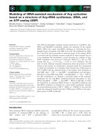
Table 1 PD candidate genes
Gene Name Accession No. Symbol Screen Position Locus
1 Tetraspanin 6 NM_003270.2 Tspan6 Chung et al. Xq22.1 PARK 12
2 ubiquitin-conjugating enzyme E2K (UBC1 homolog, yeast) NM_001111112.1 UBE2K Chung et al. 4p14 PARK 5
3 Ribosomal protein L11 NM_000975.2 RPL11 Chung et al. 1p36.1-p35 PARK 6
&7
4 Ribosomal protein L36a NM_021029.4 RPL36A Chung et al. Xq22.1 PARK 12
5 SWI/SNF related, matrix associated, actin dependent regulator of
chromatin, subfamily a, member 1
NM_003069.3 SMARCA1 Chung et al. Xq25 PARK 12
6 MARCKS-like 1 NM_023009.5 MARCKSL1 Chung et al. 1p35.1 PARK 6
7 GTPase activating protein 24 NM_001025616.2 ARHGAP24 Chung et al. 4q21.23-
4q21.3
PARK 4
8 F-box only protein 2 NM_012168.4 FBXO2 Chung et al. 1p36.22 PARK 7
9 T-complex-associated-testis-expressed 1-like NM_006520.2 DYNLT3 Chung et al. Xp21 PARK 12
10 Cyclin I NM_006835.2 CCNI Chung et al. 4q21.1 PARK 4
11 Solute carrier family 25 (mitochondrial carrier, adenine nucleotide
translocator), member 5
NM_001152.4 SLC25A5 Chung et al. Xq24-q26 PARK 12
12 Pyruvate dehydrogenase E1 alpha 1 NM_000284.2 PDHA1 Greene et al. Xp22.12 PARK 12
13 Ubiquitin carboxy-terminal hydrolase L1 PARK NM_004181.4 UCHL1 Greene et al. 4p14 PARK 5
14 G protein-coupled receptor, family C, group 5, member A NM_003979.3 GPRC5A Grimm et al. 12p13.1 PARK 8
15 Protein tyrosin phosphatase, receptor type, U NM_005704.3 PTPRU Grimm et al. 1p35.3 PARK 6
16 Klech-like 13 (drosophila) NM_001168299.1 KLHL13 Grimm et al. Xq24 PARK 12
17 Dehyrogenase/reductase (SDR family) member 3 NM_004753.4 DHRS3 Grimm et al. 1p36.22-
1p36.21
PARK 7
18 NEL-like 2 (chicken) NM_001145107.1 NELL2 Grimm et al. 12q13.11-
q13.12
PARK 8
19 Serin (or cysteine) peptidase inhibitor clade B, member 6a NM_004568.4 SERPINB6 Grimm et al. 6q25 PARK 2
20 Peripherin 1 NM_006262.3 PRPH Grimm et al. 12q12-q13 PARK 8
The list of genes, with ISH-detectable expression in midbrain dopaminergic neurons, with cytogenetic locations, falling within PARK loci are shown. Numbers
1,4,5,9,11,12 and 16 are within PARK-12, an orphan PD locus and UCHL1 (13) is a known PD gene .
Vahedi et al. Journal of Biomedical Science 2010, 17:66
/>Page 3 of 5
tool for prioritization and identification of can didate PD
gen es. Using the same approach, three other known PD
genes (so far, 10% or 4 out of 41 genes, found by two
studies , are known PD genes) were identified previously
[15]. Additional genetic and disease model studies are
needed to determine whether any of the seven genes,
which are within PARK12 can be considered a disease
gene and their degree of functional relevance to survival
and maintenance of mesDA neurons.
Additional material
Additional file 1: Table S1: The list of genes considered for
prioritization in this study. The genes showing above background
expression levels in midbrain dopaminergic neurons, confir med in using
ABA in situ hybridization database, after the removal of redundancies
with previous studies.
List of abbreviations
PD: Parkinson’s disease; mesDA: mesencephalic dopaminergic; OMIM: Online
Mendelian Inheritance in Man; BLAST: Basic Local Alignment Search Tool;
ISH: in situ hybridization; ABA: Allen Brain Atlas.
Author details
1
The Bahá’í Institute for Higher Education (BIHE), Tehran, Iran.
2
Interdisciplinary Center for Neuroscience, Ruprecht Karls Universität,
Heidelberg, Germany.
3
Department of Internal Medicine, Endocrinology, Yale
University, New Haven, CT, USA.
Authors’ contributions
This study was designed and supervised by KNA and was performed by SV,
MR. The data was analyzed by KNA and HHS. DG and AM partook in design
of the study and made critical comments. SV and KNA drafted the
manuscript and all authors read and approved the final version.
Competing interests
The authors declare that they have no competing interests.
Received: 2 July 2010 Accepted: 17 August 2010
Published: 17 August 2010
References
1. Langston JW, Ballard PA Jr: Parkinson’s disease in a chemist working with
1-methyl-4-phenyl-1,2,5,6-tetrahydropyridine. N Engl J Med 1983,
309(5):310.
2. Polymeropoulos MH, Lavedan C, Leroy E, Ide SE, Dehejia A, Dutra A, Pike B,
Root H, Rubenstein J, Boyer R, Stenroos ES, Chandrasekharappa S,
Athanassiadou A, Papapetropoulos T, Johnson WG, Lazzarini AM,
Duvoisin RC, Di Iorio G, Golbe LI, Nussbaum RL: Mutation in the alpha-
synuclein gene identified in families with Parkinson’s disease. Science
1997, 276(5321):2045-7.
3. Matsumine H, Saito M, Shimoda-Matsubayashi S, Tanaka H, Ishikawa A,
Nakagawa-Hattori Y, Yokochi M, Kobayashi T, Igarashi S, Takano H, Sanpei K,
Koike R, Mori H, Kondo T, Mizutani Y, Schäffer AA, Yamamura Y,
Nakamura S, Kuzuhara S, Tsuji S, Mizuno Y: Localization of a gene for an
autosomal recessive form of juvenile Parkinsonism to chromosome
6q25.2-27. Am J Hum Genet 1997, 60(3):588-96.
4. Gasser T, Müller-Myhsok B, Wszolek ZK, Oehlmann R, Calne DB, Bonifati V,
Bereznai B, Fabrizio E, Vieregge P, Horstmann RD: A susceptibility locus for
Parkinson’s disease maps to chromosome 2p13. Nat Genet 1998,
18(3):262-5.
5. Waters CH, Miller CA: Autosomal dominant Lewy body parkinsonism in a
four-generation family. Ann Neurol 1994, 35(1):59-64.
6. Boyer R, Auburger G, Leube B, Ulm G, Mezey E, Harta G, Brownstein MJ,
Jonnalagada S, Chernova T, Dehejia A, Lavedan C, Gasser T, Steinbach PJ,
Wilkinson KD, Polymeropoulos MH: The ubiquitin pathway in Parkinson’s
disease. Nature 1998, 395(6701):451-2.
7. Valente EM, Bentivoglio AR, Dixon PH, Ferraris A, Ialongo T, Frontali M,
Albanese A, Wood NW: Localization of a novel locus for autosomal
recessive early-onset parkinsonism, PARK6, on human chromosome
1p35-p36. Am J Hum Genet 2001, 68(4):895-900.
8. van Duijn CM, Dekker MC, Bonifati V, Galjaard RJ, Houwing-Duistermaat JJ,
Snijders PJ, Testers L, Breedveld GJ, Horstink M, Sandkuijl LA, van
Swieten JC, Oostra BA, Heutink P: Park7, a novel locus for autosomal
recessive early-onset parkinsonism, on chromosome 1p36. Am J Hum
Genet 2001, 69(3):629-34.
9. Funayama M, Hasegawa K, Kowa H, Saito M, Tsuji S, Obata F: A new locus
for Parkinson’s disease (PARK8) maps to chromosome 12p11.2-q13.1.
Ann Neurol 2002, 51(3):296-301.
10. Hampshire DJ, Roberts E, Crow Y, Bond J, Mubaidin A, Wriekat AL, Al-Din A,
Woods CG: Kufor-Rakeb syndrome, pallido-pyramidal degeneration with
supranuclear upgaze paresis and dementia, maps to 1p36. J Med Genet
2001, 38(10):680-2.
11. Hicks AA, Pétursson H, Jónsson T, Stefánsson H, Jóhannsdóttir HS, Sainz J,
Frigge ML, Kong A, Gulcher JR, Stefánsson K, Sveinbjörnsdóttir S: A
susceptibility gene for late-onset idiopathic Parkinson’s disease. Ann
Neurol 2002, 52(5):549-55.
12. Pankratz N, Nichols WC, Uniacke SK, Halter C, Rudolph A, Shults C,
Conneally PM, Foroud T, Parkinson Study Group: Genome screen to
identify susceptibility genes for Parkinson disease in a sample without
parkin mutations. Am J Hum Genet 2002, 71(1):124-35.
13. Wakabayashi K, Takahashi H: Pathology of familial Parkinson’s disease.
Brain Nerve 2007, 59(8):851-64.
14. Gasser T: Update on the genetics of Parkinson’s disease. Mov Disord 2007,
22(Suppl 17):S343-50.
15. Gherbassi D, Bhatt L, Thuret S, Simon HH: Merging mouse transcriptome
analyses with Parkinson’s disease linkage studies. DNA Res 2007,
14(2):79-89.
16. Thuret S, Bhatt L, O’Leary DD, Simon HH: Identification and
developmental analysis of genes expressed by dopaminergic neurons of
the substantia nigra pars compacta. Mol Cell Neurosci 2004, 25(3):394-405.
17. Stewart GJ, Savioz A, Davies RW: Sequence analysis of 497 mouse brain
ESTs expressed in the substantia nigra. Genomics 1997, 39(2):147-53.
18. Barrett T, Xie T, Piao Y, Dillon-Carter O, Kargul GJ, Lim MK, Chrest FJ,
Wersto R, Rowley DL, Juhaszova M, Zhou L, Vawter MP, Becker KG,
Cheadle C, Wood WH, McCann UD, Freed WJ, Ko MS, Ricaurte GA,
Donovan DM: A murine dopamine neuron-specific cDNA library and
microarray/increased COX1 expression during methamphetamine
neurotoxicity. Neurobiol Dis 2001, 8(5):822-33.
19. Greene JG, Dingledine R, Greenamyre JT: Gene expression profiling of rat
midbrain dopamine neurons: implications for selective vulnerability in
parkinsonism. Neurobiol Dis 2005, 18(1):19-31.
20. Grimm J, Mueller A, Hefti F, Rosenthal A: Molecular basis for
catecholaminergic neuron diversity. Proc Natl Acad Sci USA 2004,
101(38):13891-6.
21. Chung CY, Seo H, Sonntag KC, Brooks A, Lin L, Isacson O: Cell type-specific
gene expression of midbrain dopaminergic neurons reveals molecules
involved in their vulnerability and protection. Hum Mol Genet 2005,
14(13):1709-25.
22. Lein ES, Hawrylycz MJ, Ao N, Ayres M, Bensinger A, Bernard A, Boe AF,
Boguski MS, Brockway KS, Byrnes EJ, Chen L, Chen L, Chen TM, Chin MC,
Chong J, Crook BE, Czaplinska A, Dang CN, Datta S, Dee NR, Desaki AL,
Desta T, Diep E, Dolbeare TA, Donelan MJ, Dong HW, Dougherty JG,
Duncan BJ, Ebbert AJ, Eichele G, Estin LK, Faber C, Facer BA, et al: Genome-
wide atlas of gene expression in the adult mouse brain. Nature 2007,
445(7124):168-76.
23. Alavian KN, Simon HH: Linkage of cDNA expression profiles of
mesencephalic dopaminergic neurons to a genome-wide in situ
hybridization database. Mol Neurodegener 2009, 4(1):6.
24. Tanaka M, Kim YM, Lee G, Junn E, Iwatsubo T, Mouradian MM: Aggresomes
formed by alpha-synuclein and synphilin-1 are cytoprotective. J Biol
Chem 2004, 279(6):4625-31.
25. Dodson MW, Guo M: Pink1, Parkin, DJ-1 and mitochondrial dysfunction
in Parkinson’s disease. Curr Opin Neurobiol 2007, 17(3):331-7.
Vahedi et al. Journal of Biomedical Science 2010, 17:66
/>Page 4 of 5
26. Chandra S, Fornai F, Kwon HB, Yazdani U, Atasoy D, Liu X, Hammer RE,
Battaglia G, German DC, Castillo PE, Südhof TC: Double-knockout mice for
alpha- and beta-synucleins: effect on synaptic functions. Proc Natl Acad
Sci USA 2004, 101(41):14966-71.
27. Hauser MA, Li YJ, Takeuchi S, Walters R, Noureddine M, Maready M,
Darden T, Hulette C, Martin E, Hauser E, Xu H, Schmechel D, Stenger JE,
Dietrich F, Vance J: Genomic convergence: identifying candidate genes
for Parkinson’s disease by combining serial analysis of gene expression
and genetic linkage. Hum Mol Genet 2003, 12(6):671-7.
doi:10.1186/1423-0127-17-66
Cite this article as: Vahedi et al.: Parkinson’s disease candidate gene
prioritization based on expression profile of midbrain dopaminergic
neurons. Journal of Biomedical Science 2010 17:66.
Submit your next manuscript to BioMed Central
and take full advantage of:
• Convenient online submission
• Thorough peer review
• No space constraints or color figure charges
• Immediate publication on acceptance
• Inclusion in PubMed, CAS, Scopus and Google Scholar
• Research which is freely available for redistribution
Submit your manuscript at
www.biomedcentral.com/submit
Vahedi et al. Journal of Biomedical Science 2010, 17:66
/>Page 5 of 5