GT-repeat polymorphism in the heme oxygenase1 gene promoter and the risk of carotid atherosclerosis related to arsenic exposure ppt
Bạn đang xem bản rút gọn của tài liệu. Xem và tải ngay bản đầy đủ của tài liệu tại đây (464.43 KB, 11 trang )
RESEARC H Open Access
GT-repeat polymorphism in the heme oxygenase-
1 gene promoter and the risk of carotid
atherosclerosis related to arsenic exposure
Meei-Maan Wu
1,2,3*
, Hung-Yi Chiou
1*
, Te-Chang Lee
4
, Chi-Ling Chen
5
, Ling-I Hsu
6
, Yuan-Hung Wang
7
,
Wen-Ling Huang
1
, Yi-Chen Hsieh
1
, Tse-Yen Yang
6
, Cheng-Yeh Lee
6
, Ping-Keung Yip
8
, Chih-Hao Wang
9
,
Yu-Mei Hsueh
1
, Chien-Jen Chen
6
Abstract
Background: Arsenic is a strong stimulus of heme oxygenase (HO)-1 expression in experimental studies in
response to oxidative stress caused by a stimulus. A functional GT-repeat polymorphism in the HO-1 gene
promoter was inversely correlated to the development of coronary artery disease in diabetics and development of
restenosis following angioplasty in patients. The role of this potential vascular pro tective factor in carotid
atherosclerosis remains unclear. We previously reported a graded association of arsenic exposure in drinking water
with an increased risk of carotid atherosclerosis. In this study, we investigated the relationship between HO-1
genetic polymorphism and the risk of atherosclerosis related to arsenic.
Methods: Three-hundred and sixty-seven participants with an indication of carotid atherosclerosis and an
additional 420 participants without the indication, which served as the controls, from two arsenic exposure areas in
Taiwan, a low arsenic-exposed Lanyang cohort and a high arsenic-exposed LMN cohort, were studied. Carotid
atherosclerosis was evaluated using a duplex ultrasonographic assessment of the extracranial carotid arteries. Allelic
variants of (GT)n repeats in the 5′-flanking region of the HO-1 gene were identified and grouped into a short (S)
allele (< 27 repeats) and long (L) allele (≥ 27 repeats). The association of atherosclerosis and the HO-1 genetic
variants was assessed by a logistic regression analysis, adjusted for cardiovascular risk factors.
Results: Analysis results showed that arsenic’s effect on carotid atherosclerosis differed between carriers of the
class S allele (OR 1.39; 95% CI 0.86-2.25; p = 0.181) and non-carriers (OR 2.65; 95% CI 1.03-6.82; p = 0.044) in the
high-exposure LMN cohort. At arsenic exposure levels exceeding 750 μg/L, difference in OR estimates between
class S allele carriers and non-carriers was borderline significant (p = 0.051). In contrast, no such results were found
in the low-exposure Lanyang cohort.
Conclusions: This exploratory study suggests that at a relatively high level of arsenic exposure, carriers of the short
(GT)n allele (< 27 repeats) in the HO-1 gene promoter had a lower probability of developing carotid atherosclerosis
than non-ca rriers of the allele after long-term arsenic exposure via ground water. The short (GT)n repeat in the HO-
1 gene promoter may provide protective effects against carotid atherosclerosis in individuals with a high level of
arsenic exposure.
* Correspondence: ;
1
School of Public Health, Taipei Medical University, Taipei, Taiwan
Full list of author information is available at the end of the article
Wu et al. Journal of Biomedical Science 2010, 17:70
/>© 2010 Wu et al; licensee BioMed Central Ltd. This is an Open Access article distributed under the terms of the Creative Commons
Attribution License ( nses/by/2.0 ), which permits unrestricted use, distribution, and reproduction in
any medium, provided the original work is properly cited.
Background
Many of the health hazards caused by arsenic are carci-
nogenic effects [1,2]. Recently, attention was also paid to
the close a ssociation of ingested arsenic exposure with
the development of cardiovascular disease [3-5]. Epide-
miological studies carried out in Taiwan identified se v-
eral vascular disord ers caused by long- term exposure to
arsenic in well water. Inorganic arsenic in drinking
water is associated with increased risks of cardiovascular
mortality, peripheral vascular disease, ischemic heart
disease, and cerebral infarction in a dose-response rela-
tionship [5]. In a more-recent report, Wang e t al.
demonstrated a significant biological gradient of long-
term arsenic exposure with the prevalence of carotid
atherosclerosis [6], further providing evidence of the
presence of atherosclerosis induced by arsenic.
Despite the well-documented association between
atherosclerotic vascular disease and inorganic arsenic in
human populations, only a small percentage of arsenic-
exposed individuals develop vascular d isorders in their
lifetime [7,8]. This impliestheexistenceofmodifying
factors involved in the disease process that result in a
subg roup being susceptible to arsenic-ass ociated cardio-
vascular disorders. The nutritional status, arsenic meta-
bolite profile, and several genetic susceptibility factor s
were described [4,9]. Among these, inherited risk factors
affecting the pathogenesis of atherosclerosis underlying
the cardiovascular disorders caused by arsenic have not
been fully examined. Particula rly, potential susceptibility
genes such as those regulating the adaptive resp onse to
arsenic exposure have yet to be characterized.
Atherosclerosis is brought about by continuous oxida-
tive stress to artery walls, thereby leading to the concept
that inflammation and endogenous antioxidant pathways
may play important roles in the development of athero-
sclerosis [10]. In the initiat ion phase of atherosclerosis,
oxidant-elicited inflammation causes dysfunction of
endothelial cells, while endogenous antioxidants reduce
vascular injuries and prevent the development of athero-
sclerosis [10]. Othe r oxidant-induced gene products in
the adaptive/protective response of vessel walls to oxida-
tive stress were also proposed [11]. One such stress-
induced protein that may possibly be involved is heme
oxygenase (HO). HO is the rate-limiting enzyme in
heme degradation, decomposing heme into free iron,
biliverdi n, and carbon monoxide (CO). Biliverdin is sub-
sequently converted into bilirubin. Recent studies
showed that HO-1, an inducible isoform of H O, can be
rapidly upregulated by diverse stimulators associated
with various cardiovascular disorders [12]. Biliverdin
and bilirubin have the effect of scavenging oxygen
radicals and reducing the formation of peroxidation
products [13]. CO can down-modulate macrophage
inflammation and smooth muscle cell proliferation
which reduces vascular events [13].
Induction of HO-1 is primarily controlled at the level
of transcription initiatio n. The 5′ -flanking region con-
tains varying lengths of GT repeats 526 bp upstream of
the transcription site [14]. The number of GT repeats,
(GT)n, was shown to influence the inducibility of the
gene promoter under oxidative stimulus; the short poly-
morphic allele leads to high HO-1 inducibility [15,16].
Length polymorphism of the HO-1 gene promoter is
inversely correlated to the development of coronary
artery disease in high-risk individuals [15,17,18] and of
restenosis after clinical angioplasty [19]. However, there
are f ew studies on the relationship of HO-1 with envir-
onmentally related cardiovascular disease or subclinical
atherosclerosis. Because HO-1 is an early-response
molecule and m ay provide protectio n from cell damage,
we hypothesized that there is reduced risk of athero-
sclerotic lesions for those persons that display short
(GT)n repeats in the HO-1 gene promoter when
exposed to an environmental toxin such as arsenic.
Arsenic i s an oxidant produc er and a s trong stimulus
of HO-1 expres sion in cell cultures as a part of the cel-
lular response to oxidative stress to prevent cell damage
[20]. However to date, no human data have justified the
observationofHO-1incellculture.Wepreviously
reported on apparently healthy human subjects in
whom transcripts levels of the HO-1 gene increased
with arsenic in the blood in a dose-dependent pattern,
indicating that HO-1 induction is one of the early
responses in arsenic-exposed human beings [21]. The
relevance of HO-1 induction to arsenic-associated cardi-
ovascular disorders is not known. The aim of t he pre-
sent study was to test the hypothesis that HO-1
induction has a protective effect against atherosclerosis
in arsenic-exposed individuals. We assessed the fre-
quency of HO-1 (GT)n repeat genotypes and examined
the relationship between HO-1 gene variability and the
risk of atherosclerotic lesions in two cohorts from
arsenic-exposure areas in Taiwan.
Methods
Study areas and cohorts
This study recruited participants from two endemic
areas of arsenic exposure in Taiwan: the Lanyang
Basin in the northeastern coastal region and the Black-
foot disease (BFD)-endemic area in the southwestern
coastal region [22]. Epidemiological biomarker studies
were launched as part of a long-term follow-up study
onhealthhazardsaswellastoexploreriskfactors
other than arsenic exposure, in 1988 [23,24] and 1997
[25,26], respectively, for the two arseniasis-endemic
areas.
Wu et al. Journal of Biomedical Science 2010, 17:70
/>Page 2 of 11
The arsenic content in well water in the Lanyang
Basin area ranged from undetectable (< 0.15 μg/L) to >
3000 μg/L, with median arsenic co ncentrations of unde-
tectable to 140 μg/L [27]. Residents in this area used
their household-owned well water from the late 1940s
until the early 1990s, when a government-sponsored
water supply system was im plemented. During initial
health examinations in 1998-1999, a random sample of
687 cohort members who c ompleted an ultrasono-
graphic assessment of the extracranial carotid artery
(ECCA) was studied and reported in previous studies
[25,26]. Among them, 530 members (77.1%) gave their
consent and provided DNA samples for this research.
The study protocol was appro ved by t he Institutional
Review Board at Taipei Medical Universit y. This subco-
hort is hereafter called the Lanyang cohort.
In the BFD-endemic area, we focused on three BFD-
hyper-endemic villages, consisting of Homei (L, village
designation), Fuhsin (M), and Hsinming (N) in Putai
Township [23,24]. Residents in these three villages
began using arsenic-tainted artesian (> 300 m) well
water in the early 1910s. The arsenic level in the arte-
sian well water ranged 90-1700 μg/L, with a median of
400-874 μg/L. A public water supply system was intro-
duced in the early 1960s, and the artesian well water
was no longer used a fter the mid-1970s. In a follow-up
health examination in 1996, an ultrasonographic assess-
ment of E CCA atherosclerosis w as conducted for the
first time. In total, 436 cohort members completed the
ECCA assessment during t his examination [6]. Among
them, 383 members (87.8%) ga ve their consent and pro-
vided DNA samples for this research. The study proto-
col was approved by the Institutional Review Board at
College of Public Health National Taiwan University.
This subcohort is hereafter called the LMN cohort.
Study subjects, questionnaire data, and biochemical assay
To assess the extent and severity of atherosclerosis, we
used a high-resolution duplex ultrasound system with B-
mode and Doppler scanners (SONOS 1000, Philips,
USA) to examine the ECCA for each participant (Hew-
lett-Packard Sono 1000, Philips, USA). Duplex scanning
and operation as well as the definition of carotid athero-
sclerosis were described in previous studies [6,26].
Briefly, the presence of carotid atherosclerosis was eval-
uated based mainly on two parameters: the maximal
ECCA intimal-medial thickness (IMT) and th e presence
of ECCA plaque. The maximal IMT was measured on
the far side of the common carotid artery (CCA) at the
most stenotic location between 0 and 1 cm proximal to
the carotid bifurcation. The presence of ECCA plaque
was defined as a wall thickening ≥ 50% of the adjacent
IMT and assessed at five carotid artery segments,
including the proximal CCA, distal CCA, bulb, internal
carotid artery, and external carotid artery. Parti cipants
having carotid atherosclerosis were defined as patients
according to a maximal ECCA IMT of ≥ 1.0 mm or the
presence of observable plaque in any of the five carotid
artery segments. The remaining participants with no
indications constituted the control group.
Information on demographic and lifestyle characteristics
was obtained from the baseline questionnaire and updated
through a supplemental questionnaire if necessary. Bio-
chemical variables, including total cholesterol, triglycer-
ides, and glucose level in fasting blood were assayed in the
year of the ECCA assessment. All laboratory analyses were
performed using a standard automatic analyzer. Height,
weight, systolic blood pre ssure, and diastolic blood pres-
sure were measured according to standard protocols.
Hypertension was defined as (1) an average systolic blood
pressure of ≥ 140 mmHg, (2) an average diasto lic blood
pressure of ≥ 90 mmHg, or (3) a history of being diag-
nosed as hypertensive or having taken antihypertensive
medication. Subjects were considered to have diabetes, if
they had ever been diagnosed by a physician as being
diabetic, or had a fasting blood sugar level of ≥ 126 mg/dL.
Index for arsenic exposure
To evaluate arsenic exposure in one’s lifetime for each
study subject, a detailed history of residential addresses
and duration of artesian well water use were obtained
from a personal interview according to a structured ques-
tionnaire. In the Lanyang Basin area, well water samples
were collected from each household, and the arsenic con-
tent in the well water was determined during 1991-1994,
by a method of hydride-generation atomic absorption
spectrometr y [27] . Since residents of the Lanyang cohort
had used their own wells, on a household basis, and had
drunk water from those wells fo r more than 50 years, the
arsenic concentration i n the well water was used to esti-
mate the arsenic exposure of the Lanyang participants.
On the other hand, residents of the LMN cohort had
at one time shared one or several artesian wells because
of economic reasons, and some of the LMN participants
had even moved from one village to another within t he
BFD-endemic area. To reflect the overall exposure to
ingested arse nic for the LMN participants, a cumulati ve
arsenic exposure f rom drinking well water was applied
to represent the arsenic exposure as usually used in our
previous reports [23,24]. The cumulative arsenic expo-
sure was calculated as the sum of the products derived
by multiplying the arsenic concentration in the well
water by the number of years a participant had been
drinking that well water while living in any of the var-
ious villages. Me dian levels of arsenic in the well water
of the villages where the s tudy subjects had lived were
obtained from a report of previous studies carried out in
the 1960s [28]. To be compatible with the Lanyang
Wu et al. Journal of Biomedical Science 2010, 17:70
/>Page 3 of 11
cohort, an index of average arsenic exposure from con-
suming well water was presented, which was derived by
dividing the cumulative arsenic exposure by the years of
consuming artesian well water during the subject’s life-
time [29].
For the LMN cohort, a total of 95 participants were
exclud ed due to a lack of information regarding arsenic
exposure. The median level of arsenic concentration was
unknown for some villages in the BFD-endemic area, in
which case, the cumulative arsenic exposure or average
arsenicexposureofastudysubjectwasclassifiedas
unknown and thus removed from the analysis. The pro-
portion of missing values for arsenic exposure was
24.8%, which is similar to those reported in previous
studies [29,30].
HO-1 (GT)n repeat polymorphism
Genomic DNA was extracted from leukocytes in the
buffy coat using the Puregene DNA isolation kit (Gentra
Syst em, Minneapolis, MN, USA). The 5′-flanking region
containing the (GT)n repeats of the HO-1 gene was
amplified by the polymerase chain reaction (PCR) with a
FAM-labeled sense primer, 5′ -AGAGCCTGCAG
CTTCTCAGA-3′, and an unlabeled antisense primer,
5′-ACAAAGTCTGGCCA TAGGAC-3′ , according to a
published s equence by Kimpara et al. [ 31]. The sizes of
the PCR products were analyzed by the National Geno-
typing Center of Aca demia Sinica, Taiwan. In short, the
PCR products were mixed with the DNA ladder (35-
500-bp range; Applied Biosystems, Foster City, CA,
USA)andanalyzedonalaser-basedautomaticDNA
sequencer (ABI Prism 377). The respective sizes of the
(GT)n repeats for each participant were then calcul ated
using the software, GeneMapper vers. 3.0, ABI Prism.
To adjust for the variation resulting from different
batches of gel electrophoresis, we prepared six cloned
alleles and included them in every run of the capillary
electrophoresis for the sample allele analysis as stated
above. The repeat numbers of the cloned alleles as con-
trol DNA were 16, 20, 23, 27, 30, and 35 (GT). To con-
firm the sizes of the (GT)n repeats in the control DNA,
their PCR products were subcloned into a pCRII vector
(Invitrogen, Foster City, CA, USA), and the purified
plasmid DNA was subjected to sequence analysis. Using
the allele sizing information obtained from these control
DNAs, an adjustment to compensate for the variation in
different batches was applied to all sample data. This
external adjustment step in genotype binning with capil-
lary electrophoresis increases the precision of allele siz-
ing [ 32]. As to the genotyping accuracy, 5% of random
samples were duplicated in the PCR products sequen-
cing and binning adjustment. The agreement between
the samples was 100%.
Monocyte chemotactic protein (MCP)-1 protein levels
To examine the biological effect of HO-1 gene variants,
a random sample of 214 control participants (50% o f
total controls) was selected for the assay of MCP-1 pro-
tein levels in serum. Among them, 16 participants were
excluded because of hemolysis disturbance, resulting in
a final sample of 198. MCP-1 lev els in serum were mea-
sured by a n enzyme-linked immunosorbent assay
(Biotrak, Piscataway, NJ, USA) according to the manu-
facturers’ instructions. The lower limit of detection of
the assays was 20.5 pg/mL.
Statistical analysis
In the Lanyang cohort, HO-1 genotypes of 24 subjects
were unsuccessfully a ssayed. In the LMN cohort, the
ECCA reading values of seven study subjects were clas-
sified as unknown. We therefore excluded these
31 study subjects, resulting in a total of 281 and 506
subjects in the Lanyang and LMN cohorts, respectively,
being used throughout the analysis.
For the statistical analysis, we first used a logistic regres-
sion model to identify conventional risk factors in relation
to cardiovascular disease while adjusting for age and se x
distributions. Factors achieving p < 0.1 in the age- and gen-
der-adjusted regression were entered as possible confound-
ing variables in the subsequent analysis of arsenic’s effect
on carotid atherosclerosis. The effect of a risk factor was
expressedasanoddsratio(OR)anda95%confidence
interval (CI). All risk factors in the present study were
defined as categorical variables in the regression modeling,
unless otherwise indicated. Allele repeats were divided into
two classes, short (S) or long (L) based on the distribution
reports of previous studies [15,16] and ours of this study.
To evaluate whether there was an interactive effect
between the HO-1 length polymorphism and arsenic
exposure for the risk of developing atherosclerosis, we
first estimated the risk associated with arsenic exposure
according to the presence or absence of short (GT)n
repeat s among the participants (carriers of the S/S or S/
L genotype vs. carriers of the LL genotype). In the next
combination analysis, the relative percentage cha nge in
the risk of atherosclerosis from carriers to non-carriers
of the class S allele was also measured by arsenic expo-
sure. All analyses were performed using SAS (Win8e;
SAS, Cary, NC, USA) statistical software, and the statis-
tical significance level was defined as p < 0.05.
Results
Conventional risk factors and carotid atherosclerosis
Table 1 presents the frequency distribution and the age-
and gender-adjusted ORs with the 95% CIs for the clas-
sic risk factors for the patient and control groups of the
two cohorts. Aging and being male gender were the two
Wu et al. Journal of Biomedical Science 2010, 17:70
/>Page 4 of 11
common risk factors that had the strongest effects on
carotid atherosclerosis in the study coho rts. In the
Lanyang cohort, having a history of hypertension was
sig nificantly associated with an increased risk of carotid
atherosclerosis. Although statistically not significant, the
frequency of total cholesterol o f ≥ 200 mg/dL or trigly-
cerides of ≥ 150 mg/dL was found t o be higher in the
patient gro up compared to the control group (0.05 ≤ p
< 0.10). Other factors, including habitual smoking,
body-mass index (BMI), and a history of diabetes,
revealed no evidence of being associated with carotid
atherosclerosis in the Lanyang cohort. On the other
hand, having a history of diabetes was a significant risk
factor, and having a history of hypertension was found
to be associated with a borderline significance level
(0.05 ≤ p < 0.10), with an increased risk of carotid ather-
osclerosis in the patient group of the LMN cohort.
These factors associated with carotid atherosclerosis at a
significant or borderline level were include d in further
analyses.
HO-1 GT repeat polymorphism and the carotid
atherosclerosis index
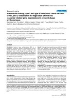
The number of GT repeats in the HO-1 gene promoter
of the participants ranged 16-38 (Figure 1). In both
cohorts, 23 and 30 GT repeats were the two most com-
mon alleles, which is consistent with findings from pre-
vious reports [15,16]. We thus selected 27 GT repeats as
Table 1 Conventional risk factors and HO-1 genotype in relation to carotid atherosclerosis
Lanyang cohort LMN cohort
Controls Patients Age- and gender-adjusted Controls Patients Age- and gender-adjusted
Characteristics n (%) n (%) OR (95% CI) n (%) n (%) OR (95% CI)
Total subjects 256 250 164 117
Age, year
< 55 70 (27.3) 25 (10.0) 1.0 90 (54.9) 22 (18.8) 1.0
55-65 117 (45.7) 94 (37.6) 2.13 (1.25-3.64)
†
57 (34.8) 49 (41.9) 3.68 (1.99-6.83)
‡
≥ 65 69 (27.0) 131 (52.4) 4.92 (2.85-8.50)
‡
17 (10.4) 46 (39.3) 11.33 (5.39-23.83)
‡
Gender
Female 154 (60.2) 116 (46.4) 1.0 91 (55.5) 40 (34.2) 1.0
Male 102 (39.8) 134 (53.6) 1.46 (1.01-2.11)* 73 (44.5) 77 (65.8) 2.47 (1.41-4.32)
†
Habitual smoking
No 182 (71.1) 146 (58.4) 1.0 133 (81.0) 79 (68.1) 1.0
Yes 74 (28.9) 104 (41.6) 1.10 (0.61-1.97) 31 (18.9) 37 (31.9) 1.29 (0.62-2.71)
Body mass index, kg/m
2
< 27 200 (79.4) 207 (83.8) 1.0 134 (81.7) 100 (85.5) 1.0
≥ 27 52 (20.6) 40 (16.2) 0.84 (0.52-1.35) 30 (18.3) 17 (14.5) 0.79 (0.38-1.67)
Triglycerides, mg/dL
< 150 197 (77.6) 174 (70.2) 1.0 121 (73.8) 78 (67.2) 1.0
≥ 150 57 (22.4) 74 (29.8) 1.48 (0.97-2.25) 43 (26.2) 38 (32.8) 1.07 (0.59-1.95)
Total cholesterol, mg/dL
< 200 136 (53.5) 115 (46.2) 1.0 78 (47.6) 40 (34.5) 1.0
≥ 200 118 (46.5) 134 (53.8) 1.43 (0.99-2.08) 86 (52.4) 76 (65.5) 1.44 (0.81-2.57)
Hypertension history
No 168 (65.9) 134 (54.0) 1.0 105 (64.0) 50 (42.7) 1.0
Yes 87 (34.1) 114 (46.0) 1.58 (1.08-2.31)* 59 (36.0) 67 (57.3) 1.65 (0.95-2.87)
Diabetes mellitus
No 222 (87.4) 221 (88.8) 1.0 137 (84.1) 82 (71.3) 1.0
Yes 32 (12.6) 28 (11.2) 0.84 (0.48-1.47) 26 (16.0) 33 (28.7) 2.57 (1.31-5.03)
†
HO-1 Genotype
L/L 65 (25.4) 72 (28.8) 1.0 45 (27.4) 36 (30.8) 1.0
L/S 129 (50.4) 131 (52.4) 0.88 (0.57-1.35) 90 (54.9) 54 (46.2) 0.49 (0.25-0.96)*
S/S 62 (24.2) 47 (18.8) 0.69 (0.41-1.17) 29 (17.7) 27 (23.1) 0.75 (0.33-1.66)
OR: odds ratio; CI: confidence interval.
Age was defined as continuous variable in the age- and gender-adjusted regression models.
Difference from the total number of patients and controls for each variable is due to missing data.
The class S allele denotes < 27 GT repeats and L allele ≥ 27 GT repeats in the HO-1 gene promoter.
* p < 0.05, † p <0.01,‡ p < 0.001.
Wu et al. Journal of Biomedical Science 2010, 17:70
/>Page 5 of 11
a cutoff to classify study subjects in the genetic analysi s.
(GT)n repeats of < 27 were designated the short (S)
allele, and repeats of ≥ 27 as the long (L) allele. Geno-
type distributions in all groups were in Hardy-Weinberg
equilibrium. Analysis results showed that there were no
statistically significant differences in the genoty pe distri-
bution of (GT)n repeats between controls and patients
with carotid atherosclerosis (Table 1). Interestingly, we
observed a higher frequency of the class S allele or a
higher frequency of g enotypes containing the class S
allele in the control groups compared to the patient
groups in both cohorts.
Association of arsenic exposure with carotid
atherosclerosis
As shown in Table 2, the age- and gender-adjusted ana-
lysis results demonstrated an increased risk of carotid
atherosclerosis with an increase in the arsenic concen-
tration in well water in a dose-response pattern for both
study cohorts. R esults from the Lanyang cohort
indicated a significant association between atherosclero-
sis and levels of arsenic exposure in well water after tak-
ing into account the logarithm of triglycerides, total
cholesterol, and hypertension history. For participants in
the LMN cohort, the association observed in the prior
age- and gender-adjusted analysis remained significant
after additional adjust ment for a hypertension history
and diabetes history.
Interaction between HO-1 (GT) repeat genotypes and
arsenic exposure
In the multivariate models inc luding conventional risk
factors, the effect of arsenic exposure seemingly differed
between carriers of the class S allele and non-carriers o f
the allele in the LMN cohort according to analysis
results of a trend test for arsenic exposure by the HO-1
genotype (Table 3). In carriers of the class S allele,
arsenic exposure had a low OR for atherosclerosis indi-
cation (OR 1.39; 95% CI 0.86-2.25; p = 0.181), whereas
in non-carriers, arsenic exposure was associated with a
Figure 1 Frequency distribution of the number of GT repe ats in patients having carotid atherosclerosis index (Black) and in controls
none the index (Grey) in (A) Lanyang cohort and (B) LMN cohort.
Wu et al. Journal of Biomedical Science 2010, 17:70
/>Page 6 of 11
high OR (OR 2.65; 95% CI 1.03-6.82; p = 0.044). In con-
trast, no such result was found in the Lanya ng cohort.
In a further analysis of the combined effect of arsenic
exposure and HO-1 genotype (carriers or non-carriers
of the class S all ele), no significant OR estimates were
found for any subdiv ided groups in either cohort (Table
4). We noted that the OR estimates were consistently
lower in carriers with the class S allele than non-carriers
in all comparisons by arsenic exposure group in both
cohorts(Table4).ThedifferenceinORestimates
between class S allele carriers and non-carriers reached
borderline significance (p = 0.051) at an level arsenic
exposure exceeding 750 μg/L in the LMN cohort.
Serum MCP-1 levels in carriers versus non-carriers of the
class S allele
We also examined the influence of the HO-1 genotype
on serum MCP-1 protein levels, an indicator of an
inflammatory vessel wall response. We divided the con-
trol participants without athero sclerosis indications into
three groups, L/L, L/S, and S/S genotypes, and com-
pared serum levels of the MCP-1 protein among the
Table 2 Arsenic exposure and carotid atherosclerosis
Average arsenic exposure,
μg/L
Controls
n (%)
Patients n (%) Age- and gender-adjusted OR
(95% CI)
Multivariate-adjusted OR
(95% CI)
Lanyang cohort
a
≤ 10 18 (7.0) 9 (3.6) 1.00 (referent) 1.00 (referent)
10.1-50 10 (3.9) 9 (3.6) 2.54 (0.71-9.06) 2.58 (0.70-9.56)
50.1-100 87 (34.0) 88 (35.2) 2.74 (1.13-6.64)* 2.98 (1.21-7.34)*
100.1-300 79 (30.9) 81 (32.4) 2.82 (1.16-6.87)* 3.07 (1.23-7.65)*
> 300 62 (24.2) 63 (25.2) 2.49 (1.01-6.15)* 2.62 (1.04-6.60)*
LMN cohort
b
≤ 300 52 (31.7) 12 (10.3) 1.00 (referent) 1.00 (referent)
300-750 65 (39.6) 56 (47.9) 2.03 (0.86-4.77) 1.93 (0.81-4.60)
> 750 47 (28.7) 49 (41.9) 2.70 (1.12-6.47)* 2.78 (1.14-6.78)*
Trend test 1.56 (1.04-2.34)* 1.61 (1.06-2.45)*
OR: odds ratio; CI: confidence interval.
a
Adjusted for age, sex, logarithm triglyceride, total cholesterol, and hypertension history.
b
Adjusted for age, sex, hypertension history, and diabetes history.
Age, triglyceride, and cholesterol were defined as continuous variables in the regression models.
* p < 0.05.
Table 3 Association of arsenic exposure with carotid atherosclerosis by carriers and non-carriers of the class S allele in
the HO-1 gene promoter
Carriers of the class S allele Non-carriers of the class S allele
Arsenic exposure, μg/L Controls
n (%)
Patients
n (%)
Multivariate-adjusted OR (95%
CI)
Controls
n (%)
Patients
n (%)
Multivariate-adjusted OR (95%
CI)
Lanyang cohort
a
≤ 10 13 (6.8) 7 (3.9) 1.0 (referent) 5 (7.7) 2 (2.8) 1.0 (referent)
10.1-50 7 (3.7) 5 (2.8) 1.57 (0.33-7.38) 3 (4.6) 4 (5.6) 8.64 (0.70-106.82)
50.1-100 66 (34.6) 64 (36.0) 2.90 (1.02-8.27)* 21 (32.3) 24 (33.3) 4.04 (0.64-25.76)
100.1-300 60 (31.4) 61 (34.3) 2.90 (1.00-8.35)* 19 (29.2) 20 (27.8) 4.47 (0.69-29.19)
> 300 45 (23.6) 41 (23.0) 2.39 (0.81-7.05) 17 (26.2) 22 (30.6) 3.97 (0.62-25.57)
Trend test 1.12 (0.91-1.38) 1.13 (0.81-1.57)
LMN cohort
b
≤ 300 39 (32.8) 10 (12.4) 1.0 (referent) 13 (28.9) 2 (5.6) 1.0 (referent)
300-750 43 (36.1) 36 (44.4) 2.04 (0.75-5.57) 22 (48.9) 20 (55.6) 1.13 (0.16-7.95)
> 750 37 (31.1) 35 (43.2) 2.20 (0.79-6.10) 10 (22.2) 14 (38.9) 4.65 (0.66-32.94)
Trend test 1.39 (0.86-2.25) 2.65 (1.03-6.82)*
OR: odds ratio; CI: confidence interval.
a
Adjusted for age, sex, logarithm triglyceride, total cholesterol, and hypertension history.
b
Adjusted for age, sex, hypertension history, and diabetes history.
Age, triglyceride, and cholesterol were defined as continuous variables in the regression models.
The class S allele denotes <27 GT repeats and L allele ≥27 GT repeats in the HO-1 gene promoter.
* p < 0.05.
Wu et al. Journal of Biomedical Science 2010, 17:70
/>Page 7 of 11
three groups. However, no significant association
between HO-1 genotypes and serum MCP-1 levels was
found (Additional file 1). Median values with the inter-
quartile range were 689 (498-895), 660 (517-833), and
643 (505-842) pg/ml for the L/L, L/S, and S/S geno-
types, respectively.
Discussion
Our previous study demonstrated that oxidative stress
levels elevate with an increasing arsenic level in the
blood of individuals consuming arsenic-contaminated
well water [33]. Among the same study subjects, tran-
script levels of an inflammation mediator gene and the
HO-1 gene increased in dose-response patterns with
arsenic exposure [21]. Whether induction of the HO-1
gene in humans is merely a biomarker responding to
arsenic exposure without influencing the health or
rather an induced response protecting against oxidative
damage caused by arsenic remains unknown. This study
investigated the relationship between (GT)n repeat poly-
morphism in the HO-1 gene promoter and the risk o f
carot id atherosclerosis in arsenic-exposed study cohorts.
The cohort members were recruited from two endemic
areas that repre sent, respectively, low- and high-arsenic -
exposure areas of Taiwan. In the low-exposu re Lanyang
cohort, the HO-1 genotype was not significantly asso-
ciated with carotid atherosclerosis. In the high-exposure
LMN cohort, however, our results suggested a
borderline significant (p = 0.051) lower risk of athero-
sclerosis indication for carriers of the class S allele (< 27
GT repeats) compar ed to non-carriers at a high level of
arsenic exposure. Analysis results of this study partially
support our hypothesis th at the short (GT)n repeat
allele in the HO-1 gene promoter, which is relevant to
high HO-1 induction levels, may protect against athero-
sclerosis in Taiwanese after long-term high-level arsenic
exposure via groundwater.
Our study results did not indicate any particular (GT)
n repeat allele in the study participants ind ependently
being associated with the risk of carotid atheros clerosis.
This finding is consistent with that of previous studies
indicat ing that HO-1 protect s against adverse cardiovas-
cular events only in the presence of c onventional risk
factors or following a clinical intervention [15,17-19].
The above studies indicated no association in the entire
sample group without stratification by risk factors
[15,18]. In our data, the HO-1 genotype was seemingly
associated with atherosclerosis risk only in the subgroup
of high-risk individuals with arsenic e xposure levels
exceeding 750 μg/L. In that particular circumstance,
arsenic e xposure presumably acted as a strong inducer,
and the apparent inability of non-carriers of the class S
allele to generate protective proteins against arsenic
toxicity may have led to a high risk probability.
HO-1 is a well-known toxicological signature of arsenic
treatment in diverse experimental conditions [20].
Table 4 Combined effect of arsenic exposure and HO-1 genotype on the risk of carotid atherosclerosis
Combination of arsenic exposure, μg/L
and HO-1 genotype
Controls n
(%)
Patients
n (%)
Multivariate-adjusted
OR (95% CI)
OR changes for carriers vs. non-carriers of
the class S allele, %
Lanyang cohort
a
≤ 50, Non-carriers of the class S allele 8 (3.1) 6 (2.4) 1.00 (Reference)
≤ 50, Carriers of the class S allele 20 (7.8) 12 (4.8) 0.60 (0.15-2.45) -40.0
50-100, Non-carriers of the class S allele 21 (8.2) 24 (9.6) 1.49 (0.39-5.69) (Reference)
50-100, Carriers of the class S allele 66 (25.8) 64 (25.6) 1.38 (0.40-4.80) -7.4
100-300, Non-carriers of the class S allele 19 (7.4) 20 (8.0) 1.72 (0.44-6.70) (Reference)
100-300, Carriers of the class S allele 60 (23.4) 61 (24.4) 1.37 (0.39-4.79) -20.3
> 300, Non-carriers of the class S allele 17 (6.6) 22 (8.8) 1.48 (0.38-5.77) (Reference)
> 300, Carriers of the class S allele 45 (17.6) 41 (16.4) 1.14 (0.32-4.08) -23.0
LMN cohort
b
≤ 300, Non-carriers of the class S allele 13 (7.9) 2 (1.7) 1.00 (Reference)
≤ 300, Carriers of the class S allele 39 (23.8) 10 (8.6) 0.49 (0.07-3.32) -51.0
300-750, Non-carriers of the class S allele 22 (13.4) 20 (17.1) 1.13 (0.18-7.16) (Reference)
300-750, Carriers of the class S allele 43 (26.2) 36 (30.8) 1.02 (0.17-6.10) -9.7
> 750, Non-carriers of the class S allele 10 (6.1) 14 (12.0) 4.45 (0.64-30.93) (Reference)
> 750, Carriers of the class S allele 37 (22.6) 35 (29.9) 1.09 (0.18-6.64) -75.5*
OR: odds ratio; CI: confidence interval.
a
Adjusted for age, sex, logarithm triglyceride, total cholesterol, and hypertension history.
b
Adjusted for age, sex, hypertension history, and diabetes history.
Age, triglyceride, and cholesterol were defined as continuous variables in the regression models.
The class S allele denotes <27 GT repeats and L allele ≥27 GT repeats in the HO-1 gene promoter.
* p = 0.051 for the OR difference between carriers vs. non-carriers of the class S allele at arsenic exposure level >750 μg/L.
Wu et al. Journal of Biomedical Science 2010, 17:70
/>Page 8 of 11
However, the molecular mechanism by which arsenic
induces HO-1 expression has not been clearly defined.
Despite the relation of several t ranscriptional factors to
HO-1 induction by arsenic [34], the precise cis-acting
elements in the 5′flanking region of the HO-1 gene have
not been identified [35,36]. Exactly how arsenic exposure
in humans interacts with the (GT)n repeats i n the HO-1
gene promoter and how the resulting interaction limits
the progression of atherosclerosis need to be elucidated
experimentally.
This study a lso evaluated whether the HO-1 genotype
affected systematic levels of inflammation by assaying
serum MCP-1 protein levels in study subjects. MCP-1 was
evaluated because our previous study involving a group of
study subjects recruited from the same cohort demon-
strated that arsenic exposure upregulates this inflamma-
tory molecule [21]. Other studies showed that HO-1
induction inhibits MCP-1 expression in cultured cells
[37,38]. Therefore, in this study, this marker was used to
reflect the anti-inflammatory effect of HO-1 induction in
blood vessels after long-term exposure to arsenic. How-
ever, analysis results revealed no differential anti-inflam-
matory response in carriers of the class S allele vs.
non-carriers after chronic arsenic exposure. No difference
in the inflammatory response between groups could be
attributed to the possibility that the MCP-1 expression
level is not sufficiently sensitive to determine the differen-
tial effect of the HO-1 genotype on arsenic-related athero-
sclerosis. HO-1 expression is seen throughout the
development of atherosclerotic lesions, from early fatty
streaks to advance d lesions, in which many cytokines
other than MCP-1 are regulated by HO-1 catalytic pro-
ducts during the inflammatory process [11,13]. As for
high-level arsenic exposure, the extent to which the HO-1
functi onal polymorphism affects inflammation molecules
in the atherosclerotic process needs to be elucidated.
We recognize that this study has certain limitations.
First, a reducing effect from the combination of the
HO-1 short (GT)n allele and arsenic exposure, if it
exists at all, is only slight and limited to high arsenic
exposure; in addition, the statistical testing was of bor-
derline significance. The resulting slight effect could
partially be attributed to the many genes involved in
atherosclerosis, possibly masking the role of the HO-1
genotype. Gene polymorphisms of p53 and glutathione-
transferase P1 (GSTP1) were related to the risk of caro-
tid atherosclerosis in the Lanyang cohort [25]. After
adjusting for the influence of the combined p53 and
GSTP1 gene polymorphism in the multivariate models,
our results indicated no essential change (data not
shown). However, we c ould not exclude the possibility
of other gen es that confounded the relation between the
HO-1 genotype and carotid atherosclerosis. In the LMN
cohort in the context of high arsenic exposure, the
possibility o f an effect from other genes linked to the
HO-1 gene or their haplotypic blocks could not be
ruled out. Thus, the suggestive borderline association in
participants with a high level of arsenic exposure of >
750 μg/L in the LMN cohort might not be attributed to
the HO-1 short (GT)n allele, but rather due to linkage
disequilibrium with a nearby gene.
Second, this study utilized a cross-sectional design,
implying some inherent limi tations. For instance, a
selection bias cannot be ruled out. This study did not
include participants who had died of fatal cardiovascular
conditions before the scheduled ECCA examination
date. However, unless HO-1 activity has a delete rious
effect on late-stage clinical events, the benefits from
short (GT)n alle les should not spuriously exist. More-
over, our control groups of both cohorts appeared to be
well represented in the allele and genotype distributions,
which occurred at a similar frequency to that in pre-
vious reports involving East Asians [15,18]. Selection of
participants might not have influenced our findings of
the relatio nship between the HO-1 genotype and carotid
atherosclerosis in the context of high arsenic exposure.
However, our findings are only applicable to survivors
of serious cardiovascular events.
Finally, given the wide 95% confidence intervals, the
risk estimates may be a chance finding. Therefore, the
attenuating effect of the HO-1 short (GT) allele must be
interpreted cautiously. Results of this study are explora-
tory. Future studies with a prospective design are war-
ranted to confirm the above preliminary observations.
Conclusions
Results o f this exploratory study suggest that at a rela-
tively high arsenic exposure level, carriers of the short
(GT)n allele (containing < 27 repeats) in the HO-1 gene
promoter may have a smaller carotid atherosclerosis risk
than non-carriers. To confirm our results, further stu-
dies are warrante d using a larger sample with improved
effect estimates, as well as samples of other arsenic-
exposed populations with different ethnic backgrounds.
A follow-up study must also be carried out on relation-
ships among the HO-1 length polymorphis m, long-term
arsenic exposure, and adverse cardiovascular events.
Additional material
Additional file 1: Supplemental figure. Supplemental figure
Acknowledgements
This work was supported by grants from the National Science Council
(NSC92-2321-B-038-011, NSC93-2321-B-038-014, and NSC94-2321-B-038-004),
Taiwan, R.O.C. We thank the National Genotyping Center, Academia Sinica,
for technical support.
Wu et al. Journal of Biomedical Science 2010, 17:70
/>Page 9 of 11
Author details
1
School of Public Health, Taipei Medical University, Taipei, Taiwan.
2
Graduate
Institute of Oncology, College of Medicine, National Taiwan University, Taipei,
Taiwan.
3
Graduate Institute of Basic Medicine, College of Medicine, Fu-Jen
Catholic University, Taipei, Taiwan.
4
Institute of Biomedical Sciences, Academia
Sinica, Taipei, Taiwan.
5
Graduate Institute of Clinical Medicine, College of
Medicine, National Taiwan University, Taipei, Taiwan.
6
Genomics Research
Center, Academia Sinica, Taipei, Taiwan.
7
Center of Excellence for Cancer
Research, Taipei Medical University, Taipei, Taiwan.
8
School of Medicine, College
of Medicine, Fu-Jen Catholic University, Taipei, Taiwan.
9
Department of
Cardiology, Cardinal Tien Hospital, College of Medicine, Fu-Jen Catholic
University, Taipei, Taiwan.
Authors’ contributions
Study concept and design: MMW, TCL, HYC. Data analysis and interpretation:
MMW, CLC, LIH. Drafting the manuscript: MMW. HO-1 genotyping data
acquisition: MMW, WLH. ECCA clinical data acquisition: PKY, CHW. MCP-1
assay data acquisition: MMW, TYY, CYL. Funding acquisition: MMW. Cohort
database management: LIH, YHW, YCH. Cohort material support: CJC, YMH,
HYC.
Competing interests
The authors declare that they have no competing interests.
Received: 13 May 2010 Accepted: 26 August 2010
Published: 26 August 2010
References
1. National Research Council: Arsenic in drinking water. 2001 update.
Washington, DC 2001.
2. World Health Organization: Arsenic and arsenic compounds. Geneva:
World Health Organization 2001.
3. Navas-Acien A, Sharrett AR, Silbergeld EK, Schwartz BS, Nachman KE,
Burke TA, Guallar E: Arsenic exposure and cardiovascular disease: a
systematic review of the epidemiologic evidence. Am J Epidemiol 2005,
162(11):1037-1049.
4. States JC, Srivastava S, Chen Y, Barchowsky A: Arsenic and cardiovascular
disease. Toxicol Sci 2009, 107(2):312-323.
5. Tseng CH: Cardiovascular disease in arsenic-exposed subjects living in
the arseniasis-hyperendemic areas in Taiwan. Atherosclerosis 2008,
199(1):12-18.
6. Wang CH, Jeng JS, Yip PK, Chen CL, Hsu LI, Hsueh YM, Chiou HY, Wu MM,
Chen CJ: Biological gradient between long-term arsenic exposure and
carotid atherosclerosis. Circulation 2002, 105(15):1804-1809.
7. Chen CJ, Chiou HY, Chiang MH, Lin LJ, Tai TY: Dose-response relationship
between ischemic heart disease mortality and long-term arsenic
exposure. Arterioscler Thromb Vasc Biol 1996, 16(4):504-510.
8. Tseng WP: Effects and dose–response relationships of skin cancer and
blackfoot disease with arsenic. Environ Health Perspect 1977, 19:109-119.
9. Hernandez A, Marcos R: Genetic variations associated with interindividual
sensitivity in the response to arsenic exposure. Pharmacogenomics 2008,
9(8):1113-1132.
10. Libby P, Ridker PM, Maseri A: Inflammation and atherosclerosis. Circulation
2002, 105(9):1135-1143.
11. Morita T: Heme oxygenase and atherosclerosis. Arterioscler Thromb Vasc
Biol 2005, 25(9):1786-1795.
12. Idriss NK, Blann AD, Lip GY: Hemoxygenase-1 in cardiovascular disease. J
Am Coll Cardiol 2008, 52(12):971-978.
13. Abraham NG, Kappas A: Pharmacological and clinical aspects of heme
oxygenase. Pharmacol Rev 2008, 60(1):79-127.
14. Lavrovsky Y, Schwartzman ML, Levere RD, Kappas A, Abraham NG:
Identification of binding sites for transcription factors NF-kappa B and
AP-2 in the promoter region of the human heme oxygenase 1 gene.
Proc Natl Acad Sci USA 1994, 91(13):5987-5991.
15. Chen YH, Lin SJ, Lin MW, Tsai HL, Kuo SS, Chen JW, Charng MJ, Wu TC,
Chen LC, Ding YA, Pan WH, Jou YS, Chau LY: Microsatellite polymorphism
in promoter of heme oxygenase-1 gene is associated with susceptibility
to coronary artery disease in type 2 diabetic patients. Hum Genet 2002,
111(1)
:1-8.
16. Yamada N, Yamaya M, Okinaga S, Nakayama K, Sekizawa K, Shibahara S,
Sasaki H: Microsatellite polymorphism in the heme oxygenase-1 gene
promoter is associated with susceptibility to emphysema. Am J Hum
Genet 2000, 66(1):187-195.
17. Dick P, Schillinger M, Minar E, Mlekusch W, Amighi J, Sabeti S, Schlager O,
Raith M, Endler G, Mannhalter C, Wagner O, Exner M: Haem oxygenase-1
genotype and cardiovascular adverse events in patients with peripheral
artery disease. Eur J Clin Invest 2005, 35(12):731-737.
18. Kaneda H, Ohno M, Taguchi J, Togo M, Hashimoto H, Ogasawara K,
Aizawa T, Ishizaka N, Nagai R: Heme oxygenase-1 gene promoter
polymorphism is associated with coronary artery disease in Japanese
patients with coronary risk factors. Arterioscler Thromb Vasc Biol 2002,
22(10):1680-1685.
19. Schillinger M, Exner M, Minar E, Mlekusch W, Mullner M, Mannhalter C,
Bach FH, Wagner O: Heme oxygenase-1 genotype and restenosis after
balloon angioplasty: a novel vascular protective factor. J Am Coll Cardiol
2004, 43(6):950-957.
20. Del Razo LM, Quintanilla-Vega B, Brambila-Colombres E, Calderon-
Aranda ES, Manno M, Albores A: Stress proteins induced by arsenic.
Toxicol Appl Pharmacol 2001, 177(2):132-148.
21. Wu MM, Chiou HY, Ho IC, Chen CJ, Lee TC: Gene expression of
inflammatory molecules in circulating lymphocytes from arsenic-
exposed human subjects. Environ Health Perspect 2003, 111(11):1429-1438.
22. Chen CJ, Wang CJ: Ecological correlation between arsenic level in well
water and age-adjusted mortality from malignant neoplasms. Cancer Res
1990, 50(17):5470-5474.
23. Chen CJ, Hsueh YM, Lai MS, Shyu MP, Chen SY, Wu MM, Kuo TL, Tai TY:
Increased prevalence of hypertension and long-term arsenic exposure.
Hypertension 1995, 25(1):53-60.
24. Lai MS, Hsueh YM, Chen CJ, Shyu MP, Chen SY, Kuo TL, Wu MM, Tai TY:
Ingested inorganic arsenic and prevalence of diabetes mellitus. Am J
Epidemiol 1994, 139(5):484-492.
25. Wang YH, Wu MM, Hong CT, Lien LM, Hsieh YC, Tseng HP, Chang SF,
Su CL, Chiou HY, Chen CJ: Effects of arsenic exposure and genetic
polymorphisms of p53, glutathione S-transferase M1, T1, and P1 on the
risk of carotid atherosclerosis in Taiwan. Atherosclerosis 2007,
192(2):305-312.
26. Wu MM, Chiou HY, Hsueh YM, Hong CT, Su CL, Chang SF, Huang WL,
Wang HT, Wang YH, Hsieh YC, Chen CJ: Effect of plasma homocysteine
level and urinary monomethylarsonic acid on the risk of arsenic-
associated carotid atherosclerosis. Toxicol Appl Pharmacol 2006,
216(1):168-175.
27. Chiou HY, Huang WI, Su CL, Chang SF, Hsu YH, Chen CJ: Dose-response
relationship between prevalence of cerebrovascular disease and
ingested inorganic arsenic. Stroke 1997, 28(9):1717-1723.
28. Kuo TL: Arsenic content of artesian well water in endemic area of
chronic arsenic poisoning. Report. Instiitute of Pathology, National Taiwan
University 1964, 60:1139-1140.
29. Chen CL, Hsu LI, Chiou HY, Hsueh YM, Chen SY, Wu MM, Chen CJ:
Ingested
arsenic, cigarette smoking, and lung cancer risk: a follow-up study in
arseniasis-endemic areas in Taiwan. Jama 2004, 292(24):2984-2990.
30. Chiou HY, Hsueh YM, Liaw KF, Horng SF, Chiang MH, Pu YS, Lin JS,
Huang CH, Chen CJ: Incidence of internal cancers and ingested inorganic
arsenic: a seven-year follow-up study in Taiwan. Cancer Res 1995,
55(6):1296-1300.
31. Kimpara T, Takeda A, Watanabe K, Itoyama Y, Ikawa S, Watanabe M, Arai H,
Sasaki H, Higuchi S, Okita N, Takase S, Saito H, Takahashi K, Shibahara S:
Microsatellite polymorphism in the human heme oxygenase-1 gene
promoter and its application in association studies with Alzheimer and
Parkinson disease. Hum Genet 1997, 100(1):145-147.
32. Ghosh S, Karanjawala ZE, Hauser ER, Ally D, Knapp JI, Rayman JB, Musick A,
Tannenbaum J, Te C, Shapiro S, Eldridge W, Musick T, Martin C, Smith JR,
Carpten JD, Brownstein MJ, Powell JI, Whiten R, Chines P, Nylund SJ,
Magnuson VL, Boehnke M, Collins FSl: Methods for precise sizing,
automated binning of alleles, and reduction of error rates in large-scale
genotyping using fluorescently labeled dinucleotide markers. FUSION
(Finland-U.S. Investigation of NIDDM Genetics) Study Group. Genome Res
1997, 7(2):165-178.
33. Wu MM, Chiou HY, Wang TW, Hsueh YM, Wang IH, Chen CJ, Lee TC:
Association of blood arsenic levels with increased reactive oxidants and
decreased antioxidant capacity in a human population of northeastern
Taiwan. Environ Health Perspect 2001, 109(10):1011-1017.
Wu et al. Journal of Biomedical Science 2010, 17:70
/>Page 10 of 11
34. Gong P, Stewart D, Hu B, Vinson C, Alam J: Multiple basic-leucine zipper
proteins regulate induction of the mouse heme oxygenase-1 gene by
arsenite. Arch Biochem Biophys 2002, 405(2):265-274.
35. Elbirt KK, Whitmarsh AJ, Davis RJ, Bonkovsky HL: Mechanism of sodium
arsenite-mediated induction of heme oxygenase-1 in hepatoma cells.
Role of mitogen-activated protein kinases. J Biol Chem 1998,
273(15):8922-8931.
36. Miralem T, Hu Z, Torno MD, Lelli KM, Maines MD: Small interference RNA-
mediated gene silencing of human biliverdin reductase, but not that of
heme oxygenase-1, attenuates arsenite-mediated induction of the
oxygenase and increases apoptosis in 293A kidney cells. J Biol Chem
2005, 280(17):17084-17092.
37. Lee PC, Ho IC, Lee TC: Oxidative stress mediates sodium arsenite-induced
expression of heme oxygenase-1, monocyte chemoattractant protein-1,
and interleukin-6 in vascular smooth muscle cells. Toxicol Sci 2005,
85(1):541-550.
38. Shokawa T, Yoshizumi M, Yamamoto H, Omura S, Toyofuku M, Shimizu Y,
Imazu M, Kohno N: Induction of heme oxygenase-1 inhibits monocyte
chemoattractant protein-1 mRNA expression in U937 cells. J Pharmacol
Sci 2006, 100(2):162-166.
doi:10.1186/1423-0127-17-70
Cite this article as: Wu et al.: GT-repeat polymorphism in the heme
oxygenase-1 gene promoter and the risk of carotid atherosclerosis
related to arsenic exposure. Journal of Biomedical Science 2010 17:70.
Submit your next manuscript to BioMed Central
and take full advantage of:
• Convenient online submission
• Thorough peer review
• No space constraints or color figure charges
• Immediate publication on acceptance
• Inclusion in PubMed, CAS, Scopus and Google Scholar
• Research which is freely available for redistribution
Submit your manuscript at
www.biomedcentral.com/submit
Wu et al. Journal of Biomedical Science 2010, 17:70
/>Page 11 of 11