Evidence of d-phenylglycine as delivering tool for improving l-dopa absorption pptx
Bạn đang xem bản rút gọn của tài liệu. Xem và tải ngay bản đầy đủ của tài liệu tại đây (704.51 KB, 8 trang )
RESEARC H Open Access
Evidence of d-phenylglycine as delivering tool for
improving l-dopa absorption
Chun-Li Wang
1
, Yang-Bin Fan
1
, Hsiao-Hwa Lu
2
, Tung-Hu Tsai
3
, Ming-Cheng Tsai
4
, Hui-Po Wang
1*
Abstract
Background: l-Dopa has been used for Parkinson’s disease management for a long time. However, its wide variety
in the rate and the extent of absorption remained challenge in designing suitable therapeutic regime. We report
here a design of using d-phenylglycine to guard l-dopa for better absorption in the intestine via intestinal peptide
transporter I (PepT1).
Methods: d-Phenylglycine was chemically attached on l-dopa to form d-phenylglycine-l-dopa as a dipeptide
prodrug of l-dopa. The cross-membrane transport of this dipeptide and l-dopa via PepT1 was compared in brush-
boarder membrane vesicle (BBMV) prepared from rat intestine. The intestinal absorption was compared by in situ
jejunal perfusion in rats. The pharmacokinetics after i.v. and p.o. administration of both compounds were also
compared in Wistar rats. The striatal dopamine released after i.v. administration of d-phenylglycine-l -dopa was
collected by brain microdialysis and monitored by HPLC. Anti-Parkinsonism effect was determined by counting the
rotation of 6-OHDA-treated unilateral striatal lesioned rats elicited rotation with (+)-methamphetamine (MA).
Results: The BBMV uptake of d-phenylglycine-l-dopa was inhibited by Gly-Pro, Gly-Phe and cephradine, the typical
PepT1 substrates, but not by amino acids Phe or l-dopa. The cross-membrane perme ability (Pm*) determined in rat
jejunal perfusion of d-phenylglycine-l-dopa was higher than that of l-dopa (2.58 ± 0.14 vs. 0.94 ± 0.10). The oral
bioavailability of d-phenylglycine-l-dopa was 31.7 times higher than that of l-dopa in rats. A sustained releasing
profile of striatal dopamine was demonstrated after i. v. injection of d-phenylglycine-l-dopa (50 mg/kg), indicated
that d-phenylglycine-l-dopa might be a prodrug of dopamine. d-Phenylglycine-l-dopa was more efficient than
l-dopa in lowering the rotation of unilateral striatal lesioned rats (19.1 ± 1.7% vs. 9.9 ± 1.4%).
Conclusion: The BBMV uptake studies indicated that d-phenylglycine facilitated the transport of l-dopa through
the intestinal PepT1 transporter. The higher jejunal permeability and the improved systemic bioavailability of
d-phenylglycine-l-dopa in comparison to that of l-dopa suggested that d-phenylglycine is an effective deliver y tool
for improving the oral absorption of drugs like l-dopa with unsatisfactory pharmacokinetics. Th e gradual release of
dopamine in brain striatum rendered this dipeptide as a potential dopamine sustained-releasing prodrug.
Background
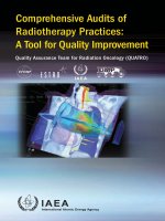
l-Dopa (Figure 1), a dopamingenic precursor, has long
been used for the treatment of Parkinson’s disease [1-4].
Clinically use of this drug was reported to have wide
range of inter- and intra-patient variations in t he rate
and the extent of absorption [5,6]. The inconsistent
pharmacokinetics remained as the major issue in design-
ing optimal regime in the disease manage ment [7,8].
The variation in oral bioavailability due to the
interaction of l-dopa with diet protein is, in part, attrib-
uted to its complicated absorption through the amino
acid transport sy stems [9-11]. Although many dopamine
agonists emerged, l-dopa in combination with metabolic
enzyme inhibitors is still the first choice for the treat-
ment of Parkinson’s disease [2,3].
Recent reports indicated that intestinal PepT1,
a member of proton-coupled oligopeptide transporter sys-
tem, is responsible for the a bsorption of a variety of di-
and tripeptide mimetic drugs such as amino-b-lactams
[12-14] and ACE inhibitors [15]. The structure feature of
PepT1 substrates was established [16-18] and the
* Correspondence:
1
Taipei Medical University College of Pharmacy, 250 Wu-Hsing St., Taipei,
110-31, Tai wan
Full list of author information is available at the end of the article
Wang et al. Journal of Biomedical Science 2010, 17:71
/>© 2010 Wang et al; licensee BioMed Central Ltd. This is an Open Access article distributed under the terms of the Creative Commons
Attribution License (http://creativecommons .org/licenses/by/2.0), which permits unre stri cted use, dis tribution, an d reproduction in
any medium, provided the original work is properly cited.
transport system has been used in the design of novel oral-
absorbable drugs [19,20].
Based on the thought that d-phenylglycine is the com-
mon moiety in the molecules of PepT1-mediated orally
absorbable amino-b-lactams [21], we thought that this
moiety might be useful as a seeing-eye dog for guiding
l-dopa to transport through the intestine via PepT1.
We therefore synthesized a series of d-phenylglycine-
containing di- and tripeptide derivatives as dopamine
prodrug [22]. Rationale behind the design of these
compounds was that the oral bioavailability of l-dopa
might be improved due to the affinity of d-phenylgl ycin e
to PepT1. Besides, the fast decarboxylation of l-dopa in
peripheral circulation might be prevented or prolonged
as the free amino group of l-dopa is blocked by
d-phenylglycine.
This report describes the transport of d-phenylglycine-
l-dopa via PepT1 by measuring the uptake in brush-border
membrane vesicles (BBMV) prepared from rat intestine.
The intestinal absorption of this compound and l-dopa
was compared by measuring the steady-state plasma con-
centration after in situ jejunal perfusion and by determin-
ing the pharmacokinetics after oral administration in rats.
Anti-Parkinsonism effects after oral administration
of d-phenylglycine-l-dopa and l-dopa were also compared
by measuring the change of the (+)-methamphetamine
induced rotation of dopamine-depleted unilateral striatal-
lesioned rats. Correlation between pharmacological activ-
ity and the pharmacokinetic profile was analyzed.
Methods
Materials
Chemicals, reagent grade for synthesis and analytical
grade for biological studies, were from Sigma-Aldrich (St.
Lou is, MO, U.S.A), E. Merck KG (Darms tadt, Germany),
Fluka Chemika (Buchs, Switzerland), Acros (NJ, U.S.A)
and Wako (Richmond, VA, U.S.A) companies. Acid-
washed alumina was purchased from RiedaL-de Haen
Company (Spring Valley, CA, U.S.A.). Melting points
were determined in Buchi (Flawil, Switzerland) 510 capil-
lary melting point apparatus and were uncorrected. IR
spectra were carried out on a Perkin-Elmer (Shelton, CT,
U.S.A) 1760 FT-IR instrument.
1
H NMR spectra were
determined on a Bruker (Wissem-bourg, France) 80
MHzorBruker400MHzspectrometer with chemical
shifts recorded in parts per million relative to tetra-
methylsilane. Mass a nd high-resolution mass (HRMS)
were measured on Finnigan (San Jose, CA. U.S.A.) MAT
4510 an d JEOL (Boston, MA, U.S.A. ) JNS-D300 spectro-
meter respectively. Branson (Danbury, CT. U.S.A.) Soni-
fier 450 sonicator, Kubota (Tokyo, Japan) 2010 or
Eppendorf AG (Hamberg, Germany) 5415C centrifuge
Model 905 incubator (Cherng Huei Instrument Co.,
Tainan, T aiwan) and Ystral (Ballrechten-Dottingen,
Germany) Laboratory series × 10/20 homogenizer were
used in the preparation of intestinal mucosal suspension.
Osmolarity of test solutions was determined with Wescor
5500 vapor pressure osmometer (Wescor Company,
Logan, UT, U.S.A.). Male Wistar rats (300 - 350 g) from
the Animal center of National Taiwan University were
used in preparing intestinal mucosal suspension, BBMV
and in per fusion studies. The same species of rats weigh-
ing 180 - 200 g were used in rotational behaviour studies.
Male Sprague-Dawley rats (280 - 320 g) were used for
determining brain dopamine. Animal studies were in
accordance with the National I nstitute of Health Guide
for the Care and Use of Laboratory Animals.
Brush-Boarder Membrane Vesicle (BBMV) Uptake
The intestinal cross-membrane transport of d-
phenylglycine-l-dopa and l-dopa was investigated using
simulated intestinal brush-boarder membrane vesicle
[23,24]. BBMV was prepare d using magnesium precipita-
tion method [25]. Protein content was determined. The
purity of BBMV was determined by measuring the activity
of the marker enzymes, alkaline phosphatase and amino-
peptidase. Generally, these two enzymes were enriched 8 -
21 folds in the preparation. The activity of Na
+
,K
+
-
ATPase, the marker enzyme of basolateral membrane, was
ver y small. Normal function of BBMV was confirmed by
measuring the uptake of d-glucose. In the presence of Na
+
gradient ([Na
+
]
in
<[Na
+
]
out
), an overshoot phenomenon of
glucose uptake with peak values of 9-11 times the equili-
brium was routinely observed. The membrane vesicles
were preloaded in the buffer solution containing 300 mM
mannitol and 16 mM HEPES/Tris (pH 7.4) before the
experiment. The uptake of test compounds in BBMV was
measured by rapid filtration.
Degradation of Compounds in Intestinal Mucosa
Suspension
Mucosa suspension was prepared from the intestine of
male Wistar rats according to established method [26]
and was stored in an ice bath before use.
In Situ Rat Perfusion
Literature procedure was followed for the preparation
of perfusion solutions and the jejunal segments [27].
To maximize the absorptio n and to prevent the test
Figure 1 The structures of d-phenylglycine-l-dopa and l-dopa.
Wang et al. Journal of Biomedical Science 2010, 17:71
/>Page 2 of 8
compounds from being oxidized during perfusion, the
experiments were performed at pH 6.0 with 0.02% (w/v)
ascorbic acid added as antioxidant and nitrogen gas was
bubbled through for 10 min before each experiment.
Perf usion solution was pumped through the jejunal seg-
mentataflowrateof0.2ml/minbyasyringepump
(Stoelting, KD Scientific, U.S.A.). The jejunal segment
was pre-washed with drug-free buffer for 10 min before
the drug solution was pumped in. Outlet tubing samples
were collected every 10 min for 6 collection periods
after water and solute transport reached steady-state.
The dimensionless membrane permeability Pm* [28]
was measured as indications for the disappearance rate
of test compound from the intestine. Plasma samples
were withdrawn from carotid artery.
Intravenous and Oral Absorption Experiments
Rats were fasted for at least 18 h prior to drug adminis-
tration. Anaesthesia was induced by i.p. injection of
urethane (0.15 g/100 g body weight). The rats were put
under a heating lamp to maintain body temperature.
Chromatography and Validation of Assay Methods
The HPLC system used in the assay of biological sam-
ples consisted of an autosampler (AS950, Jasco, Tokyo,
Japan), a Waters Model 600E solvent delivery pump
(Millipore, Milford, MA, USA), a Model LC-4C electro-
chemical detector with a glassy-carbon electrode (Bio-
analytical Systems, Inc., West Lafayette, IN, USA), and
an integrator (Macintosh LC II with Macintegrator I). A
Nucleosil® 10 SA cationic ion-exchange column (10 μm,
300 × 4.0 mm, Macherey-Nagel, Düren, Germany) with
a mobile phase comprising NaCl (50 mM)andNa
2
-
EDTA (1.0 mM)in0.1M ammonium phosphate
buffer (pH 2.0) at a flow rate of 2.0 ml/min was used
for the elution of the samples. The detection limits of
d-phenylglyc ine-l-dopa and l-dopa were 50 ng/ml and 25
ng/ml, respectively. HPLC assay methods were validated
by determining the precision a nd accuracy of intra-day
and inter-day analysis of serum standards over a period of
6 days. The coefficients of variation for inter- and intraday
assays were less than 15% for both compounds (n = 6).
Pharmacokinetic Analysis
According to the literature [29, 30], the area under the
plasma concentration-time profile (AUC) was calculated
by log-linear trapezoidal rule. Plasma concentration
after i.v. administration of drugs were also fitted to a
non-compartment model using PCNONLIN and
Akeike’s Information Criteria, sum of squared residuals,
residual plot and correlation coefficient were use for
determination of the compartment model. The residual
are a after the last observed data point was calculated as
C
last
/k, where C
last
is the last observed concentration,
and k is the corresponding terminal rate constant.
Terminal half-life (t
1/2
) was estimated compartment
model-independently. The fraction of absorption was
calculated according to Equation 1.
BA
AUC
oral
k
oral
dose
oral
AUC
iv
k
iv
dose
iv
=
⋅
⋅
× 100%
(1)
Brain Microdialysis
Single dose d-phenylglycine-l-dopa (50 mg/kg in 2.5 mL
of normal saline) was administered i. v. via femoral vein
to anesthetized male Sprague-Dawley rats (280 - 320 g).
The body temperature of the rats was maintained at
37°C with a heating pad throughout the experiment.
The rat was immobilized in a stereotaxic frame (David
Kopf Instruments, Tujunga, CA, USA), the skull was
surgically exposed, and a hole was drilled with a tre-
phine into t he skull based on stereo taxic coordinates.
The brain microdialysis system consisted of a CMA/100
microinjection pump (CMA, Stockholm, Sweden) and a
microdialysis probe. The dialysis probes (3 mm in
length) were made of silica capillary in a concentric
design with their tips covered by dialysis membrane
(Spectrum, 150 μm outer diameter with a cut-off
at nominal molecular mass of 13000, Laguna Hills,
CA,USA).Theprobewasplacedintorightstriatum
(0.2 mm anter ior to bregma and 3.2 mm lateral to mid-
line) and perfuse d with Ringer ’ s solution (147 mM Na+;
2.2 mM Ca++; 4 mM K+; pH 7.0) at a flow-rate of 1 μl
min
-1
. The position of each probe was verified at the
end of experiments. The dialysates was collected at
10 min intervals and aliquots of 10 μl was assayed by
microbore HPLC.
The HPLC system consisted of a pump (BAS PM-80,
West Lafayette, IN, USA) and an on-line injector (CMA
160, Stockholm, Sweden) equipped with a 10 μlsample
loop, a reversed phase C18 microbore column (particle
size 5 μm, 150 × 1 mm I.D.; Bioanalytical Systems, West
Lafayette, IN, USA) and an EC detector (BAS-4C
amper ometric) coupled to a glassy carbon working elec-
trode and referenced to a Ag/AgCl electrode at 750 mV
with a range set at 50 nanoamper. Output data from the
detector were integrated via an EZChrom chromato-
graphic data system (Scientific Software, San Ramon,
CA, USA). The mobile phase for analyzing striatal dopa-
mine, eluted at a flow rate of 0.05 ml/min, comprised
80 ml acetonitrile, 2.2 mM sodium 1-octanesulfonate,
14.7 mM monosodium dihydrogen orthophosphate,
30 mM sodium citrate, 0.027 mM EDTA, and 1 ml
diethyl amine in one liter double distilled water, adjusted
Wang et al. Journal of Biomedical Science 2010, 17:71
/>Page 3 of 8
topH3.5byorthosphoricacid(85%).Theelutewas
filtered through a Millipore 0.22 μm filter and degassed
prior to use.
Rotational Behavior of Rats [31-34]
Male Wistar rats (180 - 200 g) were anesthetized with
pentobarbital sodium (30 mg/kg body weight, i.p.) and the
heads were fixed in a DaviD-Kopf steric taxic frame. A
solution of 6-hydro xydopamine (6-OHDA, 2.00 mg/ml ×
8 ml) in saline was infused using Paxinos and Watson
coordinates (AP 5.3, L 2.0, H 7.8 mm, [34]) into the unilat-
eral substantia nigra compacta (SNc) of brain with a syr-
inge pump through a 30 gauge stainless steel needle at a
flow rate of 2 μl/min. After two weeks of recovery period,
the 6-OHDA treated rats were placed in a spherical bowl
(radius 20 cm) and secured by a thoracic harness which
was connected to a 486 PC computer for automatic
recording of rotation induced by (+)-methamphetamine
(MA). The rotational behavior of rats was recorded
10 min after MA treatment (MA in saline, 4.00 body
weight mg/kg of rat, s.c.). The numbers of turns recorded
were defined as the control value (T
0
) for each individual
animal. Only animals showing a T
0
greater than 400 were
chosen for further experiments. After two weeks of a
wash-out period the animals were subjected to drug treat-
ment. Single dose (0.051 mmol) of each test compound
was administered orally to rats 5 min prior to MA treat-
ment (4.00 mg/kg body weight, s.c.). The rotation counted
for a period of 110 min starting 10 min after MA treat-
ment was recorded as T
d
for each tested rat. The percen-
tage of reduction i n rotation for each animal was
calculated and presented as (T
d
-T
0
)/T
0
×100%.
Data Analysis
Data analysis were performed on Visual dBase and
SPSS/PC+ and were represented as mean ± SE for n
experiments. Treatment differen ces were eva luated by
paired-t test.
Results
d-Phenylglycine-l-dopa Uptake in BBMV
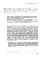
The uptake of d-phenylglycine-l-dopa in BBMV was mea-
sured. Amino acid l-Phe or l-dopa, dipeptide l-Gly-l-Pro or
l-Gly-l-Phe, or cephradine was added for investigating the
competition with d-phenylglycine-l-dopa in BBMV uptake
(Figure 2).
Stability of d-Phenylglycine-l-dopa in Intestinal Mucosal
Suspension
The stability of d-phenylglycine-l-dopa in the intestine
was determined prior to the intestinal absorption stu-
dies. In o rder to simulate intestinal microclimate pH,
the compound was incubated with the intestinal muco-
sal suspension in a pH 6.5 isotonic buffer solution.
l-Gly-l-Phe comprising essential amino acids degraded
rapidly with only 50% of recovery after 2 min of incuba-
tion. d-Phenylglycine-l-dopa, on the other hand, was
very stable with almost 100% of recovery after 90 min of
incubation (Figure 3).
Permeability of d-Phenylglycine-l-dopa in Rat Intestine
The absorption of d-phenylglycine-l-dopa and l-dopa
was compared in rats by in situ single-pass jejunal
perfusion experiments. Amidon’s dimensionless cross-
membrane permeability (P
m
*) was determined as a para-
meter of intestinal absorption [28]. The steady-state
plasma concentration was also determined (Table 1).
Pharmacokinetic Profile in Rats
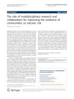
The mean plasma concentration-time profiles after single
dose oral and i.v. administration of d-phenylglycine-l-dopa
and l-dopa are depicted in Figure 4. The pharmacokinetic
parameters calculated with the data of plasma concentra-
tion-time curves based on the non-compartmental model
Figure 2 Theuptakeofd- phenylgly cine-l-dopa in BBMV with
or without the presence of l-Phe, l-dopa, l-Gly-l-Pro, l-Gly-l-Phe
and cephradine (**: p < 0.01; ***: p < 0.001.). The BBMV was
prepared according to material and methods. The BBMV preparation
(20 ml containing approximately 20 mg protein/ml) was added into
200 ml of a reaction buffer (composed of 300 mM mannitol, 25 mM
HEPES/Tris buffer pH 7.4, (pH was adjusted by adding MES) and the
test solution (to 1 - 2 mM of final conc.) was added. After
incubation at room temperature for acquired time, an ice-cold stop
solution (1.5 ml) containing NaCl (150 mM) and HEPES/Tris (16 mM,
pH 7.4) was added and the solution was filtered through a filter
paper (Whatman WCN, 0.45 μm pore size, 2.5 cm diameter) under a
vacuum. The filter paper was washed twice with 3 ml of the same
stop solution. The test compound remained on the filter paper
were extracted with 0.5 ml of 0.01 M aqueous HCl solution by virtue
of a vortex motion. The solution (100 μl) was injected onto the
HPLC column. Test compound bound on the filter paper was
determined for correction in different runs using preparations
without BBMV added.
Wang et al. Journal of Biomedical Science 2010, 17:71
/>Page 4 of 8
analysis were summarized in Table 2. The fraction of oral
absorption (BA) was calculated according to Equation 1.
The Striatal dopamine level after i.v. injection of d-phenyl-
glycine-l-dopa (50 mg/kg) is depicted in Figure 5.
Anti-Parkinsonism Activity
The in vivo anti-Parkinsonism effect was determined with
conventional rotation model m easured in 6-OHDA-treated
unilateral striatal-lesioned rats elicited rotation with
(+)-methamphetamine (MA) [32,35]. As shown in Table 3,
d-phenylglycine-l-dopa as well as l-dopa demonstrat ed
inhibition of MA-ind uced rotation of rats. With equal
molar of test compound administered, the activity of
d-phenylglycine-l-dopa in reducing the rotation of rats was
significantly higher than that of l-dopa.
Discussion
The BBMV uptake of d-phenylglycine-l-dopa was signifi-
cantly inhibited by dipeptides l-Gly-l-Pro (***p <0.001),
l-Gl y- l-Phe (**p < 0.01) and cephradin e, a typical PepT1
substrate (***p < 0.001), while was less inhibited by l-Phe
and l-dopa, suggesting that PepT1 might be involved in
the uptake of this dipeptide. We previously reported a
kinetic study on th e BBMV uptake of d-phenylglycine-a-
methyldopa. The uptake of this dipeptide was also signifi-
cantly inhibited by typical PepT1 substrate [22]. The high
value of Michaelis-Menten kinetic parameter (Vmax/Km)
in comparison to that of passive diffusion (Kd) at low
concentrations suggested that PepT1 dominates the
transport of the d-phenylglycine-containing dipeptide
through the intestine. Both results indicated that d-
phenylglycine increased the intesti nal transport of amino
acid a-methyldopa and l-dopa via PepT1.
The absorption of oral drugs in human can be evaluated
as dimensionless permeability P
m
*inin situ single-pass
perfusioninratsdespitethe complicated process of
absorption in the gastrointestinal tract [28,36,37]. The high
P
m
* demonstrated by d-phenylglycine-l-dopa (2.58 ± 0.14)
in comparison to that of l-dopa (0.94 ± 0.10) indicated the
high absorption of this dipeptide in the intestine. The
steady-state plasma concentration of d-phenylglycine-
l-dopa after the perfusion was 31.1 fold, in terms of molar
ratio, higher than that of l-dopa, indicated that this dipep-
tide was b etter absorbed than l-dopa.
The pharmacokinetic profiles upon i.v. and oral adminis-
tration of d-phenylglycine-l-dopa and l-dopawerecom-
pared. Although the volume of distribution after i.v.
injection o f d-phenylglycine-l-dopa was higher than that of
l-dopa, this dipeptide was cleared much faster than l-dopa
from the plasma. This made th e systemic bioavailability
(AUC) of d-phenylglycine-l-dopa 7 times lower than th at
of l-dopa (62.53 ± 19.68 vs. 459.81 ± 195.14 mg·min/ml).
On the contrary, the AUC of d-phenylglycine-l-dopa was
comparable to that of l-dopa upon oral administration
(28.85 ± 8.52 vs. 27.37 ± 4.60 mg·min/ml). As a result, the
fraction of oral absorption of d-phenylglycine-l-dopa was
31 fold higher th an that of l-dopa (27.58 ± 4.56% vs. 0.87 ±
0.24%).
The striatal dopamine level increased gradually after i.v.
injection of d-phenylglycine-l-dopa and had not reached
plateau 3.5 hours when the anaesthetized mice woke up.
The gradual release of dopamine in brain striatum ren-
dered this dipeptide as a dopamine sustained-releasing
prodrug.
Figure 3 Comparison of the stability of d-phenylglycine-l-dopa
and l-Gly-l-Phe in rat intestinal mucosa suspension. Each point
represents mean ± SE. of 3 experiments. A methanolic solution (100
μl) of the test compound (1 mg/ml) was diluted with an isotonic
mannitol buffer solution (pH 6.5, 2.4 ml) as the stock solution. This
stock solution (1 ml) was mixed with the freshly prepared mucosal
suspension (1 ml). The mixture was incubated in a water bath at 37°
C and subjected to sampling at intervals between zero to 90 min of
incubation. Each sampled solution (200 μl) was denatured with 0.8
ml of MeOH and centrifuged at 6,600 g for 5 min. Each of the
supernatant (20 - 100 μl) was subjected to HPLC assay.
Table 1 Plasma concentrations of d-phenylglycine-l-dopa and l-dopa measured in in situ single-pass jejunal perfusion
experiments
Compound No. of Experiments Pm* Blood concentration (μg/ml) Molar ratio of blood concentration
(μg/ml)
d-phenylglycine-l-dopa 4 2.58 ± 0.14 64.6 ± 5.40 31.10
l-dopa 3 0.94 ± 0.10 1.24
a
1.02
a
l-dopa was detected only in the plasma sample from one of the three rats tested. It was below detection limit in plasma samples of the other two rats. Data
presented are mean ± SD of n experiments.
Wang et al. Journal of Biomedical Science 2010, 17:71
/>Page 5 of 8
Figure 4 Plasma concentration-time profile of d-phenyglycine-l-dopa (a), (b) and l-dopa(c),(d)afteri.v.(a),(c)andoral(b),(d)
administration in Wistar rats (n = 6). The aqueous solution of test compound with dose equivalent to 5.97 mg/kg body weight of l-dopa was
administered either intravenously from the tail vein or orally by a feeding tube. Blood samples were collected from the carotid artery at time
intervals of from 1 to 180 min. Heparin sodium (25 I.U./ml in 0.3 ml of saline) was added to blood samples, and were then centrifuged at 6,600
g for 5 min. Plasma was stored at -78°C until being analyzed. A 200 μl of the plasma sample in a 10 ml test tube was mixed with 500 μl of 1.0
M Tris buffer (pH 8.6, adjusted by EDTA-2Na
+
) and 10 μl of 3,4-dihydroxybenzylamine (DHBA, 2 μg/ml) was added as internal standard. Alumina
100 mg was then added and then shake for 15 sec and the supernatant was decanted. The alumina was washed four times with 5 ml of water,
and the adsorbed compounds on the alumina was eluted with 200 μl of an acidic solution (0.9 ml of glacial acetic acid in 4.0 ml of 1.0 M
phosphate buffer). A 30 μl of the eluent was then analyzed by HPLC.
Table 2 Pharmacokinetic parameters derived from non-compartmental analysis after i.v. and oral administration of d-
phenylglycine-l-dopa and l-dopa in rats (mean ± SD, n = 6)
d-phenylglycine-l-dopa l-dopa
i.v. Oral i.v. Oral
AUC (mg·min/ml) 62.53 ± 19.68 28.85 ± 8.52 459.81 ± 195.14 27.37 ± 4.60
t
1/2
(min) 254.10 ± 73.05 142.50 ± 23.71 101.52 ± 27.74 184.80 ± 46.20
Cl
p
(l/kg/min) 0.18 ± 0.06 0.29 ± 0.10 0.02 ± 0.02 0.01 ± 0.00
Vd
ss
(l/kg) 11.01 ± 5.08 35.7 ± 17.1 1.22 ± 0.89 1.22 ± 0.36
t
max
(min) – 38.30 ± 17.72 – 25.02 ± 16.10
Fraction of absorption (%) – 27.58 ± 4.56 – 0.87 ± 0.24
Wang et al. Journal of Biomedical Science 2010, 17:71
/>Page 6 of 8
d-Phenylglycine-l-dopa after oral administration
demonstrated higher activity than l-dopa in reducing
the MA-induced rotation in rats with statistical signifi-
cance (19.1 ± 1.7% vs. 9.9 ± 1.4%, ***p < 0.001), suggest-
ing its anti-Parkinsonism activity. Whether the activity
came from the dipeptide per se or from the released
dopamine needs further investigation. Correlation
between the pharmacological activity and the pharmaco-
kinetic parameters indicated that the high activity
demonstrated by d-phenylglycine-l-dopa might partially
come from its better oral absorption.
Conclusion
d-P henylglycine-l-dopa was proved to be better
absorbed from the intestine than l-dopa. The BBMV
uptake suggested that d- phenylgly cine might act as a
seeing-eye dog for guiding l-dopa to tr ansport through
the intestine via intestinal PepT1 oligopeptide transpor-
ter. The higher anti-Parkinsonism activity of this dipep-
tide in comparison to that of l-dopa might come from
the improved oral bioavailability. The pharmacokinetic
profile of striatial dopamine indicated that d-phenylgly-
cine l-dopa might be useful as a slow dopamine-releas-
ing prodr ug for therapeutic use. The improved intestinal
permeability with improved oral bioavailability as a con-
sequence, suggested the potentia l use of d-phenylglycine
as an effective delivery tool for drugs with unsatisfied
oral absorption.
Abbreviations
PepT1: Intestinal peptide transporter T1; BBMV: Brush-b oarder membrane
vesicle; MA: Methamphetamine
Acknowledgements
This study was supported by grant NCS95-2320-B-039-049-MY3 of National
Science Council (2008) and DOH99-TD-C-111-008 of the Department of
Health, the Republic of China.
Author details
1
Taipei Medical University College of Pharmacy, 250 Wu-Hsing St., Taipei,
110-31, Tai wan.
2
Roche Products Ltd., Taipei, Taiwan.
3
Institute of Traditional
Medicine, School of Medicine, National Yang-Ming Uni versity, 155 Li-Nong
Street, Section 2, Taipei, Taiwan.
4
Department of Pharmacology, College of
Medicine, National Taiwan University, No. 1, Section 1, Jen-Ai Rd., Taipei,
Taiwan.
Authors’ contributions
CLW and YBF carried out PK and rat rotational studies and drafted the
manuscript. HHL carried out permeability studies. MCT designed rat
rotational behavior studies. THT carried out the brain microdialysis studies.
HPW conceived and is responsible for the study. All authors read and
approved the final manuscript.
Competing interests
The authors declare that they have no competing interests.
Received: 13 July 2010 Accepted: 6 September 2010
Published: 6 September 2010
References
1. Birkmayer W, Hornykiewicz O: The effect of l-3,4-dihydroxyphenylalanine
(= DOPA) on akinesia in Parkinsonism. Parkinsonism Relat Disord 1998,
4:59-60.
2. Hauser R: Levodopa: Past, present and future. Eur Neurol 2009, 62:1-8.
3. LeWitt PA: Levodopa for the treatment of Parkinson’s disease. N Eng J
Med 2008, 359:2468-2476.
4. Olanow CW, Stern MB, Sethi K: The scientific and clinical basis for the
treatment of Parkinson’s disease. Neurology 2009, 72(Suppl 4):S1-S136.
5. Juncos JL: Levodopa: pharmacology, pharmacokinetics, and
pharmacodynamics. Neurol Clin 1992, 10:487-509.
6. Nomoto M, Nishikawa N, Nagai M, Yabe H, Nakatsuka A, Moritoyo H,
Moritoyo T, Kubo M: Inter- and intra-individual variation in l-dopa
pharmacokinetics in the treatment of Parkinson’s disease. Parkinsonism
Relat Disord 2009, 15(Suppl 1):S21-S24.
7. Jenner P: Molecular mechanisms of l-dopa -induced dyskinesia. Nat Rev
Neurosci 2008, 9:665-677.
8. Abbruzzese G: Optimising levodopa therapy. Neurol Sci 2008, 29:
S377-S379.
9. Lennernas H, Nilsson D, Aquilonius SM, Ahrenstedt O, Knutson L,
Paalzow LK: The effect of l-leucine on the absorption of levodopa,
studied by regional jejunal perfusion in man. Br J Clin Pharmacol 1993,
35:243-250.
10. Kempster PA, Wahlqvist ML: Dietary factors in the management of
Parkinson’s Disease. Nutr Rev 1994, 52:51-58.
11. Crevoisier C, Zerr P, Calvi-Gries F, Nilsen T: Effects of food on the
pharmacokinetics of levodopa in a dual-release formulation. Eur J Pharm
Biopharm 2003, 55:71-76.
Figure 5 Striatal dopamine level after i. v. injection of d-
phenylglycine-l-dopa. Values represent the group mean ± s.e.m. (n
= 4). Single dose d-phenylglycine-l-dopa (50 mg/kg in 2.5 mL of
normal saline) was administered i.v. via femoral vein to anesthetized
male Sprague-Dawley rats (280 - 320 g). The dialysates collected
from the brain microdialysis probe was subjected to HPLC to
measure the striatal dopamine concentration at 10 min interval.
Table 3 Percent reduction of (+)-methamphetamine-
induced rotation in unilateral nigrostriatal-lesioned rats
after oral administration of d-phenylglycine-l-dopa and
l-dopa
Compound dose
(mg/kg)
no. of
experiment
Reduction in rotation
(mean ± SE %)
d-phenylglycine-
l-dopa
16.7 6 19.1 ± 1.7*
l-dopa 10.0 6 9.9 ± 1.4
***(p < 0.001).
Wang et al. Journal of Biomedical Science 2010, 17:71
/>Page 7 of 8
12. Groneberg DA, Döring F, Eynott PR, Fischer A, Daniel H: Intestinal peptide
transport: ex vivo uptake studies and localization of peptide carrier
PEPT1. Am J Physiol Gastrointest Liver Physiol 2001, 281:G697-G704.
13. Brodin B, Nielsen CU, Steffansen B, Frokjaer S: Transport of peptidomimetic
drugs by the intestinal di/tri-peptide transporter, PepT1. Pharmacol
Toxicol 2002, 90:285-296.
14. Hironaka T, Itokawa S, Ogawara K, Higaki K, Kimura T: Quantitative
evaluation of PEPT1 contribution to oral absorption of cephalexin in
rats. Pharm Res 2009, 26:40-50.
15. Knütter I, Wollesky C, Kottra G, Hahn MG, Fischer W, Zebisch K, Neubert RH,
Daniel H, Brandsch M: Transport of angiotensin-converting enzyme
inhibitors by H+/peptide transporters revisited. J Pharmacol Exp Ther
2008, 327:432-41.
16. Brandsch M, Knutter L, Leibach FH: The intestinal H+/peptide symporter
PEPT1: structure-affinity relationships. Eur J Pharm Sci 2004, 21:53-60.
17. Vig BS, Stouch TR, Timoszyk JK, Quan Y, Wall DA, Smith RL, Faria TN:
Human PEPT1 pharmacophore distinguishes between dipeptide
transport and binding. J Med Chem 2006, 49:3636-3644.
18. Larsen SB, Jørgensen FS, Olsen L: QSAR models for human H
+
/peptide
symporter, hPEPT1: Affinity prediction using alignment-independent
descriptors. J Chem Inf Model 2008, 48:233-241.
19. Brandsh M: Transport of drugs by proton-coupled peptide transporters:
pearls and pitfalls. Expert Opin Drug Metab Toxicol 2009, 5:887-905.
20. Majumdar S, Duvvuri S, Mitra AK: Membrane transporter/receptor-targeted
prodrug design: Strategies for human and veterinary drug development.
Adv Drug Deliv Rev 2004, 56:1437-1452.
21. Wang HP, Lu HH, Lee JS, Cheng CY, Mah JR, Hsu WL, Yen CF, Lin CJ,
Kuo HS: Intestinal absorption studies on peptide mimetic alpha-
methyldopa prodrugs. J Pharm Pharmacol 1996, 48:270-276.
22. Wang HP, Wang CL: Biological Transporters as Target for New Drug
Design. J Exp Clin Med 2010, 1:31-38.
23. Sheikh MI, Muller JV: Preparation and use of renal and intestinal plasma
membrane vesicles for toxicological studies. In Biochemical Toxicology: A
Practical Approach. Edited by: Snell K, Mullock B. IRL press, Oxford; , 1
1987:153-182.
24. Anand BS, Patel J, Mitra AK: Interactions of the dipeptide ester prodrugs
of acyclovir with the intestinal oligopeptide transporter: competitive
inhibition of glycylsarcosine transport in human intestinal cell line-Caco-
2. J Pharmacol Exp Ther 2003, 304:781-791.
25. Okano T, Inui K, Takano M, Hori R: H
+
gradient-dependent transport of
aminocephalosporins in rat intestinal brush-border membrane vesicles.
Biochem Pharmacol 1986, 35 :1781-1786.
26. Lu HH, Thomas J, Fleisher D: Influence of d -glucose-induced water
absorption on rat jejunal uptake of two passively absorbed drugs. J
Pharm Sci 1992, 81:21-25.
27. Hu M, Subramanian P, Mosberg HI, Amidon GL: Use of the peptide carrier
system to improve the intestinal absorption of l-alpha-methyldopa:
Carrier kinetics, intestinal permeabilities and in vitro hydrolysis of
dipeptidyl derivatives of l-alpha-methyldopa. Pharm Res 1989, 6:66-70.
28. Amidon GL, Sinko PJ, Fleisher D: Estimating human oral fraction dose
absorbed: A correlation using rat intestinal membrane permeability for
passive and carrier-mediated compounds. Pharm Res 1988, 5:651-654.
29. Reigelman S, Collier P: The application of statistical moment theory to
the evaluation of in vivo dissolution time and absorption time. J
Pharmacokinet Biopharm 1980, 8:509-534.
30. Kaplan SA, Jack ML, Cotler S, Alexander K: Utilization of area under the
curve to elucidate the disposition of an extensively biotransformed
drug. J Pharmacokinet Pharmacodyn 1973, 1:201-216.
31. Paxinos G, Waston C: The Rat Brain in Sterotaxic Coordinates New York:
Academic Press, 2 1986.
32. Hudson JL, van Horne CG, Stromberg I, Brock S, Clayton J, Masserano J,
Hoffer BJ, Gerhard GA: Correlation of apomorphine and amphetamine
induced turning with nigrostriatal dopamine content in unilateral 6-
hydroxydopamine lesioned rats. Brain Res 1993, 626:167-174.
33. Iancu R, Mohapel P, Brundin P, Paul G: Behavioral characterization of a
unilateral 6-OHDA-lesion model of Parkinson’s disease in mice. Behav
Brain Res 2005, 162 :1-10.
34. Beal MF: Experimental models of Parkinson’s disease. Nat Rev Neurosci
2001, 2:325-332.
35. Metz GA, Tse A, Ballermann M, Smith LK, Fouad K: The unilateral 6-OHDA
rat model of Parkinson’s disease revisited: an electromyographic and
behavioural analysis. Eur J Neurosci 2005, 22:735-744.
36. Lennernäs H: Animal data: The contribution of the Ussing chamber and
perfusion system to predicting human oral drug delivery in vivo. Adv
Drug Deliv Rev 2007, 59:1103-1120.
37. Johnson DA, Amidon GL:
Determination of intrinsic membrane transport
parameters from perfused intestine experiments: a boundary layer
approach to estimating the aqueous and unbiased membrane
permeabilities. J Theor Biol 1988, 131:93-106.
doi:10.1186/1423-0127-17-71
Cite this article as: Wang et al.: Evidence of d-phenylglycine as
delivering tool for improving l-dopa absorption. Journa l of Biomedical
Science 2010 17:71.
Submit your next manuscript to BioMed Central
and take full advantage of:
• Convenient online submission
• Thorough peer review
• No space constraints or color figure charges
• Immediate publication on acceptance
• Inclusion in PubMed, CAS, Scopus and Google Scholar
• Research which is freely available for redistribution
Submit your manuscript at
www.biomedcentral.com/submit
Wang et al. Journal of Biomedical Science 2010, 17:71
/>Page 8 of 8