Báo cáo y học: " Over-expression of HO-1 on mesenchymal stem cells promotes angiogenesis and improves myocardial function in infarcted myocardium" pdf
Bạn đang xem bản rút gọn của tài liệu. Xem và tải ngay bản đầy đủ của tài liệu tại đây (1.06 MB, 8 trang )
RESEA R C H Open Access
Over-expression of HO-1 on mesenchymal stem
cells promotes angiogenesis and improves
myocardial function in infarcted myocardium
Bin Zeng
1*
, Guosheng Lin
1
, Xiaofeng Ren
2
, Yan Zhang
1
, Honglei Chen
3
Abstract
Heme oxygenase-1 (HO-1) is a stress-inducible enzyme with diverse cytoprotective effects, and reported to have an
important role in angiogenesis recently. Here we investigated whether HO-1 transduced by mesenchymal stem cells
(MSCs) can induce angiogenic effects in infarcted myocardium. HO-1 was transfected into cultured MSCs using an
adenoviral vector. 1 × 10
6
Ad-HO-1-transfected MSCs (HO-1-MSCs) or Ad-Null-transfected MSCs (Null-MSCs) or PBS
was respectively injected into rat hearts intramyoc ardially at 1 h post-myocardial infarction. The results showed that
HO-1-MSCs were able to induce stable expression of HO-1 in vitro and in vivo. The capillary density and expression of
angiogenic growth factors, VEGF and FGF2 were significantly enhanced in HO-1-MSCs-treated hearts compared with
Null-MSCs-treated and PBS-treated hearts. However, the angiogenic effects of HO-1 were abolished by treating the
animals with HO inhibitor, zinc protoporphy rin. The my ocardial apoptosis was marked reduced with significantly
reduced fibrotic area in HO-1-MSCs-treated hearts; Furthermore, the cardiac function and remodeling were also
significantly improved in HO-1-MSCs-treated hearts. Our current findings support the prem ise that HO-1 transduced
by MSCs can induce angiogenic effects and improve heart funct ion after acute myocardial infarction.
Introduction
Recent pre-clinical and clinical studies have demonstrated
that mesenchymal stem cells (MSCs) transplantat ion can
attenuate ventric ular remodeling and augment cardiac
function when implanted into the infarcted myocardium.
With an emerging interest to combine cell transplantation
with ge ne therapy, MSCs are being asse ssed for their
potential as carriers of exogenous therapeutic genes[1].
Several studies have showed that genetic modification of
donor cells prior to transplantat ion may result in the ir
enhanced survival, better engraftment and improved
restoration in infarcted hearts. Genetic modification MSCs
with antiapoptotic Bcl-2 gene enhanced the survival of
engrafted MSCs in the heart after acute myocardial infarc-
tion, ameliorated LV remodeling and improved LV func-
tion[2]. Recent study shows that transplantation of MSCs
transduced with Connexin43 gene into a rat MI model
enhances MSCs surv ival, r educes infarct size, a nd i mproves
contractile performance[3]. MSCs over-expressing Akt
limit infarct size and improve ventricular function, and the
functional improvement occurs in < 72 h[4]. However,
improved survival of the cell graft may be less meaning if
regional blood flow in the ischemic myocardium is not
restored, especially expecting for long-term therapeutic
effects.
HO-1 is a stress-inducible rate-limiting enzyme that cat-
alyzes the breakdown of pro-oxidant heme into biliverdin,
carbon monoxide (CO) and free iron. Biliverdin can be
reduced to bilirubin by biliverdin reductase[5]. Several stu-
dies have shown that HO-1 is an anti-apoptotic and anti-
oxidant enzyme, possessing cytopr otective activity under
ischemic environment and increasing cell survival.
Recently, studies have implicated a role for HO-1 in angio-
genesis. Increasing expression of HO-1 can enhance prolif-
eration and tube formation in human microvascular
endothelial cells[6], and stromal cell-derived factor 1 pro-
motes angiogenesis via a HO-1 dependent mechanism[7].
Furthermore, local HO-1 inhibition blocks angiogenesis[8].
Nevertheless, whether HO-1 transduced by MSCs has an
effect on angiogenesis remains unclear. To test the hypo-
thesis, we infected MSCs with recombi nant adenovi rus
bearing human HO-1 (Adv-hHO-1) according to our
* Correspondence:
1
Department of Cardiology, Renmin Hospital of Wuhan University, Wuhan,
Hubei, China
Full list of author information is available at the end of the article
Zeng et al. Journal of Biomedical Science 2010, 17:80
/>© 2010 Zeng et al; licensee BioMed Cent ral Ltd. This is an Open Access article distributed under the terms of the Creative Commons
Attribution License ( which permits unre stricted use, distribution, and reproduction in
any medium, provided the original work is properly cited.
previous protocols[9], and transplanted MSCs over-expres-
sing HO-1 into acute myocardial infarction hearts. Our
data indicate that over-expression of HO-1 in MSCs
enhance angiogenesis and improves heart function in
ischemic myocardium.
Materials and methods
Approval of animal experiments
The animal experimen ts were conformed to the Guide
for the Care and Use of Laboratory Animals published
by the US National Institute of Health (NIH published
No.85-23, revised 1996).
Preparation of recombinant adenovirus
A recombinant adenovirus containing human HO-1
(Adv-HO-1) was constructed as previously described
[10]. Briefly, a full- length human HO-1 gene cDNA was
cloned in to th e adenovirus shuttle pla smid vector pAd-
CMV, which contains a cytomegalovirus promoter and a
polyadenylation signal of b ovine growth hormone. For
construction of adenovirus containing green fluorescent
protein (GFP), a shuttle vector containing human phos-
phoglycerate kinase gene promoter was used. The control
virus lacking the hHO-1 gene (Adv-null) was separately
prepared. Recombinant adenovirus was generate d by
homologous recombination and propagated in 293 cells.
At stipulated time, the supernatant from 293 cells w as
collected and purified on cesium chloride (CsCl) gradient
centrifugation and stored in 10 mmol/L Tris-HCl (pH
7.4), 1 mmol/L MgCl2, and 10% (vol/vol) glycerol at
-70°C until used for experiments. Virus titers were deter-
mined by a plaque assay on 293 cell monolayers.
Preparation of MSCs
MSCs were isola ted from bone marrow of adult Sprague-
Dawley male rats and expanded according to reported
protocols [2,4]. Whole marrow cells were cultured at a
densityof1×10
6
cells/cm
2
in a-mini mum essential
medium (a-MEM, Gibco, USA) with 10% fetal bovine
serum (FBS, Invitrogen, USA) and 100 μg/ml peni cillin-
streptomycin (Sigma, USA). The nonadherent cells were
removedbyamediumchangeat72handeveryfour
days thereafter. After two passages, homogeneous MSCs
that devoid of hematopoietic cells were used. A total of
1×10
6
cells/ml MSCs were plated in plates for 24 h. The
medium was then replaced with serum free a-MEM con-
taining indicated multiplicities of infection (MOI) of
Adv-HO-1 or Adv-null. After incubation for 2 h, an
equal volume of a-MEM containing 20% FBS was added
to the medium and cell culture was continued for
another 48 hours. To observe the nuclei of MSCs in
vitro, sterile 4’ ,6’ -diamidino -2’ phenylindole (DAPI)
(Sigma, USA) stock solution was added to culture med-
ium at a final concentration of 50 μg/ml for 30 min.
After labeling, cells were washed six times in D-Hanks
solution to remove unbound DAPI and then the cells
were observed using fluorescent microscopy.
Cell implantation and trafficking of the MSCs in vivo
The male rats were anesthetized with sodium pentobarbi-
tal (40 mg/kg.i.p.), and mechanical ly ventilat ed. After the
heart was exposed through a later al thoracotomy, an 6-0
polypropylene thread was passed around the left coron-
ary artery and the artery was occluded. Cy anosis an d aki-
nesia of the affected left ventricle were observed. The
ECG was recorded to confirm the presence of infarction.
One hour after myocardial infar ction (MI), rats were ran-
domly selected and approximately 1 × 10
6
HO-1MSCs or
Null-MSCs in 0.1 ml of medium or equivalent volume of
PBS alone was injected at four sites into the infarcted
border zone using a 30-gauge needle (n = 12, each
group). Some rats were given a daily intraperitoneal
injection of the HO-1 inhibitor zinc-protoporph yrin
(ZnPP, Porphyrin Products, Logan, UT, USA) at a con-
centration of 50 μmol/kg/day, starting two days before
and continuing until 7 days after the HO-1-MSCs trans-
plantation. Some rats were killed at 7 days after trans-
plantation, and the t reated hearts were harvested and
cryopreserved in OCT media. Frozen tissue sections were
used for histological examination of cell distribution.
Western blot
MSCs were lysed in electrophoresis buffe r (125 mmol/L
Tris-H Cl, pH 6.8, 12% glycerol, and 2% SDS), sonicated
and boiled. Proteins (50 μg) were separated by sodium
dodecyl sulfate polyacrylamide gel electropho resis (SDS-
PAGE), electrophoretically transferred to nitrocellulose
membranes, and blocked with 1 × PBS containing
Tween 20 (0.1%) and nonfat milk (5%) for 1 h. Then,
the membranes were incubated with anti-HO-1 antibody
(Santa Cruz, USA). Three weeks after transplantation,
border regions of infarcted heart s from different groups
were excised. Immunoblotting was performed using
antibodies against VEGF or FGF2 (Santa Cruz, USA).
Blots were de veloped by the ECL method (Pierce, USA),
and relative protein levels were quantified by scanning
densitometry and the relative gray value of protein =
protein of interest/internal reference.
RT-PCR
After 1 week of transplantation, the hearts was excised,
and total RNA was extracted from the infarcted border
zone using TRIzol reagent (Invitrogen, USA). The RT-
PCR was performed as previously described [3].
Immunohistochemistry
Three weeks after transplantation, myocardial specimens
were embedded in OCT compound (Sigma), then
Zeng et al. Journal of Biomedical Science 2010, 17:80
/>Page 2 of 8
quickly frozen in liquid nitrogen and stored at -80°C.
Cryostat sections were cut into 5-μm. For immunostain-
ing, sections were incub ated with anti a-smooth muscle
actin (abCAM, USA ). The sections were then incubated
with appropriate secondary antibody. Five fields per
section we re randomly selecte d and analyzed at a mag-
nification of 200. The number of capillaries was assessed
from photomicrographs by computerized image analysis.
TUNEL Staining
To study the degree of cell apoptosis, TUNEL staining
was performed using the In Situ Cell Death Detection
Kit, POD (Roche, Germany) according to the manufac-
turer’s instructions[11]. For each heart, the total number
of TUNEL-positive myocyte nuclei in the i nfarcted zone
was counted in ten section s. Individual nuclei were
visualized at a magnification of 200, and the percentage
of apoptotic nuclei (apoptotic n uclei/total nuclei) was
calculated in 6 randomly chosen fields per slide and
averaged for statistical analysis.
Measurement of hemodynamics
4 weeks after injection, hemodynamic measurements
were made. In brief, rats were anesthetized with pento-
barbital sodium (60 mg/kg, i. p.). Catheter (model SPR-
320, Millar, Inc.) filled with heparinized (10 U/ml) saline
solution was placed in the right carotid artery and then
advanced retrogradely into the LV. Hemo dynamic para-
meters were recorded by a phyisiogical recorder (RJG-
4122, Nihon Kohden, Japan).
Assessment of Fibrosis
After 4 weeks of injection, the hearts were harvested,
washed in PBS, and fixed in 10% formalin overnight at
4°C. Paraffin embedded tissues were cut into 5-μm sec-
tions a nd stained by Masson’ s Trichrome staining
(Sigma) for collagen determination. Five fields per section
were calculated and the collagen-delegated infarction
percentage was analyzed by a blinded investigator. The
calculation formula used for the infracted size is: %
infarct size = infarct areas/total left ventricle area × 100%.
Statistics
At least three independent experiments were carried
out. Each data point was presented as mean ± SD.
Statistical significance was evaluated using one-way
ANOVA. A value of P < 0.05 was considered statistically
significant.
Results
MSCs mediated HO-1 over-expression in vitro and in vivo
MSCs isolated from rat bone marrow were infected with
Adv-HO-1, and strong expression of GFP was observed
by fluorescence analysis (Fig.1A).Theover-expression
of HO-1 was confirmed by Western blotting (Fig. 1B).
Levels of HO-1 in HO-1-MSCs were significantly higher
than that in MSCs and Null-MSCs. At 7 days post-
transplantation, the HO-1-MSCs were embedded into
the host myocardium (Fig. 1C). The expression of HO-1
in hearts was confirmed by relative quantification of
hHO-1 mRNA (Fig. 1D). The hHO-1 mRNA was
detected in the cardiac sample extracted from cardiac
tissue of HO-1-MSCs group rather than in the Null-
MSCs and PBS group.
Effects of HO-1-MSCs transplantation on angiogenesis
Immunofluorescent staining for a-smooth muscle actin
and quantification of capillary density rev ealed that the
capillary density w as significantly e nhanced by HO-1-
MSCs transplantation compared with Null-MSCs and
PBS transplantation; and the c apillary density was also
significantly enhanced by Null-MSCs transplantation
compared with by PBS transplantation (Fig 2A, B). To
determine whether expression of HO-1 mediated by
MSCs results in angiogenesis and to minimize the
impacts on an giogenesis induced by MSCs in this study,
we inve stigated the effect of an HO inhibitor, ZnPP, on
the HO-1-MSCs group. ZnPP treatment abolished the
increase in capillary density. There was not si gnificant
difference between Null-MSCs group and ZnPP treated
HO-1-MSCs group (Fig 2A, B). Similarly, the expre s-
sions of angiogenic factors VEGF and FGF2 were signifi-
cantly higher in HO-1-MSCs group compared with
Null-MSCs group and ZnPP treated HO-1-MSCs group;
The expression of VEGF and FGF2 did not differ
between Null-MSCs group and ZnPP treated HO-1-
MSCs group (Fig. 2C).
Effects of HO-1-MSCs transplantation on myocyte
apoptosis
The degree of myocyte apoptosis as assessed by TNUEL
was significantly less in the HO-1-MSCs group than
other groups, and there was no significant difference
between Null-MSCs group and ZnPP treated HO-1-
MSCs group. TUNEL positive nuclei were also less in
Null-MSCs group and ZnPP treated HO-1-MSCs group
than that in PBS group (Fig. 3A, B).
Effects of HO-1-MSCs transplantation on ventricular
function and fibrosis
Hemodynamic parameters were measured 4 weeks after
transplantation. LV function in HO-1-MSCs and Null-
MSCs group was improved significantly compared with
that in PBS group, and there was significant difference
between HO-1-MSCs and Null-MSCs group (Fig. 4).
The typical left ventricle wall sections afte r Masson-
Trichome staining were shown on Fig. 5A, C. Th e per-
centage of fibrosis in the HO-1-MSCs and Null-MSCs
Zeng et al. Journal of Biomedical Science 2010, 17:80
/>Page 3 of 8
group was significantly re duced compared with PBS
group, which was the lowest in HO-1-MSCs group
(Fig. 5B).
Discussion
Under most circumstance, the treatment of MI by using
MSCs showed poor survival of transplanted cells. In addi-
tion to the quick loss of cells wi thin 24 h o f transplanta-
tion caused by cell leakage into the extra myocardial
space, or being flushed out in the coronary vein, the mole-
cular mechanism for cell death in ischemic myocardium
may include ischemia, ischemic/reperfusion, and more
importantly the host inflammatory response mediators
and proapoptotic facto rs in t he ischemic myocardium.
It has been showed that inflammatory process after MI
peaks at 1 week[12], and apoptosis is a major factor caus-
ing donor cell death[13]. Many studies point to the anti-
apoptotic and anti-inflammatory effects. It is clear that
angiogenesis cannot only improve the survival of trans-
planted cells, but also reduce myocardial apoptosis and
restores the heart function.
MSCs were reported to have the potential to release sev-
eral kinds of cytokines, which induce angiogenesis[4,9].
However, the number of cells at 3 weeks after transplanta-
tion decreased significantly, and almost all transplantation
cells seemed to be lost at 6 weeks[14]. Limited MSCs can-
not achieve maximum functional benefits of angiogenesis.
HO-1 has been recognized to be involved in diverse
A
D
DAPI-HO-1-MSCs
HO
-
1
B
a b c
Actin
HO
1
HO-1-MSCs
E
5
c
tion
a b c
* #
C
555 bp
hHO-1
1
2
3
4
t
ive fold indu
c
&
0
1
Rela
t
a b c
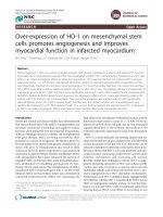
Figure 1 HO-1 expression mediated by MSCs in Vitro and Vivo. (A) HO-1 expression mediated by MSCs with GFP in Vitro (200×).
(B) Western blot analysis of HO-1 protein in MSCs with actin used as an internal control. Lane a, MSCs control (untransfected); lane b, Null-MSCs;
lane c, Adv-HO-1-MSCs. (C) Graph showing the relative fold induction of HO-1 protein levels in MSCs, n = 6. *P < 0.05 compared with MSCs
control (untransfected);
&
P > 0.05 compared with MSCs control (untransfected);
#
P < 0.05 compared with Null-MSCs. (D) Image from grafted HO-
1-MSCs in the infarcted myocardium (200×). (E) RT-PCR detection mRNA in cardiac tissue. Lane a, MSCs control (untransfected); lane b, Null-MSCs;
lane c, Adv-HO-1-MSCs.
Zeng et al. Journal of Biomedical Science 2010, 17:80
/>Page 4 of 8
cytoprotective effects, due to its mu ltiple catalytic bypro-
ducts. HO-1 was administered to improve the survival
environment of MSCs and to achieve maximum functional
benefits of MSCs[15]. Recent studies showed that over-
expression of the HO-1 gene in endothelial cell cau sed a
significant increase in angiogenesis[16]. Adenovirus-
mediated HO-1 gene transfer into t he ischemic hindlimb
facilitated a significant recovery of blood flow in the hin-
dlimb, and this effect was, at least in part, due to an
increase in the capillary density, thus, to angiogenic effects
of HO-1[17]. In our study, capillary density and the
expression of angiogenic growth factors, including vascular
endothelial growth factor (VEGF) and fibroblast growth
factor 2 (FGF2), in the border area of the infarct in HO-1-
MSCs group was significantly higher than that in Null-
MSCs group and ZnPP treated HO-1-MSCs group. How-
ever, capillary density and the expression of VEGF and
FGF2 did not show significant difference between Null-
MSCs and ZnPP treated HO-1-MSCs group, indicating
theroleofHO-1intheinductionofangiogenesis.We
confirmed that HO-1 transduced by MSCs also have posi-
tive effects on angiogenesis. It has been reported that nitric
oxide (NO) may modulate angiogen esis by upregula ting
VEGF in vascular cells, and NO inhibitors can reduce the
angiogenic potential of endothelial cells[18]. CO may also
be involve d in the expression of VEGF[ 19]. Another con-
tributor to enhance angiogenesis may be the increasing
expression of angiogenic growth factors in the ischemic
a
b
A
a
b
C
6
* #$
n
c
d
2.4
3.6
4.8
*
*
*&
*&
* #$
fold inductio
n
0
1.2
2.4
a b c d a b c d
Relative
1250
1500
* #$
VEGFFGF
n
/mm
2
)
B
500
750
1000
1250
*
*&
l
ary density (
n
0
250
a b c d
Capil
l
Figure 2 Effects of HO-1-MSCs transplantation on neovascularization and angiogenic growth factors. (A) Representative microv esse l in
the border of infarcted myocardium 3 weeks after transplantation (200×). (B) Values are means ± SD of data from 6 separate experiments,
*P < 0.05 compared with the hearts treated with PBS.
#
P < 0.05 compared with the hearts treated with Null-MSCs.
&
P > 0.05 compared with the
hearts treated with Null-MSCs.
$
P < 0.05 compared with the hearts treated with HO-1-MSCs and HO inhibitor. Lane a, hearts treated with PBS;
Lane b, hearts treated with Null-MSCs; Lane c, hearts treated with HO-1-MSCs and HO inhibitor; Lane d, hearts treated with HO-1-MSCs. (C) Blots
regarding the expression of FGF2, VEGF and actin were developed by the ECL method and relative protein levels were quantified by scanning
densitometry and the relative gray value of protein = protein of interest/internal reference. Values are means ± SD of data from 6 separate
experiments, * P < 0.05 compared with the hearts treated with PBS.
#
P < 0.05 compared with the hearts treated with Null-MSCs.
&
P > 0.05 compared with the hearts treated with Null-MSCs.
$
P < 0.05 compared with the hearts treated with HO-1-MSCs and HO inhibitor. Lane
a, hearts treated with PBS; Lane b, hearts treated with Null-MSCs; Lane c, hearts treated with HO-1-MSCs and HO inhibitor; Lane d, hearts treated
with HO-1-MSCs.
Zeng et al. Journal of Biomedical Science 2010, 17:80
/>Page 5 of 8
c
c
A
a
B
20
o
tic nuclei
t
otal nuclei)
*
d
10
15
20
*
*&
Apopt
o
(% of
t
b
d
* #
0
5
a b c d e
*#$
e
Figure 3 Effects of HO-1-MSCs tr ansplantation on apoptosis. (A) TUNE L-positive cells in the border zone of infracted myocardium 3 week s
after transplantation (100×). (B) Values are means ± SD of data from 6 separate experiments, *P < 0.05 compared with the hearts treated with
PBS.
#
P < 0.05 compared with the hearts treated with Null-MSCs.
&
P > 0.05 compared with the hearts treated with Null-MSCs.
$
P < 0.05
compared with the hearts treated with HO-1-MSCs and HO inhibitor. Lane a, normal control; Lane b, hearts treated with PBS; Lane c, hearts
treated with Null-MSCs; Lane d, hearts treated with HO-1-MSCs and HO inhibitor; Lane e, hearts treated with HO-1-MSCs.
12
16
20
80
100
120
140
m
mHg)
(
mmHg)
*
* #
* #
*
0
4
8
ab c
0
20
40
60
LVE DP (
m
LVS P
(
a b c
a
b
c
6000
3300
4400
5500
m
mHg/s)
m
mHg/s)
* #
*
3000
4000
5000
* #
*
0
1100
2200
+dp/dt
m
-dp/dt
m
0
1000
2000
ab c
a b c
a
b
c
Figure 4 Effects of HO-1-MSCs transplantation on ventricular function. (A) Hemodynamic assessment of cardiac function at 4 weeks after
transplantation. LVSP: left ventricle systolic pressure; LVEDP: left ventricle end-diastolic pressure; + dP/dtmax and -dP/dtmax: rate of rise and fall
of ventricular pressure, respectively. means ± SD of data from 6 separate experiments, *P < 0.05 compared with the hearts treated with PBS,
#
P < 0.05 compared with the hearts treated with Null-MSCs. Lane a, hearts treated with PBS; Lane b, hearts treated with Null-MSCs; Lane c,
hearts treated with HO-1-MSCs.
Zeng et al. Journal of Biomedical Science 2010, 17:80
/>Page 6 of 8
myocardium. VEGF is a strong therapeutic reagent by
inducing angiogenesis in ischemic myocardium[20], and
VEGF can mediate the ischemia-induced mobilization of
bone marrow stem cells[21]. In addition, FGF2 also have
the potential to promote angiogenesis, and regulate prolif-
eration, migration, differentiation of vascular cells[22,23].
Lin’study showed that HO-1 gene tran sfer post MI pro-
vides protection at least in part by promoting angiogenesis
through inducing angiogenic growth factors[24].
Angiogenesis contributes to the regional blood flow in
the ischemic myocardium. C ardiomyocytes death plays
an important role in the development of remodeling;
ventricular remodeling with chamber dilatation and wall
thinning are important features of post-infarction cardiac
function[25,26]. Stu dies have shown that lat e reperfusion
after infarction results in enhanced cardiac function and
remodeling. The improved blood supply may result in
salvaging of cardiomyocytes that would otherwise b e lost
or no-functional d ue to ischemia. In addition, VEGF,
may pro vide myocardial protection, blocking the pro-
grammed cell death response that is know to c ontribute
significantly to the development of ischem ic heart failure
[27,28]. In the current study, significant decrease of apop-
totic cel ls in HO-1-MSCs group was observe d as com-
pared with that of control groups, and the enlargement
of L V dilatation and fibrosis were significantly decreased
in HO-1-MSCs group wit h smaller ch ambers and thicker
LV anterior walls. Echocardiographic results f urther
confirmed our hypothesis that HO-1 modified MSCs sig-
nificantly improve LV function.
In conclusion, HO-1 transduced by MSCs can induce
angiogenic effects and improve heart function after
acute myocardial infarction
Acknowledgements
We thank Dr. Lee-young Chau for generously providing the Adv-hHO-1 and
kind experimental helps. This work was supported by the Chinese National
Nature Science Foundation (30900609)
Author details
1
Department of Cardiology, Renmin Hospital of Wuhan University, Wuhan,
Hubei, China.
2
College of Veterinary Medicine, Northeast Agricultural
University, Harbin, Heilongjiang, China.
3
Department of Pathology, School of
Basic Medical Science, Wuhan University, Wuhan, Hubei, China.
Authors’ contributions
BZ designed, carried out the main experiment and drafted the manuscript.
GS-L helped to design the experiment and drafted the manuscript. XF-R
helped to finish the statistical analysis and improve the manuscript. YZ
participated in RT-PCR and Western blot analysis. HL-Ch helped to finish
histological experiments. All authors read and approved the final
manuscript.
Competing interests
The authors declare that they have no competing interests.
Received: 21 June 2010 Accepted: 7 October 2010
Published: 7 October 2010
References
1. Valen G: Extracardiac approaches to protecting the heart. Eur J
Cardiothorac Surg 2009, 35:651-657.
A
28
35
42
a
(%)
*
B
0
7
14
21
Fibrotic are
a
* #
C
0
PBS Null-MSCs HO-1-MSCs
PBS Null-MSCs HO-1-MSCs
PBS Null
-
MSCs HO
-
1
-
MSCs
PBS Null
-
MSCs
HO
-
1
-
MSCs
Figure 5 Effects of HO-1-MSCs transplantation on ventricular remodeling. (A) The transmural slices of the left ventricle were stained with Masson
trichrome (1.25×). (B) % fibrotic area in heart with infarction was measured. Values are means ± SD of data from 6 separate experiments, *P < 0.05
compared with the hearts treated with PBS,
#
P < 0.05 compared with the hearts treated with Null-MSCs. (C) The border zone of the infarct area (100×).
Zeng et al. Journal of Biomedical Science 2010, 17:80
/>Page 7 of 8
2. Li W, Ma N, Ong LL, Nesselmann C, Klopsch C, Ladilov Y, Furlani D,
Piechaczek C, Moebius JM, Lützow K, Lendlein A, Stamm C, Li RK,
Steinhoff G: Bcl-2 engineered MSCs inhibited apoptosis and improved
heart function. Stem Cells 2007, 25:2118-2127.
3. Wang D, Shen W, Zhang F, Chen M, Chen H, Cao k: Connexin43 promotes
survival of mesenchymal stem cells in ischaemic heart. Cell Biol Int 2010,
12:415-423.
4. Gnecchi M, He H, Noiseux N, Liang OD, Zhang L, Morello F, Mu H, Melo LG,
Pratt RE, Ingwall JS, Dzau VJ: Evidence supporting paracrine hypothesis
for Akt-modified mesenchymal stem cell-mediated cardiac protection
and functional improvement. FASEB J 2006, 20:661-669.
5. Tenhunen R, Marver HS, Schmid R: The enzymatic conversion of heme to
bilirubin by microsomal heme oxygenase. Proc Natl Acad Sci USA 1968,
61:748-755.
6. Jazwa A, Loboda A, Golda S, Cisowski J, Szelag M, Zagorska A, Sroczynska P,
Drukala J, Jozkowicz A, Dulak J: Effect of heme and heme oxygenase-1 on
vascular endothelial growth factor synthesis and angiogenic potency of
human keratinocytes. Free Radic Biol Med 2006, 40:1250-63.
7. Deshane J, Chen S, Caballero S, Grochot-Przeczek A, Was H, Li Calzi S,
Lach R, Hock TD, Chen B, Hill-Kapturczak N, Siegal GP, Dulak J, Jozkowicz A,
Grant MB, Agarwal A: Stromal cell-derived factor 1 promotes
angiogenesis via a heme oxygenase 1-dependent mechanism. J Exp Med
2007, 204:605-618.
8. Malaguarnera L, Pilastro MR, Quan S, Ghattas MH, Yang L, Mezentsev AV,
Kushida T, Abraham NG, Kappas A: Significance of heme oxygenase in
prolactin-mediated cell proliferation and angiogenesis in human
endothelial cells. Int J Mol Me 2002, 10:433-440.
9. Zeng B, Ren X, Lin G, Zhu C, Chen H, Yin J, Jiang H, Yang B, Ding D:
Paracrine action of HO-1-modified mesenchymal stem cells mediates
cardiac protection and functional improvement. Cell Biol Int 2008,
32:1256-1264.
10. Juan SH, Lee TS, Tseng KW, Durante W: Adenovirus-mediated heme
oxygenase-1 gene transfer inhibits the development of atherosclerosis
in apolipoprotein E-deficient mice. Circulation 2001, 104:1519-1525.
11. Akiyama K, Gluckman TL, Terhakopian A, Jinadasa PM, Narayan S,
Singaswamy S, Massey B, Bing RJ: Apoptosis in experimental myocardial
infarction in situ and in the perfused heart in vitro. Tissue Cell 1997,
29:733-743.
12. Fishbein MC, Maclean D, Maroko PR: Experimental myocardial infarction in
the rat: qualitative and quantitative changes during pathologic
evolution. Am J Pathol 1978, 90:57-70.
13. Bironaite D, Baltriukiene D, Uralova N, Stulpinas A, Bukelskiene V,
Imbrasaite A, Kalvelyte A: Role of MAP kinases in nitric oxide induced
muscle-derived adult stem cell apoptosis. Cell Biol Int 2009, 33:711-719.
14. Müller-Ehmsen J, Krausgrill B, Burst V, Schenk K, Neisen UC, Fries JW,
Fleischmann BK, Hescheler J, Schwinger RH: Effective engraftment but
poor mid-term persistence of mononuclear and mesenchymal bone
marrow cells in acute and chronic rat myocardial infarction. J Mol Cell
Cardiol 2006, 41:876-884.
15. Tang YL, Tang Y, Zhang YC, Qian K, Shen L, Phillips MI:
Improved graft
mesenchymal stem cell survival in ischemic heart with a hypoxia-
regulated heme oxygenase-1 vector. J Am Coll Cardiol 2005, 46:1339-1350.
16. Jazwa A, Loboda A, Golda S, Cisowski J, Szelag M, Zagorska A, Sroczynska P,
Drukala J, Jozkowicz A, Dulak J: Effect of heme and heme oxygenase-1 on
vascular endothelial growth factor synthesis and angiogenic potency of
human keratinocytes. Free Radic Biol Med 2006, 40:1250-1263.
17. Suzuki M, Iso-o N, Takeshita S, Tsukamoto K, Mori I, Sato T, Ohno M,
Nagai R, Ishizaka N: Facilitated angiogenesis induced by heme
oxygenase-1 gene transfer in a rat model of hindlimb ischemia. Biochem
Biophys Res Commun 2003, 302:138-143.
18. Dulak J, Józkowicz A, Dembinska-Kiec A, Guevara I, Zdzienicka A,
Zmudzinska-Grochot D, Florek I, Wójtowicz A, Szuba A, Cooke JP: Nitric
oxide induces the synthesis of vascular endothelial growth factor by rat
vascular smooth muscle cells. Arterioscler Thromb Vasc Biol 2000,
20:659-666.
19. Józkowicz A, Huk I, Nigisch A, Weigel G, Dietrich W, Motterlini R, Dulak J:
Heme oxygenase and angiogenic activity of endothelial cells:
stimulation by carbon monoxide and inhibition by tin protoporphyrin-IX.
Antioxid Redox Signal 2003, 5:155-162.
20. Huwer H, Welter C, Ozbek C, Seifert M, Straub U, Greilach P, Kalweit G,
Isringhaus H: Simultaneous surgical revascularization and angiogenic
gene therapy in diffuse coronary artery disease. Eur J Cardiothorac Surg
2001, 20:1128-1134.
21. Jeon O, Song SJ, Bhang SH, Choi CY, Kim MJ, Kim BS: Additive effect of
endothelial progenitor cell mobilization and bone marrow mononuclear
cell transplantation on angiogenesis in mouse ischemic limbs. J Biomed
Sci 2007, 14:323-330.
22. Slavin J: Fibroblast growth factors: at the heart of angiogenesis. Cell Biol
Int 1995, 19:431-444.
23. Chen JH, Wang XC, Kan M, Sato JD: Effect of FGF-1 and FGF-2 on VEGF
binding to human umbilical vein endothelial cells. Cell Biol Int 2001,
25:257-260.
24. Lin HH, Chen YH, Chang PF, Lee YT, Yet SF, Chau LY: Heme oxygenase-1
promotes neovascularization in ischemic heart by coinduction of VEGF
and SDF-1. J Mol Cell Cardiol 2008, 45:44-55.
25. Moorjani N, Catarino P, El-Sayed R, Al-Ahmed S, Meyer B, Al-Mohanna F,
Westaby S: A pressure overload model to track the molecular biology of
heart failure. Eur J Cardiothorac Surg 2003, 24:920-925.
26. Lee SD, Kuo WW, Lin DY, Chen TH, Kuo WH, Hsu HH, Chen JZ, Liu JY,
Yeh YL, Huang CY: Role of calcineurin in Porphyromonas gingivalis-
induced myocardial cell hypertrophy and apoptosis. J Biomed Sci 2006,
13:251-260.
27. Yao Yongwei, Zhang Fumin, Wang Liansheng, Zhang Guohui,
Wang Zhaojun, Chen Jianmei, Gao Xiang: Lipopolysaccharide
preconditioning enhances the efficacy of mesenchymal stem cells
transplantation in a rat model of acute myocardial infarction. J Biomed
Sci 2009, 16:74-82.
28. Heilmann CA, Attmann T, von Samson P, Göbel H, Marmé D, Beyersdorf F,
Lutter G: Transmyocardial laser revascularization combined with vascular
endothelial growth factor 121 (VEGF121) gene therapy for chronic
myocardial ischemia–do the effects really add up? Eur J Cardiothorac Surg
2003, 23:74-80.
doi:10.1186/1423-0127-17-80
Cite this article as: Zeng et al.: Over-expression of HO-1 on
mesenchymal stem cells promotes angiogenesis and improves
myocardial function in infarcted myocardium. Journal of Biomedical
Science 2010 17:80.
Submit your next manuscript to BioMed Central
and take full advantage of:
• Convenient online submission
• Thorough peer review
• No space constraints or color figure charges
• Immediate publication on acceptance
• Inclusion in PubMed, CAS, Scopus and Google Scholar
• Research which is freely available for redistribution
Submit your manuscript at
www.biomedcentral.com/submit
Zeng et al. Journal of Biomedical Science 2010, 17:80
/>Page 8 of 8