Báo cáo y học: "Precise pattern of recombination in serotonergic and hypothalamic neurons in a Pdx1-cre transgenic mouse line" ppsx
Bạn đang xem bản rút gọn của tài liệu. Xem và tải ngay bản đầy đủ của tài liệu tại đây (11.34 MB, 13 trang )
RESEARC H Open Access
Precise pattern of recombination in serotonergic
and hypothalamic neurons in a Pdx1-cre
transgenic mouse line
Gerard Honig
1,5,7*
, Angela Liou
2,4
, Miles Berger
3,5,6
, Michael S German
4
, Laurence H Tecott
5
Abstract
Background: Multicellular organisms are characterized by a remarkable diversity of morphologically distinct and
functionally specialized cell types. Transgenic techniques for the manipulation of gene expression in specific cellular
populations are highly useful for elucidating the development and function of these cellular populations. Given
notable similarities in developmental gene expression between pancreatic b-cells and serotonergic neurons, we
examined the pattern of Cre-mediated recombination in the nervous system of a widely used mouse line, Pdx1-cre
(formal designation, Tg(Ipf1-cre)89.1D am), in which the expression of Cre recombinase is driven by regulatory
elements upstream of the pdx1 (pancreatic-duodenal homeobox 1) gene.
Methods: Single (hemizygous) transgenic mice of the pdx1-cre
Cre/0
genotype were bred to single (hemizygous)
transgenic reporter mice (Z/EG and rosa26R lines). Recombination pattern was examined in offspring using whole -
mount and sectioned histological preparations at e9.5, e10.5, e11.5, e16.5 and adult developmental stages.
Results: In addition to the previously reported pancreatic recombination, recombination in the developing nervous
system and inner ear formation was observed. In the central nervous system, we observed a highly specific pattern
of recombination in neuronal progenitors in the ventral brainstem and diencephalon. In the rostral brainstem (r1-
r2), recombination occurred in newborn serotonergic neurons. In the caudal brainstem, recombination occurred in
non-serotonergic cells. In the adult, this resulted in reporter expression in the vast majority of forebrain-projecting
serotonergic neurons (located in the dorsal and median raphe nuclei) but in none of the spinal cord-projecting
serotonergic neurons of the caudal raphe nuclei. In the adult caudal brainstem, reporter expression was
widespread in the inferior olive nucleus. In the adult hypothalamus, recombination was observed in the arcuate
nucleus and dorsomedial hypothalamus. Recombination was not observed in any other region of the central
nervous system. Neuronal expression of endogenous pdx1 was not observed.
Conclusions: The Pdx1-cre mouse line, and the regulatory elements contained in the correspon ding transgene,
could be a valuable tool for targeted genetic manipulation of developing forebrain-projecting serotonergic
neurons and several other unique neuronal sub-populations. Th ese results suggest that investigators employing
this mouse line for studies of pancreatic function should consider the possible contributions of central nervous
system effects towards resulting phe notypes.
Background
The development of methods for the experimental
manipulation of gene expression in vivo has revolutio-
nized the study of biology. Transgenes which drive
expression of recombinases within specific cell types
and/or at specific developmental time points are
valuable tools for understanding the development and
physiology of organ systems in vivo [1]. One such sys-
tem, the mammalian brain, is a remarkably complex and
heterogeneous structure comprised of many highly spe-
cialized and often rare cell types. Serotonergic neurons,
which comprise a tiny fraction of all neurons in the
mammalian brain, play an important and unique role in
many physiological functions, including the regulation
* Correspondence:
1
Neuroscience Graduate Program, University of California San Francisco, San
Francisco, CA, USA
Full list of author information is available at the end of the article
Honig et al. Journal of Biomedical Science 2010, 17:82
/>© 2010 Honig et al; licensee BioMed Central Ltd. This is an Open Access artic le distribute d under the terms of the Creative Commons
Attribution Licens e ( which permits u nrestricted use, distribution, and reproduction in
any medium, provided the original work is properly cited.
of affect in humans [2]. These neurons are themselves
anatomically and functionally diverse, although the
molecular, developmental and physiological basis for
this diversity is not completely understood [2,3].
Recently, the advent of transgenic methods to express
recombinases in all or subse ts of serotonergic neurons
has provided new insights into the diverse origins and
functions of these neurons [4-6].
Serotonergic neurons and pancreatic insulin-produ-
cing b-cells exhibit a remarkably similar and specific
cascade of transcription factor expression during devel-
opment, involving the expression of nkx2.2, lmx1b,and
nkx6.1 [7,8]. In the pancreas, pdx1, a homeodomain
transcription factor, plays a critical role in specifying the
fate of the early pancreatic primordium and, later in
development, is required for successful b-c ell develop-
ment [9]. We hypothesized that regulatory elements
which control pdx1 expression might be active in the
developing brain and might be applied to genetically tar-
get serotonergic neurons and/or other neuronal cell
types. We therefore examined the developmental pattern
of Cre-mediated recombination in the nervous system
using a widely used mouse line, Pdx1-cre (formal desig-
nation, Tg(Ipf1-cre)89.1Dam) [10-12]. This mouse line
has been employed in at least 30 published studies, as it
exhibits robust recombination in the developing endo-
crine pancreas [13-19 ,10,20-41]. Using two Cre reporter
lines, Z/EG (Tg(CAG-Bgeo/GFP)21Lbe) and rosa26R
(Gt(ROSA)26So r
tm1Sor
) [42,43,12], we found that that
Pdx1-cre also exhibits develo pmental recombination in
the inner ear; in rostral serotonergic neurons; in the
hypothalamus; and in non-serotonergic neurons of the
caudal hindbrain.
Materials and methods
Mice
Strain information is summarized in Table 1. Pdx1-cre
and Z/EG mouse lines were maintained as independent
colonies of hemizygous transgenic mice; the rosa26R
mouse line was maintained as homozygous mutant
mice. Strain background was mixed for all lines. Pdx1-
cre mice w ere kindly provided by D. Melton; Z/EG and
rosa26R mice were obtained from Jackson Labs. To gen-
erate experimental animals, transgenic hemizygous mice
from the Pdx1-cre line (genotype pdx1-cre
Cre/0
)were
bred with hemizygous transgenic mice from the Z/EG
line (Zeg
GFP/0
) or homozygous transgenic mice from the
rosa26R line (rosa26
LacZ/LacZ
). Offspring genotypes were
obtained in accor d with expected Mendelian ratios. Off-
spring of the following genotypes were used for analysis:
pdx1-cre
Cre/0
; Zeg
GFP/0
(experimental), pdx1-cre
0/0
;
Zeg
GFP/0
(control); pdx1-cre
Cre/0
; rosa26
LacZ/+
(experi-
mental) and pdx1-cre
0/0
; rosa26
LacZ/+
(control). Mice
were housed on a 12-hr light-dark cycle in a controlled
climate and were fed ad lib with Purina LabDiet 5053
mouse chow. All studies involving mice were approved
by the UCSF Institutional Animal Care and Use
Committee.
Genotyping
Ear punches or e mbryonic tails were digested in strip
tubes with 0.05 U proteinase K (03115887001, Roche) in
50 μL of DirectPCR Lysis Reagent (402-E, Viagen Bio-
tech) diluted with 50 μLwater.5μLPCRswereper-
formed using SYBR GreenER PCR mix (Invitrogen),
primers (concentration depends on the assay, generally
200 nM) and 0.15 μL of heat-inactivated genomic DNA
solution. Thermal cycling was performed on an ABI
7300 instrument with SYBR detection as follows: 95°C
for 10 min; 95°C for 15 s followed by 60°C for 1 min
(40 cycles); 95°C for 15 s; 60°C for 15 s; followed by a
melting curve step with a 2% ramp rate from 60°C to
95°C. Allele-sp ecific PCR products were identified using
melting curve analysis as described in Results and Figure
1. Primer sequences and concentrations for assays are
provided in Table 2.
Dissection and histology
4 mice or embryos were analyzed for each genotype and
developmental stage. Timed matings were carried out
with embryonic day 0.5 considered to be midday of the
day of discovery of a vag inal plug. For whole-mount
preparations, e10.5 embryos were dissected and briefly
fixed in 4% para-formaldehy de in phosphate-buffered
saline (PBS), then incubated overnight at 37°C in
b-galactosidase (LacZ) staining media (10 mM Tris-HCl
pH 7.4, 5 mM K
4
FeCN
6
,5mMK
3
FeCN
6
,2mMMgCl
2
and 0.8 mg/ml X-gal (Invitrogen)). For embryonic sam-
ples, embryos were dissected at the appropriate stage
and immediately embedded and frozen without fixation
in Optimal Cutting Temperature media (Tissue-Tek).
Embedded embryos were sectioned (transverse, 20 μm)
on a cryostat. For adult sam ples, mice were deeply
anesthetized and then perfused with phosphate-b uffere d
saline (PBS) followed by 4% para-formaldehyde in PBS;
brains were removed, cryoprotected in 30% sucrose in
PBS and sectioned (50 μm, saggital) on a freezing
microtome. Immunostaining was performed on-slide for
embryonic samples and free-floating for adult tissues.
Sections were incubated in blocking solution (4% goat
serum, 2% BSA and 0.1% Triton-X-100 in PBS) for 1 h;
incubated with primary antibody diluted in blocking
solution for 18 h at 4°C; washed in PBS; incubated for 2
h with appropriate secondary anti-IgG antibodies conju -
gated t o Alexa 488 or Alexa 594 dyes; and washed and
mounted in Vectashield media (Vector Laboratories).
Tyramide signal amplification (TSA) reagents (Invitro-
gen) were used as per manufacturer’s instructions. For
Honig et al. Journal of Biomedical Science 2010, 17:82
/>Page 2 of 13
b-galactosidase staining of sections, sections were incu-
bated overnight in staining media as described above.
Slides were imaged using a confocal, upright epifluores-
cence or brightfield dissection microscope. Red and
green fluorescence spectra were captur ed separa tely and
appropriate control experiments were performed to con-
firm specificity and lack of cross-reactivity in labeling.
Antibodies
The following primary antibodies and dilutions were
used: chicken a-GFP, 1:1000 (Aves Labs); rabbit a -sero-
tonin, 1:6000 (Immunostar); mouse a-TPH (tryptophan
hydroxylase), 1:200 (Sigma); mouse a-Nkx2.2 (Develop-
mental Studies Hybridoma B ank, clone 74.5A5); mouse
a-Mash 1, 1:100 (BD Transduction); rabbit a-Pdx1 (gen-
erated in lab??), 1:1000; and mouse a-Isl1, 1:50 (Devel-
opmental Studies Hybridoma Bank, clone 40.206).
Results and discussion
As the efficiency of Cre-mediated recombination is not
necessarily identical across different target loci, we
intercrossed Pdx1-cre transgenic mice with mice from
two distinct Cre reporter lines. The transgenes and gen-
otypes analyzed are summarized in Table 1 and in the
Methods. In brief, the re porter lines function as follows:
the Z/EG line carries a sing le-copy transgene containing
a strong and ubiquitous recombinant p romoter, fol-
lowed by a b -galactosidase and transcriptional stop cas-
sette flanked by loxP sites, followed by a GFP cassette
[42]. In Cre-negative cells, b-galactosidase only is
expressed; in Cre-expressing lineages, the b-galactosi-
dase cassette is excised, pe rmitting expression of GFP.
The Rosa26R mouse line harbors a transgene inserted
by targeted mut agenesis into the ubiquitously expressed
rosa26 locus; this transgene consists of a loxP-flanked
stop cassett e followed by a b-galactosidase cassette [43].
In Cre-negative cells, no reporter b-galactosidase trans-
gene is expressed; in Cre-expressing lineages, the
stop cassette is excised, permitting expression of
b-galactosidase.
Standard polymerase chain reaction (PCR) genotyping
protocols for the mous e lines used have been described
previously [10,42,43]. We adapted previously published
methods for s ingle nucleotide polymorphism dete ction
[44-50] to develop a gel-free genotyping metho d based
on multiplex PCR and discrimination of allele-specific
products using SYBR-Green-detected melting curve ana-
lysis. Small-product multiplex PCR reactions are per-
formed using an optical cycler with the inclusion of
SYBR Green dye, w hich fluoresces in the presen ce of
double-strandedDNA.Attheendoftheamplification
rea ction, PCR products corresponding to specific alleles
are detected by progressively heating the reaction and
plotting the der ivat ive of SYBR fluores cenc e; annealing
and melting of a specific product generates a peak at a
specific m elting temperature. The PCR reaction well is
never opened and no gels are required. Melting curve
peaks (position and shape) can be manipulated using
simple, inexpensive primer modification [45], such that
this approach can readily address most PCR based, mul-
tiple x genot yping applications. This method confers sig-
nificant advantages over existing methods, including
higher throughput, uniform and robust PCR conditions,
low cost and reduction of post-PCR contamination.
Representative data, from an assay used to genotype
mice of the Pdx1-cre line, is provided in Figure 1.
Detailed instructions for assay design and implementa-
tion are available upon request (also see [45,44,46-50]).
Primer sequences and reaction conditions are provided
in Table 2.
Table 1 Transgenic mouse lines and relevant genotypes employed
Formal
designation
Common
designation
Transgene design Trans-gene
insertion
Purpose WT
allele
Trans-
genic
allele
Geno-
types
analyzed
Initial
reference
MGI ID
Tg(Ipf1-cre)
89.1Dam
Pdx1-cre 5.5-kb portion of the
mouse pdx1 promoter
fused to cre cassette
Random insertion
(pro-nuclear
injection
Expression of Cre is
driven by regulatory
elements which regulate
pdx1 expression
0 Cre Cre/0
(hemi-
zygote);
0/0 (wild-
type
control)
Develop-
ment
2002, 129
(10):2447-
2457
2684317
Tg(CAG-
Bgeo/GFP)
21Lbe
Z/EG Ubiquitous
recombinant promoter,
followed by LoxP-
flanked LacZ cassette,
followed by GFP
cassette
Random insertion
with screening for
high expression in
ES cells (ES cell
electro-poration)
When cre is expressed in
a cell, LacZ cassette is
excised, leading to GFP
expression in that cell
and all daughter cells
0 GFP GFP/0
(hemi-
zygote)
Genesis
2000, 28
(3-4):147-
155
3046177
Gt(ROSA)26
Sor
tm1Sor
Rosa26R LoxP-flanked stop
cassette followed by
LacZ cassette
Targeted to the
ubiquitously
expressing Gt
(ROSA)26Sor locus
When cre is expressed in
a cell, stop cassette is
excised, leading to LacZ
expression in that cell
and all daughter cells
+ LacZ LacZ/+
(hetero-
zygote)
Nature
Genetics
1999, 21
(1):70-71
1861932
Honig et al. Journal of Biomedical Science 2010, 17:82
/>Page 3 of 13
Under our experimental conditions, reporter expres-
sion of GFP (Z/EG line) or LacZ (rosa26R line) provided
a specific marker of Cre-mediated recombination. No
reporter expression was evident in pdx1-cre
0/0
; Zeg
GFP/0
and pdx1-cre
0/0
; rosa26
LacZ/+
littermate control tissues
(Figures 2C; 3A; 4A; C; E; G). GFP and LacZ expression
patterns described bel ow were observed in pdx1-cre
Cre/0
;
Zeg
GFP/0
and pdx1-cre
Cre/0
; rosa26
LacZ/+
mice or
embryos. For each genotype and time point, comparable
patterns of GFP or LacZ expression were observed in all
analyzed animals (n = 4).
Recombination was first detected at e10.5 in the pan-
creas (Figure 2A), as reported [10], and in the inner ear
formation (patchy expression in the developing anterior
and posterior semicircular canal region with enriched
expression in two anterior and posterior medial domains)
(Figures 2A &2B). The earliest recombination in the cen-
tral nervous system was observed at e11.5, in the hind-
brain (Figure 3) and in the diencephalon (Figure 5).
In the e11.5 hindbrain, GFP expression was observed
to spatiotemporally coincide with serotonergic neuro-
genesis. In rhombomeres 1 (r1) and 2 (r2), GFP was
observed exclusively within a ventral zone where seroto-
nin neurons are first observed in the developing brain
[8](Figures3B,C,D).Inr1andr2,mostGFP
+
cells
were newborn serotonergic neurons, as identified by ser-
otonin (5-HT) immunoreactivity, although a small min-
ority of GFP-lab eled cells appear ed to be 5-HT
-
and
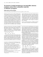
Figure 1 Transgenic mouse genotyping using multiplex allele-specific PCR and melting curve analysis. PCR was performed in an optical
cycler (ABI 7300) using 1-10 ng genomic DNA from mice of the indicated genotypes and a reagent mix containing SYBR GreenER. Amplification
plots (A, C) and melting curves (B, D) are shown. Primers were designed to amplify 2 specific products: a genomic control product, generated
from any genomic mouse template; and a transgene-specific product, generated only from genomic templates containing a cre transgene.
Normalized fluorescence (y-axis, A &C) is the baseline-subtracted ratio of SYBR signal to ROX (passive reference dye) signal during amplification
cycling (A, C). Normalized fluorescence derivative (y-axis, B & D) is the 2
nd
derivative of normalized fluorescence during the melting curve step.
Dotted lines (A, C) indicate cycle threshold. A&B.Genomic DNA from a wild-type mouse; note robust amplification of the genomic control
product with a single melting peak (allowing the distinction of a negative result from a failed PCR). C&D.Genomic DNA from a pdx1-cre
Cre/0
mouse; note robust amplification with 2 distinct melting peaks corresponding to the control and cre-specific products. Arrows indicate presence
of the genomic control product (Control) and the transgene-specific product (Cre).
Honig et al. Journal of Biomedical Science 2010, 17:82
/>Page 4 of 13
Table 2 Primer sequences and reaction conditions for gel-free genotyping assays
Assay
name
Target locus Primer
1
Conc.
(nM)
Primer
2
Conc.
(nM)
Primer
3
Conc.
(nM)
Primer
4
Conc.
(nM)
Expected results
Z/EG Any transgene
generated using the
pCAGG construct (e.g.,
Z/EG line)
TCGA
TGCA
GGAT
AACTT
CGT
400 GGT
ACC
GTC
GACT
GCA
GAAT
400 AGC
AGC
AGG
CAG
GGC
TTT
50 GTCT
GGA
CAC
GGG
AGC
ACTT
50 Primers 3 & 4 generate a single peak in all
samples (control product, gdf); primers 1 &
2 generate an additional, lower Tm peak
(transgene-specific product) in Zeg
GFP/0
or
Zeg
GFP/GFP
samples.
Cre Any transgene
containing cre (e.g.,
Pdx1-cre line)
ACATT
TGGG
CCAG
CTAAA
CAT
200 CGG
CATC
AAC
GTTT
TCTT
TT
200 GGC
GAG
AGC
AGA
GTGT
GGA
T
200 AAGT
CGG
CAG
GCA
CAG
GAG
200 Primers 3 & 4 generate a single peak in all
samples (control product, k17); primers 1 &
2 generate an additional, higher Tm peak
(transgene-specific product) in any sample
with a cre transgene.
PdxCre cre fused to 5’
regulatory region of
pdx1 gene (Pdx1-cre
line)
TAAG
GCCT
GGCT
TGTA
GCTC
200 ACC
GGT
AATG
CAG
GCA
AAT
200 AGC
AGC
AGG
CAG
GGC
TTT
30 GTCT
GGA
CAC
GGG
AGC
ACTT
30 Primers 3 & 4 generate a single peak in all
samples (control product); primers 1 & 2
generate an additional, lower Tm peak
(transgene-specific product) in pdx1-cre
Cre/
0
or pdx1-cre
Cre/Cre
samples.
Rosa26 Any rosa26 allele
targeted using a
standard targeting
allele (e.g., Rosa26R
line)
GCGC
GCGC
GCGT
GATC
TGCA
ACTC
CAGT
CTTTC
200 GCG
CGC
GCG
CGC
GCG
CGC
GCC
ACAC
CAG
GTTA
GCC
TTTA
AGC
200 GAC
AGG
ATAA
GTAT
GAC
ATCA
TCAA
GG
200 Primers 1 & 2 generate a single peak in
samples containing the wild-type rosa26
allele; primers 2 & 3 generate a lower Tm
peak (transgene-specific product) in
samples containing a targeted allele; both
peaks are observed in heterozygote
samples.
Figure 2 Cre-mediated recombination in the pancreatic primordium and inner ear in the e10.5 embryo. Whole-mount images of a pdx1-
cre
Cre/0
; rosa26
LacZ/+
embryo (A & B) and a pdx1-cre
0/0
; rosa26
LacZ/+
embryo (C) processed for b-galactosidase activity. b-galactosidase activity was
observed in the pancreatic primordium (bottom arrow, left panel) and inner ear formation (top arrow, A; region in higher magnification in B).
b-galactosidase activity was not evident in pdx1-cre
0/0
; rosa26
LacZ/+
control embryos (C). Scale bars, 1 mm (A and C) and 150 μm (B).
Honig et al. Journal of Biomedical Science 2010, 17:82
/>Page 5 of 13
some 5-HT
+
cells were GFP
-
, suggesting that recombi-
nation in this cell lineage was mosaic at this time point
(Figures 3B, C, E). GFP exp ression was obser ved adja-
cent to but not in the Nkx2.2
+
progenitor zone (Figure
3D), suggesting that Cre i s first expressed as serotoner-
gic cells differentiate and migrate out of this progenitor
zone. In rhombomere 4 (r4), serotonergic neurons are
not generated[8], although 5-HT
+
fibers can be detected
(distinguished from cell bodies by morphology, anatomi-
cal location and lack of DAPI staining); in r4, GFP
expression was observed in most 5-HT
+
fibers and rare
5-HT
-
cell bodies (Figure 3E). In the caudal hindbrain,
serotonin and reporter immunoreactivity was always
observed in the same sections, but rarely within the
same cell s; cellular GFP immunoreactivity was observed
immediately dorsal to most 5-HT
+
neurons (Figure 3F).
Figure 3 Cre-mediated recombination coincides with serotonergic neurogenesis in the e11.5 embryo. Epifluorescence images of the e11.5
developing hindbrain of embryos, transversely sectioned, immunostained for GFP (green) and 5-HT or Nkx2.2 (red). In pdx1-cre
Cre/0
; Zeg
GFP/0
embryos, GFP was always expressed in or adjacent to newborn serotonergic neurons. Both serotonergic and non-serotonergic neurons expressed
GFP in the rostral hindbrain (B, C, D) (rhombomere 1, r1; rhombomere 2, r2) and caudal hindbrain (ch) (F). The degree of co-expression of GFP and
the serotonergic phenotype was greatest in the rostral hindbrain, with little overlap in the caudal hindbrain and sparse GFP expression in
rhombomere 4 (r4) (E). GFP was not expressed in the Nkx2.2
+
progenitor zone (D). GFP expression was not evident in sections from a pdx1-cre
0/0
;
Zeg
GFP/0
embryo (A). Scale bars, 100 μm.
Honig et al. Journal of Biomedical Science 2010, 17:82
/>Page 6 of 13
Figure 4 Cre-mediated recombination in the hindbrain and diencephalon in the e16.5 embryo. Epifluorescence images from pdx1-cre
Cre/0
;
Zeg
GFP/0
(B, D, F, H) and pdx1-cre
0/0
; Zeg
GFP/0
(A, C, E, G) e16.5 embryos, transversely sectioned, immunostained for GFP (green) and 5-HT (red).
GFP was expressed in the dorsal raphe nucleus (dr) (B), caudal linear raphe (clr) (D), caudal hindbrain (ch) (F) and hypothalamus (hp) (H). In the
rostral hindbrain, GFP expression occurred in the serotonergic dorsal raphe and caudal linear nuclei (B, D). In the caudal hindbrain, GFP
expression was observed in the non-serotonergic inferior olive nucleus, adjacent to serotonergic raphe nuclei (F). GFP expression was not
evident in sections from pdx1-cre
0/0
; Zeg
GFP/0
control embryos (A, C, E, G). Scale bars, 100 μm.
Honig et al. Journal of Biomedical Science 2010, 17:82
/>Page 7 of 13
In general, we observed a rostral-caudal gradient of
overlap between GFP expression and the serotonergic
phenotype in the hindbrain.
The recombination pattern observed in the e11.5
hindbrain predicted the pa ttern we observed at later
stages of development. At e16.5, reporter expression in
the rostral hindbrain was restricted to the rostral raphe
nuclei, particularly the dorsal raphe nucleus (Figure 4B)
and caudal linear raphe nucleus (Figure 4D). In the cau-
dal hindbrain, reporter expression was observed in the
non-serotonergic inferior olive nucleus but not in adja-
cent serotonergic neurons (Figure 4F). In adult tissues,
confocal microscopy was employed for analysis of hind-
brain sections in order to more rigorously analyze the
co-expression of GFP and the serotonergic phenotype.
In the dorsal raphe nucleus, which is generated in r1,
the most rostral portion of the develop ing hindbrain
[4], the large majority of serotonergic neurons (identi-
fied by immunoreactivity for tryptophan hydroxylase, or
TPH) expressed GFP, and all GFP-positive cells were
serotonergic (Figures 6B, B’,B’’). In the median raphe
nucleus, which is generated in r1, r2 and r3 [4], there
was partial overlap between GFP and 5-HT expression
(Figures 6C, C’,C’’). In the caudal hindbrain, 5-HT and
GFP expression were completely non-overlapping,
although occurring in the same sect ions; re combination
was restricted to the inferior olive nucleus (Figures 6D,
D’ ,D’’, E). Interestingly, the inferior olive can be simi-
larly labeled using a Cre line in which the cre was intro-
duced into the locus for ptf1a , a transcription factor
which interacts with pdx1 during early pancreatic devel-
opment [51,52]. Given the anatomical localization of
GFP
+
5-HT
+
cells in the adult, it is likely that a vast
majority of forebrain-projecting serotonergic neurons,
and virtually no spinal-cord-projecting serotonergic neu-
rons, exhibit Cre-mediated recombination in the Pdx1-
cre line[2]. These data provide further evidence that
caudal and rostral serotonergic neurons, though gener-
ated through highly similar dev elopmental processes [8],
exhibit distinct patterns of gene expression regulation
[3]. This Pdx1-cre mouse line may be a useful resource
for investigators interested in manipulating gene expres-
sion in serotonergic neuron s projecting to the forebrain
but not to the brainstem and spinal cord.
In the e11.5 diencephalon , recombination occurred, as
in the hindbrain, in a restricted ventral zone of the
neural tube, adjacent to neurogenic zones (here identi-
fied by Isl1 an d Mash1 expression) (Figure 5). This
developmental pattern resulted in GFP and LacZ expres-
sion in specific, anatomically defined nuclei of the
hypothalamus, as could be observed at e16.5
(Figure 4H) and especially in adult sections. In the adult
Figure 5 Cre-mediated recombination in the ventral diencephalon in the e11.5 embryo. Epifluorescence images of the e11.5 diencephalon
of pdx1-cre
Cre/0
; Zeg
GFP/0
embryos, transversely sectioned, immunostained for GFP (green) and Isl1 (A) or Mash1 (B) (red). GFP was not expressed
at the ventral surface near the floor plate adjacent to the Isl1
+
Mash1
+
neurogenic zone. Scale bars, 100 μm.
Honig et al. Journal of Biomedical Science 2010, 17:82
/>Page 8 of 13
Figure 6 Cre-mediated recombination in forebrain-projecting serotonergic neurons, inferior olive neurons and hypothalamic neurons
in the adult brain. A-D: Individual optical sections obtained using confocal imaging of saggital sections from adult pdx1-cre
Cre/0
; Zeg
GFP/0
mice.
A: Wide-field image of the serotonergic dorsal raphe nucleus (dr) demonstrating extensive and anatomically restricted expression of TPH and
GFP in this structure. B: Higher-magnification image of the dorsal raphe nucleus: a large majority of TPH
+
neurons express GFP and that all GFP
+
cells in this region are serotonergic neurons. C: In the median raphe nucleus (mr), there was partial overlap between GFP and TPH expression.
D: In the caudal hindbrain, GFP expression was observed in the inferior olive nucleus (io), adjacent to but not overlapping with serotonergic
raphe nuclei. E-F: Brightfield images of saggital sections obtained from adult pdx1-cre
Cre/0
; rosa26
LacZ/+
mice, processed for LacZ activity. E: The
inferior olive nucleus was labeled with LacZ. F: Multiple nuclei of the hypothalamus, notably the dorsomedial, lateral and arcuate nuclei, were
labeled with LacZ. Scale bars: 80 μm (A); 60 μm (B, C, D); 200 μm (E); 150 μm (F).
Honig et al. Journal of Biomedical Science 2010, 17:82
/>Page 9 of 13
hypothalamus, reporter expression occurred in the arcu-
ate nucleus, dorsomedial nucleus and lateral hypothala-
mus (Figure 6F). T hese regions of the hypothalamus are
all critically involved in the in vivo regulation of meta-
bolic functions such as glucose homeostasis [53]. The
dorsomedial hypothalamic nucleus is relatively poorly
characterized at the molecular level, and to our knowl-
edge no transgenic mouse line has been reported which
exhibits specific transgene expressioninthissub-region
of the hypothalamus. The possible existence of specific
pdx1 regulatory sequences directing expression in the
dorsomedial hypothalamic nucleus could be used to
generate such transgenic mouse lines for the study of
this important hypothalamic cell population. Interest-
ingly, many hypothalamic neurons share specialized
physiological attributes with b-cells, such as glucose sen-
sing[54], and Cre transgenes generated using t he insulin
and ptf1a promoters produce recombination in the
hypothalamus [51,55].
Widespread expression of endogenous pdx1 in the rat
brain has been reported [56,57]. Our results suggested a
more restricted pattern of endogenous pdx1 expression
might occur in the mouse central nervous system. We
therefore attempted to detect expression in the mouse
brain of pdx1 at various developmental stages using
immunofluorescence. We were unable to detect endo-
genous pdx1 expression at any time point, including the
earliest time point at which Cre-mediated recombination
was observed: tyrami de signal amplification of Pdx1
immunoreactivity was attempted in pdx1-cre
Cre/0
;
Zeg
GFP/0
e11.5 r1 tissue. No detectable signal was
observed ( Figure 7B), despite robust expression of GFP
(Figure 7A) and reliable detection of Pdx1 in the adult
pancreas under these conditions.
While this work was under review, two publications
reported neuronal transgene expression in a variety of
mouse lines used for the study of pancreatic develop-
ment and function [58,59]. These results are consistent
with and complementary to our results. Using the Pdx1-
cre mouse line employed in our study, Wicksteed et al
and Song et al report a similar pattern of Cre-mediated
recombination in the developing and adult hypothala-
mus and brainstem. Furthermore, Wicksteed et al
observed a similar pattern of recombination in a n inde-
pendently generated mouse line in which Cre expression
is driven by a similar construct incorpora ting regulatory
sequences upstream of the pdx1 gene, suggesting that
the expression patterns we observe cannot be due solely
to i nsertion site effects. They also report a lack of galac-
tosidase activity in mice bearing a heterozygous LacZ
insertion at the pdx1 locus and a lack of a mplification
of endogenous pdx1 transcript from the brain (both
consistent with our observations, herein and u npub-
lished). Taken together, their results indicate that pdx1
regulatory elements can drive highly specific neuronal
expression, but that endogenous pdx1 is not expressed
in the mouse brain, likely due to the activity of a repres-
sor element not contained in the Pdx1-cre transgene
constructs currently employed. The reported widespread
expression of endogenous pdx1 reported in the rat brain
may reflect a difference between gene regulation
between rat and mouse or methodological differences
between the rat and mouse studies [56,57].
Conclusions
We report here, using t he widely used Pdx1-cre line, a
highly specific pattern of Cre-mediated recombination
in the central nervous system and inner e ar. This Cre
line, and the regulatory sequences that direct Cre
expression, may be a va lua ble resource for investigators
seeking to manipulate gene expression in specific sub-
sets of neurons, such as forebrain-projecting serotoner-
gic neurons and neurons of the dorsomedial
hypothalamus. To our knowledge, no other Cre mouse
lines have been described to exhibit a pattern of
hypothalamic recombination comparable to that we
observed in the Pdx1-Cre line. However, the fact that
recombination was observed in several hypothalamic
Figure 7 Lack of detectable expression of endogeno us pdx1 in the mouse hindbrain. Section from the r1 region of a pdx1-cre
Cre/0
; Zeg
GFP/0
embryo, immunostained for GFP (A) and Pdx1 (B) using TSA amplification. No detectable expression of Pdx1 was observed, despite robust
expression of GFP. Scale bars, 100 μm.
Honig et al. Journal of Biomedical Science 2010, 17:82
/>Page 10 of 13
nuclei which may have very different physiological func-
tions (such as the arcuate an d dorsomedial nuclei), a s
well as in the pancreas, may require further characteri-
zation of pdx1 regulatory elements in order to maximize
the utility of pdx1 regulatory elements for this purpose.
At present, se veral transgenic methods have bee n
developed which generate specific Cre-mediated recom-
bination in serotonergic neurons [4,5,60]. Two of these
methods involve the use of bacterial artificial chromo-
somes containing regulatory elements or genes
expressed in serotonergic neurons, and unlike the Pdx1-
cre line, result in recombination in the vast majority of
serotonergic neuron s [60,5]. However, like the Pdx1-cre
line, these Cre transgenes may also result in recombina-
tion in other cell types, such as thalamocortical neurons
[60] and pancreatic b-cells (Ohta, German et al, in sub-
mission). A technically sophisticated method has been
described which allows for highlyspecifictargeted
recombination in subsets of serotonergic neurons; how-
ever, this method may be impractical for some investiga-
tors due to the complexity of genetic manipulations
required [4].
It is import ant to note that the brain regions which
exhibit recombination in the Pdx1-cre mouse line are
well known to have important roles in in vivo metabolic
function, including glucose homeostasis [61,53]. Investi-
gators employing this line to express Cre in the develop-
ing pancreas should consider the possibility that
concomitant Cre expression in the central nervous sys-
tem may play a significant role in resulting in vivo phe-
notypes. Conversely, investigators who plan to use this
mouse line to manipulate neuronal gene expression
should consider the possible effects on pancreatic gene
expression and function. These considerations are parti-
cularly salient when analyzing behavior and physiology
in adult mice.
In conclusion, these data are consistent with the idea
that common patterns of gene expression in pancreatic
b-cells, serotonergic neurons and hypo thalamic neurons
contribute to their highly specialized and, in many cases,
similar physiology.
Acknowledgements
Elaine J. Carlson, Elaine Storm, John L. Rubenstein, Katherine Shim and Janet
Lau provided advice and reagents. L. Bogdanova assisted with mouse
genotyping. This project was supported by the Sandler Foundation and the
Howard Hughes Medical Institute (GH).
Author details
1
Neuroscience Graduate Program, University of California San Francisco, San
Francisco, CA, USA.
2
Department of Biochemistry and Molecular Cell Biology,
University of California Berkeley, Berkeley, CA, USA.
3
Biomedical Sciences
Graduate Program, University of California San Francisco, San Francisco, CA,
USA.
4
Diabetes Center, University of California San Francisco, San Francisco,
CA, USA.
5
Department of Psychiatry and Center for Neurobiology and
Psychiatry, University of California San Francisco, San Francisco, CA, USA.
6
Department of Anesthesiology, Duke University School of Medicine , NC,
USA.
7
Molecular Pathogenesis Program & Howard Hughes Medical Institute,
Kimmel Center for Biology and Medicine at the Skirball Institute, New York
University School of Medicine, New York, USA.
Authors’ contributions
GH, MB, AL, MSG and LT conceived of experiments; GH and AL performed
experiments; GH and AL analyzed data; GH wrote the manuscript; all authors
edited the manuscript.
Competing interests
The authors declare that they have no competing interests.
Received: 1 February 2010 Accepted: 17 October 2010
Published: 17 October 2010
References
1. Branda CS, Dymecki SM: Talking about a revolution: The impact of site-
specific recombinases on genetic analyses in mice. Dev Cell 2004,
6(1):7-28.
2. Jacobs BL, Azmitia EC: Structure and function of the brain serotonin
system. Physiol Rev 1992, 72(1):165-229.
3. Wylie CJ, Hendricks TJ, Zhang B, Wang L, Lu P, Leahy P, Fox S, Maeno H,
Deneris ES: Distinct transcriptomes define rostral and caudal serotonin
neurons. J Neurosci 2010, 30(2):670-684.
4. Jensen P, Farago AF, Awatramani RB, Scott MM, Deneris ES, Dymecki SM:
Redefining the serotonergic system by genetic lineage. Nat Neurosci
2008, 11(4):417-419.
5. Scott MM, Wylie CJ, Lerch JK, Murphy R, Lobur K, Herlitze S, Jiang W,
Conlon RA, Strowbridge BW, Deneris ES: A genetic approach to access
serotonin neurons for in vivo and in vitro studies. Proc Natl Acad Sci USA
2005, 102(45):16472-16477.
6. Kim JC, Cook MN, Carey MR, Shen C, Regehr WG, Dymecki SM: Linking
genetically defined neurons to behavior through a broadly applicable
silencing allele. Neuron 2009, 63(3):305-315.
7. Wilson ME, Scheel D, German MS: Gene expression cascades in pancreatic
development. Mechanisms of Development 2003, 120(1):65-80.
8. Pattyn A, Vallstedt A, Dias JM, Samad OA, Krumlauf R, Rijli FM, Brunet JF,
Ericson J: Coordinated temporal and spatial control of motor neuron and
serotonergic neuron generation from a common pool of CNS
progenitors. Genes Dev 2003, 17(6):729-737.
9. Holland AM, Gonez LJ, Naselli G, Macdonald RJ, Harrison LC: Conditional
expression demonstrates the role of the homeodomain transcription
factor Pdx1 in maintenance and regeneration of beta-cells in the adult
pancreas. Diabetes 2005, 54(9):2586-2595.
10. Gu G, Dubauskaite J, Melton DA: Direct evidence for the pancreatic
lineage: NGN3+ cells are islet progenitors and are distinct from duct
progenitors. Development 2002, 129(10):2447-2457.
11. Mouse Genome Database. [ />12. Bult CJ, Eppig JT, Kadin JA, Richardson JE, Blake JA: The Mouse Genome
Database (MGD): mouse biology and model systems. Nucleic Acids Res
2008, , 36 Database: D724-728.
13. Aguirre AJ, Bardeesy N, Sinha M, Lopez L, Tuveson DA, Horner J,
Redston MS, DePinho RA: Activated Kras and Ink4a/Arf deficiency
cooperate to produce metastatic pancreatic ductal adenocarcinoma.
Genes Dev 2003, 17(24):3112-3126.
14. Bardeesy N, Aguirre AJ, Chu GC, Cheng KH, Lopez LV, Hezel AF, Feng B,
Brennan C, Weissleder R, Mahmood U, Hanahan D, Redston MS, Chin L,
Depinho RA: Both p16(Ink4a) and the p19(Arf)-p53 pathway constrain
progression of pancreatic adenocarcinoma in the mouse. Proc Natl Acad
Sci USA 2006, 103(15):5947-5952.
15. Bardeesy N, Cheng KH, Berger JH, Chu GC, Pahler J, Olson P, Hezel AF,
Horner J, Lauwers GY, Hanahan D, DePinho RA: Smad4 is dispensable for
normal pancreas development yet critical in progression and tumor
biology of pancreas cancer. Genes Dev 2006, 20(22):3130-3146.
16. Cano DA, Sekine S, Hebrok M: Primary cilia deletion in pancreatic
epithelial cells results in cyst formation and pancreatitis. Gastroenterology
2006, 131(6):1856-1869.
17. Collombat P, Hecksher-Sorensen J, Krull J, Berger J, Riedel D, Herrera PL,
Serup P, Mansouri A: Embryonic endocrine pancreas and mature beta
cells acquire alpha and PP cell phenotypes upon Arx misexpression. J
Clin Invest 2007, 117(4):961-970.
Honig et al. Journal of Biomedical Science 2010, 17:82
/>Page 11 of 13
18. Fujikura J, Hosoda K, Iwakura H, Tomita T, Noguchi M, Masuzaki H,
Tanigaki K, Yabe D, Honjo T, Nakao K: Notch/Rbp-j signaling prevents
premature endocrine and ductal cell differentiation in the pancreas. Cell
Metab 2006, 3(1):59-65.
19. Fujikura J, Hosoda K, Kawaguchi Y, Noguchi M, Iwakura H, Odori S, Mori E,
Tomita T, Hirata M, Ebihara K, Masuzaki H, Fukuda A, Furuyama K,
Tanigaki K, Yabe D, Nakao K: Rbp-j regulates expansion of pancreatic
epithelial cells and their differentiation into exocrine cells during mouse
development. Dev Dyn 2007, 236(10):2779-2791.
20. Gupta D, Jetton TL, Mortensen RM, Duan SZ, Peshavaria M, Leahy JL: In
Vivo and in Vitro Studies of a Functional Peroxisome Proliferator-
activated Receptor {gamma} Response Element in the Mouse pdx-1
Promoter. J Biol Chem 2008, 283(47):32462-32470.
21. Heiser PW, Cano DA, Landsman L, Kim GE, Kench JG, Klimstra DS,
Taketo MM, Biankin AV, Hebrok M: Stabilization of beta-catenin induces
pancreas tumor formation. Gastroenterology 2008, 135(4):1288-1300.
22. Hezel AF, Gurumurthy S, Granot Z, Swisa A, Chu GC, Bailey G, Dor Y,
Bardeesy N, Depinho RA: Pancreatic LKB1 deletion leads to acinar polarity
defects and cystic neoplasms. Mol Cell Biol 2008, 28(7):2414-2425.
23. Ivashchenko CY, Duan SZ, Usher MG, Mortensen RM: PPAR-gamma
knockout in pancreatic epithelial cells abolishes the inhibitory effect of
rosiglitazone on caerulein-induced acute pancreatitis. Am J Physiol
Gastrointest Liver Physiol 2007, 293(1):G319-326.
24. Kojima K, Vickers SM, Adsay NV, Jhala NC, Kim HG, Schoeb TR, Grizzle WE,
Klug CA: Inactivation of Smad4 accelerates Kras(G12D)-mediated
pancreatic neoplasia. Cancer Res 2007, 67(17):8121-8130.
25. Lammert E, Gu G, McLaughlin M, Brown D, Brekken R, Murtaugh LC,
Gerber HP, Ferrara N, Melton DA: Role of VEGF-A in vascularization of
pancreatic islets. Curr Biol 2003, 13(12):1070-1074.
26. Lee JY, Hennighausen L: The transcription factor Stat3 is dispensable for
pancreatic beta-cell development and function. Biochem Biophys Res
Commun 2005, 334(3):764-768.
27. Elghazi Lynda, W AJ, G AP, H B, G DH, B-M E: Generation of a reporter
mouse line expressing Akt and EGFP upon Cre-mediated recombination.
genesis 2008, 46(5):256-264.
28. Lynn FC, Skewes-Cox P, Kosaka Y, McManus MT, Harfe BD, German MS:
MicroRNA Expression Is Required for Pancreatic Islet Cell Genesis in the
Mouse. Diabetes 2007, 56(12):2938-2945.
29. Miralles F, Hebrard S, Lamotte L, Durel B, Gilgenkrantz H, Li Z, Daegelen D,
Tuil D, Joshi RL: Conditional inactivation of the murine serum response
factor in the pancreas leads to severe pancreatitis. Lab Invest 2006,
86(10):1020-1036.
30. Murtaugh LC, Law AC, Dor Y, Melton DA: {beta}-Catenin is essential for
pancreatic acinar but not islet development. Development 2005,
132(21):4663-4674.
31. Murtaugh LC, Stanger BZ, Kwan KM, Melton DA: Notch signaling controls
multiple steps of pancreatic differentiation. Proc Natl Acad Sci USA 2003,
100(25):14920-14925.
32. Nekrep N, Wang J, Miyatsuka T, German MS: Signals from the neural crest
regulate beta-cell mass in the pancreas. Development 2008,
135(12):2151-2160.
33. Pasca di Magliano M, Sekine S, Ermilov A, Ferris J, Dlugosz AA, Hebrok M:
Hedgehog/Ras interactions regulate early stages of pancreatic cancer.
Genes Dev 2006, 20(22):3161-3173.
34. Seymour PA, Freude KK, Tran MN, Mayes EE, Jensen J, Kist R, Scherer G,
Sander M: SOX9 is required for maintenance of the pancreatic
progenitor cell pool. Proc Natl Acad Sci USA 2007, 104(6):1865-1870.
35. Siveke JT, Einwachter H, Sipos B, Lubeseder-Martellato C, Kloppel G,
Schmid RM: Concomitant pancreatic activation of Kras(G12D) and Tgfa
results in cystic papillary neoplasms reminiscent of human IPMN. Cancer
Cell 2007, 12(3):266-279.
36. Stanger BZ, Datar R, Murtaugh LC, Melton DA: Direct regulation of
intestinal fate by Notch. Proc Natl Acad Sci USA 2005, 102(35):12443-12448.
37. Stanger BZ, Stiles B, Lauwers GY, Bardeesy N, Mendoza M, Wang Y,
Greenwood A, Cheng K-h, McLaughlin M, Brown D, DePinho RA, Wu H,
Melton DA, Dor Y: Pten constrains centroacinar cell expansion and
malignant transformation in the pancreas. Cancer Cell 2005, 8(3):185-195.
38. Ueki K, Okada T, Hu J, Liew CW, Assmann A, Dahlgren GM, Peters JL,
Shackman JG, Zhang M, Artner I, Satin LS, Stein R, Holzenberger M,
Kennedy RT, Kahn CR, Kulkarni RN: Total insulin and IGF-I resistance in
pancreatic beta cells causes overt diabetes. Nat Genet 2006,
38(5):583-588.
39. Wang S, Zhang J, Zhao A, Hipkens S, Magnuson MA, Gu G: Loss of Myt1
function partially compromises endocrine islet cell differentiation and
pancreatic physiological function in the mouse. Mech Dev 2007, 124(11-
12):898-910.
40. Zhang H, Ackermann AM, Gusarova GA, Lowe D, Feng X, Kopsombut UG,
Costa RH, Gannon M: The FoxM1 transcription factor is required to
maintain pancreatic beta-cell mass. Mol Endocrinol 2006, 20(8):1853-1866.
41. Zhang W, Feng D, Li Y, Iida K, McGrath B, Cavener DR: PERK EIF2AK3
control of pancreatic beta cell differentiation and proliferation is
required for postnatal glucose homeostasis. Cell Metab 2006, 4(6):491-497.
42. Novak A, Guo C, Yang W, Nagy A, Lobe CG: Z/EG, a double reporter
mouse line that expresses enhanced green fluorescent protein upon
Cre-mediated excision. Genesis 2000, 28(3-4):147-155.
43. Soriano P: Generalized lacZ expression with the ROSA26 Cre reporter
strain. Nat Genet 1999, 21(1):70-71.
44. Akey JM, Sosnoski D, Parra E, Dios S, Hiester K, Su B, Bonilla C, Jin L,
Shriver MD: Melting curve analysis of SNPs (McSNP): a gel-free and
inexpensive approach for SNP genotyping. Biotechniques 2001,
30(2):358-362.
45. Wang J, Chuang K, Ahluwalia M, Patel S, Umblas N, Mirel D, Higuchi R,
Germer S: High-throughput SNP genotyping by single-tube PCR with
Tm-shift primers. Biotechniques 2005, 39(6):885-893.
46. Doan L, Monuki ES: Rapid Genotyping of Mouse Tissue Using Sigma’s
Extract-N-Amp Tissue PCR Kit. J Vis Exp 2008, 11.
47. Germer S, Higuchi R: Single-Tube Genotyping without Oligonucleotide
Probes. Genome Res 1999, 9(1):72-78.
48. Krizhanovsky V, Golenser E, Ben-Arie N: Genotype identification of Math1/
LacZ knockout mice based on real-time PCR with SYBR Green I dye.
Journal of Neuroscience Methods 2004, 136(2):187-192.
49. Papp AC, Pinsonneault JK, Cooke G, Sadee W: Single nucleotide
polymorphism genotyping using allele-specific PCR and fluorescence
melting curves. Biotechniques 2003, 34(5):1068-1072.
50. Sakurai T, Kamiyoshi A, Watanabe S, Sato M, Shindo T: Rapid zygosity
determination in mice by SYBR Green real-time genomic PCR of a crude
DNA solution. Transgenic Research 2008, 17(1):149-155.
51. Yamada M, Terao M, Terashima T, Fujiyama T, Kawaguchi Y, Nabeshima Y,
Hoshino M: Origin of climbing fiber neurons and their developmental
dependence on Ptf1a. J Neurosci 2007, 27(41):10924-10934.
52. Hoshino M, Nakamura S, Mori K, Kawauchi T, Terao M, Nishimura YV,
Fukuda A, Fuse T, Matsuo N, Sone M, Watanabe M, Bito H, Terashima T,
Wright CV, Kawaguchi Y, Nakao K, Nabeshima Y: Ptf1a, a bHLH
transcriptional gene, defines GABAergic neuronal fates in cerebellum.
Neuron 2005, 47(2):201-213.
53. Saper CB: Staying awake for dinner: hypothalamic integration of sleep,
feeding, and circadian rhythms. Prog Brain Res 2006, 153:243-252.
54. Levin BE, Routh VH, Kang L, Sanders NM, Dunn-Meynell AA: Neuronal
glucosensing: what do we know after 50 years? Diabetes 2004,
53(10):2521-2528.
55. Xu AW, Kaelin CB, Takeda K, Akira S, Schwartz MW, Barsh GS: PI3K
integrates the action of insulin and leptin on hypothalamic neurons. J
Clin Invest 2005, 115(4):951-958.
56. Schwartz PT, Perez-Villamil B, Rivera A, Moratalla R, Vallejo M: Pancreatic
homeodomain transcription factor IDX1/IPF1 expressed in developing
brain regulates somatostatin gene transcription in embryonic neural
cells. J Biol Chem 2000, 275(25):19106-19114.
57. Perez-Villamil B, Schwartz PT, Vallejo M: The pancreatic homeodomain
transcription factor IDX1/IPF1 is expressed in neural cells during brain
development. Endocrinology
1999, 140(8):3857-3860.
58. Wicksteed B, Brissova M, Yan W, Opland DM, Plank JL, Reinert RB,
Dickson LM, Tamarina NA, Philipson LH, Shostak A, Bernal-Mizrachi E,
Elghazi L, Roe MW, Labosky PA, Myers MM Jr, Gannon M, Powers AC,
Dempsey PJ: Conditional gene targeting in mouse pancreatic {beta}-cells:
Analysis of ectopic Cre transgene expression in the brain. Diabetes 2010.
59. Song J, Xu Y, Hu X, Choi B, Tong Q: Brain expression of Cre recombinase
driven by pancreas-specific promoters. Genesis 2010.
60. Gong S, Doughty M, Harbaugh CR, Cummins A, Hatten ME, Heintz N,
Gerfen CR: Targeting Cre Recombinase to Specific Neuron Populations
with Bacterial Artificial Chromosome Constructs. J Neurosci 2007,
27(37):9817-9823.
Honig et al. Journal of Biomedical Science 2010, 17:82
/>Page 12 of 13
61. Xu Y, Jones JE, Kohno D, Williams KW, Lee CE, Choi MJ, Anderson JG,
Heisler LK, Zigman JM, Lowell BB, Elmquist JK: 5-HT2CRs expressed by pro-
opiomelanocortin neurons regulate energy homeostasis. Neuron 2008,
60(4):582-589.
doi:10.1186/1423-0127-17-82
Cite this article as: Honig et al.: Precise pattern of recombination in
serotonergic and hypothalamic neurons in a Pdx1-cre transgenic mouse
line. Journal of Biomedical Science 2010 17:82.
Submit your next manuscript to BioMed Central
and take full advantage of:
• Convenient online submission
• Thorough peer review
• No space constraints or color figure charges
• Immediate publication on acceptance
• Inclusion in PubMed, CAS, Scopus and Google Scholar
• Research which is freely available for redistribution
Submit your manuscript at
www.biomedcentral.com/submit
Honig et al. Journal of Biomedical Science 2010, 17:82
/>Page 13 of 13