Báo cáo Y học: Concerted regulation of free arachidonic acid and hormone-induced steroid synthesis by acyl-CoA thioesterases and acyl-CoA synthetases in adrenal cells pdf
Bạn đang xem bản rút gọn của tài liệu. Xem và tải ngay bản đầy đủ của tài liệu tại đây (246.7 KB, 9 trang )
Concerted regulation of free arachidonic acid and hormone-induced
steroid synthesis by acyl-CoA thioesterases and acyl-CoA
synthetases in adrenal cells
Paula Maloberti, Rocı
´
o Castilla Lozano, Pablo G. Mele, Florencia Cano, Cecilia Colonna,
Carlos F. Mendez, Cristina Paz and Ernesto J. Podesta
´
Department of Biochemistry, School of Medicine, University of Buenos Aires
Although the role of arachidonic acid (AA) in the regulation
of steroidogenesis is well documented, the mechanism for
AA releaseis notclear. Therefore,the aim ofthis studywas to
characterize the role of an acyl-CoA thioesterase (ARTISt)
and an acyl-CoA synthetase as members of an alternative
pathway in the regulation of the intracellular levels of AA in
steroidogenesis. Purified recombinant ARTISt releases AA
from arachidonoyl-CoA (AA-CoA) with a K
m
of 2 l
M
.
Antibodies raised against recombinant acyl-CoA thioest-
erase recognize the endogenous protein in both adrenal tissue
and Y1 adrenal tumor cells by immunohistochemistry and
immunocytochemistry and Western blot. Stimulation of
Y1 cells with ACTH significantly stimulated endogenous
mitochondrial thioesterases activity (1.8-fold). Nordihydro-
guaiaretic acid (NDGA), an inhibitor of AA release known
to affect steroidogenesis, affects the in vitro activity of
recombinant ARTISt and also the endogenous mitochond-
rial acyl-CoA thioesterases. ACTH-stimulated steroid syn-
thesis in Y1 cells was significantly inhibited by a synergistic
effect of NDGA and triacsin C an inhibitor of the AA-CoA
synthetase. The apparent IC
50
for NDGA was reduced from
50 l
M
to 25, 7.5 and 4.5 l
M
in the presence of 0.1, 0.5 and
2 l
M
triacsin C, respectively. Our results strongly support
the existence of a new pathway of AA release that operates in
the regulation of steroid synthesis in adrenal cells.
Keywords: arachidonic acid; steroidogenesis; acyl-CoA thio-
esterases; acyl-CoA synthetase; arachidonoyl-CoA.
The rate-limiting step, which initiates the synthesis of all
steroids, depends on the availability of cholesterol to
cytochrome P450scc [1,2]. The involvement of a cAMP-
dependent protein kinase (PKA) phosphorylation event is
accepted as an intermediate step in the cAMP-mediated
stimulation of cholesterol availability [3–9] and particularly
in the transport of cholesterol from the outer to the inner
mitochondrial membrane [1,2,10,11]. The latter event is, in
turn, controlled by the steroidogenic acute regulatory
protein (StAR protein) [12–16].
Several reports have shown that arachidonic acid (AA)
and its metabolites play an essential role in the regulation of
steroidogenesis and both the expression and function of
StAR [17–26]. Adrenal cholesterol metabolism is inhibited
by nordihydroguaiaretic acid (NDGA) [19–25], a lipoxy-
genase inhibitor that also acts as phospholipase A
2
inhibitor
[27]. NDGA blocks the acute response of bovine cells to
ACTH particularly when cAMP release is low [24], and it
also decreases the expression of StAR mRNA in rodent
steroidogenic cells [23,25,26]. Those effects can be reversed
by AA hydroperoxides, a fact that suggests the involvement
of lipoxygenases [23,25].
Although the role of AA in trophic hormone-stimulated
steroid production in various steroidogenic cells is well
documented [17–26], the mechanism responsible for the
release of AA remains unknown. Previous studies have
reported that phospholipase A
2
(PLA
2
) inhibitors abrogate
the effect of LH- and ACTH-stimulated steroid production
thereby suggesting the involvement of PLA
2
in the mech-
anism of action of trophic hormones [18,19,23]. However,
no evidence has been reported demonstrating the activation
of PLA
2
by steroidogenic hormones.
We have previously identified a hormone-dependent
phosphoprotein involved in steroid synthesis through the
release of AA. The protein was identified by its capacity to
increase mitochondrial steroidogenesis in a cell-free assay
[28,29]. The activity of the protein was dependent on cAMP
and PKA and blocked by the use of 4-bromophenacyl-
bromide (BPB) and NDGA, both inhibitors of AA release.
Importantly, this inhibition could be overcome by the
addition of AA [29,30], an indication that this protein
regulates steroid synthesis through the (direct or indirect)
activation of AA release. The protein was later purified to
homogeneity and identified as a 43-kDa phosphoprotein
(p43) [31]. Further cloning and sequencing of a cDNA
Correspondence to E. J. Podesta
´
, Depto. de Bioquı
´
mica,
Facultad de Medicina, Paraguay 2155, 5° piso, C1121ABG
Buenos Aires, Argentina. Fax: 5411 45083672. ext. 31
E-mail:
Abbreviations: PKA, cAMP-dependent protein kinase; AA, arachi-
donic acid; AA-CoA, arachidonoyl-CoA; NDGA, nordihydro-
guaiaretic acid; StAR protein, steroidogenic acute regulatory
protein; PLA
2
, phospholipase A
2
; ATK, arachidonoyl trifluoromethyl
ketone; BPB, 4-bromophenacyl-bromide; MTE-I, mitochondrial acyl-
CoA thioesterase; CTE-I, cytosolic acyl-CoA thioesterase; ACS4,
arachidonic acid preferred acyl-CoA synthetase; Y1, murine adreno-
cortical tumor cell line; RIA, radioimmunoassay; IPTG, isopropyl
thio-b-
D
-galactoside; DBI, diazepam binding inhibitor; PBR, periph-
eral benzodiazepine receptor; HDL, high density lipoprotein; ACBP,
acyl-CoA binding protein.
(Received 1 July 2002, revised 29 August 2002,
accepted 18 September 2002)
Eur. J. Biochem. 269, 5599–5607 (2002) Ó FEBS 2002 doi:10.1046/j.1432-1033.2002.03267.x
encoding p43 revealed its primary structure [32]. The protein
resulted 100% homologous to a mitochondrial-peroxisome-
proliferator-induced acyl-CoA thioesterase (MTE-I) and
92.5% homologous to a cytosolic thioesterase (CTE-I)
[33,34]. CTE-I and MTE-I are members of an acyl-CoA
thioesterase family with very long chain and long chain acyl-
CoA thioesterase activity [33,34] that includes four isoforms
with different subcellular locations and a high degree of
homology [35]. The family includes a cytosolic (CTE-I), a
mitochondrial (MTE-I) and peroxisomal forms (PTE-Ia
and Ib) of the enzyme [35]. Recently, this gene family was
cloned and characterized in mouse showing that all isoforms
are encoded by three exons spaced by two introns [35,36].
Like StAR, p43 is targeted to the inner mitochondrial
membrane [15,32,33]. In accordance with the postulated
obligatory role of the protein in steroidogenesis, we detected
the protein and its mRNA in all steroidogenic tissues
including placenta and brain [32]. Inhibition of ACTH
release and steroid synthesis by dexamethasone produced a
dose-dependent decrease in the abundance of the adrenal
transcript. The transcript was induced by in vivo stimulation
of the adrenal with ACTH. The effect had a rapid onset
(5 min), reached maximal stimulation (62%) at 15 min and
returned to basal levels at 30 min. The effect of ACTH on
the transcript was inhibited by actinomycin D and
enhanced by cycloheximide [32]. Given the obligatory role
of the protein in the activation of steroidogenesis through
the release of AA, we proposed the name arachidonic acid-
related thioesterase involved in steroidogenesis (ARTISt)
for p43 [32].
Recently, the expression and the activity of different
isoforms of acyl-CoA synthetases was described in the rat
liver [37,38]. In addition, a report by Kang [39] implicates
the participation of an AA-preferring acyl-CoA synthetase
named ACS4, in steroidogenic tissues. The expression of
ACS4 was observed in adrenal cortex cells, luteal and
stromal cells of the ovary and Leydig cells of the testis [39].
Moreover, it was demonstrated that ACS4 expression in the
murine adrenocortical tumor cell line Y1 (Y1) is induced by
ACTH and suppressed by glucocorticoids [40].
Here, we address the question of whether AA-CoA, acyl-
CoA synthetase and thioesterase are, indeed, essential for
AA release and adrenal cholesterol metabolism. We dem-
onstrate that recombinant MTE-I and CTE-I release AA
from arachidonoyl-CoA in vitro and that ACTH increases
the activity of the endogenous enzyme and promotes AA
release from AA-CoA. ACTH-stimulated steroid synthesis
in Y1 cells was significantly inhibited by a synergistic effect
of NDGA and triacsin C, an inhibitor of the AA-CoA
synthetase. Our results are consistent with the involvement
of an acyl-CoA synthetase and an acyl-CoA thioesterase
as important regulators of AA release in the mecha-
nism of action of trophic hormone-stimulated cholesterol
metabolism.
EXPERIMENTAL PROCEDURES
Materials
The Bulk GST Purification Module and pGEX-4T3 vector
were purchased from Amersham Pharmacia Biotech, Little
Chalfont, UK. Restriction enzymes were obtained from
Promega Corp. Madison, WI, USA and [1-
14
C]arachido-
noyl-CoA from NEN Life Science Products, Inc (Boston,
MA, USA). Arachidonoyl trifluoromethyl ketone (ATK)
was purchased from Cayman Chemicals (Ann Arbor, MI,
USA). NDGA and 22R-OH-cholesterol were purchased
from Sigma Chemicals Co., St Louis, MO, USA. Triacsin C
was from BIOMOL Research Laboratories Inc. (Plymouth
Meeting, PA, USA) and Ham-F10 cell culture medium was
from Life Technologies Inc. (Gaithersburg, MD, USA). All
other reagents were of highest grade available.
Tissue culture
Murine Y1 adrenocortical tumor cells, generously provided
by B. Schimmer (University of Toronto, Toronto, Canada),
were maintained in Ham-F10 medium, supplemented with
12.5% heat-inactivated horse serum and 2.5% heat-inac-
tivated fetal bovine serum, 1.2 gÆL
)1
NaHCO
3
, 200 IUÆmL
)1
penicillin and 200 lgÆmL
)1
streptomycin sulfate [41].
ACTH stimulation was performed in culture medium
containing 0.1% bovine serum albumin. Steroids produced
were measured by radioimmunoassay (RIA). Determin-
ation of progesterone production by RIA and of steroids by
fluorometry showed comparable results. Therefore, data are
shown as progesterone production (ngÆmL
)1
)intheincu-
bation medium.
Expression and purification of CTE-I in
E. coli
The full-length CTE-I cDNA, generously provided by
S. Alexson (Karolinska Institute, Stockholm, Sweden), was
amplified and modified by PCR to include restriction sites
for BamHI and EcoRI enzymes. The subsequent amplified
cDNA was cloned into the BamHI and EcoRI sites of
vector pGEX-4T3 and used for transformation of XL1
cells. Cloning into the BamHI and EcoRI sites creates an
open reading frame for the expression of the mouse CTE-I
as a GST-tagged fusion protein. Plasmids were isolated,
partially sequenced, and subsequently used for the trans-
formation of BL21 cells. For the expression of the fusion
protein, bacteria were cultured at 30 °C in YTA medium
until an D
600
of 0.6–2.0 was reached. Induction of protein
expression was carried out for 2 h by adding 0.1 m
M
isopropyl thio-b-
D
-galactoside (IPTG). After recovery by
centrifugation, cells were resuspended and lysed by sonica-
tion (4 · 15 s) in NaCl/P
i
, containing 0.1% 2-mercaptoeth-
anol, 1 mgÆmL
)1
lysozyme, 10 l
M
leupeptin, 1 l
M
pepstatin
Aand1m
M
EGTA. The sonicate was incubated for 30 min
with 1% Triton X-100, and subsequently clarified by
centrifugation at 12 000 g for 15 min at 4 °C. The purifi-
cation of recombinant CTE-I was performed by the GST
Purification Module according to manufacturer’s instruc-
tions (Amersham Pharmacia), by thrombin cleavage and
DEAE ionic exchange chromatography. Protein content
was determined according to Bradford [42].
Expression and Purification of MTE-I in
E. coli
The recombinant MTE-I was first expressed in E. coli using
the GST tag system. The yield of protein expression
obtained by this procedure was extremely low. Therefore,
we made use of the HIS tag system for the expression and
purification of the recombinant protein. pET22b (+)
bearing MTE-I sequence was transformed into BL21 cells
5600 P. Maloberti et al. (Eur. J. Biochem. 269) Ó FEBS 2002
according to standard procedures. Transformants were
grown overnight at 37 °C in 3 mL of LB medium contain-
ing 50 lgÆmL
)1
ampicillin. These precultures were then used
to inoculate 100 mL of fresh LB/ampicillin medium.
Expression of the MTE-I was induced by the addition of
1m
M
IPTG. After a 3-h incubation period, cells were
harvested and used for protein extraction following Nova-
gen’s recommendations. The His-tag domain adjacent to
the cloning site borne by the pET22b(+) vector, allowed
purification of the expressed fusion proteins using a Ni
2+
chelation resin. Purification of the recombinant His-tagged
MTE-I enzyme was performed according to the manufac-
turer’s instructions. Even though the yield of purified
protein was again low, the amount of protein obtained was
sufficient to perform the experiments.
Production of polyclonal antibodies against
recombinant CTE-I
Rabbits were injected once with 500 lg of recombinant
CTE-I as antigen dissolved in 0.5 mL of distilled water and
mixed with equal volumes of Freund’s complete adjuvant,
andthreetimeswith500lg of antigen mixed with equal
volumes of Freund’s incomplete adjuvant. Antibody titre
against recombinant CTE-I was determined by ELISA.
Western blot
Proteins were separated on 12% SDS/PAGE and electro-
phoretically transferred to nitrocellulose membranes in a
buffer containing 25 m
M
Tris, 192 m
M
glycine, pH 8.3 and
20% methanol at a constant voltage of 15 mA for 30 min.
Membranes were then incubated with 5% fat-free powdered
milk in NaCl/Tris/Tween (500 m
M
NaCl, 20 m
M
Tris/HCl
pH 7.5; 0.5% Tween-20) for 30 min at room temperature
with gentle shaking. The membranes were then rinsed twice
in NaCl/Tris/Tween and incubated overnight with the
appropriate dilutions of primary antibody at 4 °C. Bound
antibodies were detected by chemiluminescence using the
ECL kit (Amersham Pharmacia Biotech).
Immunohistochemistry and immunocytochemistry
Rat adrenals were dissected out and fixed by immersion in
4% paraformaldehyde in 0.01
M
phosphate buffered saline
(NaCl/P
i
) for 2 h at room temperature and left overnight at
4 °C. The tissue was stored in NaCl/P
i
containing 12%
sucrose at 4 °C, sectioned at 16 lm in a cryostat and thaw-
mounted onto gelatinized glass slides.
Y1 cells grown on poly-
L
-lysine glass cover slips were
washed once with NaCl/P
i
and then fixed overnight at 4 °C
with 4% (w/v) paraformaldehyde in NaCl/P
i
. Both cover
slips and glass slides were processed for indirect immuno-
cytochemistry and immunohistochemistry, respectively.
Briefly, sections were rinsed in NaCl/P
i
and incubated with
blocking solution (1.5% goat serum in 0.3% Triton-X100
NaCl/P
i
) for 1 h at room temperature and incubated with
rabbit polyclonal antibodies against recombinant acyl-CoA
thioesterase (1/100), or vehicle (control) in a humidified
chamber for 24 h at 4 °C. Detection of the primary antibody
was done by means of a cy3-conjugated goat anti-rabbit
IgG (Molecular Probes, Eugene, OR). After rinsing with
NaCl/P
i
, the sections were mounted in FluorSave reagent
(Calbiochem, CA, USA) and examined in an Olympus BX
50 epifluorescence Microscope. Kodak T-400 C41 film was
used for photography.
Preparation of postmitochondrial and mitochondrial
fractions
Isolation of mitochondria was carried out as described
[28,43]. Briefly, Y1 cell cultures were washed with NaCl/P
i
,
scraped in 10 m
M
Tris/HCl, pH 7.4, 250 m
M
sucrose,
0.1 m
M
EDTA, 10 l
M
leupeptin, 1 l
M
pepstatin A and
1m
M
EGTA. (buffer A), homogenized with a Pellet pestle
motor homogeniser (Kontes), and centrifuged at 600 g
during 15 min. A second centrifugation at 10 000 g during
15 min rendered a mitochondrial pellet and a supernatant
(postmitochondrial fraction). The mitochondrial pellet was
washed once with buffer A, and resuspended in 10 m
M
Tris/
HCl pH 7.4, 10 l
M
leupeptin, 1 l
M
pepstatin A and 1 m
M
EGTA.
Thioesterase activity determination
Acyl-CoA thioesterase activity was determined using
[1-
14
C]arachidonoyl-CoA (specific activity: 51.6 mCiÆ
mmol
)1
, concentration: 0.02 mCiÆmL
)1
) as substrate. The
reaction was carried out at 37 °C and under vigorous
shaking, using 0.1 lg of the recombinant protein in a buffer
containing 10 m
M
Hepes, 50 m
M
KCl, pH 7.4 and 15 l
M
of
the substrate. Arachidonic acid released during the reaction
was extracted from the aqueous phase with n-hexane and
quantified by scintillation counting.
Statistical analysis
The results of the studies of different inhibitors on enzyme
activity are expressed as progesterone produced (ngÆmL
)1
).
Comparison of mean values was performed using either the
analysis of variance (
ANOVA
) or two-way analysis of
variance (two-way
ANOVA
) followed by the Student-New-
man-Kuels test.
RESULTS
Detection of recombinant CTE-I and MTE-I
by antibodies
The recombinant cytosolic and mitochondrial acyl-CoA
thioesterases were produced in E. coli and purified to
homogeneity as described in Experimental procedures. In
order to identify the recombinant proteins we used
antibodies raised against a synthetic peptide corresponding
to the catalytic domain of ARTISt (G11K antibody) [31].
The antibody recognized the protein from adrenal zona
fasciculata (Fig. 1A, lane 3) as it was previously described
[31] and also CTE-I and MTE-I recombinant proteins
(Fig. 1A, lanes 1 and 2).
In another set of experiments we obtained an antibody
using the purified recombinant CTE-I protein as described
in Experimental procedures. The immune rabbit serum
recognized both recombinant proteins (Fig. 1A, lanes 4
and 5) and the 43-kDa protein from mitochondrial and
postmitochondrial fractions of Y1 tumor cells (Fig. 1A,
lanes 6 and 7). Preadsorbed antibody with the purified
Ó FEBS 2002 Acyl-CoA thioesterases and synthetase on steroidogenesis (Eur. J. Biochem. 269) 5601
recombinant CTE-I, did not recognize the 43-kDa protein
in both fractions (Fig. 1A, lanes 8 and 9).
The expression of the thioesterase protein in Y1 cells was
also studied by immunocytochemistry. Cells were uniformly
stained with the exception of nuclei as observed in Fig. 1B,
panel A. No signal was detected when the cells were
incubated in the presence of secondary antibody alone
(Fig. 1B, panel C).
Taken together, these results clearly indicate that the
endogenous protein is indeed similar to the recombinant
proteins.
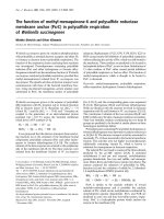
The expression of the thioesterase was also studied by
immunohistochemistry in rat adrenals. Intense fluorescence
was detected throughout the adrenal cortex, while the
medulla was devoid of signal (Fig. 2, panel A). The label
was most intense in the zona fasciculata as compared with
that shown by both zona glomerulosa and reticularis
(Fig. 2, panels C and D). The specificity of the label was
corroborated when the tissue sections were incubated with
preadsorbed primary antibody (Fig. 2, panel B).
CTE-I and MTE-I thioesterase activity
To further characterize the recombinant CTE-I and MTE-I,
we studied the kinetic parameters of the enzymes by
measuring thioesterase activity using [1-
14
C]AA-CoA as
described in Experimental procedures. Expectedly, incuba-
tion of [1-
14
C]AA-CoA in the presence of the purified
enzyme resulted in the release of AA. K
m
and Vmax values
obtained for the reactions were 4.1 and 2 l
M
and 948 and
193 nmolÆmin
)1
Æmg
)1
for CTE-I and a MTE-I, respectively.
Next, we measured the effect of ACTH on mitochondrial
thioesterase activity in Y1 adrenal cells. For that purpose,
mitochondria were isolated from cultures of confluent Y1
cells incubated in the presence or absence of 5 mIUÆmL
)1
ACTH and enzyme activity was determined as AA released
from AA-CoA. ACTH significantly stimulated enzyme
activity in the mitochondrial fraction from 1.59 ± 0.22
to 3.0 ± 0.25 and from 1.67 ± 0.28 to 4.21 ± 0.29 pmol
AAÆmin
)1
for control and ACTH at 5 and 30 min,
respectively. A similar effect in enzyme activity was
observed when the postmitochondrial fraction was used as
a source of enzyme (data not shown).
Effect of NDGA on acyl-CoA thioesterase activity
Although NDGA is commonly used as an inhibitor of the
lipoxygenase pathway, it is also known to inhibit PLA
2
activity [27]. Since NDGA strongly inhibited both steroid
production and StAR protein expression [23,25,26], we
tested here whether NDGA inhibits acyl-CoA thioesterase
Fig. 2. Immunohistochemical analysis of acyl-CoA thioesterases in the
adrenal gland. Tissue sections from rat adrenal gland were incubated
with anti-(CTE-I) (A, C and D) or antibody preadsorbed with purified
CTE-I (B). Specific binding was detected using a cy3-conjugated goat
anti-rabbit IgG as secondary antibody and observed under standard
fluorescence microscopy. Original magnifications are: A and B, · 100;
CandD· 200. Acyl-CoA thioesterase immunoreactivity was found in
the zona fasciculata (f), zona glomerulosa (g) and zona reticularis (r).
No signal was detected in the medulla (m).
Fig. 1. Immunodetection of CTE-I and MTE-I. (A) Samples of purified
recombinant CTE-I (CTE-I) and MTE-I (MTE-I), homogenates from
adrenal zona fasciculata (Fasciculata), mitochondrial (MF) or post-
mitochondrial fractions (PMF) obtained from Y1 cells, as indicated,
were resolved on SDS/PAGE and immunoblotted as described in
Experimental procedures. Antibodies used were: anti-G11K (lanes
1–3), anti-(CTE-I) (lanes 4–7) or antibody preadsorbed with purified
recombinant CTE-I (control) (lanes 8 and 9). (B) Immunocytochemi-
cal detection of acyl-CoA thioesterase in Y1 cells. Cells were fixed and
stained using anti-(CTE-I) serum as described in Experimental pro-
cedures. Specific binding was detected by means of a cy3-conjugated
secondary antibody and fluorescence observed under a standard
epifluorescence microscope (A). (C) Cells stained only with secondary
antibody. (B) and (D), phase contrast micrographs of cells observed in
(A) and (C), respectively. Original magnification · 400.
5602 P. Maloberti et al. (Eur. J. Biochem. 269) Ó FEBS 2002
activity in vitro. Figure 3 shows that a 5-min preincubation
of recombinant protein with NDGA inhibited thioesterase
activity. Interestingly, NDGA produced only a 20%
inhibition of the mitochondrial enzyme activity when
measured in mitochondria isolated from ACTH-stimulated
Y1 cells (data not shown). This later result is in agreement
with the previous observation that NDGA inhibitory effect
is manifested only when the inhibitor is added prior to
ACTH [21].
Role of arachidonoyl-CoA and arachidonoyl-CoA
synthetase on ACTH-stimulated cholesterol metabolism
Next, we tested the effect of NDGA on ACTH-induced
steroid production in Y1 cells. According with the effect
observed in isolated rat zona fasciculata cells [44], NDGA
significantly (P < 0.001) inhibited ACTH-induced steroid
production in a dose-dependent manner (Fig. 4), with an
apparent IC
50
of 50 l
M
.
The expression of an acyl-CoA synthetase specific for
AA, acyl-CoA synthetase 4 (ACS4), in steroidogenic tissues
has been reported [39]. Triacsin C has been described as an
inhibitor of acyl-CoA synthetases with a preferential effect
for AA-CoA synthetases in intact cells [45,46]. Thus, we
investigated a possible concerted regulatory role of acyl-
CoA synthetases and thioesterases in the regulation of
steroidogenesis by using triacsin C and NDGA on ACTH-
stimulated steroid synthesis in Y1 cells.
For this purpose, ACTH-stimulated Y1 cells were treated
with ineffective concentrations of NDGA (5–25 l
M
,Fig.4)
alone or in combination with increasing concentrations of
triacsin C (0.1–2 l
M
) and steroids measured as indicated in
Experimental procedures. Expectedly, NDGA alone had no
effect on ACTH-induced steroid production, while triac-
sin C alone produced a slight but significant (P ¼ 0.0445)
inhibition of steroid output (Fig. 5B). However, NDGA
significantly inhibited steroid biosynthesis when combined
with 0.1, 0.5 and 2 l
M
triacsin C (Fig. 5A). A two-way
analysis of variance rendered a highly significant
(P ¼ 0.0026) value for the combination of NDGA and
triacsin C, thereby indicating a synergistic effect on steroid
production. Moreover, the synergistic effect is evidenced
when the IC
50
for the inhibitors are analysed. Thus, the
apparent IC
50
for NDGA is reduced from 50 l
M
to 25, 7.5
and 4.5 l
M
in the presence of 0.1, 0.5 and 2 l
M
triacsin C,
respectively. The apparent IC
50
for triacsin C is also reduced
from 5.5 l
M
to 1.75, 0.275 and 0.1 l
M
in the presence of 5,
10 and 25 l
M
of NDGA, respectively (Fig. 5B).
The inhibitory effect of both triacsin C and NDGA was
clearly not due to an inhibition of P450
scc
activity since no
significant inhibition of steroid production was observed
when 22R-OH-cholesterol was added to the Y1 cell culture
(data not shown).
These results confirm the participation of ACS4 and of
AA-CoA in the regulation of steroidogenesis in Y1 cells.
DISCUSSION
AA is present in the plasma membrane of most mammalian
cells esterified to phospholipids. Free cytosolic AA can be
produced by the action of phospholipase A
2
or cholesterol
esterase which release the fatty acid by cleavage from
membrane phospholipids or cholesterol esters, respectively
[47]. Previous studies have demonstrated that inhibitors
of PLA
2
activity affect hormone-induced steroidogenesis
[17–19,25]. Those studies have raised the possibility that
PLA
2
could be involved in the mechanism of action of
hormones that control steroid production. However, there
is no direct evidence showing that AA is released by PLA
2
in
steroidogenic tissues.
The present study is the first one to provide evidence for
an alternative pathway of AA generation. Our results are
consistent with the hypothesis that, in steroidogenic cells,
AA is released by the action of an acyl-CoA thioesterase
activity. We show here that the mitochondrial acyl-CoA
thioesterase activity hydrolyses AA-CoA to release free AA
Fig. 3. Effect of NDGA on recombinant thioesterase activity. Activity
of recombinant CTE-I (open bars) and MTE-I (filled bars) was
measured in the presence of increasing doses of NDGA. Acyl-CoA
thioesterase activity was determined using [1-
14
C]AA-CoA as sub-
strate. Results are expressed as the mean ± SD of one representative
experiment performed in triplicate.
Fig. 4. Effect of NDGA on steroid hormone synthesis. Y1 cells were
preincubated with variable concentrations (5–100 l
M
)ofNDGAfor
15 min at 37 °C and further incubated in the absence or the presence of
2mIUÆmL
)1
of ACTH for 60 min. Determination of progesterone
production by RIA and of steroids by fluorometry showed comparable
results. Therefore, data are shown as progesterone production
(ngÆmL
)1
) in the incubation medium. Basal steroid production is
denoted by the dotted line. The effect of NDGA was highly significant
(P < 0.001) by
ANOVA
, and indicated values (*) significantly different
(P < 0.05) by Student-Newman-Keuls post test.
Ó FEBS 2002 Acyl-CoA thioesterases and synthetase on steroidogenesis (Eur. J. Biochem. 269) 5603
and that the activity of the mitochondrial acyl-CoA
thioesterase increases significantly after ACTH stimulation
in Y1 cells. We also demonstrate that inhibitors of AA
release and metabolism such as NDGA, are effective
inhibitors of recombinant CTE-I and MTE-I. The experi-
ment using inhibitors of acyl-CoA synthetases in combina-
tion with NDGA demonstrates that the two enzymes, the
synthetase and the acyl-CoA thioesterase act in concert and
are essential for steroidogenesis.
A possible explanation for the effect of classical PLA
2
inhibitors on thioesterase activity could be the presence of
a serine-histidine-aspartic acid catalytic triad containing ab
hydrolases as determined by site-directed mutagenesis [48].
This possibility is supported by our previous results
showing that antibodies raised against a synthetic peptide
matching a sequence that contains the serine included in
the catalytic triad inhibit steroid synthesis in a recombinant
cell-free assay [32]. These observations are also in agree-
ment with previous results showing that BPB, another
PLA
2
inhibitor, also blocks the activity of CTE-I [44]. In
addition, another specific inhibitor of PLA
2
(100 l
M
ATK)
was also effective to inhibit the activity of the enzyme (data
not shown).
NDGA is known to inhibit the activity of lipoxygenase
by binding to the reduced form (Fe
2+
)oftheenzyme
thereby keeping it inactive [49]. In our experiments NDGA
was effective in inhibiting the activity of purified recombin-
ant MTE-I in an in vitro assay performed in a Hepes-based
buffer containing only KCl. Information about the tertiary
structure and of ion requirements of acyl-CoA thioesterases
is still lacking and the mechanism of inhibition of acyl-CoA
thioesterases by NDGA is unknown. Nevertheless, we
cannot exclude an inhibitory mechanism for the thioesterase
involving electron transfer. Noteworthy, the thioesterase
displays a decreased sensitivity for NDGA after ACTH
stimulation. This latter effect could result from a confor-
mational change of the enzyme induced by protein phos-
phorylation.
Blocking lipoxygenase-mediated AA metabolism by
NDGA, greatly decreased Bt
2
cAMP-induced StAR protein
expression and reduced cholesterol metabolism [25]. How-
ever, in view of our present results, the use of NDGA only
as lipoxygenase inhibitor has to be reviewed since it can also
act as inhibitor of acyl-CoA thioesterases.
Recombinant CTE-I and MTE-I expressed in E. coli
exhibited the expected molecular mass, showed high acyl-
CoA thioesterase activity using AA-CoA as substrate and
were detected by antipeptide antibodies that recognize the
catalytic domain of the purified enzyme from adrenal. In
addition, antibodies raised against recombinant CTE-I
recognize both recombinant proteins and the protein from
cytosol and mitochondria from adrenal cells. Using this
antibody, immunohistochemical experiments show that the
protein is exclusively located in steroidogenic cells of adrenal
gland.
The expression of a cytosolic and a mitochondrial
thioesterase activity with different subcellular localizations
suggests that these enzymes have different functions in vivo,
although the role of CTE-I in steroidogenesis is currently
not known. The high degree of sequence similarity between
the CTE-I and MTE-I genes suggests that they diverged
relatively recently by gene duplication, possibly by duplica-
tion of an MTE-I gene with loss of the 5¢-end encoding the
mitochondrial targeting signal, resulting in a cytosolic
enzyme [35].
In the present study we demonstrate that AA-CoA is an
important intermediate in steroidogenesis. The observation
that triacsin C, an inhibitor of AA-CoA synthetase
[44,45], affects hormone-induced steroid synthesis in Y1
cells supports this hypothesis. The participation of the
AA-CoA-mediated pathway for AA release in steroidogen-
esis was further demonstrated by the combined inhibitory
effect of triacsin C and NDGA. Our results clearly show
that addition of triacsin C in combination with ineffective
doses of NDGA produced a marked reduction of the
IC
50
for NDGA. Therefore, the regulatory role of AA in
Fig. 5. Combined effect of triacsin C and NDGA on steroid hormone synthesis. Y1 cells were preincubated in the presence or the absence of
increasing concentrations of triacsin C (4 h, 37 °C) and of NDGA (15 min, 37 °C).Cellswerethenincubatedfor60minwith2mIUÆmL
)1
of
ACTH or its vehicle. Progesterone produced was determined in the incubation medium by RIA. Data are expressed as the mean ± SD (n ¼ 3)
of different concentrations of triacsin C, 0.1 (d), 0.5 (m), 2 (j) lMorcontrol(r) with indicated concentrations of NDGA (panel A), or variable
concentrations of NDGA, 5 (s), 10 (n), 25 (h) l
M
or control (e) in the presence of triacsin C (panel B). Basal steroid production by Y1 cells is
denoted by the dotted line.
5604 P. Maloberti et al. (Eur. J. Biochem. 269) Ó FEBS 2002
steroidogenesis needs the concerted action of the acyl-CoA
synthetase and thioesterase.
Although we cannot rule out a possible nonspecific effect
of both triacsin C and NDGA, our observation of a
synergistic effect on steroid production suggests this is
unlikely. This, along with the fact that 22R-OH-cholesterol
by-passes the effect of the inhibitors strongly indicates that
the thioesterase and the acyl CoA-synthetase act in the same
signalling pathway in a step prior to the rate-limiting
passage of cholesterol from the outer to the inner mito-
chondrial membrane.
The thioesterase activity requires an acyl-CoA pool as a
source of AA. The presence of an acyl-CoA synthetase
specific for arachidonate described in steroidogenic tissues
suggests that such a mechanism is operable in steroid
biosynthesis. The concept that long chain fatty acyl-CoA
esters are regulatory ligands as well as intermediates in
cellular metabolism is now well appreciated from results of a
number of investigations in a variety of organism and
tissues [50,51].
The question then arises as to why free cytosolic AA has
to be re-esterified in order to stimulate steroidogenesis. One
possible explanation is the need for AA in a special
compartment of the cell (e.g. mitochondria). The compart-
mentalization of long-chain acyl-CoA esters is an important
unsolved problem, and the actual cytosolic concentration of
free long-chain acyl-CoA esters is not known for any tissue
[51]. The high degree of sequestration of CoA into long
chain acyl-CoA suggests that AA is likely to become
limiting for diverse roles in specific compartments of the cell
[51]. Here we show that ACTH stimulates mitochondrial
thioesterase activity in Y1 cells. This points to a direct effect
of ACTH upon enzyme activity. However, we cannot rule
out a possible activation of the enzyme by an ACTH-
mediated increased availability of its substrate.
It is known that an acyl-CoA binding protein (ACBP)
known also as DBI (diazepam binding inhibitor) is
expressed in high concentrations in specialized cells such
as steroid producing cells of the adrenal cortex and testis
[52,53]. Thus it can be proposed that AA-CoA binds to
DBI, which in turns binds to the peripheral benzodiazepine
receptor (PBR) located in the outer mitochondrial mem-
brane [53,54]. This will possibly lead to facilitated transfer of
AA-CoA into the mitochondria.
Another important issue is the origin of cytosolic free AA
to be esterified into AA-CoA. As already mentioned, AA
could derive from plasma membrane phospholipids or from
cholesterol esters. The major source of cholesterol in the rat
adrenal is the cholesterol esterified in high-density lipopro-
teins (HDL) [55,56]. In adrenocortical cells, HDL enhances
steroid production and increases cellular cholesterol con-
tent. Rat HDL contains a high amount of arachidonate in
its cholesterol esters fatty acids. This is an agreement with
the suggestion that free AA, which will be esterified to acyl-
CoA may come from the hydrolysis of cholesterol ester [34].
Nevertheless, since it has been shown that dexamethasone
inhibits cholesterol metabolism and that this effect is
reverted by free AA, we can not rule out the possibility
that the free AA that is esterified into acyl-CoA may come
from membrane phospholipids by the action of PLA
2
.
However, in none of those studies, there is a demonstration
that dexamethasone is in fact working through the inhibi-
tion of PLA
2
.
Our current data indicate the presence of a new pathway
that regulates intracellular levels of AA, in which ACS4
could act by sequestering free AA by esterification into
AA-CoA. CoA-esterified AA may bind ACBP/DBI thus
forming an intracellular pool that could then be delivered to
an acyl-CoA thioesterase, which will, in turn, release AA in
a specific compartment of the cell upon hormone treatment.
Taken together the results shown here demonstrate the
critical role of acyl-CoA synthetases and thioesterases in the
regulation of intracellular levels of AA and steroid produc-
tion. This study further suggests a new concept in the
regulation of intracellular levels of AA, in which trophic
hormones can release AA by a mechanism different than the
classical PLA
2
-mediated pathway.
ACKNOWLEDGEMENTS
Thanks are due to Dr Stefan Alexson for the cytosolic acyl-CoA
thioesterase cDNA. This work was supported by grants from Agencia
Nacional de Promocio
´
nCientı
´
fica y Tecnolo
´
gica, Universidad de
Buenos Aires and Consejo Nacional de Investigaciones Cientı
´
ficas y
Te
´
cnicas.
REFERENCES
1. Crivello, J.F. & Jefcoate, C.R. (1980) Intracellular movement of
cholesterol in rat adrenal cells. J.Biol.Chem.255, 8144–8151.
2. Privalle, C.T., Crivello, J.F. & Jefcoate, C.R. (1983) Regulation of
intramitochondrial cholesterol transfer to side-chain cleavage
cytochrome P450
SCC
in rat adrenal gland. Proc. Natl Acad. Sci.
USA 80, 702–706.
3. Haynes, R.C. Jr, Koritz, S.B. & Peron, F.G. (1959) Influence of
adenosine 3¢,5¢-monophosphate on corticoid production by rat
adrenal glands. J. Biol. Chem. 234, 1421–1423.
4. Cooke, B.A., Lindh, M.L. & Janszen, F.H.A. (1976) Correlation
of protein kinase activation and testosterone production after
stimulation of Leydig cells with luteinizing hormone. Biochem. J.
160, 439–446.
5. Dufau, M., Tsuruhara, T., Horner, K., Podesta
´
,E.J.&Catt,K.
(1977) Intermediate role of adenosine 3¢:5¢-cyclic monophosphate
and protein kinase during gonadotropin-induced steroidogenesis
in testicular interstitial cells. Proc. Natl Acad. Sci. USA 74, 3419–
3423.
6. Podesta
´
, E.J., Dufau, M.L., Solano, A.R. & Catt, K.J. (1978)
Hormonal activation of protein kinase in isolated Leydig cells.
Electrophoretic analysis of cyclic AMP receptors. J. Biol. Chem.
253, 8994–9001.
7. Sala, G., Hayoshi, K., Catt, K. & Dufau, M. (1979) Adreno-
corticotropin action in isolated adrenal cells. The intermediate role
of cyclic AMP in stimulation of corticosterone synthesis. J. Biol.
Chem. 254, 3861–3865.
8. Podesta
´
,E.,Milani,A.,Steffen,H.&Neher,R.(1979)Ster-
oidogenesis in isolated adrenocortical cells. Correlation with
receptor-bound adenosine 3¢:5¢-cyclic monophosphate. Biochem.
J. 180, 355–363.
9. Rae, P., Gutman, U., Tso, J. & Schimmer, B. (1979) Mutations in
cyclic AMP-dependent protein kinase and corticotropin (ACTH)-
sensitive adenylate cyclase affect adrenal steroidogenesis. Proc.
Natl Acad. Sci. USA 76, 1896–1900.
10. Karaboyas, G.C. & Koritz, B.S. (1965) Identity of the site of
action of 3¢,5¢-adenosine monophosphate and adrenocorticotropic
hormone in corticosteroidogenesis in rat adrenal and beef adrenal
cortex slices. Biochemistry 4, 462–468.
11. Lambeth,J.D.,Xu,X.X.&Glover,M.(1987)Cholesterolsulfate
inhibits adrenal mitochondrial cholesterol side chain cleavage
at a site distinct from cytochrome P-450scc. Evidence for an
Ó FEBS 2002 Acyl-CoA thioesterases and synthetase on steroidogenesis (Eur. J. Biochem. 269) 5605
intramitochondrial cholesterol translocator. J. Biol. Chem. 262,
9181–9188.
12. Miller, W.L. (1988) Gene conversions, deletions and polymor-
phisms in congenital adrenal hyperplasia. Am.J.Hum.Genet.42,
4–7.
13. Clark, B.J., Wells, J., King, S.R. & Stocco, D.M. (1994) The
purification, cloning, and expression of a novel luteinizing hor-
mone-induced mitochondrial protein in MA-10 mouse Leydig
tumor cells. Characterization of the steroidogenic acute regulatory
protein (StAR). J. Biol. Chem. 269, 28314–28322.
14. Lin, D., Sugawara, T., Strauss, J.F., III, Clark, B.J., Stocco, D.M.,
Saenger, P., Rogol, A. & Miller, W.L. (1995) Role of steroidogenic
acute regulatory protein in adrenal and gonadal steroidogenesis.
Science 267, 1828–1830.
15. Stocco, D.M. & Clark, B.J. (1996) Regulation of the acute pro-
duction of steroids in steroidogenic cells. Endocrinol. Rev. 17,221–
244.
16. Artemenko, I., Zhao, D., Hales, D.B., Hales, K.H. & Jefcoate,
C.R. (2001) Mitochondrial processing of newly synthesized
steroidogenic acute regulatory protein (StAR), but not total
StAR, mediates cholesterol transfer to cytochrome P450 side
chain cleavage enzyme in adrenal cells. J. Biol. Chem. 276, 46583–
46596.
17. Dix, C., Habberfield, A., Sullivan, M. & Cooke, B. (1984)
Inhibition of steroid production in Leydig cells by non-steroidal
anti-inflammatory and related compounds: evidence for the
involvement of lipoxygenase products in steroidogenesis. Biochem.
J. 219, 529–537.
18. Didolkar, A. & Sundaram, K. (1987) Arachidonic acid is involved
in the regulation of hCG induced steroidogenesis in rat Leydig
cells. Life. Sci. 41, 471–477.
19. Jones, D., Marante, D., Williams, B. & Edwards, C. (1987)
Adrenal synthesis of corticosterone in response to ACTH in rats is
influenced by leukotriene A4 and by lipoxygenase intermediates.
J. Endocrinol. 112, 253–258.
20. Solano, A., Dada, L., Sardan
˜
ons, M., Sa
´
nchez, M. & Podesta
´
,E.J.
(1987) Leukotrienes as common intermediates in the cyclic AMP
dependent and independent pathways in adrenal steroidogenesis.
J. Steroid Biochem. 27, 745–751.
21. Solano, A.R., Dada, L.A. & Podesta
´
, E.J. (1988) Lipoxygenase
products as common intermediates in cyclic AMP-dependent and
independent adrenal steroidogenesis in rats. J. Mol. Endocrinol. 1,
147–154.
22. Mikami, K., Omura, M., Tamura, Y. & Yoshida, S. (1990) Pos-
sible site of action of 5-hydroperoxyeicosatetraenoic acid derived
from arachidonic acid in ACTH-stimulated steroidogenesis in rat
adrenal glands. J. Endocrinol. 125, 89–96.
23. Wang, X. & Stocco, D.M. (1999) Cyclic AMP and arachidonic
acid: a tale of two pathways. Mol. Cell. Endocrinol. 158, 7–12.
24. Wang, H., Walker, S.W., Mason, J.I., Morley, S.D. & Williams,
B.C. (2000) Role of arachidonic acid metabolism in ACTH-
stimulated cortisol secretion by bovine adrenocortical cells.
Endocr. Res. 26, 705–709.
25. Wang, X., Walsh, L.P., Reinhart, A.J. & Stocco, D.M. (2000) The
role of arachidonic acid in steroidogenesis and steroidogenic acute
regulatory (StAR) gene and protein expression. J. Biol. Chem. 275,
20204–20209.
26. Wang, X.J., Dyson, M.T., Mondillo, C., Patrignani, Z., Pignataro,
O. & Stocco, D.M. (2002) Interaction between arachidonic acid
and cAMP signalling pathways enhances steroidogenesis and
StAR gene expression in MA-10 Leydig tumor cells. Mol. Cell.
Endocrinol. 188, 55–63.
27. Kim, D.K. & Bonventre, J.V. (1993) Purification of a 100 kDa
phospholipase A
2
from spleen, lung and kidney: antiserum raised
to pig spleen phospholipase A
2
recognizes a similar form in bovine
lung, kidney and platelets, and immunoprecipitates phospholipase
A
2
activity. Biochem. J. 294, 261–270.
28. Neher, R., Milani, A., Solano, A. & Podesta
´
, E.J. (1982)
Compartmentalization of corticotropin-dependent steroidogenic
factors in adrenal cortex: evidence for a post-translational cascade
in stimulation of the cholesterol side-chain split. Proc. Natl Acad.
Sci. USA 101, 4350–4357.
29. Dada, L., Paz, C., Mele, P., Solano, A., Cornejo Maciel, F.
& Podesta
´
, E.J. (1991) The cytosol as site of phosphorylation of
the cyclic AMP-dependent protein kinase in adrenal steroido-
genesis. J. Steroid Biochem. Mol. Biol. 39, 889–896.
30. Mele, P., Dada, L., Paz, C., Cymeryng, C., Cornejo Maciel, F.,
Neuman, I., Finkielstein, C.F. & Podesta
´
, E.J. (1997) Involvement
of arachidonic acid and the lipoxygenase pathway in mediating
luteinizing hormone-induced testosterone synthesis in rat Leydig
cells. Endocr. Res. 23, 15–26.
31. Paz, C., Dada, L., Cornejo Maciel, F., Mele, P.G., Cymeryng, C.,
Neuman, I., Mendez, C.F., Finkielstein, C., Solano, A., Park, M.,
Fischer, W., Towbin, H., Scartazzini, R. & Podesta
´
, E.J. (1994)
Purification of a novel 43-kDa protein (p43) intermediary in the
activation of steroidogenesis from rat adrenal gland. Eur. J. Bio-
chem. 224, 709–716.
32. Finkielstein,C.,Maloberti,P.,Mendez,C.F.,Paz,C.,Cornejo
Maciel, F., Cymeryng, C., Neuman, I., Dada, L., Mele, P.G.,
Solano, A. & Podesta
´
, E.J. (1998) An adrenocorticotropin-regu-
lated phosphoprotein intermediary in steroid synthesis is similar to
an acyl-CoA thioesterase enzyme. Eur. J. Biochem. 256, 60–66.
33. Svensson, L.T., Endberg, S.T., Aoyama, T., Usuda, N., Alexson,
S.E.H. & Hashimoto, T. (1998) Molecular cloning and
characterization of a mitochondrial peroxisome proliferator-
induced acyl-CoA thioesterase from rat liver. Biochem. J. 329,
601–608.
34. Lindquist, L.G., Svensson, L.T. & Alexson, S.E.H. (1998)
Molecular cloning of the peroxisome proliferator-induced 46-kDa
cytosolic acyl-CoA thioesterase from mouse and rat liver
recombinant expression in Escherichia coli, tissue expression, and
nutritional regulation. Eur. J. Biochem. 251, 631–640.
35. Hunt, M.C., Nousiainen, S.E.B., Huttunen, M.K., Orii, K.E.,
Svenssson, L.T. & Alexson, S.E.H. (1999) Peroxisome pro-
liferator-induced long chain acyl-CoA thioesterases comprise a
highly conserved novel multi-gene family involved in lipid meta-
bolism. J. Biol. Chem. 274, 34317–34326.
36. Hunt, M.C. & Alexson, S.E. (2002) The role of acyl-CoA thio-
esterases play in mediating intracellular lipid metabolism. Prog.
Lipid Res. 41, 99–130.
37. Kim, J.H., Lewin, T.M. & Coleman, R.A. (2001) Expression and
characterization of recombinant rat acyl-CoA synthetases 1, 4 and
5. J. Biol. Chem. 276, 24667–24673.
38. Lewin, T.M., Kim, J.H., Granger, D., Vance, J.E. & Coleman,
R.A. (2001) Acyl-CoA synthetase isoforms 1, 4 and 5 are present
in different subcellular membranes in rat liver and can be inhibited
independently. J. Biol. Chem. 276, 24674–24679.
39. Kang, M., Fujino, T., Sasano, H., Minekura, H., Yabuki, N.,
Nagura, H., Iijima, H. & Yamamoto, T. (1997) A novel arachi-
donate-preferring acyl-CoA synthetase is present in steroidogenic
cells of the rat adrenal, ovary, and testis. Proc. Natl Acad. Sci.
USA 94, 2880–2884.
40. Cho, Y.Y., Kang, M.J., Ogawa, S., Yamashita, Y., Fujino, T. &
Yamamoto, T.T. (2000) Regulation by adrenocorticotropic hor-
mone and arachidonate of the expression of acyl-CoA synthetase
4, an arachidonate-preferring enzyme expressed in steroidogenic
tissues. Biochem. Biophys. Res. Commun. 274, 741–745.
41. Schimmer, B.P. (1979) Adrenocortical Y1 cells. Methods Enzymol.
58, 570–574.
42. Bradford, M.M. (1976) A rapid and sensitive method for the
quantitation of microgram quantities of protein utilizing the
principle of protein-dye binding. Anal. Biochem. 72, 248–254.
43. Stocco, D.M. & Sodeman, T.C. (1991) The 30-kDa mitochondrial
proteins induced by hormone stimulation in MA-10 mouse Leydig
5606 P. Maloberti et al. (Eur. J. Biochem. 269) Ó FEBS 2002
tumor cells are processed from larger precursors. J. Biol. Chem.
266, 19731–19738.
44. Maloberti, P., Mele, P.G., Neuman, I., Cornejo Maciel, F., Cano,
F., Bey, P., Paz, C. & Podesta
´
, E.J. (2000) Regulation of arachi-
donic acid release in steroidogenesis: role of a new acyl-CoA
thioesterase (ARTISt). Endocr. Res. 26, 653–662.
45. Tomoda, H., Igarashi, K. & Omura, S. (1987) Inhibition of
acyl-CoA synthetase by triacsins. Biochim. Biophys. Acta 921,
595–598.
46. Hartman, E.J., Omura, S. & Laposata, M. (1989) Triacsin C: a
differential inhibitor of arachidonoyl-CoA synthetase and non-
specific long chain acyl-CoA synthetase. Prostaglandins 37, 655–
671.
47. Irvine, R.F. (1982) How is the level of free arachidonic acid con-
trolled in mammalian cells? Biochem. J. 204, 3–16.
48. Huhtinen, K., O’Byrne, J., Lindquist, P.J., Contreras, J.A.
& Alexson, S.E. (2002) The peroxisome proliferator-induced
cytosolic type I acyl-CoA thioesterase (CTE-I) is a serine-
histidine-aspartic acid alpha/beta hydrolase. J. Biol. Chem. 277,
3424–3432.
49. Kemal, C., Louis-Flamberg, P., Krupinski-Olsen, R. & Shorter,
A. (1987) Reductive inactivation of soybean lipoxygenase-1 by
catechols. Biochemistry 26, 7064–7072.
50. Shrago, E., Woldegiorgis, G., Ruoho, A.E. & Di Russo, C.C.
(1995) Fatty acyl-CoA esters as regulators of cell metabolism.
Prostaglandins Leukot. Essent. Fatty Acids 52, 163–167.
51. Faergeman, N.J. & Knudsen, J. (1997) Role of long-chain fatty
acyl-CoA esters in the regulation of metabolism and in cell
signalling. Biochem. J. 323, 1–12.
52. Knudsen, J., Hojrup, P., Hansen, H.O., Hansen, H.F. & Roep-
storff, P. (1989) Acyl-CoA-binding protein in the rat. Purification,
binding characteristics, tissue concentrations and amino acid
sequence. Biochem. J. 262, 513–519.
53. Papadopoulos, V. (1993) Peripheral-type benzodiazepine/diaze-
pam binding inhibitor receptor: biological role in steroidogenic cell
function. Endocr. Rev. 14, 222–240.
54. Papadopoulos, V. (1998) Structure and function of the peripheral-
type benzodiazepine receptor in steroidogenic cells. Proc. Soc.
Exp. Biol. Medical 217, 130–142.
55. Gwyne, J.T. & Hess, B. (1980) The role of high density lipopro-
teins in rat adrenal cholesterol metabolism and steroidogenesis.
J.Biol.Chem.255, 10875–10883.
56. Andersen, J.M. & Dietschy, J.M. (1981) Kinetic parameters of
the lipoprotein transport systems in the adrenal gland of the rat
determined in vivo.Comparisonoflowandhighdensitylipopro-
teins of human and rat origin. J. Biol. Chem. 256, 7362–7370.
Ó FEBS 2002 Acyl-CoA thioesterases and synthetase on steroidogenesis (Eur. J. Biochem. 269) 5607