Báo cáo y học: " Conserved charged amino acid residues in the extracellular region of sodium/iodide symporter are critical for iodide transport activity" potx
Bạn đang xem bản rút gọn của tài liệu. Xem và tải ngay bản đầy đủ của tài liệu tại đây (1.07 MB, 9 trang )
RESEARC H Open Access
Conserved charged amino acid residues in the
extracellular region of sodium/iodide symporter
are critical for iodide transport activity
Chia-Cheng Li
1†
, Tin-Yun Ho
1†
, Chia-Hung Kao
2
, Shih-Lu Wu
3
, Ji-An Liang
4
, Chien-Yun Hsiang
5*
Abstract
Background: Sodium/iodide symporter (NIS) mediates the active transport and accumulation of iodide from the
blood into the thyroid gland. His-226 located in the extracellular region of NIS has been demonstrated to be
critical for iodide transport in our previous study. The conserved charged amino acid residues in the extracellular
region of NIS were therefore characterized in this study.
Methods: Fourteen charged residues (Arg-9, Glu-79, Arg-82, Lys-86, Asp-163, His-226, Arg-228, Asp-233, Asp-237,
Arg-239, Arg-241, Asp-311, Asp-322, and Asp-331) were replaced by alanine. Iodide uptake abilities of mutants were
evaluated by steady-state and kinetic analysis. The three-dimensional comparative protein structure of NIS was
further modeled using sodium/glucose transporter as the reference protein.
Results: All the NIS mutants were expressed normally in the cells and targeted correctly to the plasma membrane.
However, these mutants, except R9A, displayed severe defects on the iodide uptake. Further kinetic analysis
revealed that mutations at conserved positively charged amino acid residues in the extracellular region of NIS led
to decrease NIS-mediated iodide uptake activity by reducing the maximal rate of iodide transport, while mutations
at conserved negatively charged residues led to decrease iodide transport by increasing dissociation between NIS
mutants and iodide.
Conclusions: This is the first report characterizing thoroughly the functional significance of conserved charged
amino acid residues in the extracellular region of NIS. Our data suggested that conserved charged amino acid
residues, except Arg-9, in the extracellular region of NIS were critical for iodide transport.
Background
Sodium/iodide symporter (NIS) is a transmembrane glyco-
protein that is functionally expressed in thyroids, salivary
glands, gastric mucosa, and lactating mammary glands [1].
NIS mediates the active transport of iodide into the folli-
cular thyroid cells and, in turn, concentrates iodide in the
thyroid glands. The ability of cancerous thyroid cells to
actively transport iodide via NIS has provided a unique
and effective delivery system for the detection and destruc-
tion of these cells with radioiodide [2].
NIS is a member of solute-sodium symp orters. Solute-
sodium symporters are a large family of proteins that co-
transport sodium ions with sugars, amino acids, vitamins,
or iodide [3,4]. So far, more than 250 members of solute-
sodium symporters family have been identified, and sev-
eral members, including NIS, human sodium/glucose
transporter (hSGLT), Vibrio parahaemolyticus SGLT
(vSGLT) a nd Escherichia coli (E. coli) proline symporter,
have been well characterized [2-4]. NIS as well as other
members transport sodium and solute via an alternating
access mechanism with tight coupling betw een sodium
and solute transport [5-7]. However, the absence of struc-
tural/functional data of NIS may be difficult to explain this
hypothesis.
NIS mutations detected in patients with congenital
iodide transport defect (ITD) have provided the signifi-
cant structural/functional information about NIS. Twelve
ITD-causing NIS mutations, which are situated in the
transmembrane or intracellular segments of NIS, have
* Correspondence:
† Contributed equally
5
Department of Microbiology, China Medical University, Taichung 40402,
Taiwan
Full list of author information is available at the end of the article
Li et al. Journal of Biomedical Science 2010, 17:89
/>© 2010 Li et al; licensee BioMed Central Ltd. This is an Open Access article distributed under the terms of the Creative Commons
Attribution License ( es/by/2.0), which permits unrestricted use, distribution, and reproduction in
any medium, provided the original work is properly cited.
been characterized so far: V59E, G93R, Q267E, C272X,
G395R, T354P, frame-shift 515X, Y531X, G543E,
ΔM143-Q323, and ΔA439-P443 [2,8]. Mutations at the
highly conserved serine and threonine residues in the
transmembrane segment IX have shown that Thr-351,
Ser-353, Thr-354, Ser-356, and Thr-357 play a key role in
sodium/iodide co-transport [9]. Phosphorylation sites
(Ser-43, Thr-49, Ser-227, Thr-577, and Ser-581 ) of NIS
have been identified to b e important for NIS protein sta-
bility and function [10]. In addition, His-226 located in
the extracellular region of NIS is critic al for iodide trans-
port in our previous study [11]. Moreover, deletion in the
region spanning residues 233-280 of NIS loses the iodide
uptake activity [12]. In this study, we elucidated the
importance of 14 conserved charged amino acid residues,
which were located in th e extracellular region of NIS, by
site-directed mut agenesis and kinetic analysis. Our find-
ings indicated that a ll mutants, except R9A, displayed
severe defects on the iodide uptake. Moreov er, mutations
at positively charged amino acid residues led to the
decrease in Vmax, while mutations at negatively charged
residues resulted in the increase in Km. Our data sug-
gested that conserved charged amino acid residues,
except Arg-9, in the extracellular region of NIS were cri-
tical for iodide transport.
Methods
Cloning and site-directed mutagenesis
Human NIS cDNA was cloned as described previously
[11]. Briefl y, two overlapping cDNA fragments represent-
ing either the 5’ -half or the 3’ -half of the complete NIS
coding region were amplified and inserted into pBlue-
script®II KS (-) vector to create pBKS-NIS-5’ and pBKS-
NIS-3’ plasmids, respectively. A full-length NIS clone was
then const ructed by in-frame fusion of both halves using
auniqueBgl II site in the overlap of the fragments. Site-
directed mutagenesis was performed as described pre-
viously [13]. Briefly, uracil -containing single-stranded
DNA (ssDNA) was prepared by transforming pBKS-NIS-
5’ into E. coli CJ236 strain. Uracil-containing ss DNA was
annealed with 5’-kinase primer, the second-stranded
DNA was synthesized, and the double-stranded DNA
was then transformed into E. coli NM522 strain to allow
the mutated strand to be amplified. The full-length NIS
mutant clones were subcloned into pcDNA3.1 expression
vector (Invitrogen, SanDiego, CA) to create pcDNA3.1-
NIS plasmid DNA. The primers for the construction of
NIS mutants ar e shown in Additional File 1; Table S1.
All t he mutants created in this study were confirmed by
sequencing (Additional File 1; Table S2).
Cell culture and transient transfection
Human hepatoblastoma HepG2 cell line was maintained
in Dulbecco’s modified Eagle’s medium (DMEM) (Life
Technologies, Gaithersburg, MD) supplemented with
10% f etal bovine serum (HyClone, Logan. U T). HepG2
cells were transiently transfected with pcDNA3.1-NIS
wild-type, pcDNA3.1-NIS mutants, pcDNA3.1, or
pcDNA3.1 /lacZ by SuperFect® transfection reagent (Qia-
gen Inc., Valencia, CA). Transfected cells were then
kept in a humidified incubator at 37°C with 5% CO
2
for 24 h.
Total RNA extraction and reverse transcription-
polymerase chain reaction (RT-PCR)
RNA extraction and RT were performed as described pre-
viously [11]. RNA integrity was electrophoretically verified
by both the ethidium bromide staining and the absorption
ratio (OD260/OD280 > 1.95). RT mixtures were subjected
to PCR to measure the mRNAs of NIS and b-actin. PCR
amplification was performed with Taq polymerase (Pro-
mega, Madison, WI) for 20 cycles at 94°C for 45 s, 50°C for
45 s, and 72°C for 1 min. PCR primers for NIS were as
follows: sense, 5’-CTCCTCCCTGCTAACGACTC-3’; anti-
sense, 5’-CGACCACCATCATGTCCAAC-3’;PCRprimers
for b-actin were as follows: sense, 5’-TGACGGGGTCACC
CACACTGTGCCCATCTA-3’;antisense,5’-CTAGAAGC
ATTGCGGTGGACGATGGAGGG-3’.
Western blot analysis
The cellular proteins (10 μg) were separated b y 10%
sodium dodec yl sulfate-polyacrylamide gel electrophor-
esis, and the protein bands were then transferred elec-
trophoretically to nitrocellulose membranes. Membranes
were blocked in blocking buffer (20 mM Tris-HCl, pH
7.6, 140 mM NaCl, 0.1% Tween 20, and 5% skim milk)
and probed with mouse monoclonal antibody against
NIS (Lab Vision, Fremont, CA) or rabbit polyclonal
antibody ag ainst b-actin (Santa Cruz, Santa Cruz, CA).
The bound antibo dy was d etected with peroxidase-con-
jugated a nti-mouse or anti-rabbit antibody follo wed by
enhanced chemiluminescence system (Amersham, Chal-
font St. Giles, Buckinghamshire, UK) and exposed by
autoradiography.
Immunofluorescent staining
HepG2 cells were seeded in 24-well plates containing
sterilized coverslips, incubated at 37°C for 2 days, and
transiently t ransfected with DNAs. One day later, cells
were washed twice with phosphate-buffered saline (PBS)
(137 mM NaCl, 1.4 mM KH
2
PO
4
,4.3mMNa
2
HPO
4
,
2.7 mM KCl, pH 7.2), fixed with 3.7% PBS-buffered for-
maldehyde for 30 min at room temperature, and washed
three times with PBS. Coverslips were then incubated
with mouse anti-NIS monoclonal antibody overnight at
4°C, washed three times with PBS, and incubated wit h
fluorescein-conjugated goat anti-mouse IgG antibody
(Jackson ImmunoResearch, West Grove, PA) for 2 h at
Li et al. Journal of Biomedical Science 2010, 17:89
/>Page 2 of 9
37°C. Coverslips we re mounted and e xamined using a
confocal microscope (Leica, Germany), with an excita-
tion wavelength of 488 nm. Anti-NIS monoclonal anti-
body was against residues 625 to 643 mapping to the
carboxyl terminus of human NIS.
Iodide uptake and reporter assays
For steady-state analysis, cells were incubated for 1 h
with 10.2 μCi/ml carrier-free Na
125
Iin1mlDMEMat
37°C. For the inhibition of NIS-mediated uptake,
NaClO
4
, in a final concentration of 30 μM, was included
in parallel incubations. After a 1-h incubation, m edium
was completely removed and washed twice with 2 ml
ice-cold PBS. After washing, the cells w ere lysed w ith
350 μl Tr iton lysis buffer (50 mM Tris-HCl, pH 7.8, 1%
Triton X-100, 1 mM dithiothreitol). Radioactivities of
lysatesweredeterminedbyaCobraIIauto-gamma
counter (Packard BioScience, Dreieich, Germany). b-
Galactosidase activities of cell lysates were analyzed by
mixing cell lysates with O-nitrophenyl-b-D-galactopyra-
noside. After a 30-min incubation at 37°C, the absor-
bance values of the mixtures were measured at 420 nm.
For kinetic analysis, cells were incubated for 4 min with
6.25, 12.5, 25, 50, and 100 μM NaI, and uptake reactions
were determined as described aforementioned. Data were
processed using the equation: v =(Vmax × [I])/(K m+
[I]) + 0.0156 × [I] + 2.4 588. The terms 0.0156 × [I] +
2.4588 correspond to background adjusted by least
squares to the data obtained with non-transfected cells.
Molecular modeling
The three-dimensional comparative protein struc ture of
NIS was modeled using vSGLT (PDB ID: 3dh4) as the
reference protein. Protein structure was built using
SWISS-MODEL workspace [14]. ‘ Frankenstein’smon-
ster’ approach was applied to refinement of the NIS
structure [15].
Statistical analysis
Data were presented as m ean ± standard error. Stu-
dent’s t test was used for comparisons between groups.
A p value < 0.05 was considered to be statistically
significant.
Results
Characterization of expression and plasma membrane
targeting of wild-type and mutated NIS proteins in
HepG2 cells
The current NIS secondary structure model depicts NIS
as a protein with 13 transmembrane segments [16].
Multiple alignments of NIS amino acid sequences from
human, pig, mouse, and rat showed that 14 charged
residues (Arg-9, Glu-79, Arg-82, Lys-86, Asp-163, His-
226, Arg-228, Asp-233, Asp-237, Arg-239, Arg-241,
Asp-311, Asp-322, and Asp-331) were highly conserved
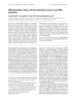
among NIS analogs (Additional File 1; Fig. S1). Addi-
tionally, all of these charged residues were located on
the extracellular region of NIS (Figure 1). Therefore, 14
conserved charged residues were then replaced with
noncharged amino acid, alanine, by site-directed
mutagenesis.
To verify the expression levels and plasma membrane
targeting of NIS mutants, HepG2 cells were transiently
transfected with wild-type or mutated NIS DNAs.
Twenty-four hours later, the mRNA level, protein level,
and plasma membrane targeting of NIS were evaluated
by RT-PCR, Western blot, and Immunofluorescent stain-
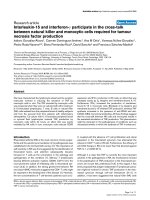
ing, respectively. As shown in Figure 2A, no apparent
difference of mRNA level was found in HepG2 cell
expressing either wild type or mutants. By using mouse
monoclonal antibody aga inst the C-terminus of NIS,
mutated NIS-expressing cells displayed the similar
protein amount and plasma membrane-associated immu-
nofluorescence staining pattern with wild-type NIS-
expressing cells (Figures 2B and 2C). These findings
indicated that NIS mutants were expressed normally in
the cells and targeted correctly to the plasma membrane.
Iodide uptake activities of NIS mutants
HepG2 cells were transiently transfected with pcDNA3.1/
lacZ and pcDNA 3.1, wild-type, or mutated NIS DNAs.
Twenty-four hours later, the iodide uptake activity was
analyzed by steady-state iodide uptake assay and the
transfection efficiency was monitored by b-galactosidase
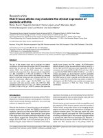
assay. As shown in Figure 3, wild-type NIS-expressing
cells exhibited a significant highly iodide uptake activity.
Perchlorate treatment led to a markedly decrease in
iodide uptake , suggesting the specificity of iodide uptake
assay. Mutation at Arg-9 displayed no defect on the
iodide uptake activity, suggesting that Arg-9 was not
involved in the iodide transport of NIS. However, repla-
cement of other charged amino acid residues with ala-
nine resulted in a large decrease in iodide uptake activity.
b-Galactosidase activities were consistent in w ild-type
and mutated NIS-expressing cells, indicatin g that the
dramatic reduced iodide uptake activities resulted from
the amino acid substitution instead of transfection varia-
tion. These findings suggested that conserved charged
amino acid residues, except Arg-9, in the extracellular
region of NIS were critical for iodide transport.
Kinetics analysis of NIS mutants
We further analyzed the kinetic properties of iodide
uptake in HepG2 cells expressing wild-type or mutated
NIS. Initial rates were assessed by measuring iodide
accumulation at 4-min time points over a range of 6.25,
12.5, 25, 50, and 100 μM NaI (Figure 4). Typical Michae-
lis-Menten kinetic was used to determine the Vmax and
Li et al. Journal of Biomedical Science 2010, 17:89
/>Page 3 of 9
Km values of NIS. The transfection efficiency was also
monitored by b-galactosidase assay. b-Galactosidase
activities were consistent in wild-type and mutated NIS-
expressing cells, indicating that the transfection efficien-
cies were consistent in wild-type and mutants (Additional
File 1; Fig. S2).A comparison of kinetic parameters for
wild type and mutants is shown on T able 1. Because R9A
displayed no defect on the iodide uptake activity, we did
not elucidate the role of Arg-9 further. Replacement of
positively charged residues (Arg-82, Lys-86, His-226,
Arg-228. Arg-239, and Arg-241) by alanine resulted in a
dramatic reduction in Vmax. Howe ver, mutations at
negatively charged residues, except Asp-331, led to a
slight change in Vmax. These findings indicated that
Asp-331- and basic residues-altered mutants displayed a
lower turnover rate . Replacement of Arg-239, Asp-163,
Asp-233, Asp-237, and Asp-322 with alanine resulted in
a s ignificant increase in Km. However, mutation at Arg-
82 showed a markedly decrease in Km. Replacement of
other residues with alanine led t o slight alternation in
Km. These findings indicated that the dissociation of the
Michaelis complex between mutants (R239A, D163A,
D233A, D237A, and D322A) and iodide was larger than
that of wild-type NIS, while the dissociation between
R82A mutant and iodide was smaller than that of wild-
type NIS.
Discussion
Mutations at the amino acid residues in the transmem-
brane or intracellular segments of NIS have identified
the roles of these residues on the iodide transport. For
examples, mutations at Val-59 in the transmembran e
segment II and Gln-267 in the intracellular loop hav e
led to severe defects on the iodide uptake [17,18]. Muta-
tions at the highly conserved serine and threonine resi-
dues in the transmembrane segment IX and intracellular
loops have revea led that these residues play key roles in
the sodiu m/iodide co-transport [9,10]. In addition to the
amino acid residues in the transmembrane or intracellu-
lar segments, some studies have shown that extracellular
loops play essential roles for the ion transport in other
transporters, such as apical sodium-dependent bile acid
transporter, serotonin transpor ter, sodium pump alpha
subunit, and chloride/bicarbonat e anion exchanger
[19-23]. Therefore, herein we analyzed the critical roles
of amino acid residues i n the extracellular segments of
NIS,andourfindingsindicatedthattheseresidues
affected the iodide transport via various mechanisms.
E79
R9
D163
R228
R82
K86
D311
D322
D331
H226
D233
D237
R239 R241
N
H
3
+
COO
-
Extracellular
Intracellular
Figure 1 Schematic representation of NIS secondary structure model. The schematic diagram shows the predicted secondary structure of
NIS. The commonly accepted topological model of NIS shows 13 transmembrane helices with N terminus located extracellularly and C terminus
located intracellularly. Transmembrane segments are represented by cylinders. Positions of 14 amino acid residues mutated in this study are
indicated by arrows.
Li et al. Journal of Biomedical Science 2010, 17:89
/>Page 4 of 9
Charged amino acid residues of some transporters
have been shown to be involved in ion transport. For
examples, charged residues of kidney electrogenic
sodium-bicarbonate cotransporter are involved in ion
recognition in putative outward-facing and inward-
facing conformation [24]. Histidine residues of E. coli
Na
+
/H
+
exchanger NhaA and Ar abidopsis cation/H
+
exchanger are important for ion transport [22,25].
(
A
)
(C)
(B)
ȕ-actin
NIS
wt R9A R82A K86A H226A R228A R239A R241A
E79A D163A D233A
D237A D311A D322A D331A
NIS
ȕ-actin
wt R9A R82A K86A H226A R228A R239A
NIS
ȕ-actin
R241A E79A D163A D233A D237A D311A D322A D331A
ȕ-actin
NIS
w
t
R9A
R82A K86A H226A
D233A D237A D311A D322A D331A
R228A R239A R241A E79A D163A
Blank Mock
Figure 2 Expression and plasma membrane targeting of NIS mutants. (A) RT-PCR. HepG2 cells were cultured in 25-cm
2
flasks and
transfected with wild-type (wt), R9A, E79A, R82A, K86A, D163A, H226A, R228A, D233A, D237A, R239A, R241A, D311A, D322A, or D331A plasmid
DNAs. Total RNAs were extracted and 1 μg of total RNA was reverse transcribed. The resulting cDNAs were then amplified by PCR. PCR products
were resolved in 1% agarose gels and visualized with ethidium bromide. (B) Western blot. HepG2 cells were cultured in 25-cm
2
flasks and
transfected with wt or mutated NIS DNAs. The NIS and b-actin proteins in the cellular lysates were detected by Western blot. (C)
Immunofluorescent staining. HepG2 cells were cultured on glass coverslips and transfected without (blank) or with pcDNA3.1 (mock), wt, or
mutated NIS DNAs for 2 days. Cells were then treated with anti-NIS antibody, stained with fluorescence-conjugated secondary antibody, and
evaluated under a confocal microscope. Magnification, 400×. Similar results were obtained in three independent experiments.
0
0.2
0.4
0.6
0.8
1
1
.
2
M
oc
k
wt
R
9
AR
8
2A K
86
AH22
6
AR22
8
AR2
39
A R241A E
79
AD1
63
AD2
33
AD2
37
AD
3
11A D
3
22A D
33
1A
Relative iodide uptake
0.0
0.2
0.4
0.6
0.8
1.0
O
D420
w/o NaClO4
w/ NaClO4
Galactosidase assay
Figure 3 Iodide uptake activities of NIS mutants. HepG2 cells were transfected with pcDNA3.1/lacZ and pcDNA3.1 (mock), wild-type (wt),
R9A, E79A, R82A, K86A, D163A, H226A, R228A, D233A, D237A, R239A, R241A, D311A, D322A, or D331A DNAs. Twenty-four hours later, iodide
uptake abilities and b-galactosidase activities were determined as described in Materials and Methods. Iodide uptake abilities are expressed as
relative iodide uptake, which is present as the comparison with the radioactivity relative to wt. b-Galactosidase activities are expressed as OD420.
Values are mean ± standard error of triplicate assays.
Li et al. Journal of Biomedical Science 2010, 17:89
/>Page 5 of 9
Mutation at histidine residues of Na
+
/bicarboxylate co-
transporter leads t o a decrease in succinate transport
[26]. Histidine residues of human proton-coupled folate
transporter SLC46A1 play an important role in
SLC46A1 protonation [27]. Moreover, His-226 is critical
for the iodide uptake activity of NIS [13]. Furthermore,
arginine residues of organic anion transporter 1 influ-
ence the binding of glutarate and interact with chloride
[28]. Arg-211 residue of rabbitproton-coupledpeptide
transporter PepT1 plays an intriguing role in the function
of PepT1 [29]. In this study, we replaced the conserved
charged amino acid residues with alanine and found that,
(A)
0
200
400
600
800
1000
0 20406080100
Iodide concentration (ȝM)
Iodide uptake (cpm/4 min)
Mock
wt
R82A
K86A
H226A
R228A
R239A
R241A
(B)
0
300
600
900
1200
020406080100
Iodide concentration (
ȝ
M)
Iodide uptake (cpm/4 min)
Mock
wt
E79A
D163A
D233A
D237A
D311A
D322A
D331A
wt
K86A
R228A
R82A
H226A
R241A
R239A
Mock
D163A
D311A
E79A
D237A
wt
D233A
D322A
D331A
Moc
k
Figure 4 Kinetic analysis of NIS mutants. HepG2 cells were transfected with pcDNA3.1 (mock), wild-type (wt), or mutated NIS DNAs. After 24
h, initial rates (4 min time points) of iodide uptake were determined at the indicated concentrations of iodide. Calculated curves were
generated using the equation v =(Vmax × [I])/(Km + [I]) + 0.0156 × [I] + 2.4588. The terms 0.0156 × [I] + 2.4588 correspond to background
adjusted by least squares to the data obtained with non-transfected cells. Values are mean ± standard error of triplicate assays.
Li et al. Journal of Biomedical Science 2010, 17:89
/>Page 6 of 9
except Arg-9, all the m utants displayed severe defects on
iodide transport. Kinetic analysis revealed that all
mutants mutated at the positively charged amino acids
showed a dramat ic reduct ion in Vmax, while most of the
mutants mutated at the negatively charged residues dis-
played an increase in Km. These findings suggested that
mutations at conserved basic amino acid residues in the
extracellular segments of NIS led to decrease NIS-
mediated iodide uptake activity by reducing the maximal
rate of iodide transport, while mutations at the conserved
acidic amino acid residues led to decrease iodide trans-
port by increasing dissociation between mutants and
iodide. Additionally, mutants in t his study displayed
reduced iodide uptake activities, suggesting that muta-
tions at the extracellular region m ay lead to the lethal
effect in vivo.ThisspeculationmayexplainwhyNIS
mutations in patients with ITD are all located in the
transmembrane and intracellular segments, but not in
the extracellular domain.
To explain why these conserved amino acid residues
affected the iodide transport, we built the three-dimen-
sional structure of NIS using vSGLT as a tem plate pro-
tein. NIS has a sequence identity of 21.8% (37.6%
similarity) to vSGLT (Additional File 1; Fig. S3). NIS
and vSGLT are the members of solute-sodium sympor-
ters that co- transport sodium ions with sugars or iodide
ions. Moreover, both share an alternating-access
mechanism with tight coupling between sodium ion and
solute transport [30]. The recognized homology sug-
gested t hat using vSGLT as the template for the model-
ing of NIS is reasonable. The three-dimensional
structure of NIS (residu es 50-443) is shown on Figure 5.
The proposed structure of NIS contained transmem-
brane helices in an inward-facing conformation. Amino
acid residues mutated in this study were located on the
extracellular segments, as expected. Interestingly, posi-
tively charged amino acid residues were situated on one
side. Structure viewed from the extracellular side dis-
played the core structure of NIS (Figure 5B). Glu-79,
Arg-82, Lys-86, His-226, Arg-228, and Asp-237 were
localized around the core. Glu-79, Arg-82, and Asp-237
were localized on one side of the core. Mutations at
these residues affected the Km values, suggesting that
these amino acid residues might influence the binding
of iodide ions. Lys-86, His-226, and Arg-228 were situ-
ated on the other side of the core. Mutations at these
residues altered the Vmax values, suggesting that these
residues might be involved in the transport of iodide
ions. Asp-233, Arg-239, and Arg-241 were also situated
around t he core. However, the side chains of t hese resi-
dues were exposed to the surface. Mutations at Asp-233
and Arg-239 affected the Km values, suggesting that
both residues might influence the e ntry or binding of
the iodide ions. Asp-163, Asp-311, and Asp-331 were
situated far from the core, and the side chain of Asp-
163 was extruded into the surface. Because mutation at
Asp-163 altered the Km dramatically, Asp-163 might
affect the entry or binding of iodide ions. It is interest-
ing to find that residues (Glu-79, Arg-82, Asp-233, A sp-
237, Arg-239, and Arg-241) involved in the entry or
binding of iodide ions were situated on one side of the
core, while residues (Lys-86, His-226, and Arg-228)
involved in the iodide transport were localized on the
other side (Figure 5C). These findings suggested that
iodide ions might be attracted by residues on one side
of the core and then transported by residues on the
other side. Previous study has shown that five hydroxyl-
containing residues (Thr-351, Ser-353, Thr-354, Ser-356,
and Thr-357) and Asn-360 play a k ey role in sodium/
iodide co-transport [9]. These residues are situated
along one face of transmembrane segment IX and
located along the cavity might explain why these resi-
dues are critical for iodide transport.
Conclusions
In conclusion, we have characterized the roles of 14 con-
served charged amino acid residues located in the extra-
cellular regions of NIS. We have shown that mutation at
these charged amino acid residues, except Arg-9, led to
the severe defects on the iodide uptake. Moreover, kinetic
analysis has shown that mutations at positively charged
residues led to decrease iodide uptake activity by redu-
cing the maximal rate of iodide transport, while muta-
tions at negatively charged residues led to decrease
iodide transport b y increasing dissociation between
mutants and iodide. This is the first report cha racterizing
thoroughly the functional significance of conserved
charged amino acid residues in the extracellular region of
Table 1 Kinetic analysis of human NIS mutants
NIS mutants Vmax
a
Km
a
Wild type 7.94 ± 0.2 81.35 ± 8.89
R82A 2.46 ± 0.31*** 32.87 ± 5.58**
K86A 5.49 ± 0.61** 68.97 ± 14.08
H226A 2.02 ± 0.48*** 71.37 ± 12.43
R228A 4.67 ± 0.45*** 91.8 ± 7.2
R239A 2.42 ± 0.45*** 171.23 ± 36.12**
R241A 2.15 ± 0.14*** 67.64 ± 14.69
E79A 8.55 ± 0.46 110.42 ± 39.87
D163A 9.46 ± 3.1 166.51 ± 37.9*
D233A 8.37 ± 1.6 207.49 ± 72.65*
D237A 7.93 ± 0.52 133.46 ± 43.93*
D311A 6.37 ± 0.69 59.89 ± 16.84
D322A 9.45 ± 1.11 496.6 ± 67.35***
D331A 3.98 ± 0.72*** 91.27 ± 15.32
a
Values are mean ± standard error of triplicate assays.
*p < 0.05, **p < 0.01, ***p < 0.001, compared with wild type.
Li et al. Journal of Biomedical Science 2010, 17:89
/>Page 7 of 9
NIS. Additional structural data are required to elucidate
the complete mechanism of iodide transport of NIS.
Additional material
Additional file 1: Supplementary Information. Table S1: DNA
oligonucleotides for the construction of human NIS mutants. Table S2:
Sequencing analysis of NIS mutants. Figure S1: Multiple alignments of NIS
homologs. Amino acid sequences of NIS from mouse, rat, and pig were
aligned with those of human by ClustalW . Residues
that are identical in all NIS homologs are indicated by asterisks. Residues
that are located on the extracellular region are highlighted in grey.
Amino acid residues mutated in this study are indicated in red. Figure S2:
b-Galactosidase activities of NIS mutants. HepG2 cells were transfected
with pcDNA3.1 (mock), wild-type (wt), or mutated NIS DNAs. After 24 h,
b-galactosidase activities were determined as described in Materials and
Methods. b-Galactosidase activities are expresse d as OD420. Values are
mean ± standard error of triplicate assays. Figure S3: Amino acid
sequence alignment and secondary structure of human NIS. Amino acid
sequences of NIS were aligned with those of vSGLT by ClustalW.
Residues that are identical in both proteins are indicated by asterisks.
Amino acid residues mutated in this study are highlighted in red. The a-
helices of vSGLT are indicated by arrows. The dashed lines represent
amino acid segments that were not visualized in the crystal structure of
vSGLT.
Acknowledgements
This work was supported by National Science Council (NSC95-2320-B-039-
046, NSC97-2320-B-039-012-MY3, NSC98-2320-B-039-030-MY2, and NSC98-
2324-B-039-004), Committee on Chinese Medicine and Pharmacy at
Department of Health (CCMP97-RD-201), and China Medical University
(CMU99-S-06 and CMU99-S-31).
Author details
1
Graduate Institute of Chinese Medicine, China Medical University, Taichung
40402, Taiwan.
2
Department of Nuclear Medicine, China Medical University
Hospital, Taichung 40447, Taiwan.
3
Department of Biochemistry, China
Medical University, Taichung 40402, Taiwan.
4
Department of Radiation
Therapy and Oncology, China Medical University Hospital, Taichung 40447,
Taiwan.
5
Department of Microbiology, China Medical University, Taichung
40402, Taiwan.
Authors’ contributions
CCL and TYH performed the experiments on the mutagenesis, iodide
uptake, kinetic, and molecular modeling. CHK participated in the design of
(A) (B)
Extracellular
D331
H226
R228
D331
D311
D233
E79
R82
R241K86
D163
D322 D237
R239
(C)
H226
R241
R228
K86
Intracellular
R82
E79
D237
R239
D233
R241
K86
R239
Figure 5 Structure modeling of NIS. (A) Structure modeling viewed in the membrane pla ne. The three-dimensional str ucture of NIS w as
modeled using vSGLT as the reference protein. Mutated residues are represented by sticks. Positively charged and negative charged residues
mutated in this study are colored as red and blue, respectively. (B) Core structure viewed from the extracellular side. Residues in the
transmembrane segment IX, which have been identified to be involved in sodium/iodide co-transport, are displayed as sticks and colored as
yellow. (C) Close-up view of the core structure. Residues involved in the entry or binding of iodide ions are colored as green. Residues involved
in the iodide transport are colored as magenta.
Li et al. Journal of Biomedical Science 2010, 17:89
/>Page 8 of 9
this study and interpretation of data. SLW carried out the mutagenesis and
drafted this manuscript. JAL participated in the design of this study. CYH
conceived of this study, participated in its design and coordination, and
drafted this manuscript. All authors read and approved the final manuscript.
Competing interests
The authors declare that they have no competing interests.
Received: 25 June 2010 Accepted: 23 November 2010
Published: 23 November 2010
References
1. Dohan O, Carrasco N: Advances in Na
+
/I
-
symporter (NIS) research in the
thyroid and beyond. Mol Cell Endocrinol 2003, 213:59-70.
2. Dohan O, De la Vieja A, Paroder V, Riedel C, Artani M, Reed M, Ginter CS,
Carrasco N: The sodium/iodide Symporter (NIS): characterization,
regulation, and medical significance. Endocr Rev 2003, 24:48-77.
3. Jung H: The sodium/substrate symporter family: structural and functional
features. FEBS Lett 2002, 529:73-77.
4. Wrigh EM, Loo DD, Hirayama BA, Turk E: Surprising versatility of Na
+
-glucose cotransporters: SLC5. Physiology (Bethesda) 2004, 19:370-376.
5. Eskandari S, Loo DD, Dai G, Levy O, Wright EM, Carrasco N: Thyroid Na
+
/I
-
symporter. Mechanism, stoichiometry, and specificity. J Biol Chem 1997,
272:27230-27238.
6. Loo DD, Hirayama BA, Karakossian MH, Meinild AK, Wright EM:
Conformational dynamics of hSGLT1 during Na
+
/glucose cotransport.
J Gen Physiol 2006, 128:701-720.
7. Mackenzie B, Loo DD, Wright EM: Relationships between Na
+
/glucose
cotransporter (SGLT1) currents and fluxes. J Membr Biol 1998,
162:101-106.
8. De la Vieja A, Dohan O, Levy O, Carrasco N: Molecular analysis of the
sodium/iodide symporter: impact on thyroid and extrathyroid
pathophysiology. Physiol Rev 2000, 80:1083-1105.
9. De la Vieja A, Reed MD, Ginter CS, Carrasco N: Amino acid residues in
transmembrane segment IX of the Na
+
/I
-
symporter play a role in its Na
+
dependence and are critical for transport activity. J Biol Chem 2007,
282:25290-25298.
10. Vadysirisack DD, Chen ES, Zhang Z, Tsai MD, Chang GD, Jhiang SM:
Identification of in vivo phosphorylation sites and their functional
significance in the sodium iodide symporter. J Biol Chem 1997,
282:36820-36828.
11. Wu SL, Ho TY, Liang JA, Hsiang CY: Histidine residue at position 226 is
critical for iodide uptake activity of human sodium/iodide symporter.
J Endocrinol 2008, 199:213-219.
12. Liang JA, Chen CP, Huang SJ, Ho TY, Hsiang CY, Ding HJ, Wu SL: A novel
loss-of-function deletion in sodium/iodide symporter gene in follicular
thyroid adenoma. Cancer Lett 2005, 230:65-71.
13. Wu SL, Li CC, Chen JC, Chen YJ, Lin CT, Ho TY, Hsiang CY: Mutagenesis
identifies the critical amino acid residues of human endonuclease G
involved in catalysis, magnesium coordination, and substrate specificity.
J Biomed Sci 2009, 16:6.
14. Arnold K, Bordoli L, Kopp J, Schwede T: The SWISS-MODEL Workspace: A
web-based environment for protein structure homology modeling.
Bioinformatics 2006, 22:195-201.
15. Kosinski J, Cymerman IA, Feder M, Kurowski MA, Sasin JM, Bujnicki JM: A
“FRankenstein’s monster” approach to comparative modeling: merging
the finest fragments of Fold-Recognition models and iterative model
refinement aided by 3 D structure evaluation. Proteins 2003, 53:369-379.
16. Spitzweg C, Morris JC: The sodium iodide symporter: its
pathophysiological and therapeutic implications. Clin Endocrinol 2002,
57:559-574.
17. De la Vieja A, Ginter CS, Carrasco N: The Q267E mutation in the sodium/
iodide symporter (NIS) causes congenital iodide transport defect (ITD)
by decreasing the NIS turnover number. J Cell Sci 2004, 117:677-687.
18. Reed-Tsur MD, De la Vieja A, Ginter CS, Carrasco N: Molecular
characterization of V59E NIS, a Na
+
/I
-
symporter mutant that causes
congenital I
-
transport defect. Endocrinology 2008, 149:3077-3084.
19. Banerjee A, Hussainzada N, Khandelwal A, Swaan PW: Electrostatic and
potential cation-pi forces may guide the interaction of extracellular loop
III with Na
+
and bile acids for human apical Na
+
-dependent bile acid
transporter. Biochem J 2008, 410:391-400.
20. Mao Y, Mathewson L, Gesmonde J, Sato Y, Holy M, Sitte HH, Rudnick G:
Involvement of serotonin transporter extracellular loop 1 in serotonin
binding and transport. Mol Membr Biol 2008, 25:115-127.
21. Schneider H, Scheiner-Bobis G: Involvement of the M7/M8 extracellular
loop of the sodium pump alpha subunit in ion transport. Structural and
functional homology to P-loops of ion channels. J Biol Chem 1997,
272:16158-16165.
22. Shigaki T, Barkla BJ, Miranda-Vergara MC, Zhao J, Pantoja O, Hirschi KD:
Identification of a crucial histidine involved in metal transport activity in
the Arabidopsis cation/H
+
exchanger CAX1. J Biol Chem 2005,
280:30136-30142.
23. Sterling D, Alvarez BV, Casey JR: The extracellular component of a
transport metabolon. Extracellular loop 4 of the human AE1 Cl
-
/HCO
3
-
exchanger binds carbonic anhydrase IV. J Biol Chem 2002,
277:25239-25246.
24. Abuladze N, Azimov R, Newman D, Sassani P, Liu W, Tatishchev S,
Pushkin A, Kurtz I: Critical amino acid residues involved in the
electrogenic sodium-bicarbonate cotransporter kNBC1-mediated
transport. J Physiol 2005, 565:717-730.
25. Wiebe CA, Dibattista ER, Fliegel L: Functional role of polar amino acid
residues in Na
+
/H
+
exchangers. Biochem J 2001, 357:1-10.
26. Pajor AM, Sun N, Valmonte HG: Mutational analysis of histidine residues
in the rabbit Na
+
/dicarboxylate co-transporter NaDC-1. Biochem J 1998,
331:257-264.
27. Unal ES, Zhao R, Chang MW, Fiser A, Romero MF, Goldman ID: The
functional roles of the His247 and His281 residues in folate and proton
translocation mediated by the human proton-coupled folate transporter
SLC46A1. J Biol Chem 2009, 284:17846-17857.
28. Rizwan AN, Krick W, Burckhardt G: The chloride dependence of the
human organic anion transporter 1 (hOAT1) is blunted by mutation of a
single amino acid. J Biol Chem 2007, 282:13402-13409.
29. Pieri M, Hall D, Price R, Bailey P, Meredith D: Site-directed mutagenesis of
Arginine282 suggests how protons and peptides are co-transported by
rabbit PepT1. Int J Biochem Cell Biol 2008, 40:721-730.
30. Faham S, Watanabe A, Besserer GM, Cascio D, Specht A, Hirayama BA,
Wright EM, Abramson J: The crystal structure of a sodium galactose
transporter reveals mechanistic insights into Na+/sugar symport. Science
2008, 321:810-814.
doi:10.1186/1423-0127-17-89
Cite this article as: Li et al.: Conserved charged amino acid residues in
the extracellular region of sodium/iodide symporter are critical for
iodide transport activity. Journal of Biomedical Science 2010 17:89.
Submit your next manuscript to BioMed Central
and take full advantage of:
• Convenient online submission
• Thorough peer review
• No space constraints or color figure charges
• Immediate publication on acceptance
• Inclusion in PubMed, CAS, Scopus and Google Scholar
• Research which is freely available for redistribution
Submit your manuscript at
www.biomedcentral.com/submit
Li et al. Journal of Biomedical Science 2010, 17:89
/>Page 9 of 9