Báo cáo y học: "Hedgehog overexpression leads to the formation of prostate cancer stem cells with metastatic property irrespective of androgen receptor expression in the mouse model" pps
Bạn đang xem bản rút gọn của tài liệu. Xem và tải ngay bản đầy đủ của tài liệu tại đây (9.49 MB, 11 trang )
RESEA R C H Open Access
Hedgehog overexpression leads to the formation
of prostate cancer stem cells with metastatic
property irrespective of androgen receptor
expression in the mouse model
Han-Hsin Chang
1†
, Bo-Yie Chen
2†
, Chia-Yung Wu
1
, Zih-Jay Tsao
1
, Ying-Yu Chen
1
, Chin-Pao Chang
3
, Chi-Rei Yang
4
,
David Pei-Cheng Lin
2,5,6*
Abstract
Background: Hedgehog signalling has been implicated in prostate tumorigene sis in human subjects and mouse
models, but its effects on transforming normal basal/stem cells toward malignant cancer stem cells remain poorly
understood.
Methods: We produced pCX-shh-IG mice that overexpress Hedgehog protein persistently in adult prostates,
allowing for elucidation of the mechanism during prostate cancer initiation and progression. Various markers were
used to characterize and confirm the transformation of normal prostate basal/stem cells into malignant cancer
stem cells under the influence of Hedgehog overexpression.
Results: The pCX-shh-IG mice developed prostatic intraepithelial neoplasia (PIN) that led to invasive and metastatic
prostate cancers within 90 days. The prostate cancer was initiated through activation of P63
+
basal/stem cells
along with simultaneous activation of Hedgehog signalling members, suggesting that P63
+
/Patch1
+
and P63
+
/Smo
+
cells may serve as cancer-initiating cells and progress into malignant prostate cancer stem cells (PCSCs). In the
hyperplastic lesions and tumors, the progeny of PCSCs differentiated into cells of basal-intermedia te and
intermediate-luminal characteristics, whereas rare ChgA
+
neuroendocrine differentiation was seen. Furthermore, in
the metastatic loci within lymph nodes, kidneys, and lungs, the P63
+
PCSCs formed prostate-like glandular structures,
characteristic of the primitive structures during early prostate development. Besides, androgen receptor (AR)
expression was detected heterogeneously during tumor progression. The existence of P63
+
/AR
-
,CK14
+
/AR
-
and CD44
+
/
AR
-
progeny indicates direct procurement of AR
-
malignant cancer trait.
Conclusions: These data support a cancer stem cell scenario in which Hedgehog signalling plays important roles
in transforming nor mal prostate basal/stem cells into PCSCs and in the progression of PCSCs into metastatic tumor
cells.
Background
Adult prostate epithelial s tem cells reside within the
basal cell layer and possess high self-renewal capacity,
leading to the generation of intermediate, luminal, and
neuroendocrine cell lineages [1,2]. Normally, most of
the adult prostate epithelial stem cells differentiate into
intermediate cell s without requirement of androgen
receptor (AR) act ivity. The process is charac terized by
loss of P63 and gain of CK14 or CD44 expression [3-5].
Then, t he intermediate cells undergo terminal differen-
tiation as they form the luminal cells with CK8 expres-
sion and become dependent on AR activity for
maintenance and fulfilling of their functions [3,4].
Like many other cancer stem cells, a hypothesis of
prostate cancer stem cells (PCSCs) originated from nor-
mal stem cells has been proposed based on their highly
* Correspondence:
† Contributed equally
2
School of Optometry, Chung Shan Medical University, Taichung 402, Taiwan
Full list of author information is available at the end of the article
Chang et al. Journal of Biomedical Science 2011, 18:6
/>© 2011 Chang et al; licensee BioMed Central Ltd. This is an Open Access article distributed u nder the terms of the Creative Commons
Attribution L icense ( licenses/by/2.0), which permits unrestricted us e, dis tribution, and reproduction in
any medium, provided the origina l work is properly cited.
tumorigenic trait and basal/stem cell-like properties of
self-renew and differentiation [6-9]. The hypothesis has
been supported by some recent studies indicating that
androgen-refractory prostate cancer cells contain an
apparent basal/stem cell-like signature [9-12], suggesting
that these cancer cells may not be derived from the AR
+
luminal cell population [9]. I n other words, AR signal-
ling may be entirely bypassed during the transformation
of prostate stem cells into PCSCs [9]. Alternatively, AR
signalling may remain active a t early stages of transfor-
mation but become repressed as the cancer cells even-
tually progress into an AR-independent status [13]. The
entire bypass pathway has attracted much attention
recently, since the basal/stem cells in human prostates
are AR
(- or low)
[14], likely to be the direct origin of
androgen- independent cancer cells through tumorigenic
transformation, although there has been no evidence so
far to support this hypothesis.
Hedgehog (Hh) signalling plays a key role in stem cell
plasticity and in many developmental, physiological, and
pathogenetic processes [15]. Binding of the Hedgehog
ligand to the Patched 1 (Patch1) receptor releases the
Patch1-associated Smoothened (Smo) G-protein, which
triggers a cascade of intracellular signalling activations
that lead to the binding of downstream transcription
factors, e.g., Gli1, Gli2 and Gli3 to their target sequences
and then expression of target genes involved in the con-
trol of cell division or differentiation [16]. Aberrant Hh
signalling activation has been implicated in prostate
tumorigenesis in human subjects and mouse models
[17-22]. Previously, we had confirmed that Hh signalling
members are expressed in tumorigenic P63
+
basal cells
in human specimens and these cells are capable of dif-
ferentiation into multiple lineages, suggesting that Hh
signalling may promote primary prostatic cancer stem
cell s [20]. However, the tumori geni c activation of basal/
stem cells and their progression toward a metastatic sta-
tus under the influence of Hh signalling remain to be
further elucidated. To further characterize the basal cells
during tumorigenic activation, we established a mouse
prostate cancer m odel in whic h prostate tumorigenesis
was induced from a normal s tatus through persistent
Hh overexpression [21], taking a dvantage of using
mouse models to elucidate tumor formation and evalu-
ate candidate therapeutic agents [23,24].
In this study, we used the Hh overexpressi on mouse
model to elucidate whether the PCSCs arise from P63
+
basal/stem cells and to examin e whether these cancer
cells c an maintain stem cell characteristics af ter metas-
tasis. More importantly, we intended to elucidate
whether P63
+
basal/stem cells can be directly trans-
formed into AR
-
cancer cells. We demonstrated that Hh
overexpression initiated malignant transformation of
P63
+
basal/stem cells that subsequently differentiated
into both AR
+
and AR
-
progeny of the basal-intermediate
and intermediate-luminal progeny, but rare ChgA
+
neuroendocrine cells. The Hh-initiated P63
+
basal/stem
cells were characteristic of PCSCs, as they were able to
form primitive prostate-like glandular structures in the
metastatic loci. Besides, androgen receptor (AR) expres-
sion was detected heterogeneously in PCSCs when they
were differentiated into intermediate and luminal cells,
indicating that androgen were not necessar y for these
PCSCs (AR
-
).ThesedatachallengethemodelofAR
+
transition into androgen-independent PCSCs and suggest
a potentially better treatment strategy by inhibition of
Hedgehog signalling prior to androgen-deprivation
therapy.
Methods
Plasmid vectors
Mouse Shh -expre ssing pCX-shh-IG vector and pCX-IG
vehiclecontrolvectorswerekindlyprovidedbyDr.
Kerby C. Oberg, Loma Linda University [25]. The pCX-
shh-IG vector contains a Shh insert tagged with green
fluorescence protein (GFP) sequence driven by a CMV
promoter. T he vehicle control pCX-IG vector contains
the same CMV promoter and the GFP tag, but without
the Shh insert. Therefore, presence of GFP in prostates
indicated successful introduction of pCX-shh-IG vector
and expression of Hh protein.
Intraprostatic injection and electroporation
ICR s train male mice a ged 8-10 weeks were purchased
from National Laboratory Animal Center, Academia
Sinica, Taipei for use in this study. The mice were
anesthetized and exposed of their prostate glands by
surge ry, followed by intrap rostatic injection and electro-
poration to introduce the pCX-shh-IG or pCX-IG vec-
tors as described in our previous study [21]. All animal
procedures were performed following the Guide for the
Care and Use of Laboratory Animal that had been pro-
mulgated by the Institute of Laboratory Animal
Resources and had been approved by the animal care
and use committee of Chung Shan Medical University.
Immunohistochemical staining, double-
immunofluorescence staining and TUNEL assay
Standard procedures were follo wed to prepare prostate
tissue sections for immunohistochemistry. Antigen
retrieval was achieved by boiling tissue in citrate buffer
(pH 6.0) for 20 min. Primary antibodies (all at 1:50 dilu-
tion) were goat anti-Shh antibody (N-19), rabbit anti-
Patch1 (H-267), goat anti-Patch1 (G-19), rabbit anti-
Smo (H-300), rabbit anti-Gli1 (H-300), goat anti-Gli2
(N-20), goat anti-Gli3 (N-19), goat anti-CK14 (C-14),
and mouse anti-CD44 (DF-1485); all were purchased
from Santa Cruz Biotechnology (Santa Cruz, CA).
Chang et al. Journal of Biomedical Science 2011, 18:6
/>Page 2 of 11
The mouse anti-p63 (4A4), mouse anti-CK8 (TS1), rab-
bit anti-AR (RB-9030), mouse anti-PCNA (MS-10 6),
rabbit anti-ChgA (RB-9003) and mouse anti-tubulin
(MS-581) were purchased fro m Lab Vision Corporation
(LabVision,Fremont,CA).The secondary antibodies
were horseradish peroxidase- conjugated anti-mouse,
anti-goat, and anti-rabbit IgG (all at 1:200) purchased
from Jackson ImmunoResearch Laboratories, Inc., PA.
For double-immunofluorescence detection, primary anti-
bodies were applied simultaneously, followed by incuba-
tion with donkey anti-mouse rhodamine Red-X and
FITC, anti-rabbit rhodamine Red-X and FITC anti-goat
FITC (1:50) (Jackson ImmunoResearch Laboratories,
Inc., PA) and counterstained with DAPI. Standard
brightfield and immunofluorescence microscopy were
performed for photo graphy using a Zeiss Axioskop2
Plus microscope and SoftWoRx software. In situ apopto-
sis assay ( TUNEL assay) was processed following the
manufacturer’s instruction (Millipore, Billerica, MA).
Western blot analysis
Standard procedures were performed for western blot
analysis. Briefly, protein extract (150 μg) was fractio-
nated on SDS-polyacrylamide electrophoresis gel and
transferred to a polyvinylidine difluoride membrane
(Immobilon-P, Millipore). The membrane was then
incubated with primary antibody (described above) over-
nightat4°C,followedbyincubationwithsecondary
antibody (horseradish peroxidase-conjuga ted an ti-
mouse, anti-rabbit, or anti-goat IgG) for 1 h our. The
immune complexes on membranes were detected by
chemiluminescence methods (ECL, Amersham).
Quantification of positive or double-positive cells
Tissue sections from five prostates of the pCX-IG-
injected vehicle controls and five pCX-shh-IG-injected
mouse prostates were used. Each prostat e specimen was
from an individual mouse. In each specimen, three ran-
domly picked 1000 μm
2
boxes showing the normal, PIN
or the CaP sites were used for quantification. The
results were presented as the average percentage of dou-
ble-positive cells over total counted cells. The difference
between the normal, PIN, and the CaP sites was ana-
lyzed by Student’s t test (significant when p < 0.001).
Results
Persistent Hedgehog overexpression induced mouse
prostate tumorigenesis
The pCX-shh-IG-injected mouse prostates exhibited dis-
cernible tumors on day 90 after the preparation, which
was not observed in those of the pCX-IG-injected vehi-
cle controls and the sham injection controls (with 0.9%
NaCl). The tumors, found exclusively in the prostates,
showed characteristics of progressive tumorigenesis
through stages of prostatic gland hyperplasia, prostatic
intraepithelial neoplasia (PIN), and prostate cancer
(CaP) (Figure 1A). The pCX-shh-IG-injected prostates
exhibited strong Hh protein expression in contrast to
the vehicle controls (Figure 1C). Evident Patch1, Smo,
Gli1, Gli2, and Gli3 expressions were found in PIN
lesions (Figure 2F to 2J) and CaP (Figure 2K to 2O) of
the pCX-shh-IG-injected prostates, in contrast to the
almost absence of expression in the vehicle controls
(Figure 2A to 2E), except that few basal cells and stro-
mal cells were Smo
+
(Figure 2B). The Patch1 and Smo
staining pa tterns were consistent with their membrane
localizations (Figure 2F to 2G and 2K to 2L), while Gli
transcription factors were pred ominantly detected in the
nucleus and cytoplasm (Figure 2H to 2J and 2M to 2O).
Moreover, Hh signal ing proteins were expressed hetero-
geneously, i.e. only in some cell lineages of the PIN
lesions (Figure 2F to 2J; indicated by arrowheads) and
particularly evident in the round-shaped or accumulated
basal cells (indicated by arrowheads in the magnified
are as of Figure 2F to 2J), in contrast to the normal slim
and flat basal cells (indicated by arrows in the magnified
areas of Figure 2A to 2E). T hese data strongly suggest
that the prostate cancer cells are likely to be trans-
formed from quiescent basal cells under the influence of
Hh overexpression. The data were further solidified by
immunoblotting assay showing similar results (Figure 2P).
Patch1, Smo, Gli1, Gli2 a nd Gli3 proteins were found
highly expressed in the pCX-shh-IG-injected prostates
even on day 90 after the preparation, in contrast to the
absence or minimal expression in the vehic le controls
or sham controls (Figure 2P). Activated forms of Gli2
and Gli3 proteins were dominantly detected in the
pCX-shh-IG-injected prostates (Figure 2P; indicated
by Gli2-act and Gli3-act), but not in the vehicle and
sham controls. Thus, this preparation offers a suitable
mouse model to study the effects of persistent Hh
overexpression during prostate cancer initiation and
progression.
P63
+
basal/stem cells were activated by Hedgehog
overexpression
To understand whether P63
+
basal/stem cells were acti-
vated under the influence of Hedgehog overexpression,
we examined the pCX-shh-IG-injected prostates during
the progression of PIN toward CaP to gain further
insights (Figure 3A). The P63
+
basal/stem cells in the
pCX-shh-IG-injected prostates showed characteristic
features of activation, including increased cell density,
bigger cell size, disoriented polarity, and displaced locali-
zation (Figure 3A; arrow-indicated in (b)). These fea-
tures are comparable to those observed in BCH (basal
cell hyperplasia) of human prostate specimens [25], in
contrast to the few P63
+
cells lying flat along the
Chang et al. Journal of Biomedical Science 2011, 18:6
/>Page 3 of 11
basement membrane of the vehicle controls (Figure 3A;
arrowhead-indicated in (a)). Moreover, apart from
nucleus localization in the normal (Figure 3A; arrowhead-
indicated in (a)) and hyperplasic basal cells (Figure 3A;
arrow-indicated in (b)), P63 was detected in the cytoplasm
of cells in the HGPIN (high grade PIN) lesions (Figure 3A;
arrow-indicated in (c)) and CaP (Figure 3A; arrow-
indicated in (d)). Interestingly, P63 was expressed in
some but not all populations of prostate cancer cells
(Figure 3A; (d)), similar to that observed in human pros-
tate cancers [20].
Hedgehog overexpression promoted P63
+
basal/stem cell
hyperplasia toward malignant transformation
Since prostate tumorigenesis was induced and P63
+
basal/stem cells exhibited human BCH-like features of
cellular activation in the pCX-shh-IG-injected prostates
[20], it is tempting to examine whether Patch1 receptor
and its co-receptor Smo are activated in the P63
+
basal/
stem cells. Patch1 and Smo were found highly expressed
in the P63
+
basal/stem cells of the pCX-shh-IG-injected
prostates, being located in the cell membrane of P63
+
basal cells in regions of primary PIN lesions (Figure 3B;
arrow in (b) indicated P 63
+
/Patch1
+
and arrow in (e)
indicated P63
+
/Smo
+
cells). In contrast, only very limited
Patch1 or Smo expression w as detected in the quiescent
P63
+
basal cells in the vehicle controls (Figure 3B;
arrowhead-indicated in (a) and (d)). In advanced CaP
lesions, P63
+
cancer cells were seen persistently co-
expressed with Patch1 or Smo protein and contributed to
a certain portion of the heterogeneous cancer cell p opu-
lations (Figure 3B; arrow in (c) indicated P63
+
/Patch1
+
and arrow in (f) indicated P63
+
/Smo
+
cells). These data
support Hedgehog involvement in promotion of basal
cell hyperplasia toward malignant transformation.
Metastatic P63
+
cancer cells recapitulated prostate-like
primitive glandular structure formation in the metastatic
loci
To understand whether the P63
+
cancer cells induced by
Hedgehog overexpression were characteristic of PCSCs,
we examined their s temness property and capacity of
metastasis. Since pCX-shh-IG vector was only intro-
duced in the prostates, the GFP signals were expected
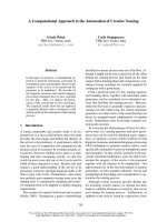
Figure 1 Persistent Hedgehog overexpression induces mouse prostate tumorigenesis. (A) Histopathological analysis with hematoxylin-
eosin stain showing characteristics of progressive tumor formation through stages of PIN and CaP in a pCX-shh-IG-injected prostate at day 90 as
compared to sham (0.9%NaCl) and vehicle (pCX-IG) injection controls. The lower pictures are higher magnifications corresponding to the tissue
sections shown in the upper pictures. (B) Tumor formation induced by Hedgehog overexpression (arrowheads in (b) indicate tumor mass), as
compared to vehicle control (arrowhead in (a)). The prostate gland displayed GFP signals at day 90 after pCX-shh-IG injection (arrowhead-
indicated in (c) and (d), also magnified in the inlets). (C) The pCX-shh-IG-injected prostate tissue sections were stained strongly for Hedgehog
protein in PIN and CaP, in contrast to those of the sham and vehicle controls. Scale bars: 50 μm in upper (d) of panel A; 10 μm in lower (d) of
panel A and in (d) of panel C. CaP: prostate cancer; HE: hematoxylin-eosin stain; PIN: prostatic intraepithelial neoplasia; HGPIN: high grade
prostatic intraepithelial neoplasia.
Chang et al. Journal of Biomedical Science 2011, 18:6
/>Page 4 of 11
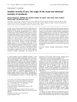
Figure 2 Overexpression of Hedgehog signaling members, including Patch1, Smo, Gli1, Gli2, and Gli3, in pCX-shh-IG-injected
prostates at day 90 after injection. The lower pictures are magnifications of the boxed areas shown in the upper pictures. Hedgehog
signalling members were expressed in the PIN (F-J) and CaP (K-O) of the pCX-shh-IG-injected prostates, in contrast to the absence of evident
signal in the pCX-IG-injected vehicle controls (A-E; basal/stem cells indicated by arrows). Note that some cells positive for Hedgehog signalling
(indicated by arrowheads in F-J) appeared to be basal/stem cells in morphology. Western blot analysis (P) confirmed that Hedgehog signalling
members were highly expressed in the pCX-shh-IG- injected prostates, as compared to those sham injections with 0.9% saline or vehicle controls
with pCX-IG vector. Tubulin detections served as loading controls. Scale bars: 10 μm in lower E, lower J and lower O. CaP: prostate cancer; PIN:
prostatic intraepithelial neoplasia; Gli2-act: active form of Gli2; Gli2-rep: repressed form of Gli2; Gli3-act: active form of Gli3.
Chang et al. Journal of Biomedical Science 2011, 18:6
/>Page 5 of 11
to be detected only within the prostates (Figure 1B; (c)
and (d)). Ectopic GFP signals in other organs would
indicate prostate cancer cell metastasis (Figure 3C; indi-
cated by arrowhead in (b)). Based on the presence of
GFP signals, we found metastasis in the mouse lymph
nodes (19/27), kidneys (7/27), and lungs (5/27) following
90 days after introducing pCX-shh-IG vector, but not i n
thevehicleandshamcontrols (Figure 3C; (a) indicated
two small lymph nodes (on the left) from the sham con-
trols and two enlarged lymph nodes (on the right) from
the pCX-shh-IG-injected mice). The metastatic loci of
lymph nodes (Figure 3C; (a), (b), (d) and (e)), kidneys
(Figure 3C; (f), (g) and (h)), and lungs (Figure 3C; (i))
were infiltrated with P63
+
cells (Figure 3C; indicated by
arrows in (e), (h) and (i)). The increase of P63
+
cells in
the pCX-shh-IG-injected prostates and lymph nodes
after metastasis was con firmed by western blot analysis
at day 90 after the preparation (Figure 3D). Further-
more, the P63
+
cells within most of the metasta tic loci
displayed a prostate-like primitive glandular structure
(Figure 3C; indicated by arrowheads in (d), (f), (g), (h)
and (i)). This finding demonstrated recapitulation of
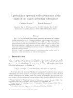
Figure 3 P63
+
basal cells are involved in prostate tumorigenesis and progress ed into metastatic cancer cells with Hedgehog
overexpression. The boxed areas in (A) and (C) are magnified respectively and inlets in (B) correspond to arrow-indicated areas. (A) Increased
P63
+
basal/stem cell density and altered cellular morphology, including bigger cell size, disoriented polarity, and displaced localization were
found along with tumor initiation and progression in the pCX-shh-IG-injected prostate (arrow-indicated in (b), (c) and (d)), in contrast to the
normal P63
+
cells in the pCX-IG-injected prostate (indicated by arrowhead in (a)). (B) Patch1 and Smo proteins were located within P63
+
basal
cells in the PIN and CaP of pCX-shh-IG-injected prostate. The P63
+
/Patch1
+
and P63
+
/Smo
+
cells are arrow-indicated respectively in (b) and (e),
as compared to those in the pCX-IG-injected prostate (arrowhead in (a) and (d)). Patch1 or Smo was co-expressed with P63
+
in some cancer
cells of the advanced prostate cancer (arrow-indicated respectively in (c) and (f)). (C) P63
+
cancer cells recapitulated prostate-like glandular
structure formation in the metastatic loci (arrowhead-indicated in (d), (f), (g), (h) and (i)). Lymph node metastasis is shown by two enlarged
specimens on the right of (a) and in (b), (d), (e), as compared to the normal small lymph node in (c) and the two other small specimens on the
left side of (a). Note that GFP signals can be detected in the lymph node in (b). Kidney metastasis is shown in (f-h) and lung metastasis in (i).
Arrows in (e), (h), and (i) indicate P63
+
metastatic cancer cells. (D) Western blot analysis confirmed increased P63
+
cells in the prostates and the
lymph nodes of the pCX-shh-IG-injected mice. All scale bars: 10 μm. CaP: prostate cancer; PIN: prostatic intraepithelial neoplasia; HGPIN: high
grade prostatic intraepithelial neoplasia.
Chang et al. Journal of Biomedical Science 2011, 18:6
/>Page 6 of 11
prostate formation by the transformed P63
+
basal/stem
cells in the metastatic loci. The data evidently demon-
strated both cancer c ell and stem cell characteristics of
the t ransformed P63
+
basal/stem cells, supporting that
they could be the origin of PCSCs under the influence
of persistent Hedgehog overexpression.
P63
+
basal/stem cells were transformed into AR
+
or AR
-
cancer cells but rarely into ChgA
+
neuroendocrine cancer
cells
Conventional human prostate cancer ce lls in clude
malignant cells of luminal, basal or neuroendocrine ori-
gin at various proportions and unlike the ChgA
-
luminal
cancer cells, tumor cells of neuroendocrine origin are
ChgA
+
[26]. We found only few ChgA
+
neuroendocrine
cells in the PIN lesions and CaP of the pCX-shh-IG-
injected prostates (Figure 4A; (b) and (c); indicated by
arrowheads), not much different to what was found in
the vehicle controls (Figure 4A; (a); indicated by arrow-
head). In the pCX-shh-IG-injected prostates, both the
AR
+
and AR
-
cells were detected, with the AR protein, if
present, located in the nucleus or cytosol in the cells of
PIN lesions a nd CaP (Figure 4A; (e-g)). Wereas, in the
vehicle controls, AR was predominantly located in the
nucleus of luminal cells (Figure 4A; (d); indicated by
arrowhead) and of some basal cells (Figure 4A; (d);
arrow (1) indicated AR
+
basal cells and arrow (2) indi-
cated AR
-
basal cells). This finding was confirmed by
western blot analysis at day 90 after the injection (Figure
4A; (h)). Since cytosolic or no AR protein was observed
in so me of the cancer cells, it appeared that androgens
were not necessarily required for survival of these ce lls
or otherwise they should had been programmed into
cell death. To elucidate the situation , we performed
TUNEL assay and found no significant apoptosis in PIN
lesions and CaP of pCX-shh-IG-injected prostates (Fig-
ure 4B; (b), (c) and (g)), as com pared to normal vehicle
controls (Figure 4B; (a) and (g)). In contrast , PCNA
+
proliferative cells were increased in the PIN and CaP
lesions (Figure 4B; (e), (f) and (h)), but rare in the vehi-
cle controls (Figur e 4B; (d) and (h)). These observat ions
indicated that the PCSCs, as induced by persisten t
Figure 4 Characterization of mouse prostate cancer under the influ ence of Hedgehog overexp ression. (A) The ChgA
+
neuroendocrine
cells were rarely detected in the pCX-IG-injected vehicle control prostate (arrowhead-indicated in (a)) and in PIN and CaP lesions of the pCX-
shh-IG-injected prostates (arrowhead-indicated in (b) and (c)). All luminal cells (arrowhead-indicated in (d)) and some basal cells (arrow (1)-
indicated in (d)) in the normal prostate expressed AR in the nucleus, in contrast to the nucleus or cytosolic AR localization in the PIN and CaP
lesions ((e), (f) and (g)). Some AR
-
basal cells (arrow (2)-indicated in (d)) and AR
-
progeny of PCSCs were found in the CaP ((f) and the magnified
boxed area of (f) as shown in (g)). Western blot analysis confirmed the status of ChgA and AR expression in the vehicle control and pCX-shh-IG-
injected prostate ((h)). (B) The PIN and CaP lesions ((b), (c) and (g)) showed similar apoptosis status as compared to the normal prostate ((a) and
(g)). PCNA
+
proliferative cells were increased in the PIN and CaP lesions ((e), (f) and (h)) as compared to normal prostate ((d) and (h)). All scale
bars represent 10 μm in length. CaP: prostate cancer; PIN: prostatic intraepithelial neoplasia; HGPIN: high grade prostatic intraepithelial neoplasia.
Chang et al. Journal of Biomedical Science 2011, 18:6
/>Page 7 of 11
Hedgehog overexpression, did not commit to differenti-
ate into ChgA
+
neuroendocrine cells and might bypass
AR signalling for survival and proliferation.
PCSCs progeny differentiated into basal-intermediate and
intermediate-luminal cells
Since our data had indicated that ChgA
+
neuroendo-
crine cells did not constitute the main cellular lineage
under the influence of Hh overexpression, the differen-
tiation status o f PCSCs was examined with some cell
markers, including P63 (primitive basal cells), CK14
(advanced and hyperplastic basal cells), CD44 (inter-
mediate cells), and CK8 (mature luminal cells). In the
normal vehicle control prostates, fewer CK14
+
basal
cells lay flat along the basement membrane (Figure 5A;
arrowhead-indicated in (a)), whereas in the PIN and
CaP lesions of pCX-shh-IG-injected prostates, CK14
+
cells were increased with neoplastic transformation
(Figure 5A; arrow-indicated in (b ), (c) and (d)). By wes-
tern blot anal ysis, we found that CK14, CD44, and CK8
markers were up-regulated in the prostate tumors as
compared to the normal prostates (Figure 5B). Double
labelling of P63 a nd CK14 markers showed P63
+
/CK14
(low or -)
cells in the normal prostates (Figure 5C; arrow-
head-indicated in (a)). In the pCX-shh-IG-injected pros-
tates, as the prostates were induced into PIN and
progressed into CaP status, these P63
+
/CK14
(low or -)
cells appeared to be differentiated into P63
+
/CK14
+
(Fig-
ure 5C; arrowhead-indicated in (b)) and further into P63
(low)
/CK14
+
(Figure 5C; arrowhead-indicated in (c)) and
P63
-
/CK14
+
(Figure 5C; arrow-indicated in (b ) and (c))
cells. The loss of P63 expression revealed that the
PCSCs were differentiated toward the lumi nal progeny.
Comparably, the CK14
+
/CD44
+
cancer cells in the PIN
and CaP lesions (Figure 5C; arrow (2)-indicated in (e)
and (f)) were likely to be originated from CK14
(low or -)
/
CD44
(low or -)
cells in the normal prostates (Figure 5C;
arrowhead-indicated in (d)) or from CK14
+
/CD44
(low or -)
cells (Figure 5C; arrow (1)-indicated in (e) and (f)) and
CK14
(low or -)
/CD44
+
cells (Figure 5C; arrow (3)-indicated
in (e) and (f)), as the cells were transformed into PIN and
progressed into CaP conditions. Additionally, the CK14
+
/CK8
+
cancer cells in the PIN and CaP lesions (Figure
5C; arrow-indicated in (h) and (i)) might be originated
from CK14
(low or -)
/CK8
-
cells in the normal prostates
(Figure 5C; arrowhead-indicated in (g)), and then diffe r-
entiated into CK14
low
/CK8
(high or +)
progeny (Figure 5C;
arrowhead-indicated in (i)). The increase of basal-inter-
mediate (CK14
+
/CD44
+
)(Figure5D)andintermediate-
luminal (CK14
+
/CK8
+
)(Figure5E)progenyinthePIN
and CaP lesions of pCX-shh-IG-injected prostates were
significant when compared to the vehicle controls. The
involvement of Hh signalling during PCSCs differentia-
tion in the PIN and CaP lesions was indicated by double
labelling of Patch1 and CK14 (Figure 5C; arrow-indicated
in (k) and (l)), in contrast to the only few Patch1
+
cells
detected in the normal vehicle controls (Figure 5C;
arrowhead-indicated in (j)).
The PCSCs progeny differentiation was not totally
dependent on androgen-AR axis
To examine whether malignant differentiat ion of PCSCs
might be independent from the andro gen-AR axis in the
pCX-shh-IG-injected prostates, we co-localized AR with
different markers, including P63, CK14, CK8, CD44, and
Patch1 (Figure 5F). Apart from the AR
+
cells, we detected
P63
+
/AR
-
,CK14
+
/AR
-
,CD44
+
/AR
-
and even th e CK8
+
/AR
-
cell s in the CaP (Figure 5F). The concurrent existence of
AR
+
and AR
-
cell populations indicated that androgen-AR
axis was not indispensably required for PCSCs and their
progeny to undergo further differentiation (Figure 5F; (f)).
The localization of Patch1 protein in both AR
+
and AR
-
cancer cells (Figure 5F; (e)) revealed the involvement of
Hedgehog signalling in both cell lineages.
Discussion
Basal/stem cells are the origin of all prostate intra-
glandular cells [1,27] and P63 has been proposed to be
required for prostate stem cell plasticity and differenti a-
tion [27,28]. Thus, PCSCs are potentially originated
from normal P63
+
prostate stem cells, although there
hasbeennoevidencesofartosupport this hypothesis.
In our previous study analyzing the human specimen
[20], we demonstrated t hat Hh protein is expressed in
P63
+
basal cells, in correlation with basal cell hyperpla-
sia a nd CaP formation. Our previous data support nor-
mal prostate stem cells transformation into PCSCs,
although based only on retrospective observations.
In this study, we provide the evidence to further sup-
port PCSCs derivation from normal p rostate stem cells,
based on several lines of observations. Firstly, the entire
process of prostate tumorigenesis was reconstituted in
vivo and the effects of Hh overexpression on the normal
prostate stem cells was shown by the transformation of
P63
+
/Patch1
(Low or -)
and P63
+
/Smo
(Low or -)
quiescent
basal cells into the P63
+
/Patch1
+
and P63
+
/Smo
+
hyper-
plastic basal cells, comparable to the human BCH condi-
tion that had been previously observed [20]. Secondly,
the P63
+
cells showed major cancer cell and stem cell
peculiarity on the metastatic sites. The P63
+
cells were
not only present within various metastatic loci, but also
differentiated into prostate-like glandular structures
where they were located within the basal compartment.
Thirdly, the P63
+
basal/stem cells, after being trans-
formed into malignant cells, were capable of differentia-
tion into the basal-intermediate (P63
+
/CK14
+
)and
intermediate-luminal (CK14
+
/CD44
+
and CK14
+
/CK8
+
)
progeny, and rarely the ChgA
+
neuroendocrine lineage.
Chang et al. Journal of Biomedical Science 2011, 18:6
/>Page 8 of 11
Figure 5 Differentiation status and AR expression profile of PCSCs under the influence of Hedgehog overexpression. The boxed areas
in the pictures are further magnified and shown in the corresponding lower or right pictures. (A) Increased CK14
+
cells along with tumorigenic
progression in the pCX-shh-IG-injected prostate ((b), (c) and (d)), as compared to the normal prostate ((a)). (B) Western blot analysis indicated up-
regulation of CK14, CD44 and CK8 in the pCX-shh-IG-injected prostates as compared to the normal pCX-IG-injected prostates. (C)
Characterization of PCSCs by double-immunofluorescence staining showing differentiation toward CK14
+
progeny ((a), (b) and (c)), CK14
+
cells
toward CD44
+
progeny ((d), (e) and (f)), and CK14
+
cells toward CK8
+
progeny ((g), (h) and (i)). CK14
+
differentiation involved Hedgehog
signalling activation, as indicated by co-localized Patch1 expression ((j), (k) and (l)). The basal-intermediate (CK14
+
/CD44
+
) and intermediate-
luminal (CK14
+
/CK8
+
) populations were increased in PIN and CaP of pCX-shh-IG-injected prostates as compared to those of the pCX-IG-injected
vehicle controls (D and E). (F) Some PCSCs were AR
-
as indicated by arrows in (a), (b), (c), (d), and (e), even though they were P63
+
, CK14
+
, CK8
+
,
CD44
+
, or Patch1
+
. The relative proportions of AR
+
and AR
-
cells among P63
+
, CK14
+
, CK8
+
, CD44
+
, and Patch1
+
cell populations were shown
respectively in (f). All scale bars represent 10 μm in length. CaP: prostate cancer; PIN: prostatic intraepithelial neoplasia; HGPIN: high grade
prostatic intraepithelial neoplasia.
Chang et al. Journal of Biomedical Science 2011, 18:6
/>Page 9 of 11
PCSCs derivation from normal prostate stem cells has
also been supported by several lines of evidence. Purified
cells such as Sca-1
+
and CD49f
+
cells from the mouse
prostates [29,30] or CD44
+
, CD133
+
, and a2/b1 integrin
+
cells from the human prostates [31,32] were used in renal
capsule transplantation studies. These normal cells could
generate prostate-like structures in the kidney, support-
ing the presence of prostate progenitor/stem cells in
these purified cell populations and their ability to regen-
erate in ectopic sites. Such ectopic regeneration capacity
has been pr oven to retain even after the transformation
of normal prostate stem cells into PCSCs. For example,
AR
-
P63
-
CD44
+
Nestin
+
HPET-5 cells were purified from
prostate cancer cell l ines [33] and shown to recapitulate
prostate-like structures in the mouse kidney after trans-
plantation. Besides, a previous report showed that Hedge-
hog may recapitulate embryonic gene expression in
tumor myofibroblasts [34]. Despite the aforementioned
studies, in vivo prostate cancer cell metastasis with a
stem cell pe culiarit y to generate prostate-like structures
in the metastatic loci has not been reported. In this
study, we demonstrated the infiltration of P63
+
cancer
cells in the metastatic loci and generation of prostate-like
glandular structures. Our data support the prostate can-
cer stem c ell characteristics observed in previous studies
and, to our knowledge, these are the first data to confirm
that PCSCs metastasis occurs under in vivo conditions.
Clinically, it is known that advanced metastatic andro-
gen-independent prostate cancers exhibit more basal/
stem cell-like differentiation although the underlying
mechanism remains unclear [9,10]. The transformati on
of basal/stem cells into PCSCs was proposed as a possi-
ble mechanism. In such case, the PCSCs are supposed
to maintain high proliferation and differentiation capaci-
ties w ithout AR activity [35,36], since androgen is con-
sidered not necessary for the survival of basal/stem cells;
in co ntrast to the ter minally-differentiat ed luminal cells
which require androgen and AR for survival [7,9,37].
Our data showed transition of nuclear to cytoplasmic
P63 expression and such transition had been reported
to associate with higher prol iferative activity, reduced
apoptosis, and increased mortality [35]. This may
explain the abundant AR
-
cancer cells found in many
high grade prostate cancers [38-40] and the failure of
and rogen deprivation therapies . Alternatively, persistent
AR activation due to loss-of-function mutations has
been reported in some androgen-refractory prostate can-
cers [13,41]. Therefore, both AR
+
and AR
-
cells were
capable of forming androgen-independent prostate can-
cer cells as the tumorigenesis progresses to the more
advanced stages. In this study, both the AR
+
and AR
-
prostate cancer cells were generated under the effects of
persistent Hedgehog signalling activation in the mouse
model (Figure 4A and Figure 5F). Our data is consistent
with the findings of Hedgehog signalling activation in
several human prostate cancers [17-20,22], especially in
the androgen-independent prostate cancers [22,42,43].
Particularly, we showed that some basal/stem cancer
cells were AR
-
,e.g.P63
+
/AR
-
,CK14
+
/AR
-
and CD44
+
/
AR
-
(Figure 5F; arrowhead-indicated in (a), (b) and (d)).
These sub-populations of AR
-
cancer cells were most
likely to contribute directly to the androgen-independent
tumors. In fact, androgen ablation by castration could
not reduce tumor mass in this model (data not shown).
Here, the key findings in this study have confirmed that
overexpression of Hedgehog can transform prostate basal
cells in vivo and lead the transformed cells to progress
into aggressive AR
-
PCSCs progeny. Although the
nuclear AR
+
(active form) and cytoplasmic AR
+
(inactive
form) cells were both observed in the aggressive tumors
in this in vivo model and our data showed that Hedgehog
signalling activation may substitute the androgen-AR axis
for tumor survival or malignant transformation, the
underlying mechanisms remain to be further investigated
by using in vitro studies.
Conclusions
Our data support the hypothesis that P63
+
hyperplastic
basal cells targeted by Hh overexpression may be the true
cellular origin of primary prostate cancer. This study also
supports that inhibition of Hedgehog signalling may be a
better treatment strategy for androgen-independent
tumors prior to androgen-deprivation therapy.
Acknowledgements
This work was supported by a grant (NSC 96-2321-B-040-007-MY3) to HH
Chang and partly by a grant (NSC 97-2321-B-040-004) to DP Lin, from
National Science Council, Taiwan. The immunofluorescence microscopy was
performed in the Instrument Center of Chung Shan Medical University,
which is supported by National Science Council, Ministry of Education and
Chung Shan Medical University.
Author details
1
School of Nutrition, Chung Shan Medical University, Taichung 402, Taiwan.
2
School of Optometry, Chung Shan Medical University, Taichung 402,
Taiwan.
3
Division of Urology, Changhua Christian Hospital, Changhua 500,
Taiwan.
4
Division of Urology, Taichung Veterans General Hospital, Taichung
407, Taiwan.
5
School of Medical Laboratory and Biotechnology, Chung Shan
Medical University, Taichung 402, Taiwan.
6
Department of Urology, Chung
Shan Medical University Hospital, Taichung 402, Taiwan.
Authors’ contributions
HHC, BYC, and DPL designed the study, carried out production of pCX-shh-
IG and pCX-IG mice, and contributed to the writing of manuscript. CYW
helped with animal maintenance, plasmid vector preparation,
immunohistochemical and double-immunofluorescence staining. ZJT
performed TUNEL assay and western blot analysis. CPC and CRY carried out
the quantification of positive and double-positive cells.
Competing interests
The authors declare that they have no competing interests.
Received: 2 August 2010 Accepted: 18 January 2011
Published: 18 January 2011
Chang et al. Journal of Biomedical Science 2011, 18:6
/>Page 10 of 11
References
1. Wang Y, Hayward S, Cao M, Thayer K, Cunha G: Cell differentiation lineage
in the prostate. Differentiation 2001, 68:270-279.
2. Uzgare AR, Xu Y, Isaacs JT: In vitro culturing and characteristics of transit
amplifying epithelial cells from human prostate tissue. J Cell Biochem
2004, 91:196-205.
3. Tokar EJ, Ancrile BB, Cunha GR, Webber MM: Stem/progenitor and
intermediate cell types and the origin of human prostate cancer.
Differentiation 2005, 73:463-473.
4. Liu AY, True LD, LaTray L, Nelson PS, Ellis WJ, Vessella RL, Lange PH, Hood L,
van den Engh G: Cell-cell interaction in prostate gene regulation and
cytodifferentiation. Proc Natl Acad Sci USA 1997, 94:10705-10710.
5. Schalken JA, van Leenders G: Cellular and molecular biology of the
prostate: stem cell biology. Urology 2003, 62:11-20.
6. Bonkhoff H: Role of the basal cells in premalignant changes of the
human prostate: a stem cell concept for the development of prostate
cancer. Eur Urol 1996, 30:201-205.
7. Collins AT, Maitland NJ: Prostate cancer stem cells. Eur J Cancer 2006,
42:1213-1218.
8. Lawson DA, Witte ON: Stem cells in prostate cancer initiation and
progression. J Clin Invest 2007, 117:2044-2050.
9. Lang SH, Frame FM, Collins AT: Prostate cancer stem cells. J Pathol 2009,
217:299-306.
10. van Leenders GJ, Aalders TW, Hulsbergen-van de Kaa CA, Ruiter DJ,
Schalken JA: Expression of basal cell keratins in human prostate cancer
metastases and cell lines. J Pathol 2001, 195:563-570.
11. Klarmann GJ, Hurt EM, Mathews LA, Zhang X, Duhagon MA, Mistree T,
Thomas SB, Farrar WL: Invasive prostate cancer cells are tumor initiating
cells that have a stem cell-like genomic signature. Clin Exp Metastasis
2009, 26:433-446.
12. Patrawala L, Calhoun T, Schneider-Broussard R, Li H, Bhatia B, Tang S,
Reilly JG, Chandra D, Zhou J, Claypool K, Coghlan L, Tang DG: Highly
purified CD44+ prostate cancer cells from xenograft human tumors are
enriched in tumorigenic and metastatic progenitor cells. Oncogene 2006,
25:1696-1708.
13. Debes JD, Tindall DJ: Mechanisms of androgen-refractory prostate cancer.
N Engl J Med 2004, 351:1488-1490.
14. Guzman-Ramirez N, Voller M, Wetterwald A, Germann M, Cross NA,
Rentsch CA, Schalken J, Thalmann GN, Cecchini MG: In vitro propagation
and characterization of neoplastic stem/progenitor-like cells from
human prostate cancer tissue. Prostate 2009, 69:1683-1693.
15. Theunissen JW, de Sauvage FJ: Paracrine Hedgehog signaling in cancer.
Cancer Res 2009, 69:6007-6010.
16. Kasper M, Regl G, Frischauf AM, Aberger F: GLI transcription factors:
mediators of oncogenic Hedgehog signalling. Eur J Cancer 2006,
42:437-445.
17. Sheng T, Li C, Zhang X, Chi S, He N, Chen K, McCormick F, Gatalica Z, Xie J:
Activation of the hedgehog pathway in advanced prostate cancer. Mol
Cancer 2004, 3:29.
18. Sanchez P, Hernandez AM, Stecca B, Kahler AJ, DeGueme AM, Barrett A,
Beyna M, Datta MW, Datta S, Ruiz i Altaba A: Inhibition of prostate cancer
proliferation by interference with SONIC HEDGEHOG-GLI1 signaling. Proc
Natl Acad Sci USA 2004, 101:12561-12566.
19. Karhadkar SS, Bova GS, Abdallah N, Dhara S, Gardner D, Maitra A, Isaacs JT,
Berman DM, Beachy PA: Hedgehog signalling in prostate regeneration,
neoplasia and metastasis. Nature 2004, 431:707-712.
20. Chen BY, Liu JY, Chang HH, Chang CP, Lo WY, Kuo WH, Yang CR, Lin DP:
Hedgehog is involved in prostate basal cell hyperplasia formation and
its progressing towards tumorigenesis. Biochem Biophys Res Commun
2007, 357:1084-1089.
21. Chen BY, Lin DP, Liu JY, Chang H, Huang PH, Chen YL, Chang HH: A mouse
prostate cancer model induced by Hedgehog overexpression. J Biomed
Sci 2006, 13:373-384.
22. Azoulay S, Terry S, Chimingqi M, Sirab N, Faucon H, Gil Diez de Medina S,
Moutereau S, Maillé P, Soyeux P, Abbou C, Salomon L, Vacherot F, de la
Taille A, Loric S, Allory Y: Comparative expression of Hedgehog ligands at
different stages of prostate carcinoma progression. J Pathol 2008,
216:460-470.
23. Sanchez P, Clement V, Ruiz i Altaba A: Therapeutic targeting of the
Hedgehog-GLI pathway in prostate cancer. Cancer Res 2005,
65:2990-2992.
24. Stecca B, Mas C, Ruiz i Altaba A: Interference with HH-GLI signaling
inhibits prostate cancer. Trends Mol Med 2005, 11:199-203.
25. Oberg KC, Pira CU, Revelli JP, Ratz B, Aguilar-Cordova E, Eichele G: Efficient
ectopic gene expression targeting chick mesoderm. Dev Dyn 2002,
224:291-302.
26. Bonkhoff H, Stein U, Remberger K: Endocrine-paracrine cell types in the
prostate and prostatic adenocarcinoma are postmitotic cells. Hum Pathol
1995, 26:167-170.
27. Signoretti S, Pires MM, Lindauer M, Horner JW, Grisanzio C, Dhar S,
Majumder P, McKeon F, Kantoff PW, Sellers WR, Loda M: p63 regulates
commitment to the prostate cell lineage. Proc Natl Acad Sci USA 2005,
102:11355-11360.
28. Kurita T, Medina RT, Mills AA, Cunha GR: Role of p63 and basal cells in the
prostate. Development 2004, 131:4955-4964.
29. Burger PE, Xiong X, Coetzee S, Salm SN, Moscatelli D, Goto K, Wilson EL:
Sca-1 expression identifies stem cells in the proximal region of prostatic
ducts with high capacity to reconstitute prostatic tissue. Proc Natl Acad
Sci USA 2005, 102:7180-7185.
30. Lawson DA, Xin L, Lukacs RU, Cheng D, Witte ON: Isolation and functional
characterization of murine prostate stem cells. Proc Natl Acad Sci USA
2007, 104:181-186.
31. Xin L, Lawson DA, Witte ON: The Sca-1 cell surface marker enriches for a
prostate-regenerating cell subpopulation that can initiate prostate
tumorigenesis. Proc Natl Acad Sci USA 2005, 102:6942-6947.
32. Richardson GD, Robson CN, Lang SH, Neal DE, Maitland NJ, Collins AT:
CD133, a novel marker for human prostatic epithelial stem cells. J Cell
Sci 2004, 117:3539-3545.
33. Gu G, Yuan J, Wills M, Kasper S: Prostate cancer cells with stem cell
characteristics reconstitute the original human tumor in vivo. Cancer Res
2007, 67:4807-4815.
34. Shaw A, Gipp J, Bushman W: The Sonic Hedgehog pathway stimulates
prostate tumor growth by paracrine signaling and recapitulates
embryonic gene expression in tumor myofibroblasts. Oncogene 2009,
28(50):4480-90.
35. Dhillon PK, Barry M, Stampfer MJ, Perner S, Fiorentino M, Fomari A, Ma J,
Fleet J, Kurth T, Rubin MA, Mucci LA: Aberrant cytoplasmic expression of
p63 and prostate cancer mortality. Cancer Epidemiol Biomarkers Prev 2009,
18:595-600.
36. Kelly K, Yin JJ: Prostate cancer and metastasis initiating stem cells. Cell
Res 2008, 18:528-537.
37. Collins AT, Berry PA, Hyde C, Stower MJ, Maitland NJ: Prospective
identification of tumorigenic prostate cancer stem cells. Cancer Res 2005,
65:10946-10951.
38. Miyamoto KK, McSherry SA, Dent GA, Sar M, Wilson EM, French FS,
Sharief Y, Mohler JL: Immunohistochemistry of the androgen receptor in
human benign and malignant prostate tissue. J Urol 1993, 149:1015-1019.
39. Chodak GW, Kranc DM, Puy LA, Takeda H, Johnson K, Chang C: Nuclear
localization of androgen receptor in heterogeneous samples of normal,
hyperplastic and neoplastic human prostate. J Urol 1992, 147:798-803.
40. Huang J, Yao JL, di Sant’Agnese PA, Yang Q, Bourne PA, Na Y:
Immunohistochemical characterization of neuroendocrine cells in
prostate cancer. Prostate 2006, 66:1399-1406.
41. Yuan X, Balk SP: Mechanisms mediating androgen receptor reactivation
after castration. Urol Oncol 2009, 27:36-41.
42. Chen M, Tanner M, Levine AC, Levina E, Ohouo P, Buttyan R: Androgenic
regulation of hedgehog signalling pathway components in prostate
cancer cells. Cell Cycle 2009, 8
:149-157.
43. Shaw G, Price AM, Ktori E, Bisson I, Purkis PE, McFaul S, Oliver RT,
Prowse DM: Hedgehog signalling in androgen independent prostate
cancer. Eur Urol 2008, 54:1333-1343.
doi:10.1186/1423-0127-18-6
Cite this article as: Chang et al.: Hedgehog overexpression leads to the
formation of prostate cancer stem cells with metastatic property
irrespective of androgen receptor expression in the mouse model.
Journal of Biomedical Science 2011 18:6.
Chang et al. Journal of Biomedical Science 2011, 18:6
/>Page 11 of 11