Báo cáo y học: " Colorectal carcinoma: nucleosomes, carcinoembryonic antigen and ca 19-9 as apoptotic markers; a comparative study" potx
Bạn đang xem bản rút gọn của tài liệu. Xem và tải ngay bản đầy đủ của tài liệu tại đây (396.4 KB, 5 trang )
RESEARCH Open Access
Colorectal carcinoma: nucleosomes,
carcinoembryonic antigen and ca 19-9 as
apoptotic markers; a comparative study
Jehad M Al-Shuneigat
1*
, Samir S Mahgoub
1
and Fazlul Huq
2
Abstract
Background: Colorectal carcinoma is a common and often fatal disea se in which methods of early detection and
monitoring are essential. The present study was conducted for measuring serum levels of nucleosomes,
carcinoembryonic antigen (CEA) and CA 19-9 in patients newly diagnosed with colorectal carcinoma and
confirmed by clinicopath ological study.
Method: Thirty subjects were included in the current study: six normal subjects as a control group with mean age
(45.6 ± 7.9) and twenty four colorectal carcinoma patients with mean age (46.9 ± 15.6), which were classified
pathologically according to the degree of malignant cell differentiation into well differentiated (group I),
moderately differentiated (group II) and poorly differentiated (group III). Fasting venous blood samples were
collected preoperative.
Results: The results revealed a significant increase in serum level of nucleosomes in patients with poorly
differentiated tumors versus patients with well differentiated tumors (p = 0.041). The levels of CEA and CA19-9
showed no significant increase (p = 0.569 and 0.450, respectively).
Conclusion: In conclusion, serum level of nucleosomes provides a highly sensitive and specific apoptotic marker
for colorectal carcinoma.
Keywords: colorectal carcinoma, nucleosomes, carcinoembryonic antigen (CEA) and CA 19-9, tumor marker
Background
Colorectal carcinoma is one of the leading causes of
cancer-related death [1,2]. The 5-year survival rate for
colon tumours in Europe ranges from 26% to 56% for
men and from 29% to 59% for women. These differ-
ences in survival have been attributed to the stage and
timing of diagnosis and, in some regions, to the quality
of medical care [3]. Hence the need for early detection
methods in colorectal cancer [4].
Tumor markers are used clinically for diagnosis, sta-
ging, and monitoring of the disease. They are proteins
released from dying tumor cells or produced by neo-
plastic cells. There are two subcategories of these pro-
teins, specific and non-specific. The specific proteins
are expressed only in the tumor cells and are very use-
ful for the detection and diagnosis of specific malig-
nant tumors. Non-specific proteins or markers related
to malignant cells are oncofetal or carcinogenic anti-
gens, such as carcinoembryonic antigen (CEA), alpha-
fetoprotein (AFP), prostate specific antigen (PSA),
carbohydrate antigens CA15.3 and CA19-9. Recently
nucleosomes, cytokeratine 18, and cyto-c in serum
have been examined as markers for the evaluation of
apoptotic death [5].
The basic unit of chr omatin, known as a nucleosome,
is composed of local wrapping of a short stretch of dou-
ble stranded (ds) DNA (147 bp in length) around an
octameric histone protein core of two molecules each of
histones H
2
A, H
2
B, H
3
and H
4
(known as nucleosome
core particle (NCP)), and the so-called linker DNA that
varies between 10 and 100 bp and connects neighboring
nucleosomes in a chain like pattern [6].
* Correspondence:
1
Faculty of Medicine, Department of Pharmacology and Biochemistry Mu’tah
University, Al Karak, Jordan
Full list of author information is available at the end of the article
Al-Shuneigat et al. Journal of Biomedical Science 2011, 18:50
/>© 2011 Al-Shuneigat et al; licensee BioMed Centr al Ltd. This is an Open Access article distrib uted under the terms of the Creative
Commons Attribution License ( which permits unrestricted use, distribution, and
reproduction in any medium, provided the original work is properly cited.
Apoptosis or programmed cell death has evolved in
multicellular organisms to remodel tissue during devel-
opment. It maintains tissue homeostasis (proliferation
and apoptosis balance) by removing senescent cells and
deleting cells with irreparable genetic damage. It is a
highly regulated process with distinct morphologic and
biochemical features [7,8].
The mechanism of apoptosis in tumors is unclear.
However, it is known to be p53 and caspases dependent
[9], a hallmark of cancer cells [10]. During apoptosis,
caspases are activated leading to degradation of cell con-
stituents [11]. High levels of circulating mono- and
oligo-nucleosomal fragments are expected. In blood, cir-
culating nucleic acids are not digested by endonucleases
due to their close association with histone protein. High
levels of circulating DNA fragments have been found in
the blood of patients with most types of malignancy
including colorectal, lung, gastrointestinal, breast, gyne -
cological, renal, and nasophary ngeal cancers as we ll as
lymphoma [12,13].
For colorectal cancer many biomarkers have been
assessed but only a small number have been recom-
mended for clinical use. The aim of the present study
was to compare the circulating levels of nucleosomes,
CEA and CA 19-9 as apoptotic markers of colorectal
car cinoma and to determine which one is more specific
and sensitive for clinical use.
CEA is a product of columnar and goblet cells in the
normal colon cells as well as colonic cancer cells with a
half life of 3-11 days. It is a glycoprotein with a molecu-
lar weight of 200 kDa; most of its carbohydrate content
is composed of mannose, galactose, N-acetylglucosa-
mine, fructose and sialic acid [14]. The serum l evels of
CEA may increase 4.5 to 8 months before the develop-
ment of cancer symptoms. Therefore, CEA monitoring
is the most cost-effective indicator for the disease [15].
CA 19-9 is an antigen originally isolated for the first
time from human colorectal carcinoma, which is identi-
fied by monoclonal antibody designated 19-9. It was
postulated that CA 19-9 could not be recommended for
early diagnosis of colorectal carcinoma and its serial
determination appears to provide little information to
that of CEA in monitoring patients [16].
Methods
The present study was conducted in patients with colorec-
tal cancer admitted to the Gastroenterology Department
of EI-Minia University Hospital. Twenty four patients
were included in the study (ten females and fo urteen
males) with ages ranging from 34 to 72 years (mean 46.9 ±
15.6). Six normal subjects were selected as a control group.
All patients proved to have colorectal carcinoma by his-
tory, examination, investigations and biopsy. The blood
samples were collected before surgical interference.
The circulating levels of nucleosomes were estimated
by cell death detection ELISA (CDDE)
plus
supplied from
Roche Diagnostic-Germany. CEA and CA 19-9 serum
levels were determined by electrochemi-luminescence
immunoassay (ECLISA) on Roche Elecsys 1010 immu-
noassay analyzer. The kits were used according to the
manufacturer’ s instructions. The chemicals were sup-
plied by Roche Diagnostics GmbH, D 68298, Mannheim,
Germany. The patien ts were classified into three groups,
nine patients with well differentiated tumor, nine
patients with moderately differentiatedtumorandsix
patients with poorly differentiated tumor according to
The Modified Dukes cla ssification of Astler and Coller
[17].
Statistical methods
To test for normal distribution, frequency of data was
plotted against normal distribution curve. Nonpara-
metric statistical methods were used. Frequency, med-
ian, range and standard error of means were used to
describe data. Kruskal-Wallis, nonparamet ric analysis of
variance was used to test for variability between groups
in quantitative variables while, Mann-Whitney u test
was used to test for significance of difference in quanti-
tative variables between each two groups. Nonpara-
metric Kendall’s correlation was used to test for linear
relationship between different quantitative variables.
Regression analysis was used for further analysis of the
linear relationship between nucleosomes, CEA and CA
19-9. Receiver operating characteristic (ROC) curve ana-
lysis was done using MedCalc software for evolution of
sensitivity and specificity of different markers. P value
was considered significant if less than 0.05. These tests
were run on an IBM compatible personal computer
using the Statistical Package for Social Scientists (SPSS)
for windows 7.5 (SPSS Inc., Chicago, IL, USA).
Results
Table (1) shows range, median and SE of the studied
parameters in control versus patient groups (well,
moderately and poorly differentiated tumors). There is
significant increase in nucleosomes levels in all groups
versus the control group (P < 0.001). CEA levels show
a significant increase in all groups except the poorly
differentiated tumor group versus the control group (P
< 0.001 for the well differentiated tumor group, 0.037
for the moderately differentiated tumor group and
0.106 for the poorly differentiated tumor group). There
is no significant increase in the levels of nucleosomes,
also, no significant decrease in the levels of CEA with
decreased grade of tumor differentiation. There is
significant increase in the levels of nucleosomes in
poor differentiated versus well differentiated tumors
(p = 0.041).
Al-Shuneigat et al. Journal of Biomedical Science 2011, 18:50
/>Page 2 of 5
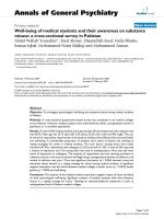
The overall positive rates obtained from ROC curve
(Figure 1) of circulating CEA and CA 19-9 (using the
cutoff values of 3.56 ng/ml and 28 U/ml, respectively)
were 56.2% and 36.4% (Table 2).
Table (2) shows statistical evaluation of nucleosomes
in comparison to CEA and CA 19-9 for colorectal carci-
noma. By all statistical parameters, nucleosomes show
the best values. As regards kappa it shows an excellent
agreement between laboratory and actual diagnosis for
nucleosomes while the values of CEA and CA 19 -9
show only weak agreement.
The curve shows the true positive rate (sensitivity)
plotted versus false positive rate (100-specificity) for dif-
ferent cutoff points for nucleosomes, CEA and CA 19-9.
Discussion
Nuclear fragmentation as one of the morphologic fea-
tures of apoptosis, results in a characteristic pattern of
DNA complexed with histone proteins known as
nucleosomes [18]. The measurement of these nucleo-
somes constitutes a feasible parameter for late stage of
apoptosis [19]. Circulating nucleosomes can be quanti-
fied by real time PCR of the DNA. Moreover they can
be estimated by stable immunologic assays that are par-
ticularly well suited for serial measurements.
CEA is a member of the immunoglobulin superfamily
which was originally identified in human fetal colon and
colorectal cancer. It is widely used as a tumor marker.
However, little is known about its function except that
it acts as a homotypic adhesion molecule that is impli-
cated in cell aggregation [20]. It is over-expressed in
Table 1 Comparison between serum levels of nucleosomes, CEA and CA 19-9 in the three patient groups versus the
control group and each group of patients versus the other groups
Parameters
Groups
Nucleosomes
(AU)
CEA
(ng/ml)
CA 19-9
U/ml
Control group (n = 6) Median 282.6 2.3 20.9
S.E ± 26.4 0.39 3.2
Range Min. 181.5 0.8 7.8
Max. 519.2 4.2 41.4
Group I Patients (well differentiated tumor) (n = 9) Median 1081 6.6 32.86
S.E ± 209.5 9.00 101.6
Range Min. 712.4 1.29 8.4
Max. 3382.5 1729 1895
Group II Patients Median 1776.2 4.61 23.9
(moderate differentiated tumor) (n = 9) S.E ± 171.3 75.6 64.6
Range Min. 772.2 1.6 1.6
Max. 3426.9 2400 2246
Group III Median 1860 4.6 34
Patients (poor differentiated tumor) (n = 6) S.E ± 260.6 6.26 44.66
Range Min. 1149.7 1.72 3.16
Max. 3482.5 52.6 386.2
P control versus group I <0.001 <0.001 0961
P control versus group II <0.001 0.037 0.666
P control versus group III <0.001 0.106 0.816
P group I versus group II 0.086 0.464 0.116
P group I versus group III 0.041 0.569 0.450
P group II versus group III 0.566 0.861 0.627
Figure 1 Receiver operating characteristic curve (ROC) of
nucleosomes, CEA and CA 19-9 in colorectal carcinoma
diagnosis.
Al-Shuneigat et al. Journal of Biomedical Science 2011, 18:50
/>Page 3 of 5
numerous human cancers where it is present on the
surface of cancer cells. It was reported that the over
expression of CEA can protect cancer cells from apopto-
sis while a decrease in expression might lead to new
approaches for management of cancer colon and other
organs [21]. CEA is produced by more than 90% of col-
orectal cancers and contributes to the malignant charac-
teristics of this type of cancer [22].
Although colorectal cancer screening is recommend ed
for persons over 50 years and older its use is still low
especially among older individuals [23].
An ideal tumor marker would be inexpensive screen-
ing that may help for early diagnosis in population at
risk of cancer. Unfortunately curren tly available serolo-
gical markers for colorectal carcinoma have not proven
to be ideal [24].
The levels of serum nucleosomes show significant
increase in patients as compared to control group
(Table 1) with excellent discrimination between the two
(area under curve equals 1 in table 2) that agrees with
other published data [25]. There is an association of
high nucleosomes levels with advanced stages of color-
ectal carcinoma. This may be due to a delayed clearance
of nucleoso mes from circulation where the bulky tumor
tends to undergo peripheral apoptosis and central
necrosis that in its turn would elicit local and systemic
inflammatory responses and hence higher nucleosomes
production [26].
The relationship between apoptosis and the degree of
cell differentiation may play an important role in the
susceptibility of the tumor cells to apoptosis [27]. An
immature phenotype represents a block in the normal
differentiation pathway [28]. Also, another study
observed that the more the colon cells are stimulated to
differentiate, the less likely they are to proliferate and
hypothetically, the higher their rate of apoptosis [29].
After complete tumor resection of colorectal cancer, it
was noticed that the level of nucleosomes increased in
most patients rapidly reaching a maximum level during
the first day. T his was followed by a subsequent
decrease, while the level was lower than in patients with
postoperative or relapse therapy [30].
In the current study, the over all positive rates
obtained from ROC curve (Figure 1) of circulating CEA
and CA 19-9 (using the cutoff values of 3.56 ng/ml and
28 U/ml, respectively) were 56.2% and 36 .4% (Table 2).
These levels are higher than those reported by Zheng et
al [24] who gave positive values of 29.2% and 25.2% for
CEA and CA 19-9, their cutoff values of 5 ug/l and 31
Ku/l respectively. While the positive rates reported by
Chan and Sell,[31], were 70% and 30% for CEA and CA
19-9, the cutoff values were of 3.5 ug/l and 37 Ku/l,
respectively. CEA in the current study can provide a
better discrimination between control subjects and
patients than CA 19-9 (the areas under the ROC curve
were 0.808 and 0. 524 for CEA and CA 19-9,
respectively).
Table (1) shows no significant difference in the levels
of CA 19-9 in patients as compared to the control
group while CEA shows a difference. Bhatnagar et al
[32] noticed that in the well differentiated colorectal
carcinoma there is more production of CEA/gram of
total protein than in the poorly differentiated tumors in
agreement with results obtained in the present study.
On the other hand, it was observed that there is no sig-
nificant correlation between levels of serum CA 19-9
and CEA [22] and the differentiation degree of the
tumor.
Conclusion
In conclusion, our study confirms that the levels of
nucleosomes provide highly specific and sensitive apop-
totic marker for colorectal carcinoma which should be
applied on a lar ge scale of cancers with respect to clini-
copathological variables. It can be used - for diagnosis,
screening, prognosis, and in therapy monitoring.
List of abbreviations
CEA: Carcinoembryonic antigen; AFP: Alphafetoprotein; PSA: Prostate specific
antigen; NCP: Nucleosome core particle; ECLISA: Electrochemi-luminescence
immunoassay; ROC: Receiver operating characteristic.
Author details
1
Faculty of Medicine, Department of Pharmacology and Biochemistry Mu’tah
University, Al Karak, Jordan.
2
Faculty of Medicine, Discipline of Biomedical
Science, The University of Sydney, Australia.
Authors’ contributions
All authors contributed equally to this work and read and approved the final
manuscript.
Competing interests
The authors declare that they have no competing interests.
Table 2 Statistical evaluation of nucleosomes compared to CEA and CA 19-9 for colorectal carcinoma
Parameter Cutoff value Sensitivity % Specificity % J index Accuracy % Kappa PPV % NPV % ROC
curve area
Value P
CA 19-9 28 u/ml 36.4 88.9 26 41 0.121 0.088 91.56 21.62 0.524
CEA 3.56 ng/ml 56.2 100 55.8 62.2 0.290 <0.001 100 30.40 0.808
Nucleosomes 421 100 100 100 100 1.00 <0.001 100 100 1.0
Al-Shuneigat et al. Journal of Biomedical Science 2011, 18:50
/>Page 4 of 5
Received: 2 March 2011 Accepted: 25 July 2011 Published: 25 July 2011
References
1. Chan CC, Fan CW, Kuo YB, Chen YH, Chang PY, Chen KT, Hung RP,
Chan EC: Multiple serological biomarkers for colorectal cancer detection.
Int J Cancer 2010, 126:1683-1690.
2. Zhao-Hut H, Li-Hua L, Fan Y, Jin-Fu W: Detection of apparent methylation
in fecal DNA as a molecular screening tool for colorectal cancer and
precancerous lesions. World J Gastroenterol 2007, 14:950-954.
3. Ramos M, Esteva M, Cabeza E, Campillo C, Llobera J, Aguilo A: Relationship
of diagnostic and therapeutic delay with survival in colorectal cancer: A
review. European journal of cancer 2007, 43:2467-2478.
4. Ahlquist DA: Molecular Detection of Colorectal Neoplasia.
Gastroenterology 2010, 138:2127-2139.
5. Osaka A, Hasegawa H, Yamada Y, Yanagihara K, Hayashi T, Mine M,
Aoyama M, Sawada T: A novel role of serum cytochrome c as a tumor
marker in patients with operable cancer. J Cancer Res Clin Oncol 2009,
135:371-377.
6. Holdenrieder S, Stieber P, Boden MH, Busch M, Pawel JV, Schalhom A,
Nagel D, Seidel D: Circulating nucleosomes in serum. Ann N Y Acad Sci
2001, 945:93-102.
7. Tan ML, Ooi JP, Ismail N, Moad AIH, Muhammad TST: Programmed Cell
Death Pathways and Current Antitumor Targets. Pharmaceutical Research
2009, 26:1547-1560.
8. Hengartner MO: The biochemistry of apoptosis. Nature 2000, 407:770-774.
9. Aleman MJ, De Young MP, Tress M, Ketting P, Perry GW, Nara YR: Inhibition
of single minded 2 gene expression mediates tumor - selective
apoptosis and differentiation in human colon cancer cells. Proc Natl Acad
Sci 2005, 102:12765-12770.
10. Remacle-Bonnet M, Garrouste F, Baillat G, Andre F, Marvaldi J, Pommier G:
Membrane rafts segregate pro-and anti-apoptotic insulin like growth
factor- 1 receptor signaling in colon carcinoma cells stimulated by
membranes of tumor necrosis factor superfamily. Am J Pathol 2005,
167:761-773.
11. Brandt D, Volkmann X, Anstatt M, Langer F, Manns MP, Schulze-Osthoff K,
Bantel H: Serum biomarkers of cell death for monitoring therapy
response of gastrointestinal carcinomas. European journal of cancer 2010,
4:1464-1473.
12. Holdenrieder S, Stieber P: Apoptotic markers in cancer. Clinical
Biochemistry 2004, 37:605-617.
13. Van Nieuwenhuijze AEM, Van Lopik T, Smeenk RJT, Aarden LA: Time
between onset of apoptosis and release of nucleosomes from apoptotic
cells: putative implications for systemic lupus erythematosus. Ann Rheum
Dis 2003, 62:10-14.
14. Haier J, Nicolson GL: The role of tumor cell adhesion as an important
factor in formation of distant colorectal metastasis. Dis Colon Rectum
2001, 44:876-884.
15. Flamen P, Hoekstra OS, Homans F, Van Cutsem E, Maes A, Stroobants S,
Peeters M, Penninckx F, Filez L, Bleichrodt RP, Mortelmans L: Unexplained
rising carcinoembryonic antigen (CEA) in the postoperative surveillance
of colorectal cancer: the utility of positron emission tomography (PET).
European Journal of Cancer 2001, 37:862-869.
16. Duffy MJ, Van Dalen A, Haglund C, Hansson L, Klapdof R, Lamerz R,
Nilsson O, Sturgron C, Toplocan O: Clinical utility of biochemical markers
of CRC: European Group on Tumor marker (EGTM) guidelines. Eur J
Cancer 2003, 39:718-727.
17. Astler VB, Coller FA: The prognostic significance of direct extension of
carcinoma of the colon and rectum. Ann Sur 1954, 139:846-852.
18. Campbell PN, Smith AD: Biochemistry illustrated Edinburgh: Churchill
Livingstone; 2000.
19. Zeerleder S, Zwart B, Wuillemin WA, Aarden LA, Groeneveld ABJ, Caliezit C,
Van Nieuwenhuijze AEM, Van Mierlo GJ, Eerenberg AJM, Lammle B,
Hack CE: Elevated nucleosome levels in systemic inflammation and
sepsis. Crit Care Med 2003, 31:1947-1951.
20. Benchimol S, Fuks A, Jothy S, Beauchemin N, Shirota K, Stanners CP:
Carcinoembryonic antigen, a human tumor marker, functions as
intercellular molecule. Cell 1989, 57:327-334.
21. Ueda K, Iwahashi M, Nakamori M: Improvement of carcinoembryonic
antigen-specific prodrug gene therapy for experimental.cancer colon.
Surgery 2003, 133:309-317.
22. He Z, Shi C, Wen H, Li F, Wang B, Wang J: The potential of
carcinoembryonic antigen, p53, Ki-67 and glutathione S-transferase-π as
clinico-histopathological markers for colorectal cancer. Journal of
Biomedical Research 2010, 24:51-57.
23. Koroukian SM, Xu F, Dor A, Woper GS: Colorectal cancer screening in the
elderly population: disparities by dual Medicare - Medicaid enrollment
status. Health Ser Res 2006, 41:2136-2154.
24. Zheng CX, Zhan WH, Zhao JZ, Zheng D, Wang DP, He YL, Zheng ZQ: The
prognostic value of pre-operative serum levels of CEA, CA 19-9 and CA
72-4 in patients with colorectal cancer. World J Gastroenterol 2001,
7:431-434.
25. Holdenrieder S, Nagel D, Schalhorn A, Heinemann V, Wilkowski R, Von
Pawel J, Raith H, Feldmann K, Kremer AE, Muller S, Geiger S, Hamann GF,
Seidel D, Stieber P: Clinical Relevance of Circulating Nucleosomes in
Cancer. Ann N Y Acad Sci 2008, 1137:180-189.
26. Wigmore SJ, McMahon AJ, Sturgeon CM, Fearon CH: Acute-phase protein
response, survival and tumor recurrence in patients with colorectal
cancer. Br J Surg 2001, 88:255-260.
27. Shoji Y, Saegusa M, Tanako Y, Ohbu M, Okayasu I: Correlation of apoptosis
with tumor cell differentiation, progression and HPV infection in cervical
carcinoma. J Clin Pathol 1996, 49:134-138.
28. Beere HM, Hickman JA: Differentiation: A suitable strategy for cancer
chemotherapy. Anticancer drug Des 1993, 8:299-322.
29. Jenab M, Thompson LU: Phytic acid in wheat bran affects colon
morphology, cell differentiation and apoptosis. Carcinogenesis 2000,
21:1547-1552.
30. Andreas K, Stefan H, Petra S, Ralf W, Dorothea N, Dietrich S: Nucleosomes
in colorectal cancer patients during radiochemotherapy. Tumor Biology
2006, 27:235-242.
31. Chan DW, Sell S: Tumor markers. In Tietz Textbook of Clinical Chemistry.
Edited by: Burtis CA, Ashwood ER. Philadelphia: W B Saunders Company;
1999:722-724.
32. Bhatnagar J, Tewari H, Bhatnagar M, Austin GE: Comparison of
carcinoembryonic antigen in tissue and serum with grade and stage of
colon cancer. Anticancer Res 1999, 19:2181-2187.
doi:10.1186/1423-0127-18-50
Cite this article as: Al-Shuneigat et al.: Colorectal carcinoma:
nucleosomes, carcinoembryonic antig en and ca 19-9 as apoptotic
markers; a comparative study. Journal of Biomedical Science 2011 18:50.
Submit your next manuscript to BioMed Central
and take full advantage of:
• Convenient online submission
• Thorough peer review
• No space constraints or color figure charges
• Immediate publication on acceptance
• Inclusion in PubMed, CAS, Scopus and Google Scholar
• Research which is freely available for redistribution
Submit your manuscript at
www.biomedcentral.com/submit
Al-Shuneigat et al. Journal of Biomedical Science 2011, 18:50
/>Page 5 of 5