Báo cáo y học: "Immune restoration disease and changes in CD4+ T-cell count in HIV- infected patients during highly active antiretroviral therapy at Zewditu" ppsx
Bạn đang xem bản rút gọn của tài liệu. Xem và tải ngay bản đầy đủ của tài liệu tại đây (268.57 KB, 7 trang )
RESEARC H Open Access
Immune restoration disease and changes in CD4+
T-cell count in HIV- infected patients during
highly active antiretroviral therapy at Zewditu
memorial hospital, Addis Ababa, Ethiopia
Kahsay Huruy
1,2*
, Afework Kassu
3,4
, Andargachew Mulu
2,3
, Yemataw Wondie
5,6
Abstract
Background: Highly active antiretroviral therapy (HAART) improves the immune function and decreases morbidity,
mortality and opportunistic infections (OIs) in HIV-infected patients. However, since the use of HAART, immune
restoration disease (IRD) has been described in association with many OIs. Our objective was to determine the
proportion of IRD, changes in CD4+ T-cell count and possible risk factors of IRD in HIV-infected patients.
Methods: A retrospective study of all HIV- infect ed patients starting HAART between September 1, 2005 and
August 31, 2006 at Zewditu memorial hospital HIV clinic, Addis Ababa, Ethiopia was conducted. All laboratory and
clinical data were extracte d from computerized clinic records and patient charts.
Results: A total of 1166 HIV- infected patients with mean ± SD age of 36 ± 9.3 years were on HAART. IRD was
identified in 170 (14.6%) patients. OIs diagnosed in the IRD patients were tuberculosis (66.5%, 113/170),
toxoplasmosis (12.9%, 22/170), herpes zoster rash (12.9%, 22/170), Pneumocystis jirovecii pneumonia (4.1%, 7/170),
and cryptococcosis (3.5%, 6/170). Of the 170 patients with IRD, 124 (72.9%) patients developed IRD within the first
3 month s of HAART initiation. Low baseline CD4+ T-cell count (odds ratio [OR], 3.16, 95% confidence interval [CI],
2.19-4.58) and baseline extra pulmonary tubercu losis (OR, 7.7, 95% CI, 3.36-17.65) were associated with
development of IRD. Twenty nine (17.1%) of the IRD patients needed to use systemic anti-inflammatory treatment
where as 19(11.2%) patients required hospitalization associated to the IRD occurrence. There was a total of 8 (4.7%)
deaths attributable to IRD.
Conclusions: The proportion and risk factors of IRD and the pattern of OIs mirrored reports from other countries.
Close monitoring of patients during the first three months of HAART initiation is important to minimize clinical
deterioration related to IRD.
Background
Highly active antiretroviral therapy (HAART) improves
the immune function and decreases morbidity, mortality
and opportunistic infections (OIs) in HIV-infected
patients [1,2]. However, the introduction of HAART
presents new clinical problems, including adverse drug
effects, and the event of diseases that are as the result of
the restoration of the immune response. When clinical
deterioration occurs during immune r ecovery and is
associated with the host inflammatory response to
pathogens, the clinical presentation has been de scribed
as immune restoration disease (IRD), immune reconsti-
tution inflammatory syndrome or immune reconstitu-
tion disease [3,4].
IRDs usually occur within a few weeks to months after
the initiation of HAART and majority of patients with
IRD present with unusual manifestations of OIs, most
often while the number of CD4+ T lymphocytes is
increasing and the viral load is decreasing [5,6]. Even if
no consistent definition exists for IRD, its diagnosis
* Correspondence:
1
Department of Medical Laboratory Technology, College of Medicine and
Health Sciences, University of Gondar, Ethiopia
Full list of author information is available at the end of the article
Huruy et al. AIDS Research and Therapy 2010, 7:46
/>© 2010 Huruy et al; licensee BioMed Central Ltd. This is an Open Access article distributed under the terms of the Creative Commons
Attribution License ( which permits unrestricted use, distribution, and reproduction in
any medium, provide d the origin al work is properly cited.
requires the worsening of a recognized (paradoxical) or
unrecognized (unmasking) pre-existing infection in the
setting of improving immunologic function [7].
Previous studies of IRD in association with initiations
of HAART to treat HIV infection differ widely and
reports have indicated IRD ranges from 10-30% in
patients who started HAART [8-10]. Majority of IRDs
described in adults are commonly reported in a ssocia-
tion with Mycobacterium tuberculosis (MTB), a m ajor
cause o f morbidity and mortality among patients living
with HIV/AIDS worldwide [11,12]. IRD has also been
associated with a range of OIs, including cytomegalo-
virus, hepatitis B and C viruses, Pneumocystis jirovecii,
Cryptococcus neoformans, herpes viruses, progressive
multifocal leucoencephalopathy, leishmaniasis, and cere-
bral toxoplasmosis [12].
In Ethiopia, antiretroviral therapy (ART) has been
made available to HIV/AIDS patients since 2004.
Although over 250,000 HIV/AID S patients require ART
in the country, only 24% of the eligible adults were on
ART by the end of 2007 [13]. Studies on IRDs and
Changes in CD4+ T-cell count among HIV- infected
patients during HAART in Ethiopia are very scarce.
Therefore, retrospective study was conducted to deter-
mine the proportion of IRD, changes in CD4+ T-cell
count and possible risk factors of IRD during HAART.
Methods
All HIV- i nfected subjects (≥18 years) who were seen at
Zewditu memorial hospital H IV clinic, Addis Ababa,
Ethiopia between September 1, 2005 and A ugust 31,
2006 and who were naive to antiretroviral-treatment at
thetimetheystartedHAARTwereretrospectively
recruited. Patients who did not have adherence to
HAART, who had previous antiretroviral exposure and
subjects with incomplete c linical and laboratory data
were excluded from the study. The hospital ethical
review board and national ethical committee approved
the protocol. Treatment initiat ion was in compliance
with Ethiopian National Antiretroviral Treatment
Guidelines [13]. The HAART was a combination of tri-
ple regimen with 2 nucleoside reverse-transcriptase inhi-
bitors and a non- nucleoside reverse-transcriptase
inhibitor.
After initiation of HAART, the study subjects were
followed every 0.5, 1, 2, 3, 6, 9 and 12 months for any
clinical complaints during the study period. Socio-demo-
graphic characteristics, previous clinical data, HAART,
CD4+ T-cell count, white blood cell (WBC) count,
hemoglobin (Hgb) level, alanine aminotransferase
(ALT), aspartate aminotransferase (AST), and alkaline
phosphatase (ALP) values were collected from compu-
terized clinic records and patient charts at the initiation
and 6 months of HAART time. Moreover, two senior
physicians reviewed the patients’ chart records to iden-
tify any clinical events (IRD) after commencing HAART
(including date of onset, diagnostic methods, clinical
history, etc).
With freshly collected bl ood samples, CD4+ T-cell
count (cells/μl) was determined using FACSCount appa-
ratus (Becton Dickinson, Sparks, MD., USA) following
the manufacturer’s protocol. WBC count (cells/μl), Hgb
level (gm/dl), and ALT, AST and ALP (IU/L) values were
also determined following the standard procedures [14].
Sputum or aspirates were collected from patients with
clinical features suggestive of tuberculosis (TB).TB was
diagnosed by smear microscopy to detect acid-fast
bacilli (AFB), chest X-ray and/or Ultrasonic and clinical
methods.
Diagnosis of cryptococcosis was based on laboratory
and clinical features of the organism. Cerebrospinal fluid
was examined microscopically for the detection of cryp-
tococcal capsule using Indian ink follo wing the standard
procedure [14]. Toxoplasmosis was diagnosed by detect-
ing immunoglobulin G using Enzyme-linked immuno-
sorbent assay in addition to its clinical features and
Pneumocystis jirovecii pneumonia (PCP) was identified
using clinical and chest X-ray assessments and herpes
zoster rash was diagnosed by clinical examination.
Diagnosis of IRD was based on previously published
definitions [15-18]. In brief, subjects with HIV infection,
low CD4+ T-cell count at baseline (most of the patients
had < 90 CD4+ T-cell count/μl), and clinical symptoms
consistent with inflammatory process after starting
HAART considered to have developed IRD. Since viral
load determination was not available in the country, it
was not used as criterion to diagnose IRD. Patients who
developed IRD were treated and managed as per routine
clinical practice of the HIV clinic.
All data were entered and analyzed using SPSS version
15 packages (SPSS, Chicago,II., USA). Student’ st-test
and chi-square tests were employed for a nalysis of con-
tinuous and categorical data, respectively. Risk factors
related to the development of IRD following HAART
initiation were identified using binary logistic regression
analyses and a p value of less than 0.05 was considered
statistically significant.
Results
A total of 1166 HIV- infected patients with mean ± SD
age of 36 ± 9.3 year were included for this retrospective
study. Majority of the patients were females (55.3%) and
married (47.7%). Most of the study sub jects had history
of previous OIs and the predominant OIs investigated
were herpes zoster rash (43.2%) followed by TB (27.6%).
At time of HAART initiation, the patients were also
diagnosed for OIs and the majority of the patients had
candidiasis (37%) followed by TB (22.1%) (Table 1).
Huruy et al. AIDS Research and Therapy 2010, 7:46
/>Page 2 of 7
At time of HAART initiation, the mean ± SD of CD4+
T-cell count, WBC count and Hgb value of the total 1166
subjects, respectively, were 113.6 ± 71, 47671 ± 1824,
12 ± 2.4 and 28%, 23.6%, and 22.9% of the patients had an
elevated AST, ALT, and ALP, respectively. According to
the WHO AIDS clinical staging criteria, 55.7%, 31.7%,
11.4% and 1.2% of the patients, respectively, were classi-
fied under stage III, stage IV, stage II and stage I and the
predominant HAART regimen given was 1b (combination
of lamivudine, stavudine andefavirenz)followedby1a
(combinations of lamivudine, stavudine and nevirapine),
1d (combination zidovudine, lamivudine and efavirenz)
and 1c (combination of zidovudine, lamivudine and
nevirapine) for 34%, 23.3%, 22% and 20.6% of the patients,
respectively at time of HAART initiation.
One hundred seventy (14.6%) of the study subjects
developed IRD. Table 2 shows the baseline characteris-
tics of patients with and without IRD. The patients with
IRD at HAART initiation were younger, had low CD4+
T-cell count, low WBC count and higher proportion of
extra pulmonary tuberculosis(EPTB) (P <0.05).How-
ever, there were no significantl y differences in body
weight, regimen, marital status, gender, pulmonary
tuberculosis (PTB) and disseminated tuberculosis (DTB)
between patients with and without IRD (P > 0.05). The
interval between the start of HAART and the onset of
Table 1 Pattern of past opportunistic infections and opportunistic infections at time of HAART initiation in
HIV- infected patients, at Zewditu Memorial Hospital, Addis Ababa, Ethiopia
Types of previous OIs Frequency (%) Types of OIs at time of HAART initiation Frequency (%)
Herpes zoster 504 (43.2) Candidiasis 431 (37.0)
PTB 236 (20.2) PTB 150 ( 12.9)
EPTB 83 (7.1) EPTB 64 (5.5)
DTB 3 (0.26) DTB 44 ( 3.8)
Candidiasis 189 (16.2) Toxoplasmosis 30 (2.6)
PCP 36 (3.1) Herpes zoster 20(1.7)
Toxoplasmosis 35 (3.0) PCP 7 (0.6)
Cryptococcosis 30(2.6) Herpes simplex 2 (0.17)
Herpes simplex 14 (1.2) Total 748 ( 64.2)
Total 1130(96.9%)
Keys: OI, opportunistic infection; HAART, highly active antiretroviral therapy; PTB, pulmonary tuberculosis; EPTB, extra pulmonary tuberculosis; DTB, disseminated
tuberculosis; PCP, Pneumocystis jirovecii pneumonia.
Table 2 Baseline characteristics of study subjects at Zewditu Memorial Hospital, Addis Ababa, Ethiopia
Characteristic Patients with IRD (n = 170) Patients without IRD (n = 996) P-value
Age(years),mean ± SD 33.9 ± 7.7 36.4 ± 9.5 0.001
Body weight (kg), mean ± SD 48.5 ± 7.3 50.7 ± 15.5 0.13
CD4+ (cells/μl), mean ± SD 84 ± 57.8 116 ± 69.4 <0.001
WBC(cells/μl),mean ± SD 4246 ± 1948 4814 ± 1729 <0.001
HAART regimens (%)
Lamivudine/Stavudine/Efavirenz 31.7 34.4 0.50
Lamivudine/Stavudine/Nevirapine 27.1 22.7 0.21
Zidovudine/Lamivudine and Efavirenz 21.8 22.1 0.92
Zidovudine/Lamivudine/Nevirapine 19.4 20.8 0.68
Marital status (%)
Single 28.8 31.9 0.42
Married 47.0 49.1 0.62
Divorced 11.8 9.2 0.30
Widowed 12.4 9.7 0.30
Gender (%)
Male 49.4 43.9 0.18
Female 50.6 56.1
Site of TB (%)
PTB 29.4 33.7 0.27
EPTB 52.4 5.8 <0.001
DTB 4.7 3.9 0.63
Huruy et al. AIDS Research and Therapy 2010, 7:46
/>Page 3 of 7
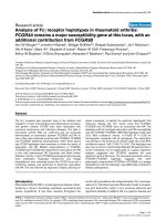
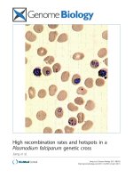
IRD was variable and ranged from 11 to 329 days with a
mean ± SD of 96 ± 89 days. Maj ority (72.9%) of the
patients developed IRD within the first three months of
HAART initiation (Figure 1).
Of the 170 IRD cases, 132 (77.6%) were new presenta-
tions (unmasking) and the 38 (22.4%) were due to wor-
sening of a recognized infections (paradoxi cal). The
most frequent OI associated with IRD in the study was
TB (66.5%, 113/170) of which 47.8% (54/113), 46% (52/
113) and 6.2% (7/113) were EPTB, PTB and DTB,
respectively. Sixty nine point nine percent (79/113) of
TB episodes were new presentations (PTB (57%, 45/79),
EPTB (39.2%, 31/79) and DTB (3.8%, 3/79), and 30.1%
(34/113) cases were due to worsening of a recognized
infection ( EPTB (67.6%, 23/34), PTB (20.6%, 7/34) and
DTB (11.8%, 4/34)). Of the total TB/IRD patients 54%
were positive for AFB and the source of specimens were
from sputum (67%) and fine needle aspiration (33%).
IRDs other than TB/IRD were toxoplasmosis (12.9%,
22/170), herpes zoster rash (12.9%, 22/170), PCP (4.1%,
7/170), and cryptococcosis (3.5%, 6/170), and the
unmasking infections involved were toxoplasmosis (22/
170), herpes zoster rash (22/170), cryptococcosis (6/170)
and PCP (3/170).
AIDS clinical stage shift was observed in 27.6% (47/
170) of the IRD patients: 32 from clinical stage III to IV,
11 from clinical stage II to III, and 4 from clinical stage
II to IV. Treatment shift w as also observed in 21.2%
(36/170) of the IRD patients, 7 from 1a to 1b, 6 from 1c
to 1d, 5 from 1a to 1c, 5 from 1b to 1c, 4 from 1b to
1d, 3 from 1a to 1d, 3 from 1c to 1a, 2 from 1d to 1b,
and 1 from 1b to 1a.
There was also a treatment shift in 6.6% (66/996) of
the non IRD patients due to peripheral neuropathy
(3.3% from 1b to 1d and 3.3% from 1a to 1c). Three
point one percent (31/996) and 1.6% (16/996) of the
non IRD patients had developed severe anemia (with a
Hgb value of less than 6.9 gm/dl) and hepatotoxicity,
respectively. Forty percent of the non-IRD patients had
developed anemia with a Hgb value of less than or
equal to 11 gm/dl.
For all study subjects, six months after initiation of
HAART, the mean ± SD CD4+ T-cell count (230 ±
118), Hgb value (13.2 ± 3.8) and WBC count (6409 ±
1998 ), showed statistically significant elevation from the
values at HAART initiation (P < 0.001). In addition,
34.5%, 31.4% and 26% of patients had significantly ele-
vated values of AST, ALT and ALP respectively com-
pared to the values at the initiation of HAART (P <
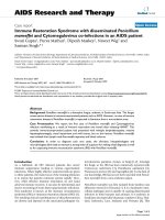
0.001). At nine months after initiation of HAART, both
IRD (73%) and non IRD (27.4%) patients had a third
CD4+ T-cell count with mean ± SD values of 220 ±
0
10
20
30
40
50
60
0-30 31-60 61-90 91-120 121-240 >240
Days after initiation of HAART
Number of patients with IRD
Figure 1 Time (days) to diagnosis of IRD after initiation of HAART.
Huruy et al. AIDS Research and Therapy 2010, 7:46
/>Page 4 of 7
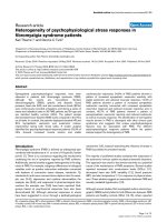
97.3 and 292 ± 145.6, respectively. The trend in CD4+
T-cell count changes ver sus number of months of treat-
ment in patients with and without IRD is shown in
Figure 2.
After commencement of HAART, laboratory values of
patients with and without IRD were compared and there
were significant increases in CD4+ T-cell count, WBC
count, ALT and AST in IRD and non IRD patients, and
ALP and Hgb values in non IRD patient s (P <0.05)
(Table 3).
Of the IRD patients 17.1% (29/170) needed to use sys-
temic anti-inflammatory treatment to alleviate symp-
toms of IRD. There were eight deaths attributable to
IRD and the causes of deaths were PTB, EPTB and DTB
in 3, 3, and 2 of them, in that order. The mean ± SD
baseline CD4+ T-cell count for these who died of IRD
was 46 ± 17.6 and 19(11.2%) of IRD pa tients required
hospitalization associated to their IRD occurrence.
Binary logistic regression was employed to assess if
age, CD4+ T-cell count, WBC count, PTB, EPTB and
DTB are possible risk factors for development of IRD.
Low CD4+ T-cell counts (odds ratio [OR], 3.16, 95%
confidence interval [CI], 2.19-4.58) and EPTB (OR, 7.7,
95% CI, 3.36-17.65) were found to be risk factors for
development of IRD.
Discussion
HAART improves immune function by suppressing HIV
viral replication and increasing CD4+ T-cell counts [19].
Since its usage, IRD has been described in association
with many concomitant infections such as mycobacterial,
fungal and viral infections. In this retrospective study,
from 1166 HIV/AIDS patients treated with HAART dur-
ing the defined period of time, the proportion of IRD was
14.6% (170/1166). This finding is consiste nt with studies
done elsewhere where the occurrence of IRD was
between 10% - 25% [3,5,8,9,20,21]. In this study most of
the IRD cases occurred within the first three months of
HAART initiation, which is in agreement with prior
reports [3,9,10].
Of the 170 IRD cases, 77.6% were new presentations,
and 22.4% were due to paradoxical episodes. This report
0
50
100
150
200
250
300
350
0
6
9
CD4 cells/microlitre
Time receiving therapy (months)
Cases
Non Cases
Figure 2 Changes in CD4+ T-cell count for IRD (cases) and non IRD (non cases) patients versus number of months of treatment.
Table 3 Laboratory values of patients with and without immune restoration disease before and after HAART
commencement, at Zewditu Memorial Hospital, Addis Ababa, Ethiopia
Variables Patients with IRD (n = 170) Patients without IRD (n = 996)
Mean (SD) values
at baseline
Mean (SD) values
after 6 months
P-value Mean (SD) values
at baseline
Mean (SD) values
after 6 months
P-value
CD4+ (cells/μl) 84 (57.8)* 185(94.8) 0.001 116 (69.4) 236(120) 0.001
WBC (cells/μl) 4246(1948) 5725 (3124) 0.001 4814 (1729) 6516(2072) 0.001
Hgb (gm/dl) 11.5(2.9) 12.1 (4.7) 0.400 12.2 (2.7) 13.9 (3.8) 0.001
AST (IU/L) 30.7(23.2) 37.8(28.8) 0.020 30(25.7) 43 (34.2) 0.001
ALT (IU/L) 25.3(20.2) 33.4(28.9) 0.003 25.1(23) 39 .2(34) 0.001
ALP (IU/L) 208(160) 213(167) 0.770 190 (149.9) 251 (212) 0.001
* Mean (SD); IRD, immune restoration disease; WBC, white blood cell; Hgb, hemoglobin; AST, aspartate aminotransferase; ALT, alanine aminotransferase; ALP,
alkaline phosphatase.
Huruy et al. AIDS Research and Therapy 2010, 7:46
/>Page 5 of 7
is in line with a previous study conducted elsewhere (8).
Our finding of TB/IRD in majority of the IRD patients
(9.7%) is in accordance with studie s conducted in India,
Thailand and Texas in which 7.6%, 12.6% and 14.4%
IRD was caused by MTB [2,10 ,22]. However, our report
is relatively low as compared with studies done in
Thailand and Texas. Our low ra te of MTB infecti on
might be exp lained partly due to genetic polymorphism
and racial differences of the study subjects [23]. And the
nature of retrospective studies that may result differences
in documenting and interpreting data in different settings
also might play a role in variation of IRD reports.
In the study, 1.9% (22/1166) of the patients developed
herpes zoster rash with m ild and unc omplicated clinical
manifestation. This finding is not consistent with a pre-
vious study in which a relatively high proportion of
herpes zoster rash was indicat ed [8]. This variation may
be due to the nature of our retros pective study. S oon
after the initiation of HAART, it was observed that
some patients presented with initial or recurrent episode
of cryptococcal meningitis during the first weeks to
months of therapy [24]. In the current study, cryptococ-
cal meningitis was observed in 0.5% (6/1166) of the
study subjects. This finding is in agreement with a pre-
vious study conducted somewhere else [8]. However, the
report is low compared to a study conducted in France
in which 8.3% cryptococcosis associated IRD was
reported [25]. This discrepancy might be due to the dif-
ference in method employed for diagnosing of
cryptococcosis.
In the study, 1.9% (22/1166) of the subjects develo ped
toxoplasmosis and this figure is similar compared to the
previous study [20]. In addition, 0.6% (7/1166) of the
patients had developed PCP and this is comparable with
a study conducted elsewhere [9].
In comparison with patients who did not develop IRD,
the IRD patients had significantly low CD4+T- cell
count and WBC count, and higher proportion of EPTB
and younger age at baseline (P < 0.05). However, in bin-
ary logistic regression analyses low CD4+ T-cell count
and EPTB were found to be risk factors for development
of IRD. Previous studies also described that both low
baseline CD4+ T-cell count and EPTB as the possible
risk factors that were associated with the occurrence of
IRD [22,26].
Thirty-one (3.1%, 31/996) patients had developed
severe anemia with Hgb value below 6.9 gm/dl [27].
Thismightbeduetothenatureofsomeantiretroviral
drugs which have myelosuppressive effect, especially
with respect to the red blood cells which eventually lead
to the development of anemia [28]. Sixteen (1.6%,16/
996) of the study subjects also developed hepatotoxicity
with three to five fold increments i n serum levels of
AST and ALT. This finding is in accordance with a
study conducted by Becker [29]. This might be due to
the direct effect of antiretroviral drugs, mainly nevira-
pine, that induce the development of hepatotoxicity
[30]. Consistent with a previous report [8], in the pre-
sent study we observed a 4.7% mortality rate after initia-
tion of HAART among IRD patients.
Conclusions
In this retrospective study, 14.6% of the patients had
clinical deterioration (IRD) during immune recovery and
eight deaths were attributable to IRD. Most IRDs were
observed within the first three months of HAART initia-
tion, primarily affecting patients with lower baseline
CD4+ T-cel l counts and the majority of IRD cases were
TB/IRD. Low baseline CD4+ T-cell count and EPTB
were associated with development of IRD. Therefore,
strict following of patients during the first three months
of HAART initiation and diagnosis of latent TB [31]
would help to prevent complications related to TB/IRD.
Acknowledgements
We thank: University of Gondar, Ethiopia and ART staffs of Zewditu memorial
hospital, Addis Ababa Ethiopia, particularly Dr. Aster Shewa-Amare and Dr.
Addis Akalu for kind support during the data collection period and Mr.
Wubet Birhan for help during data entry.
Author details
1
Department of Medical Laboratory Technology, College of Medicine and
Health Sciences, University of Gondar, Ethiopia.
2
Institute of Virology, Faculty
of Medicine, University of Leipzig, Germany.
3
Department of Microbiology
and Parasitology, College of Medicine and Health Sciences, University of
Gondar, Ethiopia.
4
Division of Allergy and Clinical Immunology, Department
of Medicine, University of Colorado, Denver, USA.
5
Faculty of Social Sciences
and Humanities, University of Gondar, Gondar, Ethiopia.
6
Institute of
Psychology II, Clinical and Health Psychology, University of Leipzig, Germany.
Authors’ contributions
KH: Study design, data collection, data analysis and write up; AK: Data
analysis and write up; AM: Study design and write up; YW: write up. All
authors read and approved the final manuscript.
Competing interests
All authors declared that no competing interest. The content of this
manuscript has not been published and/or submitted for consideration of
publication elsewhere.
Received: 18 May 2010 Accepted: 21 December 2010
Published: 21 December 2010
References
1. Palella FJ Jr, Delaney KM, Moorman AC, Loveless MO, Fuhrer J, Satten GA,
Aschman DJ, Holmberg SD: Declining morbidity and mortality among
patients with advanced human immunodeficiency virus infection. HIV
Outpatient Study Investigators. N Engl J Med 1998, 338:853-860.
2. Kumarasamy N, Chaguturu S, Mayer KH, Solomon S, Yepthomi HT,
Balakrishnan P: Incidence of Immune Reconstitution Syndrome in HIV/
Tuberculosis -Co-infected Patients After Initiation of Generic Antiretroviral
Therapy in India. J Acquir Immune Defic Syndr 2004, 37:1574-76.
3. Shelburne SA, Hamill RJ, Greenberg SB, Atmar RL, Musher DW, Gathe JC Jr,
Visnegarwala F, Trautner BW: Immune reconstitution inflammatory
syndrome: emergence of a unique syndrome during highly active
antiretroviral therapy. Medicine 2002, 81:213-217.
4. Shelburne SA, Hamill RJ: The immune reconstitution inflammatory
syndrome. AIDS Rev 2003, 5:67-79.
Huruy et al. AIDS Research and Therapy 2010, 7:46
/>Page 6 of 7
5. DeSimone JA, Pomerantz RJ, Babinchak TJ: Inflammatory reactions in HIV-
infected persons after initiation of highly active antiretroviral therapy.
Ann.Intern.Med 2000, 133:447-453.
6. Cheng VC, Yuen KY, Chan WM, Wong SS, Ma ES, Chan RM: Immune-
restitution disease involving the acute and adaptive response. Clin. Infect.
Dis 2000, 30:882-890.
7. Foudraine NA, Hovenkamp E, Notermans DW, Meenhorst PL, Klein MR,
Lange JM, Miedema F, Reiss P: Immunopathology as a result of highly
active antiretroviral therapy in HIV-1-infected patients. AIDS 1999,
13:177-184.
8. Murdoch DM, Venter WD, Feldman C, Van Rie A: Incidence and risk factors
for the Immune reconstitution inflammatory syndrome in HIV patients
in South Africa: Prospective study. AIDS 2008, 22:601-610.
9. Ratnam I, Chiu C, Kandala NB, Easterbrook PJ: Incidence and risk factors
for immune reconstitution inflammatory syndrome in an ethnically
diverse HIV type 1-infected cohort. Clin Infect Dis 2006, 42:418-427.
10. Shelburne SA, Visnegarwala F, Darcourt J, Graviss EA, Giordano TP, White AC
Jr, Hamill RJ: Incidence and risk factors for immune reconstitution
inflammatory syndrome during highly active antiretroviral therapy. AIDS
2005, 19:399-406.
11. Zampoli M, Kilborn T, Eley B: Tuberculosis during early antiretroviral-
induced immune reconstitution in HIV-infected children. Int J Tuberc
Lung Dis 2007, 11:417-423.
12. Lawn SD, Myer L, Bekker LG, Miller FR: Immune reconstitution disease
associated with mycobacterial infections in HIV-infected individuals
receiving antiretrovirals. Lancet Infect Dis 2005, 5:361-373.
13. Federal HIV/AIDS prevention and control office: Guidelines for
management of opportunistic infections and antiretroviral treatment in
adolescents and adults in Ethiopia. MOH 2007.
14. Cheesbrough M: District laboratory practice in tropical countries. Part I.
Cambridge University Press, Cambridge, England; 1998, 191-239.
15. Klotz SA, Aziz Mohammed A, Girmai Woldemichael M, Worku Mitku M,
Handrich M: Immune Reconstitution Inflammatory Syndrome in a
Resource-Poor Setting. JIAPAC 2009, 8:122-127.
16. Robertson J, Meier M, Wall J, Ying J, Fichtenbaum CJ: Immune
reconstitution syndrome in HIV: validating a case definition and
identifying clinical predictors in persons initiating antiretroviral therapy.
Clin Inf Dis 2006, 42:1639-46.
17. Shelburne S, Montes M, Hamill RJ: Immune reconstitution inflammatory
syndrome: more answers more questions. J Antimicrob Chemother 2006,
57:167-170.
18. Meintjes G, Lawn SD, Scano F, Maartens G, French MA, Worodria W,
Elliott JH, Murdoch D, Wilkinson RJ, Seyler C, Laurence J, Loeff MSV, Reiss P,
Lynen L, Janoff EN, Gilks C, Colebunders R: Tuberculosis-associated
immune reconstitution inflammatory syndrome: case definitions for use
in resource-limited settings. Lancet Infect Dis 2008, 8:516-523.
19. Phillips P, Bonner S, Gataric N, Bai T, Wilcox P, Hogg R, O
’Shaughnessy M,
Montaner J: Non tuberculosis Mycobacterial Immune Reconstitution
Syndrome In HIV- Infected Patients: Spectrum of disease and Long -
Term Follow Up. Clin Infec Dis 2005, 41:1483-94.
20. Jevtović DJ, Salemovic D, Ranin J, Pesic I, Zerjav S, Djurkovic-Djakovic O:
The prevalence and risk of immune restoration disease in HIV-infected
patients treated with highly active antiretroviral therapy. HIV Med 2005,
6:140-143.
21. French MA, Price P, Stone SF: Immune restoration disease after
antiretroviral therapy. AIDS 2004, 18:1615-27.
22. Manosuthi W, Kiertiburanakul S, Phoorisri T, Sungkanuparph S: Immune
reconstitution inflammatory syndrome of tuberculosis among HIV-
infected patients receiving antituberculous and antiretroviral therapy.
Journal of Infection 2006, 53:357-363.
23. Price P, Morahan G, Huang D, Stone E, Cheong KY, Castley A, Rodgers M,
Mclntyre MQ, Abraham LJ, French MA: Polymorphisms in cytokine genes
define subpopulations of HIV-1 patients who experienced immune
restoration diseases. AIDS 2002, 16:2043-47.
24. Woods ML, MacGinley R, Eisen D, Allworth AM: HIV combination therapy:
partial immune reconstitution unmasking latent cryptococcal infection.
AIDS 1998, 12:1491-94.
25. Lortholary O, Fontanet A, Memain N, Martin A, Sitbon K, Dromer F:
Incidence and risk factors of immune reconstitution inflammatory
syndrome complicating HIV-associated cryptococcosis in France. AIDS
2005, 19:1043-49.
26. Grant PM, Komarow L, Andersen J, Sereti I, Pahwa S, Lederman MM, Eron J,
Sanne I, Powderly W, Hogg E, Suckow C, Zolopa A: Risk Factor Analyses
for Immune Reconstitution Inflammatory Syndrome in a Randomized
Study of Early vs. Deferred ART during an Opportunistic Infection. PLoS
ONE 2010, 5:e11416.
27. WHO/UNU/UNICEF: Iron deficiency anaemia. Assessment, prevention and
control. A guide for programme managers. Geneva, World Health
Organization; 2001, (WHO/NHD/01.3).
28. DeJesus E, Herrera G, Teofilo E, Gerstoft J, Buendia CB, Brand JD,
Brothers CH, Hernandez J, Castillo SA, Bonny T, Lanier ER, Scott TR: Abacavir
versus zidovudine combined with lamivudine and efavirenz, for the
treatment of antiretroviral-naive HIV-infected adults. Clin Infect Dis 2004,
39:1038-46.
29. Becker S: Liver toxicity in epidemiological cohorts. Clin Infect Dis 2004, 38:
S49-55.
30. Wit FW, Weverling GJ, Weel J, Jurriaans S, Lange JM: Incidence and risk
factors for severe hepatotoxicity associated with antiretroviral
combination therapy. J Infect Dis 2007, 186:23-31.
31. Jiang W, Shao L, Zhang Y, Zhang S, Meng C, Xu Y, Huang L, Wang Y,
Wang Y, Weng X, Zhang W: High-sensitive and rapid detection of
Mycobacterium tuberculosis infection by IFN-γ release assay among HIV-
infected individuals in BCG-vaccinated area. BMC Immunol 2009, 10:31-37.
doi:10.1186/1742-6405-7-46
Cite this article as: Huruy et al .: Immune restoration disease and
changes in CD4+ T-cell count in HIV- infected patients during highly
active antiretroviral therapy at Zewditu memorial hospital, Addis Ababa,
Ethiopia. AIDS Research and Therapy 2010 7:46.
Submit your next manuscript to BioMed Central
and take full advantage of:
• Convenient online submission
• Thorough peer review
• No space constraints or color figure charges
• Immediate publication on acceptance
• Inclusion in PubMed, CAS, Scopus and Google Scholar
• Research which is freely available for redistribution
Submit your manuscript at
www.biomedcentral.com/submit
Huruy et al. AIDS Research and Therapy 2010, 7:46
/>Page 7 of 7