Anal and rectal diseases explained - part 8 pot
Bạn đang xem bản rút gọn của tài liệu. Xem và tải ngay bản đầy đủ của tài liệu tại đây (641.01 KB, 23 trang )
7.1 Diarrhea 155
7.2 Fecal impaction 159
7.3 Ileoanal pouch anastomosis 161
7.4 Pilonidal sinuses 167
7.5 Rectal foreign bodies 169
Miscellaneous anorectal
conditions
Chapter 7
This is trial version
www.adultpdf.com
154
This is trial version
www.adultpdf.com
Epidemiology
Self-limited diarrhea is extremely common. The passage of loose or watery stools
without abdominal pain was found to occur in 4.3% of males and 2.2% of females
surveyed in Bristol, UK during a 1-year period. Chronic diarrhea is thought to
affect 5% of the adult population annually in the United States, and approximately
450,000 patients are hospitalized.
Patients at risk
People with diabetes, celiac sprue, pancreatic disorders, or small intestinal
disorders; travelers to Third World countries; HIV-infected patients; people on
antibiotics; patients undergoing or having had radiation therapy; patients who have
had surgery of the stomach, small intestine, or colon; and individuals receiving
enteric formula feedings. A variety of medications and herbal preparations have
laxative effects.
Pathophysiology
Diarrhea occurs when the normal absorptive mechanism of the small intestine and
colon is overwhelmed by excessive fluid secretion and hypermotility. The overall
result is the passage of multiple frequent stools. Diarrhea is most objectively
defined as the passage of more than 200 mL (200 g) of stool per day. Diarrhea can
be divided into several categories, which are outlined below, together with
common causes of each.
Acute
Symptoms lasting from several days to 4 weeks. The majority of cases of acute
diarrhea are due to viral, bacterial, or parasitic infection.
Chronic
Symptoms lasting >4 weeks. A large number of conditions can result in the
development of chronic diarrhea. Chronic diarrhea may be further divided into two
main categories: osmotic diarrhea and secretory diarrhea.
155
Diarrhea
Chapter 7.1
This is trial version
www.adultpdf.com
Osmotic diarrhea
Malabsorbed or poorly absorbed sugars, other carbohydrates, and other
osmotically active substances (such as magnesium) produce laxative effects by
inducing the secretion of water. Since the overall osmolality of stool must remain
at approximately 290 mosm/L, the presence of osmotically active substances in the
colonic lumen results in net water secretion and increased stool volume.
Secretory diarrhea
A variety of conditions – including hypermotility, infectious and inflammatory
disorders, excessive secretion of chloride or bicarbonate, or decreased absorption
of sodium – result in release of fluids and electrolytes.
Symptoms
Passage of frequent watery or soft stools. Severe diarrhea may be associated with
dehydration and consequent electrolyte disturbance. Frequent small stools with
cramping and urgency suggest proctitis or left-sided colitis (see Figure 1). Large
volume stools suggest a small intestinal source of diarrhea. Bloating, flatulence, and
foul smelling and oily stools occur in malabsorptive states. Recent foreign travel
suggests the presence of an infectious source.
Diagnostic testing
A stool sample should be obtained, checked for parasites and Clostridium difficile
toxin, and cultured. Other evaluations include fecal volume, fecal fat, electrolyte
and pH measurement, complete blood count, serum chemistries, celiac sprue
panel, thyroid stimulating hormone, flexible sigmoidoscopy, colonoscopy, small
intestine biopsy, and 24-hour urine test for 5-HIAA (5-hydroxyindole acetic acid).
Figure 1. Infectious
colitis due to
cytomegalovirus in a
patient with chronic
myelogenous leukemia.
Chapter 7
156
This is trial version
www.adultpdf.com
Treatment
Identification of the underlying source of this symptom is critical for initiating
proper therapy. It is best to control diarrhea by direct treatment of the cause.
Treatments may include anti-inflammatory agents for inflammatory bowel disease
and a gluten-free diet for celiac sprue. Treatments that may provide relief of the
symptoms of diarrhea in the presence or absence of organic disorders include fiber,
opioids, cholestyramine, octreotide, and anticholinergic agents.
Clinical pearls
A careful history will assist in differentiating various causes of diarrhea. The
possibility of laxative abuse should not be ignored. Patients with diarrhea and fecal
incontinence generally experience improvement in their symptoms of incontinence
when their diarrhea is under control.
Diarrhea
157
This is trial version
www.adultpdf.com
158
This is trial version
www.adultpdf.com
Definition
Fecal impaction is the development of a colonic obstruction due to filling of the
lumen with a large, hard stool. It occurs most commonly in the rectum.
Epidemiology
The rising incidence of fecal impaction parallels the increasing prevalence of
chronic constipation. Fecal impaction is the cause of colonic obstruction in up to
50% of bedridden patients in nursing homes and patients with spinal cord injuries.
Patients at risk
Patients with spinal cord injuries and bedridden patients. Constipation may occur
in up to 25% of the elderly population, and is three-times more common in
women than in men. A variety of medications – including calcium channel
blockers, anticholinergics, opioids, antidepressants, and antipsychotics –
predispose to constipation and, therefore, the development of fecal impaction. A
number of neurologic diseases (Parkinson’s disease, dementia, multiple sclerosis)
are associated with decreased colonic function and constipation, therefore placing
patients at risk for fecal impaction. Endocrine disorders including diabetes and
hypothyroidism are additional risk factors. Dehydration increases the likelihood of
developing fecal impaction in high-risk patients.
Symptoms
Constipation, rectal pain, and a sensation of a rectal mass are common symptoms.
Other symptoms, including diarrhea and fecal incontinence due to overflow of
liquid stool past the impacted fecal bolus, may be present. Patients with neurologic
diseases or spinal cord injury may be unaware of the presence of the fecal
impaction. In addition, fecal impaction in patients with spinal cord injury may lead
to autonomic dysreflexia, a medical emergency characterized by the acute
development of symptomatic hypertension with hyperactive reflexes. Rectal
bleeding may occur in patients with stercoral ulcers (see Pathophysiology). In
extreme cases of fecal impaction, colonic obstruction with abdominal distention
and signs and symptoms of bowel perforation or peritonitis may be present.
159
Fecal impaction
Chapter 7.2
This is trial version
www.adultpdf.com
Pathophysiology
Decreased neuromuscular function of the colon results in colonic hypomotility,
prolonged transit time in the colon, and fecal retention. Increased contact time
between fecal material and the colon results in firm, dehydrated stools. A vicious
cycle may develop in which increasing stool retention further delays motility and
produces even drier, firmer stools. Altered sensorium may exacerbate the problem
through the loss of normal impulses to defecate. A hard stool may be retained for
such a prolonged period of time in a single segment of the colon that ischemic
ulceration – a stercoral ulcer – may occur.
Diagnostic testing
Examination of the abdomen may reveal the presence of soft or firm masses,
particularly over the left colon. Digital rectal examination will reveal a firm, mobile
mass in the rectum. An abdominal x-ray will demonstrate the presence of stool
accumulation in the colon. A sigmoidoscopy may be required to rule out other
types of rectal mass, eg, carcinoma.
Treatment
Most forms of fecal impaction can be treated with digital fecal disimpaction. However,
this procedure may produce marked discomfort and even hypotension in some
patients, and, therefore, some form of sedation should be considered. Following the
removal of the largest and most obstructive fecal boluses, follow-up with gentle
enema therapy is performed. In patients who have developed fecal impaction, a
bowel regimen including laxatives and enemas on a regular basis is suggested.
Clinical pearls
It is particularly important to remind patients who are on medications that cause
constipation to consume large volumes of liquid on a daily basis, for example 5–8
glasses of water or other nonalcoholic fluids daily. Patients who have an episode of
fecal impaction should be placed on a regular regimen of stool softeners and/or
osmotic laxatives as a prophylaxis against further episodes.
Chapter 7
160
This is trial version
www.adultpdf.com
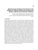
General description
In patients requiring proctocolectomy, ileoanal pouch anastomosis has obviated
the need for ileostomy as it preserves fecal continence. A direct anastomosis
between the ileum and anus was first performed in 1968. In 1978, creation of an
“S pouch”, which functions as a reservoir, was incorporated into the procedure.
The J pouch, which is currently the most commonly performed procedure, was
introduced in 1980. Ileoanal pouch anastomosis after creation of a J or S pouch is
now the procedure of choice in appropriate patients requiring complete removal of
the colon (see Figure 1).
Figure 1. Diagram of ileoanal pouch anastomosis.
161
Ileum
Sutured
to dentate
line
Anal
canal
External anal
sphincter
Rectal
tunica
muscularis
Ileoanal pouch anastomosis
Chapter 7.3
This is trial version
www.adultpdf.com
The indications, relative contraindications, and contraindications to this procedure
are outlined in Table 1.
Table 1. Indications and contraindications for ileoanal pouch anastomosis.
Alternative procedures
End ileostomy (Brook ileostomy) or continent ileostomy (Kock pouch).
How the procedure is performed
This procedure may be carried out in one, two, or three stages depending on the
preference of the performing surgeon and the general condition of the patient. For
example, in a patient with severe acute colitis, a colectomy and loop ileostomy may
be created for the first stage. After 3–6 months, when the patient is physically and
nutritionally improved, a proctectomy is performed with creation of a J pouch.
Finally, in the third stage of the procedure, the loop ileostomy is closed.
The first stage in the procedure is a total abdominal colectomy (some centers
perform the colectomy laparoscopically). The rectum is then dissected within the
pelvis through the dilated anal canal; the surgeon must be especially cautious
during this stage of the procedure to preserve the anal sphincter, local portions of
the genitourinary systems, and perineal nerves. The distal 15 cm of the ileum is
divided and then folded back onto itself (in a J shape) and opened to produce a
reservoir. Temporary ileostomy may be performed to protect the pouch and then
closed on a later occasion. The ileoanal anastomosis may be hand sewn or stapled;
a double-stapled technique is utilized for stapled anastomosis (see Figure 2).
Indications Contraindications Relative contraindications
Chronic ulcerative colitis Crohn’s disease Massive obesity
Familial adenomatous Cancer of the distal Emergency operation
polyposis rectum
Multiple colorectal Poor anal sphincter Use of steroid medication
malignancies function
Anal sphincter excised Indeterminate colitis
Age >65 years
Chapter 7
162
This is trial version
www.adultpdf.com
Ileoanal pouch anastomosis
163
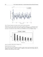
Figure 2. Construction of J-shaped ileal pouch. (A, B) The terminal ileum is divided and
the colon is removed.
(C) The terminal ileum is fashioned into a J-shape with 15-cm
limbs.
(D, E) The antimesenteric border of ileum is divided. (F) The posterior mucosal
layer of pouch is sutured.
(G) The pouch is completed.
A
B
CD E
FG
Terminal
ileum
15
cm
Incision
Sutures in
mucosa
Completed
ileal
pouch
Sutures
in serosa
Serosa
Mucosa
This is trial version
www.adultpdf.com
Results obtained
More than 90% of patients report satisfaction with their procedure. Mild fecal
incontinence, particularly with spotting of stool in the underclothing at night,
occurs chronically in up to 50% of patients having this procedure, and
approximately 25% of patients with an ileoanal pouch anastomosis will wear a pad
to prevent soiling of underclothing.
Complications
Surgical
Approximately 30% of patients who undergo this ileoanal pouch anastomosis will
experience a surgical complication. Anastomotic leakage occurs in about 10% of
patients undergoing this procedure (see Figure 3). This is managed with
intravenous antibiotics, drainage of pelvic fluid, and bowel rest. Abdominal sepsis,
which occurs in <5% of patients who have an ileoanal pouch anastomosis, often
results in pouch failure and excision. These patients then require a permanent
ileostomy. Small bowel obstruction is seen in approximately 20% of patients and
requires additional surgery in about half of these. Anastomotic strictures are
common (5%–15% of patients) and are usually easily managed with digital
dilatation or insertion of Hegar’s dilators. Surgical repair of strictures with revision
of the ileoanal anastomosis is necessary in a small number of cases.
Figure 3. Ileoanal pouch
anastomosis. Several small
anastomotic leaks are
demonstrated on dynamic
proctography (arrows).
Chapter 7
164
This is trial version
www.adultpdf.com
Long-term
Fecal leakage and incontinence as described in the Results obtained section
above. The other long-term complication of ileoanal pouch anastomosis is sexual
dysfunction. Male patients report a 2% prevalence of impotence and a 2%–4%
prevalence of retrograde ejaculation. Although a small percentage of women
complain of dyspareunia (difficult or painful coitus), 50% report improved sexual
function following the procedure.
Pouchitis
This is a nonspecific inflammatory disorder. Symptoms include watery diarrhea,
passage of blood, and cramping of the abdomen. In some patients, pouchitis is
associated with systemic symptoms such as fever and arthralgia.
Mucosal edema, granularity, and/or ulcerations may be seen endoscopically in the
affected pouch. The etiology of pouchitis has not been fully determined. The
condition may represent overgrowth of anaerobic bacteria within the pouch,
decreased mucosal exposure to intraluminal nutrients, or autoimmune induced
inflammation. Pouchitis occurs in 20%–50% of patients who receive ileoanal
pouch anastomosis for ulcerative colitis, but rarely in patients who undergo the
procedure due to familial polyposis coli. Episodes occur most commonly within
the first 6 months following creation of the pouch. In 39% of patients, only a single
acute episode occurs.
Five percent of patients develop recurrent or chronic pouchitis. About half of these
patients will need resection of the pouch. The standard treatment is metronidazole
(10–20 mg/kg/day), sometimes in combination with ciprofloxacin (500 mg, bid).
Treatment duration is usually 2–4 weeks. Chronic pouchitis has been treated with
5-ASA (5-acetylsalicylic acid) containing agents such as Pentasa or mesalamine
enemas, and immunosuppressant drugs, eg, corticosteroids azathioprine, short-
chain fatty acid enemas, and probiotics. Administration of live probiotic bacteria
has been demonstrated to maintain remission in patients with chronic pouchitis.
Dysplasia and cancer
Depending on the type of rectal dissection performed, a small portion of the rectal
mucosa from the anal transition zone can be left at the site of the anastomosis. This
cuff of rectal tissue is larger when a double-stapled technique is used. Although
rare, dysplasia and carcinoma have been reported in this remaining portion of
rectum mucosa. Current recommendations include surveillance sigmoidoscopy
with biopsies at the site of the anastomosis every 1–3 years.
Ileoanal pouch anastomosis
165
This is trial version
www.adultpdf.com
Clinical pearls
Patients with symptoms of pouchitis require a careful history and endoscopic
evaluation with biopsies to confirm the diagnosis. Other entities that mimic
symptoms of pouchitis include acute gastroenteritis and recurrent inflammatory
bowel disease (Crohn’s disease). Adaptation with improvement of pouch function,
as demonstrated by decreased stool frequency and increased fluid and electrolyte
absorption, occurs during the first 6–12 months following pouch construction.
Chapter 7
166
This is trial version
www.adultpdf.com
Epidemiology
Symptomatic pilonidal sinuses generally develop between the ages of 20 and 30
years. Three-quarters of cases are seen in males. There is some suggestion that
trauma to the skin overlying the sacrococcygeal region (such as strenuous activity
and sitting in vehicles in rugged environments – as seen in military personnel) may
increase the likelihood of development of the condition.
Symptoms
If an abscess is present, pain may be the predominant symptom. Otherwise,
patients will notice swelling, drainage, and tenderness of the affected area.
Pathophysiology
Pilonidal sinuses develop in the intergluteal cleft and in the skin overlying the
sacrum and coccygeal bone (see Figure 1). The condition develops when a sinus
tract forms following an episode of folliculitis and abscess formation. The initiating
factor may be a plug of keratin that develops in the hair follicle. Shafts of hair
entering a previously developed sinus may also initiate the condition. Recurrent
abscesses, infection, and multiple sinus tracts may be seen.
Figure 1. Pilonidal sinuses. On examination, pits or external openings in the intergluteal
cleft are seen. The openings often communicate with each other, as shown on the right.
167
Pilonidal sinuses
Chapter 7.4
This is trial version
www.adultpdf.com
Diagnosis
Physical examination reveals an area of inflammation, tenderness, and erythema in
the gluteal crease, usually 5–7 cm from the anal opening. Hair follicles will often
be noted at the site of the lesion and there is often more than one sinus opening.
The presence of hair follicles and the lack of an opening from within the anorectal
region differentiate pilonidal cysts from anal and rectal fistulas.
Treatment
If an abscess is present, incision and drainage are the treatments of choice,
followed by complete excision when the acute process is resolved. Shaving hair
from the intergluteal cleft on a weekly basis decreases the chance of recurrence.
Clinical pearls
Recurrences following excision of the pilonidal sinus need additional excisions.
In refractory cases, more extensive excision surgery may be required.
Chapter 7
168
This is trial version
www.adultpdf.com
Epidemiology
The majority of the foreign bodies found in the rectum have been placed there
rectally; however, swallowed objects may occasionally lodge in the rectum. Foreign
bodies may be introduced either intentionally (autoeroticism, sexual activity, rape)
or unintentionally (as a means of dislodging impacted stool).
Patients at risk
Homosexuals; individuals practicing receptive anal sex utilizing foreign objects;
rape victims; children; people with altered sensorium; and patients with rectal or
anal strictures (in the case of swallowed objects).
Pathophysiology
After placing large objects in the rectum, intense anal spasm and/or pain
sometimes prevent simple removal.
A sharp swallowed object may lodge itself in normal rectal mucosa. Other swallowed
objects may become impacted at the rectosigmoid junction. Narrowed luminal caliber
secondary to strictures or diverticular disease increases the likelihood of impaction.
Symptoms
Pain in the abdomen or rectum, rectal bleeding, discharge, and symptoms of
peritonitis (abdominal distention, fever, peritoneal signs).
Diagnostic testing
Physical examination should include careful abdominal examination to rule out
peritonitis. The abdomen should be palpated for masses and the anus should be
carefully inspected for evidence of fissure and/or anal trauma. Prior to the
performance of a rectal examination, an abdominal x-ray should be performed (see
Figure 1). Subsequently a digital rectal exam can be carefully performed (as long
as a sharp object is not suspected). Assessment of anal sphincter tone is
recommended. Patients may require anesthesia for adequate examination.
169
Rectal foreign bodies
Chapter 7.5
This is trial version
www.adultpdf.com
Treatment
Following adequate anesthesia, small objects may be digitally removed. Endoscopic
removal may be possible in selected cases; removal of sharp objects requires an
overtube. General anesthesia is used for the removal of larger objects. This can be
performed using a variety of devices including obstetric forceps and modified
padded pliers. After the insertion of a rigid proctoscope, a Foley catheter or a
Sengstaken–Blakemore tube may be inflated proximal to the object, which is then
pulled down towards the anus. Patients who have evidence of peritonitis will
require laparotomy.
Clinical pearls
The rectosigmoid region should be visualized endoscopically following removal of
a foreign object to ensure that it has been completely removed and to rule out
mucosal injury.
Figure 1. Rectal foreign
body (light bulb)
demonstrated on
abdominal radiograph.
Chapter 7
170
This is trial version
www.adultpdf.com
8.1 Anal fissure 173
8.2 Fecal incontinence 175
8.3 Hemorrhoids 177
8.4 Kegel exercises 179
8.5 Nonrelaxing puborectalis syndrome 181
8.6 Perianal Crohn’s disease 185
8.7 Pruritus ani 189
8.8 Radiation proctopathy 191
8.9 Rectal prolapse 193
8.10 Solitary rectal ulcer syndrome 195
8.11 Ulcerative proctitis 197
8.12 Venereal warts 199
Patient information
Chapter 8
This is trial version
www.adultpdf.com
172
This is trial version
www.adultpdf.com
What is an anal fissure?
An anal fissure is a tear or crack in the lining of the anal canal.
How does an anal fissure develop?
Anal fissures are believed to start with passage of a large, hard bowel movement
that results in tearing of the skin (anoderm) of the anal canal. Excess contraction
or spasm of the anal sphincter and weakening of the area where the fissure
develops can contribute to the condition. In time, if the fissure does not heal, a
chronic anal fissure or ulcer may develop.
What are the symptoms of an anal fissure?
The most common symptoms of an anal fissure are pain during the process of
defecation and after completion of the bowel movement. Minor bleeding from the
anus may occur. With time, pain and pressure in the anal area may become more
continuous and severe.
Are there any other conditions that cause the
same symptoms as an anal fissure?
Thrombosed hemorrhoids may mimic the symptoms of an anal fissure. Other
diseases of the anus, such as infections and tumors, may have similar symptoms.
What factors increase the risk of developing an anal fissure?
Anal fissures may occur in association with Crohn’s disease, anal and rectal
infections, AIDS, and tumors of the anus. Constipation, straining, and passage of
hard bowel movements may initiate the development of an anal fissure. A low fiber,
high fat diet may predispose to the condition.
Can anal fissures predispose to cancer?
No.
What tests are performed to diagnose anal fissures?
A physical examination of the anus is often enough to make the diagnosis.
Sometimes, an anoscope or sigmoidoscope may be used to assist with the diagnosis.
173
Anal fissure
Chapter 8.1
This is trial version
www.adultpdf.com
What over-the-counter treatments or home remedies
can be used for anal fissures?
Stool softeners such as dioctyl sodium sulfosuccinate (Colace); bulking agents
such as psyllium (Metamucil, Konsyl), methylcellulose (Citrucel), or calcium
polycarbophyl (FiberCon, Konsyl); local anesthetic creams containing lidocaine
(Analpram, Lidomantle, Tronolane).
What prescription medications are used for anal fissures
and how do they work?
Nitroglycerin ointment may relax the anal sphincter and allow the fissure to heal.
What nonsurgical procedures can be used to treat anal fissures?
Botulinum toxin (Botox) injections into the anal sphincter may be used. These
cause relaxation of the sphincter and allow for better healing of the fissure. Anal
dilatation (stretching of the anal muscle) using a finger or dilating device is
sometimes used.
Is surgery ever used as a treatment for anal fissures?
Yes.
When is surgery used to treat anal fissures?
When symptoms persist despite medical therapy and/or Botox injections.
What surgical procedures are performed for the
treatment of anal fissure?
Lateral sphincterotomy (making a small incision into the internal anal sphincter).
This may be accompanied by surgical removal of the fissure itself.
What additional information should I know about anal fissures?
Anal fissures may occur at any age and are even seen in infants. New medical
therapies (nitroglycerin ointment, Botox) have helped many patients avoid the
need for surgery for this condition.
Chapter 8
174
This is trial version
www.adultpdf.com
What is fecal incontinence?
Fecal incontinence involves the loss of rectal contents including gas, mucus, or
stool without awareness or control of the occurrence.
How does fecal incontinence occur?
As a result of damage to the nerves and muscles that normally control the function
of the anus and rectum. Fecal incontinence may also be present in persons with
brain or spinal cord damage. Severe or frequent diarrhea may overwhelm the anal
sphincter and cause leakage.
What are the symptoms of fecal incontinence?
Inability to control passage of stool or gas. Fecal incontinence is often characterized
by soiling of clothing and bedding.
Are there any other conditions that cause the same
symptoms as fecal incontinence?
Colitis, proctitis, and anal or rectal infection.
What factors increase the risk of developing fecal incontinence?
Aging, prior surgery of the anus, trauma to the anus, injury to the anal muscles
during delivery of a baby, neurologic diseases, brain damage, mental retardation.
Can fecal incontinence predispose to cancer?
No.
What tests are performed to diagnose fecal incontinence?
Anorectal manometry, anorectal ultrasound, anorectal electromyography (EMG),
defecography.
What over-the-counter treatments or home remedies
can be used for fecal incontinence?
Fiber supplements, especially calcium polycarbophyl tablets (FiberCon, Konsyl), or
loperamide (Imodium). The elimination of dairy products from the diet may
improve symptoms.
175
Fecal incontinence
Chapter 8.2
This is trial version
www.adultpdf.com