ANEMIAS AND OTHER RED CELL DISORDERS - PART 9 ppt
Bạn đang xem bản rút gọn của tài liệu. Xem và tải ngay bản đầy đủ của tài liệu tại đây (734.18 KB, 39 trang )
300 HEMOGLOBIN DISORDERS SECTION V
of these patients.
27
A former childhood disorder is increasingly a disease of adults.
Widespread screening of individuals from populations where β-thalassemia trait is
prevalent has enabled prenatal diagnosis of affected fetuses. In Greek American and
Italian American communities, the number of affected infants has sharply decreased.
The number of children with thalassemia in the United States is rising however
as a result of the increase in the number of Americans of Southeast Asian ancestry.
These more recent arrivals to the country have not yet achieved the cohesive approach
to thalassemia that proved so successful in other American communities. Until this
goal is reached, thalassemia will remain an important issue in American medicine
(Tables 14-3 and 14-4).
References
1
Cooley TB, Witwer ER, Lee P. 1927. Anemia in children with splenomegaly and peculiar
changes in bones: Report of cases. Am J Dis Child 34:347–355.
2
Olivieri NF. 1999. The beta-thalassemias. N Engl J Med 341:99–109.
3
Wasi P, Winichagoon P, Baramee T, Fucharoean S. 1982. Globin chain synthesis in het-
erozygous and homozygous hemoglobin E. Hemoglobin 6:75–78.
4
Fairbanks VF, Gilchrist GS, Brimhall B, Jereb JA, Goldston EC. 1979. Hemoglobin E trait
revisited: A cause of microcytosis and erythrocytosis. Blood 53:109–115.
5
Fairbanks VF, Oliveros R, Brandabur JH, Willis RR, Fiester RF. 1980. Homozygous
hemoglobin E mimics b-thalassemia minor without anemia or hemolysis: Hematologic,
functional and biosynthetic studies of first North American cases. Am J Hematol 8:109–
121.
6
Rees DC, Styles L, Vichinsky EP, Clegg JB, Weatherall DJ. 1998. The hemoglobin E
syndromes. Ann N Y Acad Sci 850:334–343.
7
Fucharoen S, Ketvichit P, Pootrakul P, Siritanaratkul N, Piankijagum A, Wasi P. 2000.
Clinical manifestation of beta thalassemia/hemoglobin E disease. J Pediatr Hematol Oncol
22:552–557.
8
Chui DH, Fucharoen S, Chan V. 2003. Hemoglobin H disease: Not necessarily a benign
disorder. Blood 101:791–800.
9
Chen FE, Ooi C, Ha SY, et al. 2000. Genetic and clinical features of hemoglobin H disease
in Chinese patients. N Engl J Med 343:544–550.
10
Scopinaro F, Banci M, Vania A, et al. 1993. Radioisotope assessment of heart damage in
hypertransfused thalassaemic patients. Eur J Nucl Med 20:603–608.
11
Lattanzi F, Bellotti P, Picano E, et al. 1993. Quantitative ultrasonic analysis of myocardium
in patients with thalassemia major and iron overload. Circulation 87:748–754.
12
Prennell DJ, Bland JM. 2003. Deferiprone versus desferrioxamine in thalassemia, and T2
∗
validation and utility. Lancet 361:182–184.
13
Strickland GT, Elhefni H, Salman T, et al. 2002. Role of hepatitis C infection in chronic
liver disease in Egypt. Am J Trop Med Hyg 67(4):436–442.
14
Darwish MA, Faris R, Darwish N, et al. 2001. Hepatitis C and cirrhotic liver disease in the
Nile delta of Egypt: A community-based study. Am J Trop Med Hyg 64:147–153.
15
Seeff LB, Hoofnagle JH. 2003. Appendix: The National Institutes of Health Consensus
Development Conference Management of Hepatitis C 2002. Clin Liver Dis 7:261–287.
REFERENCES 301
16
Gupta S, Bent S, Kohlwes J. 2003. Test characteristics of alpha-fetoprotein for detecting
hepatocellular carcinoma in patients with hepatitis C. A systematic review and critical
analysis. Ann Intern Med 139:46–50.
17
McCord JM. 1993. Human disease, free radicals, and the oxidant/antioxidant balance. Clin
Biochem 26:351–353.
18
Farber JL. 1994. Mechanisms of cell injury by activated oxygen species. Environ Health
Perspect 102:17–24.
19
Enright HU, Miller WJ, Hebbel RP. 1992. Nucleosomal histone protein protects DNA from
iron-mediated damage. Nucleic Acids Res 20:3341–3346.
20
Bonkovsky, HL. 1991. Iron and the liver. Am J Med 301:32–43.
21
Link G, Konijn AM, Hershko C. 1999. Cardioprotective effect of alpha-tocopherol, ascor-
bate, deferoxamine, and deferiprone: Mitochondrial function in cultured, iron-loaded heart
cells. J Lab Clin Med 133:179–188.
22
Aldouri MA, Wonke B, Hoffbrand AV, et al. 1990. High incidence of cardiomyopathy in
beta-thalassaemia patients receiving regular transfusion and iron chelation: Reversal by
intensified chelation. Acta Haematol 84:113–117.
23
Olivieri NF, Nathan DG, MacMillan JH, et al. 1994. Survival in medically treated patients
with homozygous beta-thalassemia. N Engl J Med 331:574–578.
24
Davis B, J. Porter. 2000. Long-term outcome of continuous 24-hour deferoxamine infusion
via indwelling intravenous catheters in high-risk beta-thalassemia. Blood 95(4):1229–1236.
25
Lucarelli G, Galimberti M, Polchi P, et al. 1993. Marrow transplantation in patients with
thalassemia responsive to iron chelation therapy. N Engl J Med 329:840–844.
26
Lucarelli G, Galimberti M, Giardini C, et al. 1998. Bone marrow transplantation in tha-
lassemia. The experience of Pesaro. Ann N Y Acad Sci 850:270–275.
27
Pearson HA, Giardina P, Cohen A. 1996. The changing profile of thalassemia major. Pedi-
atrics 97:352–356.
This page intentionally left blank
SECTION
VI
Enzymopathies
Copyright © 2008 by The McGraw-Hill Companies, Inc. Click here for terms of use.
This page intentionally left blank
CHAPTER
15
G6PD DEFICIENCY
CLASSIFICATION OF G6PD DEFICIENCY SYNDROMES 306
CLASS I G6PD DEFICIENCY 307
CLASS II G6PD DEFICIENCY 308
CLASS III G6PD DEFICIENCY 309
CLASS IV AND V G6PD VARIANTS 311
LABORATORY MANIFESTATIONS OF G6PD DEFICIENCY 311
G6PD AND RED CELL METABOLISM 313
THE MOLECULAR BIOLOGY OF G6PD DEFICIENCY 316
DIAGNOSIS OF G6PD DEFICIENCY 317
Glucose-6-phosphate dehydrogenase (G6PD) deficiency is the most common ery-
throcyte enzyme defect, affecting over 400 million people. The initial descriptions of
the disorder arose in the wake of peculiar outbreaks of hemolytic anemia in military
personnel in the Pacific theater during World War II following prophylactic treatment
with the antimalarial drug Primaquine.
1
A number of unusual attributes characterized
the hemolytic episodes and whetted interest in the relationship between Primaquine
and hemolysis. Although the problem involved a significant number of people, only
a minority of the group who used the drug were affected. The episodes of hemolysis
were self-limiting, with a sometimes explosive early phase followed by spontaneous
recovery. After the initial hemolytic episode, susceptible people could continue Pri-
maquine treatment without further problem. A break of several months in Primaquine
exposure saw a recrudescence in hemolytic sensitivity.
The demographics of the problem also were unusual. The hemolytic episodes
occurred almost exclusively in people of African heritage, pointing to an ethnic com-
ponent of the susceptibility. The familial pattern of drug sensitivity reinforced belief
305
Copyright © 2008 by The McGraw-Hill Companies, Inc. Click here for terms of use.
306 ENZYMOPATHIES SECTION VI
TABLE 15-1
CLINICAL CLASSIFICATION OF G6PD SYNDROMES
Residual G6PD
Class Activity (% normal) Clinical Characteristics
I <20 Chronic nonspherocytic hemolytic anemia (CNSHA).
Neonatal jaundice.
II <10 Severe episodic hemolysis often related to drugs or other
oxidant exposure. Fava bean mediated hemolysis.
Neonatal jaundice.
III 10–60 Episodic hemolysis often related to drug exposure.
Hemolysis with infections. Neonatal jaundice in
premature infants.
IV 90–100 No clinical symptoms.
V >100 No clinical symptoms.
in the genetic nature of the disorder. Furthermore, men were the almost sole victims
of Primaquine-induced hemolysis, indicating an X-linked pattern of inheritance. In
retrospect the episodes of hemolysis were a result of a form of G6PD deficiency that
originated in sub-Saharan Africa and spread to the New World as part of the African
diaspora driven by the slave trade.
Further investigation uncovered the existence of many other types of G6PD defi-
ciencies with varying manifestations and severity. Over time, more than 400 descrip-
tions of mutations in the gene encoding G6PD appeared in the literature.
2
While some
of the mutations, such as those affecting African Americans, are common, most are
extremely rare. Also, the clinical characteristics of the G6PD deficiency varied sig-
nificantly. The WHO devised a classification that divided G6PD deficiency into five
groups, depending on the clinical manifestations (Table 15-1). The strikingly different
presentations, clinical characteristics, and manifestations seen within these subgroups
reflect the degree to which the G6PD enzyme levels fall below the normal range.
CLASSIFICATION OF G6PD DEFICIENCY SYNDROMES
Table 15-1 outlines the five clinical classes of G6PD variant syndromes as categorized
by the WHO. The Class I G6PD deficiency phenotype derives from a marked erythro-
cyte enzyme deficiency. The extreme sensitivity of these cells to oxidant stress pro-
duces ongoing hemolysis with a moderately severe chronic nonspherocytic hemolytic
anemia (CNSHA), with an associated marked reticulocytosis as the most common
CHAPTER 15 G6PD DEFICIENCY 307
manifestation. Class II syndromes exhibit extreme enzyme deficiency in which G6PD
values average less than 10% of the normal value. Affected patients are healthy at
baseline but are susceptible to fulminant and sometimes life-threatening episodes
of hemolysis when exposed to certain oxidizing agents. The Class III syndromes
have mild to moderate enzyme deficit. This group was the primary subject of the
Primaquine studies. The Class IV G6PD deficiency syndromes show at most only
mildly depressed enzyme levels and no clinical symptoms. Class V, a phenotype also
without clinical manifestations, rounds out the categorization of the condition with
G6PD enzymes whose activity exceeds the normal value.
The G6PD deficiency syndromes show patterns of geographic and ethnic clus-
tering. The Class III variety is the most prevalent form in people of African ethnic
heritage, as noted earlier. Class II G6PD deficiency occurs commonly, but not exclu-
sively, in people of Mediterranean background.
3
Both Class II and Class III G6PD
deficiency variants are common in China and Southeast Asia.
4
Knowledge of a pa-
tient’s ethnic background greatly aids in the evaluation and treatment of hemolytic
episodes where G6PD deficiency is the possible culprit.
CLASS I G6PD DEFICIENCY
Patients who fall under this rubric typically have chronic hemolytic anemia that often
is moderate in severity, thanks to a robust compensatory reticulocyte response. Class I
G6PD deficiency manifests most commonly as congenital nonspherocytic hemolytic
anemia. The very low G6PD levels in the erythrocytes render these cells incapable
of withstanding the normal levels of oxidant stress visited upon all erythrocytes. The
compensating reticulocytosis commonly ranges between 10% and 40%. Interestingly,
splenomegaly is not a major manifestation of the condition. Occasional patients with
congenital nonspherocytic hemolytic anemia due to G6PD deficiency develop recur-
rent infections. This problem apparently reflects dampening of neutrophil-generated
reactive oxygen species that are essential to the bacteriocidal capability of these
phagocytes.
5,6
The marked elevation of bilirubin levels that sometimes occurs in the neonatal
period raises the risk of kernicterus. Although hemolysis contributes to the high
bilirubin levels, an additional factor is a neonatal deficiency of the hepatic glucuronyl
transferase enzyme with consequent impairment of bilirubin metabolism. Neonatal
hyperbilirubinemia often arises in association with Gilbert’s syndrome.
7–9
Supportive
treatment including phototherapy or even exchange transfusions may be necessary to
control hyperbilirubinemia.
Most patients with CNSHA hemolytic anemia due to G6PD deficiency maintain
hemoglobin levels in the range of 8–10 g/dL and compensate for anemia reasonably
well. Epistatic factors may influence the degree of the anemia associated with the
condition, since siblings sometimes have very disparate courses. Rarely, an anemia
arises whose severity necessitates chronic transfusion to maintain a reasonable level
of activity and comfort. The lack of splenic enlargement associated with congenital
nonspherocytic hemolytic anemia due to Class I G6PD deficiency parallels the failure
of splenectomy to benefit most such patients.
308 ENZYMOPATHIES SECTION VI
TABLE 15-2
REPRESENTATIVE AGENTS CAUSING HEMOLYSIS IN
G6PD DEFICIENCY
∗
Class Agents
Antibiotics Sulphonamides, Co-trimoxazole (Bactrim, Septrin), Dapsone,
Chloramphenicol, Nitrofurantoin, Nalidixic acid
Antimalarials Chloroquine, Hydroxychloroquine, Primaquine, Quinine,
Mepacrine
Other drugs Aspirin, Phenacitin, Sulphasalazine, Methyldopa, pharmacological
doses of vitamin C, Hydralazine, Procainamide, Phenothiazine,
vitamin K, penicillamine
Anthelmintics β-Naphthol, Stibophen, Niridazole
Chemicals Mothballs (naphthalene), Methylene blue
Foods Raw fava beans (broad beans)
Infections Bacterial infection; viral infection, hepatitis
∗
The drugs and agents listed are representative examples only. Susceptibility to hemolytic episodes varies
depending on the subtype of G6PD deficiency. Definitive information comes from the WHO Glucose-6-
phosphate Working Group, Bull WHO 1989, 67:610.
The maintenance of a hemoglobin value that is both stable and substantive depends
on the brisk and ongoing production of new red cells. Any phenomenon or process that
heightens hemolysis can precipitate life-threatening anemia. Even small quantities of
oxidant drugs such as those listed in Table 15-2 can trigger massive hemolysis and a
dramatic fall in hematocrit. Equally dangerous are events that dampen production of
new erythrocytes. Parvovirus B19 infection, for instance, shuts off erythropoiesis for
several days, creating an anemia of life-threatening severity.
CLASS II G6PD DEFICIENCY
Favism is a striking phenomenon that occurs in many people with Class II G6PD de-
ficiency where spectacular and occasionally life-threatening hemolysis follows con-
sumption of raw, uncooked fava beans (Vicia fava). These innocent legumes of the
broad bean family are common throughout the world and are nutritional staples for
millions of people. The form of G6PD deficiency common in the Mediterranean ren-
ders red cells particularly susceptible to hemolysis after consumption of raw fava
beans. The syndrome has been known for centuries without, of course, an under-
standing of its basis. In some Mediterranean countries, hospitals geared up each year
during planting or harvesting of the beans for the expected influxes of people stricken
CHAPTER 15 G6PD DEFICIENCY 309
with favism. In some regions, such as Sardinia, the economic and social burden of
favism was enormous.
The particulars of the syndrome are puzzling and suggest that factors other than
exposure to the bean contribute to or modify the degree of hemolysis. Perhaps the most
curious aspect of favism is that not every exposure to the beans produces hemolysis
in people with the Mediterranean variety of G6PD deficiency. Some people consume
fava beans for years without difficulty and then are felled by a hemolytic episode.
Others are so sensitive to the active agent in fava beans that hemolysis can develop
after mere exposure to pollen from the plant. Interestingly, hemolysis is more common
in children than in adults, with as many as three-quarters of episodes occurring in
children between the ages of 2 and 10 years.
As many as 48 unremarkable hours commonly pass following ingestion of the
beans. The child then develops lethargy, sometimes in conjunction with confusion and
a mild fever. Nausea, abdominal pain, and diarrhea are other nonspecific manifesta-
tions. A more telling feature (in retrospect) is back pain, which commonly accompa-
nies acute hemolytic episodes of any cause. The alarming and illuminating aspect of
the illness that usually brings the child to medical attention is the passage of dark red
or brown urine. Physical examination at this point reveals frank jaundice, pallor, and
tachycardia. Moderately severe anemia quickly follows, often with the hemoglobin
falling to range of 4–7 g/dL. Although extremely severe cases that threaten circulatory
collapse sometimes demand transfusion therapy,
10
most children recover without this
intervention. The hemoglobin level usually returns to normal in 4–6 weeks.
The precise basis of hemolysis induced by fava beans in people with the Mediter-
ranean variety of G6PD deficiency is unclear. The likely mediators are vicine and
convicine, constituents in the beans that are metabolized to compounds capable of
generating reactive oxygen species. The extreme deficiency of G6PD leaves the red
cells compromised and very susceptible to oxidant damage and consequent hemol-
ysis. The delay in the onset of manifestations following consumption of fava beans
probably represents the time required to convert the latent compounds to their active
metabolites. Favism is only one clinical manifestation of Class II G6PD deficiency.
People with the condition are also susceptible to oxidizing compounds and drugs such
as those listed in Table 15-2. Neonatal jaundice with possible kernicterus is another
issue in Class II G6PD deficiency. The problem of hyperbilirubinemia in the newborn
reflects both decreased hepatic Conjugation of bilirubin as well as hemolysis.
An interesting neonatal syndrome sometimes designated “Greek Baby Jaundice”
is common in newborns from Greece and the islands of the Aegean Sea, characterized
by extreme hyperbilirubin and a risk of kernicterus. Affected babies can be shown to
have Type II G6PD deficiency; however, unknown environmental factors must also be
operative, because the syndrome is not seen with any frequency in children of Greek
families who have emigrated to North America or Australia.
CLASS III G6PD DEFICIENCY
Class III G6PD deficiency is associated with only a modest depression of the ery-
throcyte enzyme level (Table 15-1). The most commonly encountered variety of
310 ENZYMOPATHIES SECTION VI
condition in the United States is that seen in African Americans, which is designated
as the A
−
subtype of G6PD deficiency. People with the condition ordinarily have a
normal hematological profile and most are unaware of any issue with respect to their
red cells. Problems arise only with stressful conditions such as exposure to drugs
(Table 15-2) that can produce significant red cell oxidant damage.
Most drugs and chemicals that produce problems do so after an initial conver-
sion to metabolites that then generate oxidant species. The result is a delay of 1–2
days between drug exposure and the manifestations of hemolysis. An investigation
of hemolysis possibly related to G6PD deficiency must include potential exposures
occurring in the days preceding the attack. The severity of hemolytic reactions varies
substantially between patients. Furthermore, separate episodes of hemolysis experi-
enced by a single patient can vary in course and severity. Other as yet poorly under-
stood modifying factors likely contribute to the inconsistent hemolytic reactions in
patients with G6PD deficiency of the A
−
variety.
On the first day of the hemolytic reaction, patients commonly experience jaundice
and dark urine. Some develop back pain and a few experience low-grade fever. A
general malaise afflicts the patient often without specific localizing features. Pallor
is a prominent aspect of the clinical complex, but detection often requires more
than a glancing observation in patients with dark skin. Pale conjunctivae, mucous
membranes, and nail beds are the features most easily assessed. The low hemoglobin
value often produces prominent retinal pallor in which the eye grounds take on a
salmon color.
The hemoglobin often falls to levels that are 3–4 g/dL below baseline. Palpitations
and dyspnea are common complaints and tachycardia accompanies these symptoms.
The precipitous decline in the hemoglobin value produces lightheadedness and dizzi-
ness in many patients. Older people can experience serious secondary side effects
including angina and peripheral edema. Nausea and sometimes vomiting are less
frequent clinical features.
The focus on drugs as agents of hemolysis in G6PD deficiency is an understand-
able result of the key role played by drug exposure in bringing the disorder to the
medical spotlight. Worldwide, however, infection likely is the most common cause of
hemolysis due to deficiency of the enzyme. Bacterial processes, such as Streptococcus
pneumoniae infections, are quite prominent in this regard. Viral hepatitis often is at
the root of episodes of hemolysis in people with G6PD deficiency. The variability
in the association between infection and hemolysis exceeds that seen with the drug
insult. The precise mechanism by which infection produces hemolysis in people with
G6PD deficiency is unknown. Many factors undoubtedly contribute in small ways to
the final effect. One speculation on mechanism posits that reactive oxygen species
generated by phagocytes in the battle with bacteria somehow set off a chain reaction
with inadvertent oxidant injury to susceptible erythrocytes.
11
Hemolytic episodes in people with Class III G6PD deficiency rarely produce the
life-threatening scenarios that can arise with Class I or Class II deficiency. The pro-
portion of cells in the circulation that are susceptible to oxidant-mediated hemolysis
is much lower than in the latter two conditions. Recovery from the acute hemolytic
reaction proceeds due to replenishment of the circulation with young erythrocytes
CHAPTER 15 G6PD DEFICIENCY 311
containing sufficient G6PD to resist further oxidant assault. A recovery phase occurs
within about 10 days of the initial insult even with continued administration of the
offending drug. By 3–4 weeks of chronic exposure to the oxidant drug, a new steady
state ensues wherein a higher reticulocyte count (usually about twofold greater than
baseline) maintains a normal hemoglobin value. Interestingly, the K
m
of the G6PD
A
−
variant for G6P is normal. The problem for the red cell is poor enzyme stability
that leads to depletion in older erythrocytes (see below). The fresh reticulocytes have
abundant and active G6PD with which to resist oxidant assault. Neonatal jaundice
associate with Class III G6PD may occur in babies whose mothers ingested mothballs
prior to delivery; and hyperbilirubinemia may occur in premature African American
babies, even without known drug exposures.
CLASS IV AND V G6PD VARIANTS
Class IV and V G6PD variants are rare anomalies that do not produce clinical prob-
lems. People with Class IV variants have G6PD activity that is either normal or only
slightly below normal. The very unusual Class V variants have G6PD activity that ex-
ceeds the normal value. The lack of clinical symptoms associated with these variants
means that serendipity commonly underlies their discovery.
LABORATORY MANIFESTATIONS OF G6PD
DEFICIENCY
Hemolysis due to G6PD deficiency invariably lowers the hemoglobin level. The
degree of decline depends on the class of the deficiency state and the magnitude of
the oxidant insult. Mild episodes might register a fall in hemoglobin of only 1 or
2 g/dL to values of 9 or 10 g/dL. In contrast, the hemoglobin might plunge to values
as low as 3 or 4 g/dL in people with severe G6PD deficiency.
The intravascular red cell destruction produces prominent hemoglobinemia and
hemoglobinuria. Massive release of hemoglobin into the circulation reduces hap-
toglobin levels to zero. Methemalbumin is prominent when looked for. Urinary
hemosiderin appears within several days of the hemolytic episode and can persist
for weeks following the insult. While assay for hemopexin is usually a research tool,
the protein is invariably absent in the aftermath of hemolysis due to G6PD deficiency.
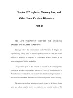
Hemolysis due to G6PD deficiency dramatically alters erythrocyte morphology.
Some deformed red cells appear to have bites taken out of them, producing the
characteristic “bite cells” that mark this type of hemolysis (Figure 15-1). Occasional
spherocytes and other nonspecifically deformed red cells also appear in the peripheral
blood. Prominent polychromasia reflects the large number of reticulocytes that pour
out in the wake of the hemolytic episode.
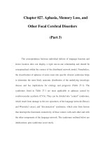
Clumps of denatured hemoglobin, called Heinz bodies, form within the raddled
red cells. Visualization of Heinz bodies by ordinary microscopy is difficult due to the
subtle nature of these inclusions. Wright-Geimsa staining shows dark, irregular bulges
or blisters at the edges of some cells presumably due to clusters of Heinz bodies in
312 ENZYMOPATHIES SECTION VI
FIGURE 15–1 Peripheral blood with drug-induced G6PD hemolysis. The smear is 3 days into
the acute hemolytic episode. Several bite cells appear on the smear along with other nonspecific
shape anomalies. Small bulges exist along the edge of some of the cells. (From Kapff CT and
Jandl JH. 1991. Blood: Atlas and Sourcebook of Hematology, 2nd edn. Boston: Little, Brown
and Company. Figure 23-1, p. 53. Reproduced with permission of the publisher.)
the region. Although phase contrast microscopes easily show these anomalous bod-
ies within erythrocytes, most clinical laboratories lack these relatively sophisticated
instruments. A more readily available means of visualizing Heinz bodies involves
staining the red cells with crystal violet or methyl violet in supravital preparations
(Figure 15-2). However, Heinz bodies are not evident after 24–48 hours.
Heinz bodies cause a number of problems for red cells. Methemoglobin accu-
mulates due to the dearth of NADPH to support methemoglobin reductase activity.
Hemoglobin denatures and the oxidized heme slips out of its usual site in the heme-
binding pocket. The resulting amalgam forms hemichromes that can themselves fa-
cilitate further formation of reactive oxygen species. This downward spiral due to
oxidant injury dooms the G6PD-deficient erythrocytes.
Heinz bodies also adhere to the red cell membrane where they disrupt architec-
ture, promote oxidation and cross-linking of lipids and proteins, and impair function
of membrane-associated enzymes including ion channels. Cross-links of membrane
structures produce rigid red cells with impaired ability to pass through capillaries.
CHAPTER 15 G6PD DEFICIENCY 313
FIGURE 15–2 Heinz body prep of G6PD deficient cells. The red cells are ghostly outlines with
the Heinz bodies appearing as dark inclusions.
The spleen briskly clears the circulation of these decrepit red cells. Disturbed ion
channel activity produces erythrocyte swelling and rupture. Some cells show bizarre
distributions of hemoglobin with protein confined to one portion of the cell and a
ghostly appearance to the remainder.
Reticuloendothelial cells rapidly remove these spavined erythrocytes from the
circulation. Sometimes an erythrocyte rips free of the reticuloendothelial cell, leaving
a large rent in its membrane. The cells appear to have bites taken out of them, giving
rise to the common moniker “bite cells.” Many such cells are scalloped and some
have thin peduncles reflecting narrow escapes from reticuloendothelial cell assault.
The spleen eventually clears the circulation of these aberrant erythrocytes.
Levels of unconjugated bilirubin rise during the hemolytic acute attack, mirroring
the jaundice on physical examination. Small rises in the level of conjugated bilirubin
occur occasionally, particularly with massive hemolysis. Transient, small elevations
in BUN occur most often in the wake of marked hemolysis.
The high RDW in the condition reflects the tremendous size variation of red cells
that include reticulocytes, cell fragments, and distorted cells. The augmented marrow
activity that produces the reticulocytosis also raises production of neutrophils. This
at times confuses the distinction between hemolysis due to infection and that due to
oxidant agents. The platelet count typically varies little from baseline.
G6PD AND RED CELL METABOLISM
The monomer subunit of G6PD is a 59-kDa polypeptide. Two subunits join to create
a protein dimer that includes a tightly bound NADP molecule. The enzymatically
314 ENZYMOPATHIES SECTION VI
Auto-oxidant drugs;
Natural oxidant
reactions
Methemoglobin reductase;
other reduction reactions
G6P
Dehydrogenase
G6P
NADP
GSSG
H
2
O
2
H
2
O
GSH
NADPH
6PG
GSSG
Reductase
GSH
Peroxidase
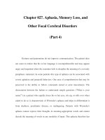
FIGURE 15–3 Schematic representation of the hexose monophospate (HMP) shunt. G6PD
dehydrogenase converts glucose-6-phosphate (G6PD), the initial product of glycolysis, into
6-phosphogluconate (6PG) with the coupled conversion of NADP to NADPH. This reaction is
the sole source of NADPH in the red cell, which is needed for a number of important metabolic
functions including the rescue of hemoglobin from constant oxidation to methemoglobin. A
host of normal metabolic processes generate hydrogen peroxide (H
2
O
2
) as a usual byproduct.
This compound, along with other pernicious reactive oxygen species that it produces, has the
potential of wrecking widespread cell injury including the cross-linking of lipids and proteins.
In the last step of the HMP shunt, glutathione peroxidase converts this destructive chemical to
water.
inactive dimer isin equilibrium with thefully functional tetramer form of the molecule.
Structural studies show that the NADP lodges near the dimer contact point and is
essential to molecular stability.
12
G6PD provides red cells with crucial protection against oxidant damage. Glucose-
6-phosphate is the first metabolite produced in glycolysis. The Embden-Meyerhof
pathway is the primary metabolic route for the compound, producing ATP along the
way and generating lactate in the final step. A portion of the glucose-6-phosphate takes
an alternate route involving the hexose monophosphate (HMP) shunt. The metabolic
steps of the HMP eventually swing back to merge with the Embden-Meyerhoff path-
way leading to lactate. Along the way, however, the HMP shunt produces several
compounds important to the health of the cell.
Figure 15-3 is a simplified representation of the HMP shunt. G6PD catalyzes
the first step that converts glucose-6-phosphate to 6-phosphogluconate. The NADPH
CHAPTER 15 G6PD DEFICIENCY 315
generated from NADP in the process provides the cell with a metabolite that is
central to many reductive biochemical reactions. Hydrogen peroxide (H
2
O
2
) produced
by natural processes in the cell oxidizes heme iron from the Fe(II) valance state
to Fe(III), thereby daily converting as much as 3% of hemoglobin to functionally
inactive methemoglobin. The NADPH-dependent enzyme, methemoglobin reductase,
regenerates hemoglobin from methemoglobin allowing continued normal oxygen
delivery.
H
2
O
2
transforms into other reactive oxygen species including the highly volatile
hydroxyl radical (
•
OH). These destructive molecules attack carbon–carbon double
bonds of unsaturated fatty acids, producing fatty acid hydroperoxides that promote
further cleavage and cross-linking of carbon–carbon bonds in membrane lipids. The
damaged regions of membrane loose their fluidity and accumulate aggregates of pro-
teins that normally are integral to or closely associated with the membrane. These
changes alter both the biophysical and biochemical properties of red cell mem-
branes and promote hemolysis. As Figure 15-2 shows, glutathione peroxidase defuses
H
2
O
2
before it can wreck havoc on the cell, a job shared with catalase. Glutathione
peroxidase performs the additional valuable function of decomposing fatty acid
peroxides.
G6PD deficiency blunts the efficiency of this entire cascade of events. The degree
of enzyme deficiency markedly influences the clinical character of the condition.
Syndromes in which the G6PD deficit is only mild or moderate, such as occurs with
Class III deficiency, show hemolysis only with major insults that rain vast quantities
of H
2
O
2
on the cell. People with Class I G6PD deficiency in contrast are in such
delicate balance with the forces producing oxidant cell stress that a relatively minor
upswing in cellular oxidant activity can mean disaster.
Red cell G6PD levels normally decline slowly with age, due to among other things
protein inactivation by oxidant damage. Reticulocyte levels of G6PD in fact exceed
by nearly fivefold those of the oldest erythrocytes. Figure 15-4 schematically shows
that the normal decay of G6PD maintains sufficient levels of the enzyme to protect
erythrocytes from exorbitant oxidant damage out to the end of the cell life span. The
enzyme produced by the G6PD A
−
gene is unstable and decays much more rapidly
than normal. Consequently, older cells fall below the protective threshold and are
vulnerable to the type of oxidant stress produced by Primaquine, for instance. After
the initial bout of hemolysis following exposure to an oxidant drug, the compensatory
reticulocytosis produces young cells containing sufficient enzyme to defend the cell
integrity. A new steady state maintains the hemoglobin in the normal range even with
continued exposure to the oxidant drug.
The G6PD Mediterranean deficiency, in contrast, results from an extremely unsta-
ble enzyme whosehalf-life in the cell is exceedingly short. As indicated inFigure 15-3,
most of the cells fall into the oxidant danger zone soon after their production. The
massive hemolysis seen with this form of G6PD deficiency reflects the large number
of vulnerable erythrocytes in the circulation. Maximally augmented erythropoiesis of-
ten fails to keep pace with the rate of hemolysis creating a potentially life-threatening
situation.
316 ENZYMOPATHIES SECTION VI
Red Cell
G6PD Level
100 %
0 %
Red Cell
Life Span
Normal (G6PD B)
G6PD Mediterranean
G6PD A
−
"Protecctive"
G6PD Level
Time (days) 140
FIGURE 15–4 Schematic representation of the time-dependent decay of red cell G6PD. Normal
G6PD (G6PD B) decays naturally over the life of the red cell but maintains a level sufficient to
protect against oxidant assault such as that produced by Primaquine. In contrast to G6PD B,
G6PD A
−
begins in the normal range but decays more rapidly due to a structural mutation. A
cohort of older cells comprising up to 30% of those in circulation are susceptible to hemolysis
when bombarded with excessive quantities of oxidants. The more severe G6PD Mediterranean
is very unstable and falls quickly to low levels that leave a large fraction of circulating red
cells susceptible to destruction by oxidant assault.
THE MOLECULAR BIOLOGY OF G6PD DEFICIENCY
The gene encoding G6PD located on the X-chromosome, X(q28), is over 20 Kb in
length and contains 13 exons.
13
Most of the hundreds of known mutations are point
mutations that producesome degree of reduced enzymaticactivity. A few mutants have
normal or even enhanced enzymatic activity. The absence of a mutant that produces
complete enzyme loss suggests that complete G6PD deficiency is incompatible with
life.
Table 15-3 provides information on some common and representative examples of
G6PD deficiency. The designation “G6PD B” attached to the normal enzyme derives
from its mobility on gel electrophoresis relative to the more rapidly migrating band
seen commonly in African Americans, which is designated “G6PD A
+
.” The altered
mobility of G6PD A
+
derives from a nucleotide and amino acid difference relative
to G6PD B, but the protein has full enzymatic activity. A mutation on the G6PD
A
+
background that lowers G6PD enzymatic activity is the basis of the G6PD A
−
deficiency seen in African Americans.
14
The mutations that produce the Class II G6PD variants tend to cluster in the region
of the NADP binding site or the interface between the two dimers. Protein structural
alterations in these regions appear to be particularly detrimental to enzymatic activity.
NADP binds tightly to the G6PD dimer behaving more as a structural component than
CHAPTER 15 G6PD DEFICIENCY 317
TABLE 15-3
REPRESENTATIVE G6PD VARIANTS
WHO Clinical Common Amino Acid
Common Distribution Classification Designation Substitution
Worldwide IV Normal; G6PD B None
African ethnicity IV G6PD A
+
68 Val → Met
Sporadic I G6PD Marion 213 Val → Leu
Mediterranean II G6PD Mediterranean 188 Ser → Phe
Southeast Asia II G6PD Canton 463 Arg → Leu
African Ethnicity III G6PD A
−
68 Val → Met
126 Asn → Asp
as a simpleenzyme substrate. Mutations that interrupt NADP binding severely dampen
enzyme activity. The fact that G6PD activity is confined to the tetramer means that
mutants disrupting the interface between dimers are particularly deleterious.
DIAGNOSIS OF G6PD DEFICIENCY
A number of enzyme assays exist that assess G6PD activity. Such tests have limitations
in certain clinical settings, but are very useful and reliable within the parameters set
by those limitations. The most discriminating assay detects the conversion of NADP
to NADPH wherein a resonating structure forms in one of the molecule’s rings and
produces a characteristic UV absorption band. The assay is precise and provides a
quantitative readout of G6PD activity, but is relatively complex. A simplified variant
of that is particularly suitable for population screening is the fluorescent spot test.
The test takes advantage of the change in UV absorption profile by an approach that
requires a simple UV lamp rather than a sophisticated spectrophotometer.
A number of other simple screening tests for G6PD rely on biochemical changes
produced by the active enzyme, including dye decolorization and reduction of methe-
moglobin. These semiquantitative tests are best at providing a “positive” or “negative”
answer, which requires setting proper cutoffs for the reading. Used correctly, these
tests are very useful in the detection of G6PD deficiency. A number of caveats exist,
however.
The key drawback to all these approaches when applied to patients suspected of
having hemolysis due to G6PD deficiency is that an acute hemolytic episode destroys
the cohort of cells that are deficient in G6PD (Figure 15-3). Patients with the G6PD
A
−
deficiency, for instance, often show normal testing readouts for more than 3 weeks
318 ENZYMOPATHIES SECTION VI
after drug exposure. Patients with the more severe Class II defects are less of a problem
since even very young red cells have substantial enzyme deficits. Physicians who order
testing for G6PD deficiency must be aware of the laboratory procedure employed and
alert to the possibility that it will elide a Class III G6PD deficiency in particular.
The ascorbate-cyanide test provides a broad screen for red cells that are sensitive
to oxidant stress. The test involves inactivating catalase with cyanide followed by
treatment with sodium ascorbate. The ascorbate produces reactive oxygen interme-
diates that convert hemoglobin to methemoglobin, seen as a brown pigment that is
readily visible to the unaided eye. The ascorbate-cyanide test is a sensitive but not
specific screen with respect to G6PD deficiency. Positive results occur in a variety of
settings with red cells that are sensitive to oxidant stress, including pyruvate kinase
deficiency. The test can be very useful in screening for Class III G6PD deficiency
in the immediate aftermath of a hemolytic episode, however. The extreme oxidant
stress of the ascorbate-cyanide test overwhelms red cells with low levels of G6PD that
managed to survive the in vivo hemolysis. The positive test supports the existence of
a red cell metabolic defect whose nature later can be definitely documented.
Depending on the urgency of obtaining a diagnosis of G6PD deficiency, one pos-
sible solution to the dilemma of testing in the aftermath of hemolysis is to wait for a
few weeks and perform the assay after the patient has returned to steady state. When
time is an issue, another approach is to test the patient’s mother who is an obligate het-
erozygote for this X-linked disorder. However, heterozygotes sometimes have normal
enzyme levels due to selective lyonization of the affected X-chromosome. Testing of
the patient’s brothers, who have a 50% chance of also having G6PD deficiency, can
also provide indirect support for the diagnosis. The size and availability of the family
dictates the usefulness of this approach to the issue.
TABLE 15-4
KEY DIAGNOSTIC POINTS WITH G6PD DEFICIENCY
Issue Manifestation Approach
Congenital hemolysis Anemia, reticulocytosis,
elevated bilirubin
r
Assess G6PD level.
r
Heinz body prep.
r
Osmotic fragility test.
Acute hemolysis
following drug exposure
Falling hematocrit,
reticulocytosis, elevated
bilirubin, hematuria
r
Obtain history of drug
exposure over preceding
several days.
r
Urine hemosiderin if
hemolytic episode occurred
several weeks before
evaluation.
r
G6PD level. Low in Class I
and II. Speciously normal in
Class III.
REFERENCES 319
TABLE 15-5
KEY MANAGEMENT POINTS WITH G6PD DEFICIENCY
Issue Comments
Infection and hemolysis Infection can produce severe hemolysis in Class III G6PD deficiency.
Control of the infection and supportive care for anemia and hemolysis are
indicated. Infection induces hemolysis inconsistently, even for a single
patient.
Drug-induced hemolysis Removing the offending drug is extremely important with Class I and II
G6PD deficiency due to their very low enzyme levels. Continued use of the
drug is possible with Class III G6PD deficiency. The moderate enzyme
deficiency allows attainment of a new steady state with a mild increase
over baseline of the reticulocyte count.
Diagnosis of G6PD
deficiency following acute
hemolysis
The severe enzyme deficiency of Class I and II G6PD deficiency shows up
as a low enzyme levels even after acute hemolysis. Class III G6PD
deficiency can show normal values in G6PD testing immediately after
hemolysis due to the higher enzyme levels in the surviving cells. The test
becomes abnormal only after 6–8 weeks when aged cells with low G6PD
content again exist in the circulation.
Analysis of genomic DNA obtained from the circulating lymphocytes allows di-
rect assay of the gene encoding G6PD and provides powerful insight into the patient’s
status. Most often, the procedure involves polymerase chain reaction that amplifies
the gene encoding the mutant enzyme allowing its subsequent analysis by a number
of sophisticated techniques. Despite its power and precision, DNA analysis has limi-
tations. More than 400 mutants involving the G6PD gene are known, including many
that are essentially genetic polymorphisms. This shortcoming is balanced by the fact
that a few subtypes of true G6PD deficiency dominate in specific populations or ethnic
groups, allowing a directed search for the most likely candidates. In the real world,
the search for G6PD deficiency will be determined by factors such as the prevalence
of the disorder and the availability of the various testing methods. Genetic testing is
not an option in many regions of the world where G6PD deficiency is common and
medical resources are scarce. As long as the physician remains aware of the probable
culprit, the likelihood is good that an accurate determination will result irrespective
of the final analytical approach (Tables 15-4 and 15-5).
References
1
Beutler E. 1994. G6PD deficiency. Blood 84:3613–3636.
2
Beutler E. 1992. The molecular biology of G6PD variants and other red cell enzyme defects.
Annu Rev Med 43:47–59.
320 ENZYMOPATHIES SECTION VI
3
Cappellini MD, Martinez di Montemuros F, De Bellis G, Debernardi S, Dotti C, Fiorelli G.
1996. Multiple G6PD mutations are associated with a clinical and biochemical phenotype
similar to that of G6PD Mediterranean. Blood 87:3953–3958.
4
Chiu DT, Zuo L, Chao L, et al. 1993. Molecular characterization of glucose-6-phosphate
dehydrogenase (G6PD) deficiency in patients of Chinese descent and identification of new
base substitutions in the human G6PD gene. Blood 81:2150–2154.
5
van Bruggen R, Bautista JM, Petropoulou T, et al. 2002 Deletion of leucine 61 in glucose-
6-phosphate dehydrogenase leads to chronic nonspherocytic anemia, granulocyte dysfunc-
tion, and increased susceptibility to infections. Blood 100:1026–1030.
6
Iancovici-Kidon M, Sthoeger D, Abrahamov A, et al. 2000 A new exon 9 glucose-6-
phosphate dehydrogenase mutation (G6PD “Rehovot”) in a Jewish Ethiopian family with
variable phenotypes. Blood Cells Mol Dis 26:567–571.
7
Kaplan M, Renbaum P, Levy-Lahad E, Hammerman C, Lahad A, Beutler E. 1997. Gilbert
syndrome and glucose-6-phosphate dehydrogenase deficiency: A dose-dependent genetic
interaction crucial to neonatal hyperbilirubinemia. Proc Natl Acad Sci USA 94:12128–
12132.
8
Kaplan M, Hammerman C. 2002. Glucose-6-phosphate dehydrogenase deficiency: A po-
tential source of severe neonatal hyperbilirubinaemia and kernicterus. Semin Neonatol
7:121–128.
9
Huang CS, Chang PF, Huang MJ, Chen ES, Chen WC. 2002. Glucose-6-phosphate de-
hydrogenase deficiency, the UDP-glucuronosyl transferase 1A1 gene, and neonatal hyper-
bilirubinemia. Gastroenterology 123:127–133.
10
Shibuya A, Hirono A, Ishii S, Fujii H, Miwa S. 1999. Hemolytic crisis after excessive
ingestion of fava beans in a male infant with G6PD Canton. Int J Hematol 70:233–235.
11
Baehner RL, Nathan DG, Castle WB. 1971. Oxidant injury of caucasian glucose-6-
phosphate dehydrogenase-deficient red blood cells by phagocytosing leukocytes during
infection. J Clin Invest 50:2466–2473.
12
Au SW, Gover S, Lam VM, Adams MJ. 2000. Human glucose-6-phosphate dehydrogenase:
The crystal structure reveals a structural NADP(+) molecule and provides insights into
enzyme deficiency. Structure Fold Des 8:293–303.
13
Takizawa T, Huang IY, Ikuta T, Yoshida A. 1986. Human glucose-6-phosphate dehydroge-
nase: Primary structure and cDNA cloning. Proc Natl Acad Sci USA 83:4157–4161.
14
Vulliamy TJ, Othman A, Town M, et al. 1991. Polymorphic sites in the African population
detected by sequence analysis of the glucose-6-phosphate dehydrogenase gene outline the
evolution of the variants A and A
−
. Proc Natl Acad Sci USA 88:8568–8571.
CHAPTER
16
PYRUVATE KINASE
DEFICIENCY
CLINICAL MANIFESTATIONS 322
LABORATORY FEATURES OF PK DEFICIENCY 323
BIOLOGY OF PK DEFICIENCY 324
DIAGNOSIS OF PK DEFICIENCY 325
TREATMENT OF PK DEFICIENCY 327
In 1953, John Dacie and colleagues provided the first cogent description of patients
with a congenital hemolytic anemia who were distinct from those with the previ-
ously recognized disorder hereditary spherocytosis.
1
This heterogeneous collection
of patients had a number of important common features including the absence of
spherocytosis, no detectable abnormal hemoglobin, and no antibody directed against
the red cell. The osmotic fragility of freshly isolated cells was normal. Spleen size
was normal or only modestly enlarged and splenectomy provided no clinical benefit.
Later, investigators identified pyruvate kinase (PK) deficiency as the basis of this
constellation of findings.
2
PK deficiency is the second most common human erythrocyte enzyme deficiency,
following glucose-6-phosphate dehydrogenase (G6PD) deficiency. These two condi-
tions account for most cases of congenital nonspherocytic hemolytic anemia (CN-
SHA). While the number of people with G6PD deficiency ranges in the millions,
however, only several hundred cases of PK deficiency exist in the literature. Most
reported cases involve people of Northern European background with an estimated
incidence of 50 cases per million, but sporadic reports involving people through-
out the world suggest a wider distribution to the condition than has generally been
appreciated in the past.
3
321
Copyright © 2008 by The McGraw-Hill Companies, Inc. Click here for terms of use.
322 ENZYMOPATHIES SECTION VI
CLINICAL MANIFESTATIONS
PK deficiency has a wide expression spectrum, ranging from severe neonatal hemol-
ysis and jaundice to mild conditions that evade discovery until adulthood. Typically,
anemia, jaundice, or both appear in childhood. A few cases of very severe PK defi-
ciency have appeared in utero and some have produced recurrent episodes of hydrops
fetalis.
4,5
Most often, however, the fetuses survive the period of pregnancy with rel-
atively little difficulty.
6
Early recognition that a fetus has PK deficiency, often the
result of a good family history, allows appropriate application of the tools available
to high-risk obstetrics teams. Occasionally, mild hemolysis characterizes CNSHA,
for which the reticulocyte response compensates fully, leaving jaundice as the only
clinical manifestation. Anemia when present is lifelong and varies little over time.
Hyperbilirubinemia occurs commonly in newborns with PK deficiency and can
necessitate exchange transfusion. Some children with severe manifestations subse-
quently require transfusions to maintain an acceptable hemoglobin level. Splenec-
tomy, usually after 5 years of age, typically reduces or eliminates the transfusion
requirement for most such children. A paradoxical rise in the reticulocyte count
commonly follows splenectomy, with values sometimes reaching 70% or more. The
hyperactive erythroid production partially compensates for the anemia. Marrow ex-
pansion in the maxilla and facial bones can produce severe cosmetic deformity in
children with extreme marrow erythroid hyperplasia. Prominent malar eminences
give the eyes a mongoloid slant reminiscent of that seen with thalassemia. Expan-
sion of the marrow cavity of the cranial bones caused enlargement of the skull with
frontal and parietal bossing. In contrast to thalassemia, PK deficiency does not stunt
general growth and maturation. Cholelithiasis is a common sequela of long-standing
hemolysis and hyperbilirubinemia.
Iron overload is a concern in PK deficiency, but generally the serum iron and fer-
ritin levels are normal or only mildly elevated.
7,8
The picture differs strikingly from
that seen with thalassemia where iron absorption exceeds normal and commonly pro-
duces severe overload in the absence of transfusion. The difference likely represents
the fact that PK deficiency is a hemolytic disorder with little ineffective erythro-
poiesis. The destruction of erythroid precursors in the marrow, which is the core of
ineffective erythropoiesis, appears to be the key signal for augmented gastrointestinal
iron uptake. Recurrent transfusion in severely affected patients can of course produce
iron overload to a degree requiring chelation therapy.
Events that disturb the balance between erythropoiesis and hemolysis can be
profoundly deleterious to patients with PK deficiency. A well-recognized threat is
infection with human parvovirus B19. This adeno-associated virus causes “Fifth Dis-
ease,” a normally benign childhood disorder associated with fever, malaise, and a
mild rash. The virus has a tropism for erythroid progenitor cells and impairs cell
division for a few days during the infection. Reticulocyte counts often fall literally to
zero. Normal people experience, at most, a slight drop in hematocrit since the half-
life of erythrocytes in the circulation is 40–60 days. The viral infection resolves in a
few days with no long-lasting problem. Anemia of life-threatening severity occurs in
CHAPTER 16 PYRUVATE KINASE DEFICIENCY 323
children with PK deficiency whose survival depends on brisk reticulocyte production.
9
Early recognition of the problem along with transfusion support allows the child to
weather the storm. Fortunately, the virus induces a profound immune response that
prevents further episodes of infection. Nonetheless, other viral (and sometimes bac-
terial) infections that only modestly suppress erythropoiesis can pose a significant
danger.
With proper management, most patients with PK deficiency reach adulthood.
Women with PK deficiency can become pregnant and carry their fetuses to term, but
the course is variable and depends greatly on the severity of the particular case of PK
deficiency. The dangers to pregnancy posed in the past by PK deficiency have subsided
with improved skill at obstetrical management.
10
Women with mild disease often
tolerate pregnancy without intervention. Even some women with relatively severe PK
deficiency can stay the course without the need for transfusion.
11
In severe cases,
prophylactic blood transfusions aim at maintaining the hemoglobin concentration
above an arbitrary threshold of 7–8 g/dL. Puerperal jaundice is an occasional feature
in otherwise unremarkable pregnancies.
12
PK deficiency occasionally arises in people with acquired defects in hematopoiesis
including myelodysplasia and myelogenous leukemia.
13,14
The changes likely reflect
the profound alterations in the basic metabolism of erythroid precursors, which oc-
curs with these disorders. Acquired enzyme deficiencies of many types as well as
thalassemic disturbances in globin production occur in some patients. These are rare
events. However, myelodysplasia is far more common than hereditary PK deficiency,
making acquired PK deficiency relatively prominent in the overall picture of the
enzymopathy.
LABORATORY FEATURES OF PK DEFICIENCY
The anemia associated with PK deficiency commonly produces a hemoglobin value in
the range of 5–11 g/dL. Some patients require transfusions while others are asymp-
tomatic. Splenectomy usually raises the hemoglobin value in patients with severe
disease. The robust rise in the reticulocyte number following splenectomy sometimes
makes these cells the primary conveyors of oxygen in the body.
The erythrocyte morphology often is surprisingly bland in cases of PK deficiency.
Macrocytosis and polychromophilia are invariant. Red cells with spicules or irregular
contractions occur with more severe disease, and rare cases can display acanthocytes.
More prominent morphological alterations occur following splenectomy. Howell-
Jolly bodies and nucleated red cells make an appearance following the procedure. The
astonishing rise in reticulocyte count that often follows splenectomy sometimes gives
the smear a complete polychromatophilic cast. Occasional target cells, spherocytes,
as well as cells with Pappenheimer bodies can also appear on the stage following
removal of the spleen (Figure 16-1).
Neutrophil and platelet morphology are normal with PK deficiency. Splenectomy
produces a visible increase in platelets on the smear that includes some giant platelets
324 ENZYMOPATHIES SECTION VI
FIGURE 16–1
Pyruvate kinase deficiency after splenectomy. Reticulocytes are prominent on
the smear and crenated cells are numerous. The bristled edges seen in many of the cells are
characteristic of the condition. (From Jandl JH. 1987. Blood: Textbook of Hematology. Boston:
Little, Brown and Company. Figure 12-2, p. 353.)
and occasional megakaryocyte fragments. The WBC often is slightly higher than
normal.
Hyperbilirubinemia is invariant with PK deficiency, with most in the unconjugated
state. Values in the range of 4–6 mg/dL are common, but some patients with severe
disease have values as high as 20 mg/dL. As noted earlier, the bilirubin level commonly
rises with pregnancy.
BIOLOGY OF PK DEFICIENCY
As shown in Figure 16-2, PK generates two equivalents of ATP during the conversion
of phosphoenol-pyruvate to pyruvate in the penultimate step of the Embden-Meyerhof
pathway. The block produced by PK deficiency not only deprives the cell of needed
ATP but also disrupts the NAD cycle. Mitochondria in developing erythroid cells
provide an alternate energy source, but are lost as the red cells mature. The severity of
the PK deficiency determines the length of survival of the erythrocytes. The lifespan
of severely defective cells is only a few days while those with more modest metabolic
disturbances can survive for weeks.
Glycolytic intermediates produced prior to the PK block, such as 2,3-DPG, accu-
mulate in the erythrocytes while the decline in NAD levels impairs important steps
in intermediary metabolism. The factors that doom the cells are failed maintenance
activities due to the energy deficit. ATP-dependent cation pumps fail, leading to potas-
sium loss and cell shrinkage. Pathological levels of calcium accumulate in the cells
and activate the Gardos channel with consequent further loss of cell potassium. The
shrunken, crenated cells become rigid and are quickly cleared from the circulation by
reticuloendothelial cells.