Atlas of Clinical Hematology - part 8 potx
Bạn đang xem bản rút gọn của tài liệu. Xem và tải ngay bản đầy đủ của tài liệu tại đây (3.27 MB, 44 trang )
V
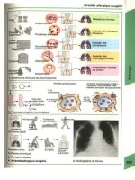
Fig. 135 a – d. Lymph node hyperplasia
a Two immunoblasts and an eosinophil
in lower half of field
b Plasmablast at top center, several
plasma cells at bottom, tissue basophil
at left
c Starry sky macrophage at center,
several immunoblasts at right
d Immunoblast at upper left, above it a
centroblast, at right a reticulum cell
301
6 · Cytology of Lymph Node and Splenic Aspirates
V
Fig. 136 a – d. Lymph node hyperplasia
in toxoplasmosis
a Epitheloid cell lymphadenitis (Piringer-
Kuchinka type). Epitheloid cell cluster at
left, an immunoblast at center, to its right
a binucleated plasma cell
b Starry sky macrophage at center
c Starry sky macrophage. Typical speci-
men with many coarse granular inclu-
sions and cytoplasmic vacuoles
d Epitheloid cell shows typical fine but
dense chromatin pattern with one or two
blue, sharply defined nucleoli
302 Chapter V · Lymph Nodes and Spleen
V
Fig. 137 a, b. Tuberculous lymphade-
nitis
a Langhans giant cell
b Syncytium of epitheloid cells
Fig. 138 a, b. Sarcoidosis
(Boeck disease), lymph node
a Numerous epitheloid cells resembling
a school of fish
b Epitheloid cells in a “footprint”
configuration
303
6 · Cytology of Lymph Node and Splenic Aspirates
V
6.2 Infectious Mononucleosis
Infectious mononucleosis is caused by the Ep-
stein-Barr virus. Cytologically it is the prototype
of lymph node hyperplasias of viral etiology. The
clinical picture may be dominated by lymph node
enlargement and splenomegaly or by a pseudo-
membranous, lacunar, or ulcerative sore throat.
Fever may reach 39 8C and persists for several
days or as long as 2 to 3 weeks. Often the fever
precedes the onset of glandular swelling. The
red blood count is usually normal, and mild an-
emia is rare. Generally there is a leukocytosis of
10,000 to 15,000/lL, which in rare cases exceeds
50,000/lL. Sporadic cases present with leukocy-
topenia; this finding plus the pharyngitis can mi-
mic agranulocytosis. Thrombocytopenia is occa-
sionally seen and may be so pronounced that a
hemorrhagic diathesis results. The leukocyte dis-
tribution is characteristic. Examination of the
blood smear shows a predominance of pleo-
morphic mononucleated cells (60 %–90 %),
which give the disease its name. Morphologically
these cells may resemble young lymphocytes or
lymphoblasts. In the early phase of the disease
one finds granulated lymphocytes or lympho-
cytes with small vacuoles at the circumference
of the cytoplasm. Some appear as large blast-
like cells with strongly basophilic cytoplasm. A
significant increase in monocytes is not observed.
Because similar cells can occur in other viral dis-
eases, they have also been referred to as “viro-
cytes” or lymphoid cells. The nuclei are pleo-
morphic, often kidney-shaped or irregular, and
the nuclear chromatin forms a coarse meshwork
of loosely arranged strands. One or more nucleoli
are usually present. Azurophilic granules are
found in some cells, which represent transformed
T-lymphocytes. Bone marrow findings are gener-
ally nonspecific, and thus the changes are typical
of infection. The mononuclear elements of the
bone marrow may be increased in a few cases,
but never to a high degree.
By contrast, large numbers of typical cells are
found in lymph node aspirates (Fig. 139). The pic-
ture is dominated by mononuclear cells very si-
milar or identical to those in the peripheral blood.
Particularly striking are the large basophilic cells
whose “transformed” state is evidenced by their
enlarged, blue-tinged nucleoli. The cytologic
changes can even cause confusion with Hodgkin
cells (“Hodgkin-like cells”). Isolated epitheloid
cells are also found.
The diagnosis of infectious mononucleosis is
established by detecting antibodies against the
Epstein-Barr virus (EBV). While the rapid tests
give a general impression as to whether infectious
mononucleosis is present, their capabilities are
otherwise limited.
304 Chapter V · Lymph Nodes and Spleen
V
Fig. 139 a – d. Infectious mononucleo-
sis, lymph node
a Marked increase of immunoblasts,
macrophage at lower right
b At center an immunoblast, several
plasma cells
c Various immunoblasts, with several
pleomorphic lymphoid cells in lower half
of field
d Two strongly stimulated immuno-
blasts, Hodgkin-like cells
305
6 · Cytology of Lymph Node and Splenic Aspirates
V
Fig. 140 a – d. Blood smears in
infectious mononucleosis
a–c Typic al pleomorphic lymphatic
cells. Often these cells have irregular
nuclear contours and basophilic cyto-
plasm, an d some resemble bla sts
b
c
d Detection of CD3, APAAP method. CD3
is detectable in the typical cells of in-
fectious mononucleosis (red). These cells
represent transformed T lymphocytes
306 Chapter V · Lymph Nodes and Spleen
V
6.3 Persistent Polyclonal B Lymphocytosis
This is a lymphocytosis stable for years, and bi-
nucleated lymphocytes are detected in peripheral
blood smears (about 3 %). These changes are
almost only found in cigarette-smoking women
under 50 years of age. In most cases there is a
polyclonal increase of IgM; an association with
HLA-DR 7 seems to exist. Of 25 women with
this anomaly, 77 % had an isochromosome
3q(+i(3q)) (Fig. 140 e).
Fig. 140 e. Lympho-
cytes with two nuclei
in persistent polyclo-
nal B lymphocytosis
307
6 · Cytology of Lymph Node and Splenic Aspirates
V
6.4 Maligne Non-Hodgkin-Lymphome
und Hodgkin-Lymphom
Wa¨hrend das Hodgkin-Lymphom ein eigenes
Krankheitsbild (mit großer Var iationsbreite in
Symptomatik und Prognose) darstellt, bildet
die Gruppe der malignen Non-Hodgkin-Lym-
phome eine hinsichtlich Erscheinungsbild, Prog-
nose und Therapie recht heterogener Krankheits-
gruppe, deren U
¨
bersichtlichkeit zudem durch die
wechselnden Klassifizierungsvorschla¨ge noch zu-
sa¨tzlich erschwert wird. Tabelle 14 und 15 geben
einen U
¨
berblick u¨ber die Non-Hodgkin-Lym-
phome. In Tab. 16 sind typische Immunmar-
ker-Profile zusammengestellt.
Nach der Herkunft der erkrankten Zellen ko¨n-
nen B- und T-Zell-Lymphome unterschieden wer-
den. B-Zell-Lymphome sind in Europa und Ame-
rika ha¨ufiger, wa¨hrend die T-Zell-Lymphome in
Asien u¨berwiegen. Unter Einbeziehung von mor-
phologischen und zytochemischen Befunden, Im-
munpha¨notyp, Zytogenetik und Molekulargene-
tik sowie klinischen Daten hat die WHO-Arbeits-
gruppe nach den Prinzipien der Revised Euro-
pean-American Classification of Lymphoid Neo-
plasms (REAL) die neue Einteilung der malignen
Non-Hodgkin-Lymphome und verwandter Er-
krankungen konzipiert. Nach wie vor stu¨tzt
sich der Prima¨rbefund auf die Morphologie
und manche Erkrankungen sind alleine morpho-
logisch definiert, ha¨ufig ist aber die Definition
einer Entita¨t ohne Immunpha¨notyp oder gene-
tisch-molekulargenetisches Profil nicht mo¨glich.
B-, T- und NK-Zell-Neoplasien werden weiter
in Vorstufen-(Precursor) und reife (mature) Er-
krankungen unterteilt. Auf die Einteilung nach
prognostischen Kriterien (niedrig-maligne – in-
dolent, intermedia¨r – aggressiv, hoch-maligne –
sehr aggressiv) wurde verzichtet.
Es ist nicht immer mo¨glich, allein mit Hilfe der
Ausstrichzytologie eine eindeutige Klassifizie-
rung der verschiedenen Lymphome vorzuneh-
men. Trotzdem gibt es eine Reihe von zytomor-
phologischen Kriterien, die in den meisten Fa¨llen
zumindest eine Verdachtsdiagnos e gestatten. Sie
sollen in den folgenden Abbildungen dargestellt
werden.
Die aktuelle Diagnostik und Klassifizierung
der malignen NHL stu¨tzt sich auf die histologi-
sche und immunologische Untersuchung von ex-
stirpierten Lymphknoten oder anderen befalle-
nen Geweben. Wie bei den akuten Leuka¨mien
sind zytogenetische oder molekulargenetische
Befunde wichtige Erga¨nzungen oder sogar ent-
scheidend fu¨r die genaue Einordnung. Lymph-
knotenpunktate spielen in der Prima¨rdiagnostik
eine untergeordnete Rolle, es sei denn, periphere
Lymphknoten ko¨nnen aus technischen Gru¨nden
nicht exstirpiert werden. Andererseits ist die
Punktatdiagnostik eine wichtige Erga¨nzung,
wenn Organe im Abdomen, Thorax oder intraab-
dominell gelegene Lymphknoten unter sonogra-
phischer oder computertomographischer Kon-
trolle gezielt punktiert werden ko¨nnen. Auch
zur schnell eren Orientierung oder Verlaufskon-
trolle sind sie hilfreich. In unterschiedlichem Gra-
de ist auch das Knochenmark befallen, und/ oder
es kommt zur leuka¨mischen Ausschwemmung in
das periphere Blut. Wir werden in diesem Ab-
schnitt auf die Beteiligung von Knochenmark
und Blut bei NHL eingehen, im u¨brigen verweisen
wir auf die Publikation zur WHO-Klassifizierung.
Beim Hodgkin-Lymphom ist der Knochenmark-
befall am ehesten nach histologischer Untersu-
chung von Biopsien, selten auc h im Ausstrich
von Aspiraten nachzuweisen. Das periphere
Blut liefert nur indirekte Hinweise, wenn eine
Lymphopenie oder selten eine Eosinophilie be-
stehen. Wir richten uns bei der Nomenklatur
nach der WHO-Klassifizierung.
Bei der histologischen Beurteilung von Kno-
chenmark-Schnittpra¨paraten spielt die Art des
Infiltrationsmuster s speziell in den fru¨hen Sta-
dien der Erkrankung eine wichtige Rolle, da es
teilweise recht spezifisch ist.
In der Schemazeichnung 1 ist die Topographie
von Lymphominfiltraten im Knochenmark sche-
matisch dargestellt (Schaefer, H. E.).
308 Kapitel V · Lymphknoten und Milz
V
Paratrabekular: Splenic marginal zone lympho-
ma, mantle cell lymphoma
Paratrabekular osteoclastic: Plasma cell myelo-
ma, adult T-cell leukemia/lymphoma
Random interstitial: Advanced hairy cell leuke-
mia, large B-cell lymphoma
Random tumorforming: Lar ge B-cell lymphoma,
Hodgkin-lymphoma, advanced hairy cell leuke-
mia
Courtesy Prof. Dr. H. E. Schaefer, Freiburg. Up
until now, he has only presented his schematic
drawings publicly once previously, during a Japa-
nese-Korean workshop in 1995.
Suggested Further Reading
World Health Organisation (2001) Classification
of Tumours: Pathology and Genetics. IARC Press,
Lyon (Tumou rs of Haematopoietic and Lym-
phoid Tissues. Ed.: E. S. Jaffe, N. L. Harris, H.
Stein, J. W. Vardiman)
Table 14. WHO classification of mature B-cell neoplasms
B-cell neoplasms
Precursor B-cell neoplasm
Precursor B lymphoblastic leukemia lymphoma
Mature B-cell neoplasms
Chronic lymphocytic leukemia / small lymphocytic lymphoma
B-cell prolymphocytic leukemia
Lymphoplasmacytic lymphoma
Splenic marginal zone lymphoma
Hairy cel l leukemia
Plasma cell myeloma
Monoclonal gammopathy of undetermined significance (MGUS)
Solitary plasmacytoma of bone
Extraosseous plasmacytoma
Primary amyloidosis
Heavy chain diseases
Extranodal marginal zone B-cell lymphoma of mucosa-associated lymphoid tissue (MALT-lymphoma)
Nodal marginal zone B-cell lymphoma
Follicular lymphoma
Mantle cel l lymphoma
Diffuse large B-cell lymphoma
Mediastinal (thymic) large B-cell lymphoma
Intravascular large B-cell lymphoma
Primary effusion lymphoma
Burkitt lymphoma/leukemia
B-cell proliferations of uncertain malignant potential
Lymphomatoid granulomatosis
Post-transplant lymphoproliferative disorder, polymorphic
309
6 · Cytology of Lymph Node and Splenic Aspirates
V
Table 15. Immune markers in leukemic forms of non-Hodgkin lymphoma
Marker B-CLL B-PLL HCL FL MCL SLVL T-CLL / LGL SS T-PLL ATLL
SIg (þ) þþ þþ þþ þþ þþ À À À À À
CD2 ÀÀÀÀÀÀþþþþþ
CD3 ÀÀÀÀÀÀþÀþþþ
CD4 ÀÀÀÀÀÀÀÀþþ/ Àþ
CD5 þþÀÀÀþÀÀÀþþþ
CD7 ÀÀÀÀÀÀÀÀþ/ Àþ À/ þ
CD8 ÀÀÀÀÀÀþþ/ Àþ/ Àþ/ Àþ/ À
CD19 / 20 / 24 þþ þþ þþ þ þ þþ À À À À À
CD22 þ / Àþþ þþ þ þ þþ À À À À À
CD10 ÀÀÀþ/ ÀÀÀÀÀÀÀÀ
CD25 ÀÀþþÀÀþ/ ÀÀ À À À þþ
CD56 ÀÀÀÀÀÀÀþÀÀÀ
CD103 ÀÀþþÀÀÀÀÀÀÀÀ
HL-DR þþ þþ þþ þþ þþ þþ À À À À À
À¼negative; (+) ¼ weakly positive; + ¼ positive; ++ ¼ markedly positive; +++ ¼ strongly positive;
+/À¼majority of cases positive; À /+ ¼ majority of cases negative. CLL, chronic lymph ocytic leukemia;
PLL, prolymphocytic leukemia; HCL, hairy cell leukemia; FL, follicular lymphoma; MCL, mantle cell lymphoma;
SLVL, splenic lymphoma with villous lymphocytes; LGL, large granulated lymphocyte leukemia; SS, Se´zary
syndrome; ATLL, adult T-cell leukemia/lymphoma
310 Chapter V · Lymph Nodes and Spleen
V
6.4.1 Some Mature B-cell Lymphomas
(Table 14)
Chronic Lymphocytic Leukemia
(Chronic Lymphadenosis, CLL)
CLL is a chronic disease characterized by hema-
tologic changes, multiple or generalized lymph
node enlargement, splenomegaly, and frequent
hepatomegaly. It predominantly affects older in-
dividuals. Peripheral blood examination usually
reveals a leukocytosis with a strong preponder-
ance of lymphocytes. Most of the cells appear
morphologically as small, mature lymphocytes
with a dense, coarse nuclear structure. Nucleoli
are found only in a few “immature” cells. The cy-
toplasmic rim is narrow with a medium blue col-
oration. Gumprecht shadows or “smudge cells”
(Fig. 141 a – c) is the term applied to crushed cells
that, while often present in B-CLL, are not specific
for that disease. A large percentage of the lympho-
cytes show marked, usually fine to moderate
PAS granulations on cytochemical staining
(Fig. 141 f). Small lymphocytes also predominate
in the bone marrow (Fig. 141 d), lymph nodes,
and spleen. Granulocytopoiesis, erythropo iesis,
and thrombocytopoiesis are quantitatively de-
pressed in the bone marrow, resulting in a pro-
gressive anemia, absolute granulocytopenia,
and thrombocytopenia with their associated ef-
fects (which could also result from autoantibo-
dies). The depletion of normal B-lineage cells
from the bone marrow and lymph nodes almost
always produces an increasing hypogammaglo-
bulinemia that may culminate in an antibody de-
ficiency syndrome.
The lymphocytes of CLL have B-cell properties
(B-CLL) in the majority of cases seen in Europe
and America. The existence of a T-CLL is contro-
versial. The mature-cell type of T-cell leukemia or
NK-cell leukemia is referred to as LGL (large
granulated lymphocyte) leukemia or T-cell lym-
phocytosis. It is characterized by neutropenia
and a chronic course. The abnormal cells have
fine to medium-sized azurophilic granules in
their cytoplasm and should be further identified
by immunologic or molecular genetic criteria. T-
cell leukemias (except for T-ALL) are classified
mainly as T-cell prolymphocytic leukemias. The
cells are frequently small and thus may be mista-
ken for the cells of B-CLL in improperly prepared
smears. Most have nucleoli, however, and some
have an irregular nuclear contour. A variant
with a broader cytoplasmic rim and sharply de-
fined vesicular nucleoli resembles B-cell prolym-
phocytic leukemia. Immunophenotyping is es-
sential. The other leukemic forms of NHL are de-
scribed in the figure legends.
Either of two staging systems may be used for
evaluating the course of CLL and planning ther-
apy:
1. Staging system of Rai et al.:
Stage 0: Blood lymphocytosis < 15,000/lL,
bone marrow infiltration
Stage I: Stage 0 plus enlarged lymph nodes
Stage II: 0 with or without I, plus hepatome-
galy and/or sp lenomegaly
Stage III: 0 with or without I and II, plus ane-
mia (Hb < 11g/dl)
Stage IV: 0 with or without I– III, plus throm-
bocytopenia (<100,000/lL)
2. Staging system of Binet:
A. Lymphocytosis > 4000/lL,
bone marrow infiltration > 40 %,
hemoglobin > 10 g/dL,
platelets > 100,000/lL,
two lymph node regions involved (enlarged)
B. Same as stage A,
plus enlargement of at least three
lymph node regions
C. Same as A,
plus anemia (Hb<10 g/dL) and/or
thrombocytopenia <100,000/lL,
with involvement of any number
of lymph node regions
Since the introduction of fluorescence in-situ hy-
bridization (FISH) it has been possible to detect
prognostic important cytogenetic aberrations in
B-CLL. Today the 13q(-) aberration is thought
to be favorable, the 17p(-) aberration (by muta-
tion or loss of p53) is unfavorable, the 11q deletion
is unfavorable (but less so), and the trisomy 12 has
a median position, with a median survival of 114
months [Do¨hner, H. et al. (2000) N Engl T Med
343: 1910].
Immunocytoma (IC)
In the Kiel classification, this group of low-grade
non-Hodgkin lymphomas includes lymphoplas-
mocytoid and lymphoplasmacytic immunocyto-
ma. Cases that are CD5–positive on immunologic
testing are presently classified as a form of B-CLL,
whereas CD5-negative cases, often associated
with an IgM type of monoclonal gammopathy
and featuring lymphatic cells transitional with
plasma cells, are classified as Waldenstro¨m dis-
ease. In up to 50 % of the patients, the transloca-
tion t(9;14)(p13;q32) and rearrangements of the
PAX-5 gene are found, as in other lymphomas,
with plasmacytoid differentiation. The abnormal
cell forms range from small lymphocytes to plas-
311
6 · Cytology of Lymph Node and Splenic Aspirates
V
ma cells. Bone marrow examination sometimes
shows an increase in tissue mast cells
(Fig. 142 d, h).
Prolymphocytic Leukemias
Prolymphocytic leukemias can be classified phe-
notypically as having a B-cell or T-cell lineage. B-
cell prolymphocytic leukemia (B-PLL) is diag-
nosed if at least 55 % prolymphocytes are found
on examination of the peripheral blood. These
cells are larger than lymphocytes, have a broader
cytoplasmic rim, and a round nucleus with a well-
defined, vesicular nucleolus that is usually cen-
tered within the nucleus. Generally the leukocyte
count is greatly elevated. Splenomegaly is present
but there is little or no lymph node enlargement.
Immunophenotypic analysis shows strong ex-
pression of the surface immunoglobulin (SJg).
The B-cell markers CD19, CD20, CD24, and
CD22 are markedly positive, FMC7 is positive,
and CD5 is negative. No specific cytogenetic aber-
rations are known (Fig. 150).
T-cell prolymphocytic leukemia (T-PLL) is
also associated with an elevated leukocyte count
and splenomegaly. In con trast to B-PLL, however,
lymph node enlargement is usually present and is
sometimes accompanied by cutaneous infiltrates
and effusions. The cells resemble those of B-PLL
in approximately 50 % of cases bu t usually show
an irregular nuclear contour. In other cases the
cells are smaller and have a narrower and mark-
edly basophilic cyto plasmic rim. In approxi-
mately one-third of cases the nucleoli are difficult
to detect with light microscopy but are clearly de-
fined by electron microscopy. At one time such
cases were frequently diagnosed as T-CLL, and in-
deed there are sporadic cases that do not display
the features of LGL leukemia and must be classi-
fied as T-CLL. The T-cell markers CD2, CD3, CD5,
and CD7 are positive on immunophenotypic ana-
lysis. CD4 and the T-cell receptor are positive in
more than 50 % of cases, and CD8 is rarely detect-
able.
Cytogenetic analysis frequently demonstrates
an inv(14) or aberrations of chromosome 8
(Figs. 157, 158).
Mantle Cell Lymphoma (MCL)
This lymphomatous disease was previously re-
cognized by the Kiel group as a separate entity
(“centrocytic lymphoma”) because of its poor
prognosis, but the entity was accepted interna-
tionally only after the cytogenetic detection of
the (11;14) translocation (Fig. 151 f). The abnormal
cells are derived from the mantle zone of the lym-
phoid follicle and require differentiation from
true follicular cells (from the center of the folli-
cle). They may equal or greatly surpass lympho-
cytes in size. The smaller cells usually have an ir-
regular nuclear contour but are sometimes diffi-
cult to distinguish from the cells of CLL; even the
immunologic detection of CD5 and standard B-
cell markers cannot p rovide reliable differentia-
tion. These cases are diagnosed by using cytoge-
netic analysis or the FISH technique to detect the
(11;14) translocation. The variant with larger and
sometimes anaplastic cells is identified by a con-
spicuous pleomorphism of nuclear contours and
a very coarse (“rocky”) chromatin pattern
(Fig. 151).
Follicular Lymphoma
The cells are derived from the center of the folli-
cle. Mature lymphatic forms predominate, and
blasts are rarely observed in blood and bone mar-
row smears. Deep nuclear clefts are typically
found in the more mature forms and may almost
completely divide the nucleus (“cleaved cell,”
Fig. 152 e). Additionally there are cells with round
nuclei and isolated blasts. Depending on the pro-
portion of centroblasts in the histological section
per high-power microscopic field, three grades
are distinguished. This method cannot be applied
to smears. Cytogenetic analysis reveals a t(14;18)
in approximately 70 % of cases, and molecular
analysis shows the hyperexpression of bcl-2.
The t(14;18) can be detected with high sensitivity
by FISH and PCR and therefore makes a useful
differentiating criterion. On immunophenotyp-
ing, the cells of low-grade follicular lymphoma
may express CD10 in addition to standard B-
cell markers. CD5 is negative.
Hairy Cell Leukemia (HCL)
Hairy cell leukemia is always associated with sple-
nomegaly, which tends to progress slowly during
the course of the disease. Lymph node enlarge-
ment is usually absent. Most cases present hema-
tologically with leukopenia, neutropenia, and ty-
pically with monocytopenia. Lymphocytes and
only small numbers of hairy cells are found in
blood films (Fig. 144 a). The hairy cells are
roughly the size of lymphocytes or slightly larger.
The grayish-blue cytoplasm appears finely honey-
combed and has irregular bord ers with typical
hairlike projections that give this disease its
name. Other cells may present denser, pseudo-
pod-like cytoplasmic extensions. Azurophil ic
312 Chapter V · Lymph Nodes and Spleen
V
granules are occasionally present in the cyto-
plasm. The nucleus is usually oval, and its chro-
matin is finer than in lymphocytes (Figs. 144 –
146). A single, small nucleolus is rarely present.
Cytochemical staining demonstrates a strong
acid phosphatase activity that is not inhibited
by tartrate (see method on p. 14). This is due
to the presence of isoenzyme 5 in the hairy cells
(Fig. 147 a – d). When these cells are seen in the
bone marrow, which may be difficult or impossi-
ble to aspirate (dry tap), they are frequently ar-
ranged in clusters. If a dry tap is obtained, a
core biopsy is necessary for a definitive diagnosis
(Fig. 146 c – e). The hairy cells are a special variant
of the B-lymphocyte line that have a typical im-
munophenotype. They express the standard B-
cell markers in addition to CD11c and CD103. Be-
sides typical hairy cell leukemia, there is a variant
(HCL-V) characterized by elevated white cell
counts in the peripheral blood. In contrast to ty-
pical HCL, a decrease in monocytes is usually not
observed. Most of the cel ls have a broader and
more basophilic cytoplasm than the typical hairy
cells. Their denser nuclear chromatin and distinct
nucleoli create a picture that resembles the nuclei
in B-PLL. Tartrate-resistance acid phosphatase is
usually absent (Fig. 148).
Splenic Marginal Zone Lymphoma (SMZL)
Formerly known as “splenic lymphoma with vil-
lous lymphocytes (SLVL)”, this is rare disease that
accounts for less than 1 % of the lymphatic neo-
plasms. Most patients are over 50 years of age.
Spleen, hilary lymph nodes of the spleen, bone
marrow and, in most cases, peripheral blood
are involved, liver rarely, and peripheral lymph
nodes are usually not infiltrated. In peripheral
blood one finds lymphocytes with polar villi, nor-
mal appearing and plasmocytoid lymphatic cells.
On immunphenotyping the cells have surface IgM
and IgD, they are CD20 and CD79a positive, and
CD5, CD10, and CD23 negative, which makes it
easier to distinguish from CLL and HCL. In up
to 40 % of the patients there is an allelic loss at
7q21-32; trisomy 3 and the t(11;18), which can be
demonstrated in extranodal marginal zone lym-
phoma, are not found.
313
6 · Cytology of Lymph Node and Splenic Aspirates
V
Figs. 141 – 152. Mature B-cell lympho-
mas
Fig. 141 a – h. Chronic lymphocytic
leukemia of B lineage (B-CLL)
a–c Peripheral blood smears show pre-
dominantly mature lymphocytes, some
with a clumped chromatin pattern (c).
Occasional crushed nuclei (Gumprecht
nuclear shadows) are seen
b
c
d Bone marrow smear in B-CLL, with
diffuse infiltration of the bone marrow
314 Chapter V · Lymph Nodes and Spleen
V
Fig. 141 e – h
e Bone marrow smear from the same
patient. Immunocytochemical detection
of CD5. All the lympho cytes are positive
(red) – a characteristic feature of B-CLL.
Standard B-lineage markers are also
found
f Peripheral blood, PAS reaction. Nu-
merous cells show a strong, finely gran-
ular PAS reaction
g Schematic diagram and interphase
FISH of a deletion 13q. In cases of CLL
deletions in the chromosome band
13q14 are often observed. They can be
detected with FISH on interphase nuclei.
While a normal cell shows two signals, a
cell with deletion 13q has only one signal.
h Interphase FISH of a trisomy 12. It can
be detected by FISH on interphase nuclei.
While a normal cell shows two signals, a
cell with a trisomy 12 has three signals.
315
6 · Cytology of Lymph Node and Splenic Aspirates
V
Fig. 142 a – g
a Cellular pleomorphism is greater than
in Fig. 141, and there are scattered
younger forms with well-defined nu-
cleoli. This is an atypical form of CLL
(formerly classified as immunocytoma)
b Different case with vacuoles in the
cytoplasm
c Different case with rod-shaped
crystalline inclusions in the cytoplasm
d Two mature tissue mast cells sur-
rounded by lymphocytes, which often
have a somewhat broader and more
basophilic cytoplasm. Similar findings are
seen in Waldenstro¨ m disease
316 Chapter V · Lymph Nodes and Spleen
V
Fig. 142 e – g
e, f Filiform, spaghetti-like inclusions in
the cytoplasm
f
g Bone marrow smear showing granu-
loma-like infiltration and five tissue mast
cells. Sample from a patient with Wal-
denstro¨m disease
317
6 · Cytology of Lymph Node and Splenic Aspirates
V
Fig. 143 a – d. Transitional form of CLL/
PLL or atypical CLL
a Besides small lymphocytes, there are
more than 10 % prolymphocytes with
distinct nucleoli
b Larger cells with broader, basophilic
cytoplasm
c Different case with small lymphocytes
and large r lymphocytes containing a
distinct nucleolus (prolymphocytes)
d Different site in smear c. Here all the
cells have nucleoli, showing that the
distribution of cells in a smear can be very
heterogeneous
318 Chapter V · Lymph Nodes and Spleen
V
Fig. 144 a – g. Hairy cell leukemia (HCL)
a Typical hairy cell, shown with a normal
lymphocyte for comparison
b Hairy cell with abundant, agranular,
very “hairy” cytoplasm
c More hairy cells with abundant cyto-
plasm, which has a shredded appearance
d Exceptionally cellular bone marrow
smear in HCL. The nuclear indentation at
center is not unusual. The cytoplasm
shows a fine honeycomb structure
319
6 · Cytology of Lymph Node and Splenic Aspirates
V
Fig. 144 e – g
e Large-cell type of HCL with abundant
cytoplasm
f Very conspicuous “hairs” are seen in the
leukocyte concentrate from a different
case
g Different case with a broad, smooth
cytoplasmic rim
320 Chapter V · Lymph Nodes and Spleen
V
Fig. 145 a, b. Hairy cell leukemia. The
cytoplasm contains pale, streaklike,
relatively well-defined structures
corresponding to the ribosome-lamel-
lar complex (RLC) seen on electron
microscopy (upper left in b)
321
6 · Cytology of Lymph Node and Splenic Aspirates
V
Fig. 145 c, d. Electron microscopic ap-
pearance of the RLC, whose light mi-
croscopic equivalent is shown in a and
b.
c RLC in cross section
d RLC in longitudinal section. The pa-
rallel structures are each composed of
five lines. (Fig. 145 c, d courtesy of Prof.
F. Gudat, Institute of Pathology, Basel)
322 Chapter V · Lymph Nodes and Spleen
V
Fig. 146 a – d. Hairy cell leukemia
a Unusual nuclear form (“flower cell”) not
normally seen in HCL. Typical hairy cells
were found elsewhere in the sample
b Bone marrow smear in HCL shows
loosely arranged nuclei of hairy cells and
interspersed tissue mast cells (violet
“blotches”)
c Bone marrow section, Giemsa stain.
The loose arrangement of the hairy cells
contrasts with the dense arrangement
seen in B-CLL
d Higher-power view of an isolated hairy
cell at the center of the field. Below it is a
tissue ma st cell
323
6 · Cytology of Lymph Node and Splenic Aspirates
V
Fig. 146 e. Appearance of reticular fi-
bers in histologic section. Note how the
hairy cells are trapped in the reticul ar
fiber meshwork. This explains the
generally poor aspirability of the bone
marrow (dry tap)
324 Chapter V · Lymph Nodes and Spleen
V
Fig. 147 a – g. Cytochemistry and
immunocytochemistry of HCL
a Tartrate-resistant acid phosphatase
(TRAP) activity in three hairy cells
b Different smear shows varying degrees
of positive TRAP staining
c TRAP in bone marrow from a different
patient. Neutrophils and lymphocytes are
negative
d TRAP demonstrated by a different
technique (Sigma)
325
6 · Cytology of Lymph Node and Splenic Aspirates