A–Z of Haematology - part 5 potx
Bạn đang xem bản rút gọn của tài liệu. Xem và tải ngay bản đầy đủ của tài liệu tại đây (452.2 KB, 25 trang )
duplication duplication of a gene or
DNA sequence encompassing several
genes or duplication of a segment of a
chromosome, the latter detectable by
conventional cytogenetic analysis and
designated ‘dup’
Dutcher body an intranuclear inclusion
in a plasma cell caused by invagination of
immunoglobulin-containing cytoplasm
into the nucleus
DVT deep vein thrombosis
dyscrasia a generic term used to refer to
plasma cell and related neoplasms
(plasma cell dyscrasia) or, more generally,
to any disorder of the blood (blood
dyscrasia)
dyserythropoiesis morphologically
abnormal erythropoiesis
dysfibrinogenaemia presence of a dys-
functional fibrinogen, usually an inher-
ited, autosomal recessive abnormality
resulting from mutation in one of the
three fibrinogen genes, FGA, FGB and
FGC at 4q28
dysgranulopoiesis morphologically
abnormal granulopoiesis
dyskeratosis congenita an inherited
syndrome characterized by abnormal
skin pigmentation, dystrophic nails,
mucosal leukoplakia and aplastic
anaemia; inheritance may be X-linked
recessive (the great majority of cases),
autosomal recessive or autosomal domin-
ant; X-linked recessive cases can result
from mutation in the DKC1 gene and
autosomal dominant cases from muta-
tion in the hTR gene
dysmegakaryopoiesis dysplasia
affecting megakaryocytes and platelets
dysmorphism abnormal development
leading to abnormal physical charact-
eristics
dysmyelopoiesis an alternative term
for myelodysplasia
dysmyelopoietic syndromes an ear-
lier designation of the myelodysplastic
syndromes
dysplasia morphologically abnormal
cells or tissues
double helix the tertiary structure of DNA
double minute chromosome (dm)
an abnormal chromosome with two
chromatids and no centromere, i.e. an
acentric fragment of a chromosome
down regulation a reduction in the
number of receptors of a specific type on
a cell surface as the result of reduced
expression of the relevant gene
Down’s syndrome a congenital syn-
drome of mental retardation with char-
acteristic dysmorphic features as a
consequence of trisomy 21 (or of triplica-
tion of a specific critical region of chro-
mosome 21); in neonates may cause
polycythaemia and transient abnormal
myelopoiesis; in infants and older chil-
dren is associated with an increased incid-
ence of acute lymphoblastic leukaemia
and acute myeloid leukaemia (particu-
larly acute megakaryoblastic leukaemia)
doxorubicin an anthracycline antibiotic
used in the treatment of lymphoma and
various carcinomas and sarcomas
2,3-DPG 2,3-diphosphoglycerate
drumstick a nuclear appendage in
females that contains the inactive X
chromosome
dry tap jargon for an attempted bone
marrow aspiration that yields nothing
dsDNA double-stranded DNA
D
u
see RHD
Duffy a system of blood group antigens
(CD234); Duffy antigens are receptors
for Plasmodium vivax and for several
classes of pro-inflammatory cytokines
(see also FY )
Duncan’s syndrome a sex-linked reces-
sive condition in which there is abnormal
susceptibility to Epstein–Barr virus
infection, resulting from mutation of the
SAP gene at Xq25, now generally
known as the X-linked lymphoprolifer-
ative syndrome
duodenum the most proximal part of
the small intestine that connects the stom-
ach to the jejunum, the site of maximal
iron absorption
dup a cytogenetic abbreviation indicat-
ing a duplication
90 double helix
HAE-D 01/13/2005 05:10PM Page 90
plexes actively repress transcription
from promoters with E2F binding sites;
deregulation of E2F1 activity is seen in
a variety of neoplasms
EAP a gene, Epstein–Barr Associated
P
rotein, correctly known as Ribosomal
P
rotein L22, RPL22; gene map locus
3q26; encodes a ribosomal protein; EAP
contributes to the AML1-EAP fusion
gene in acute myeloid leukaemia, the
myelodysplastic syndromes and blast
crisis of chronic granulocytic leukaemia
associated with t(3;21)(q26;q22)
EBV Epstein–Barr virus
ecchymosis a large subcutaneous haem-
orrhage, a form of purpura
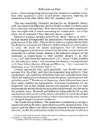
echinocyte an erythrocyte the surface of
which is covered by a large number of
short regular spicules (Fig. 24)
εε
the epsilon gene and epsilon globin
chain, the latter being synthesized during
early embryonic life and forming part of
haemoglobin Gower 1 and haemoglobin
Gower 2
εε
(epsilon) aminocaproate
a cyto-
chemical reaction which can be used to
identify basophils
E2A a gene, E-box 2A, correctly known as
T
ranscription Factor 3, TCF3, also
known as I
mmunoglobulin Transcrip-
tion F
actor 1, ITF1, E12 and E47; gene
map locus 19p13.3, encodes two bHLH
proteins, E12 and E47, by differential
splicing; these proteins bind a motif in the
immunoglobulin gene enhancer and are
essential for B-cell development; E2A
contributes to:
• the E2A-PBX fusion gene in B-lineage
acute lymphoblastic leukaemia associ-
ated with t(1;19)(q23;p13)
• the E2A-HLF fusion gene in B-lineage
acute lymphoblastic leukaemia associ-
ated with t(17;19)(q21-22;p13)
There are at least two types of E2A-HLF
rearrangements at the molecular level,
which result in different fusion proteins,
both of which lead to arrested early B-cell
development
E2F a gene, adenovirus E2 promoter
binding transcription F
actor 1, gene map
locus 20q11.2, encodes a widely expressed
DNA-binding protein whose consensus
binding sites are found in the promoters
of many genes encoding proteins involved
in cell proliferation and specifically in
DNA synthesis; the archetypal member
of a family of related proteins essential
for the G1/S phase transition of the
cell cycle; E2F proteins complex with
unphosphorylated RB1; RB1–E2F com-
E
Figure 24 An echinocyte.
An echinocyte, a cell covered with numerous short
regular spicules.
91
HAE-E 01/13/2005 05:10PM Page 91
nucleic acids by application to a mem-
brane followed by exposure to a charge
gradient, e.g. serum protein electrophore-
sis or haemoglobin electrophoresis; the
separation of particles is determined
mainly by their size and their charge
elephantiasis non-pitting oedema, par-
ticularly of the legs, as a consequence of
filariasis
ELISA enzyme-linked immunosorbent assay
ELL a gene, Eleven nineteen Lysine-rich
L
eukaemia gene, also known as MEN;
gene map locus 19p13.1, encodes a ubiq-
uitous transcription elongation factor
which suppresses transient pauses by
RNA polymerase II during transcription;
ELL contributes to the MLL-ELL fusion
gene in acute myeloid leukaemia associ-
ated with t(11;19)(q23;p13.1)
elliptocyte an elliptical erythrocyte
elliptocytosis presence of elliptical
erythrocytes
elliptogenic giving rise to elliptocytosis
eluate a solution of a substance that is
eluted
elution (i) removal of an absorbed sub-
stance from a chromatography column
(ii) removal of immunoglobulin from
the surface membrane of an erythrocyte
EMA epithelial membrane antigen
Embden–Meyerhof pathway see
glycolytic pathway
embolism (i) the process of movement of
a thrombus to another organ or site (ii)
movement of a piece of tissue, such as
bone marrow or atheromatous material,
or extraneous material, such as air,
through the bloodstream
embolus (i) a blood clot that breaks free
and is transported by the flowing blood to
another organ or site (ii) any solid or
cohesive material that moves through the
bloodstream, usually causing obstruction
of arteries
embryo the earliest stage of develop-
ment of the fertilized ovum, from implan-
tation up to about 8 weeks
emesis vomiting
emetic causing vomiting
emperipolesis active entry of haemo-
poietic cells into the surface-connected
canalicular system of megakaryocytes
eclampsia pregnancy-associated hyper-
tension complicated by convulsions, may
cause microangiopathic haemolytic anaemia
ectasia dilation, e.g. of a bone marrow
sinusoid
EDTA ethylenediaminetetraacetic acid
EEN a gene, Extra Eleven Nineteen leuk-
aemia fusion gene, correctly known as
SH3
domain, Grb2-Like, 1, SH3GL1;
gene map locus; 19p13, encodes a ubi-
quitously expressed adapter molecule
involved in intracellular signalling; a
member of a recently described sub-
family of Src-homology-3 domain (SH3)-
containing proteins; EEN contributes
to the MLL-EEN fusion gene in acute
myeloid leukaemia associated with
t(11;19)(q23;p13)
EGIL European Group for the Immuno-
logical Characterization of Leukemias
ehrlichiosis disease caused by infection
by Ehrlichia species
ELA2 gene, gene map locus 19p13.3,
encoding neutrophil elastase, mutations
of which may cause both severe con-
genital neutropenia (some autosomal
cases and the majority of sporadic cases)
and cyclical neutropenia (some autosomal
dominant and some sporadic cases)
elastase an enzyme present in neutro-
phils and in other cells and tissues
electrolyte a solute that forms ions in
solution and conducts electricity
electron a negatively charged elemen-
tary particle associated with the nucleus
of an atom
electronic issue the issuing of ABO-
compatible blood on the basis of a com-
puter programme, identification of suit-
able units being by bar-code reading,
applicable to patients with no atypical
antibodies and with two concordant
results for ABO and Rh groups, one of
which is on a current sample
electron microscopy the production
of an image of a cell by using a beam of
electrons, rather than light, to produce a
photographic image (see scanning elec-
tron microscopy, transmission electron
microscopy)
electrophoresis separation of charged
suspended particles such as proteins or
92 eclampsia
HAE-E 01/13/2005 05:10PM Page 92
lymphoblastic leukaemia associated with
t(11;19)(q23;p13.3)
enolase an enzyme in the erythrocyte
glycolytic pathway (see Fig. 33, p. 113)
enoxaparin a low molecular weight
heparin
enzyme a protein produced by living
cells that catalyses chemical reactions
enzyme-linked immunosorbent assay
(ELISA)
a method of quantifying an
antigen or an antibody by means of an
enzyme-labelled immunoreactant and a
solid-phase support
eosin an orange dye, named for Eos—the
goddess of the dawn—used in Romanow-
sky stains of blood or bone marrow films
and in haematoxylin and eosin (H & E)
stains of histological sections
eosinopenia a reduction in the
eosinophil count
eosinophil a granulocyte with aci-
dophilic granules which stain orange with
the acid stain, eosin
eosinophilia an increased eosinophil
count
eosinophilic (i) showing increased up-
take of eosin by a cell or tissue component
(ii) pertaining to the eosinophil lineage
eosinophilic granuloma a form of
Langerhans cell histiocytosis in which the
lesions are infiltrated by eosinophils
eosinophilic leukaemia a leukaemia
with prominent eosinophilic differentia-
tion
eotaxin a chemokine (of the CC family),
which is produced by eosinophils,
some epithelial cells, lymphocytes and
macrophages, and is a powerful chemoat-
tractant for eosinophils
EPB41 a gene, gene map locus 1p33-34.2,
encoding protein 4.1 of the red cell mem-
brane (see Fig. 33, p. 113); mutation may
result in hereditary elliptocytosis
EPB42 a gene, gene map locus 15q15,
encoding pallidin, also known as protein
4.2 (see Fig. 33, p. 113), a component of
the red cell membrane; mutations can
result in hereditary spherocytosis
epidemic occurring in episodic out-
breaks
epidermis the squamous cells forming
the most superficial layer of the skin
emphysema a chronic lung disease in
which there is destruction of air sacs or
alveoli leading to loss of oxygen-exchang-
ing capacity; may lead to chronic hypoxia
and therefore secondary polycythaemia
empirical based on experience without
the scientific basis necessarily being
understood
endemic constantly present in a com-
munity
endocrine secreting a hormone that has
an effect on distant tissues or organs
endocytosis the process by which the
surface membrane of a cell, usually with a
specific particle bound to its receptor, is
invaginated forming a vesicle containing
the particle and some extracellular fluid;
phagocytosis is a specialized form of
endocytosis in which larger particles are
engulfed
endogenous coming from within
endonuclease an enzyme that cleaves
DNA or RNA within the strands rather
than at the ends
endoplasmic reticulum a cytoplasmic
organelle composed of a fluid-filled
membrane system that is concerned with
synthesis and transport of proteins and
lipids; composed of the rough and the
smooth endoplasmic reticulum
endosteal cells cells lining the inner sur-
face of bone, osteoblasts and osteoclasts
endosteum the inner lining of bone
endothelial cells cells lining blood and
lymphatic vessels
endotoxic shock an acute illness with
hypotension mediated by endotoxin
endotoxin a toxin contained in the walls
of certain bacteria
enhancer a DNA sequence that
influences the promoter of a nearby gene
to increase or decrease the initiation of
transcription; an enhancer acts on a gene
in cis and may be sited upstream, down-
stream or within a gene
ENL a gene, Eleven Nineteen Leukaemia
gene also known as M
ixed Lineage
L
eukaemia, Translocated to, 1, MLLT1;
gene map locus 19p13.3, encodes a
nuclear transactivating protein; ENL
contributes to the MLL-ENL fusion gene
in acute myeloid leukaemia and acute
epidermis 93
HAE-E 01/13/2005 05:10PM Page 93
Family; amplification of this gene has
been reported in a case of myelodysplas-
tic syndrome transforming to acute
myeloid leukaemia; expression has been
reported in some cases of B-lineage acute
lymphoblastic leukaemia; overexpression
of ERBB2 has been reported in prostate
cancer and in 25–30% of breast cancer,
where it confers Taxol resistance, increas-
ing the aggressiveness of the tumour;
however a recombinant monoclonal anti-
body against ERBB2 (trastuxumab)
increases the clinical benefit of first-line
chemotherapy in metastatic breast cancer
that overexpresses the protein
ERFC E-rosette forming cell
ERG a gene, Early Response Gene, also
known as v-ets, avian erythroblastosis,
avian E
26 oncogene-Related Gene,
ERG1 and ERG2; gene map locus
21q22.3; encodes an ETS family tran-
scription factor which interacts in in
vitro assays with SAP18, a transcriptional
repressor in haemopoietic cells; ERG
contributes to the FUS-ERG fusion gene
in acute myeloid leukaemia associated
with t(16;21)(p11;q22)
E-rosette forming cell (ERFC) a T cell,
defined by its ability to form rosettes with
sheep red blood cells; such cells express
CD2
erythema redness of the skin or mucous
membrane caused by vascular dilation
erythremic myelosis a neoplasm
characterized by increased and abnormal
erythropoiesis
erythroblast a nucleated red cell precur-
sor
erythroblastic pertaining to erythro-
blasts
erythroblastic island a cluster of ery-
throblasts surrounding a central
macrophage in the bone marrow
erythroblastosis fetalis an alternative
designation of haemolytic disease of the
newborn
erythrocyte a red cell, a non-nucleated
peripheral blood cell containing
haemoglobin and having oxygen trans-
port as its major function
erythrocyte sedimentation rate (ESR)
the rate at which erythrocytes sediment in
epidermotropism having a tendency to
infiltrate the epidermis
epinephrine adrenaline, the main hor-
mone secreted by the adrenal medulla; a
platelet agonist
epistaxis bleeding from the nose
epithelial cell the surface cell of skin or
mucous membrane
epithelial membrane antigen (EMA)
an antigen expressed by cells of epithelial
origin and by cells of anaplastic large cell
lymphoma
epithelioid cell an altered macrophage
with abundant eosinophilic cytoplasm
epithelioid granuloma a cohesive
collection of altered macrophages,
referred to as epithelioid cells, with or
without other cells; this term covers all
granulomas with the exception of lipid
granulomas
epithelium the surface covering of the
body and of the gastrointestinal, respirat-
ory and genitourinary tracts
epitope an antigenic determinant, part
of an antigen which can be specifically
recognized by a cell or antibody, e.g. by
binding to a receptor on a B or T lympho-
cyte or by binding to a highly specific
monoclonal antibody
EPO erythropoietin
EPOR the gene at 7q11-22, encoding the
erythropoietin receptor, mutated in some
types of familial polycythaemia
Epstein–Barr virus (EBV) a herpesvirus
which causes infectious mononucleosis
and is also one of the aetiological factors
in a number of lymphomas including
endemic Burkitt’s lymphoma
Epstein’s syndrome an inherited syn-
drome of renal failure, sensorineural
deafness and thrombocytopenia resulting
from a mutation in the non-muscle
myosin heavy chain 9 gene (NMMHC-A
or MYH9) at 22q11-13 (or 22q12.3-
q13.2); a variant of Alport’s syndrome
ERBB2 a gene, avian Erythroblastic
Leukaemia viral oncogene homologue 2
,
also known as Her
statin (Her2), Neu
and T
yrosine Kinase-type cell surface
R
eceptor, TKR1; gene map locus
17q21.1; encodes an orphan receptor
tyrosine kinase of the EGF Receptor
94 epidermotropism
HAE-E 01/13/2005 05:10PM Page 94
erythropoietin receptor a receptor for
erythropoietin, which is abundant on red
cell precursors, encoded by the EPOR gene
ESR erythrocyte sedimentation rate
essential primary, having no recognized
external cause
essential cryoglobulinaemia cryo-
globulinaemia occurring as a manifesta-
tion of a plasma cell neoplasm which is
otherwise occult
essential erythrocytosis polycyth-
aemia for which no cause can be found;
many cases represent an early stage of
polycythaemia rubra vera
essential thrombocythaemia a myelo-
proliferative disorder with thrombocy-
tosis without coexisting polycythaemia
ester a chemical compound formed by
bonding of an alcohol and one or more
organic acids; fats are esters
esterase an enzyme catalysing the
hydrolysis of an ester
ET essential thrombocythaemia
ethnic origin deriving from a group
with a common culture and sharing
genetic characteristics
ethylenediaminetetraacetic acid
(EDTA)
a chelator of bivalent cations
anticoagulated blood; more precisely, the
number of millimetres which red cells
have sedimented after one hour
erythrocytosis an increased red cell
count, haemoglobin and haemato-
crit; the term is synonymous with
‘polycythaemia’
erythroderma an abnormality of the
skin associated with redness, not due to
simple vasodilation
erythroid pertaining to erythroblasts or
erythropoiesis
erythroleukaemia acute myeloid leu-
kaemia with prominent erythroid dif-
ferentiation; as defined by the FAB
group, erythroleukaemia or M6 AML
is AML with more than 50% of bone
marrow nucleated cells being erythroid
(see Tables 3 and 4, pp. 7 and 8)
erythrophagocytosis phagocytosis of
erythrocytes
erythropoiesis the process by which
erythroid progenitors gives rise to mature
erythrocytes or red cells (Fig. 25)
erythropoietin (EPO) a hormone,
secreted mainly by the kidney, which pro-
motes erythropoiesis, available in recom-
binant form for therapeutic use
ethylenediaminetetraacetic acid (EDTA) 95
Myeloblast
Promyelocyte
Myelocyte
Metamyelocyte Band
cell
Neutrophil
Proerythroblast Early
erythroblast
Intermediate
erythroblast
Late
erythroblast
Polychromatic
erythrocyte
Mature
erythrocyte
Common
erythroid/granulocytic
precursor
Figure 25 Erythropoiesis and granulopoiesis.
A diagrammatic representation of the various stages of erythropoiesis and granulopoiesis.
HAE-E 01/13/2005 05:10PM Page 95
and repression functions; it is essential for
yolk sac angiogenesis and adult haemo-
poiesis; ETV6 has been rearranged in
at least 41 different translocations and
many of the partner genes have been
cloned; involvement of the PNT domain
of ETV6 in these fusion genes permits
oligomerization of any resulting chi-
maeric proteins; ETV6:
• contributed to an ETV6-ARNT fusion
gene in a case of M2 acute myeloid leuk-
aemia associated with t(1;12)(q21;p13)
• contributed to the ETV6-ARG
(ABL2) fusion gene in t(1;12)(q25;p13),
occurring as a second event in a case of
M3 acute myeloid leukaemia associated
with t(15;17)
• contributed to the ETV6-MDS1/EVI1
in a case of chronic myeloid leukaemia
associated with t(3;12)(q26;p13)
• contributes to a BTL/CHIC2-ETV6
fusion gene in acute myeloid leukaemia
associated with t(4;12)(q11-12;p13)
• contributes to an ETV6-ACS2 fusion
gene in myelodysplastic syndrome and
acute myeloid leukaemia associated with
t(5;12)(q31;p13)
• contributes to the ETV6-PDGFRB
fusion gene in chronic myelomonocytic
leukaemia with eosinophilia associated
with t(5;12)(q33;p13)
• contributed to an ETV6-STL fusion
gene in a B-lineage acute lymphoblastic
leukaemia cell line with t(6;12)(q23;p13)
• contributed to an ETV6-AF7p15 fusion
gene in a patient with acute myeloid leuk-
aemia associated with t(7;12)(p15;p13)
• contributed to an HLXB9-ETV6 fusion
gene in infant acute myeloid leukaemia
associated with t(7;12)(q36;p13)
• contributes to the ETV6-JAK2 fusion
gene is rare cases of acute lympho-
blastic leukaemia or atypical chronic
myeloid leukaemia associated with either
t(9;12)(p24;p13) or with a complex chro-
mosomal rearrangement with the same
breakpoints
• contributed to an ETV6-SYK fusion
gene in a patient with a myelodysplastic–
myeloproliferative syndrome associated
with t(9;12)(q22;p12)
• contributes to the ETV6-ABL fusion
gene in rare cases of acute lymphoblas-
which is used, in the form of its sodium or
potassium salt, as an anticoagulant for
blood samples for a haemoglobin estima-
tion and blood count
ETO a gene, Eight Twenty One, also
known as C
ore-Binding Factor (see Fig.
29, p.107), A
lpha subunit 2, Translocated
to, 1
(CBFA2T1) and Myeloid Trans-
location G
ene on 8q22, MTG8; gene map
locus 8q22; named for the chromosomes
involved in the t(8;21)(q22;q22) translo-
cation in which part of this gene is fused
to part of the AML1 gene to form AML1-
ETO; homologous to the Drosophila gene
nervy; encodes a non-DNA-binding nuclear
protein normally expressed in gut, testes
and central nervous system which is
involved in the recruitment of histone
deacetylases to the transcriptional complex
etoposide an anticancer drug, which is a
topoisomerase-II interactive agent, used
in treating lymphoma
ETS a family of transcription factor
regulators related to the product of v-ets
(E
26-Transformation Specific), a viral on-
cogene encoded by the avian erythroblas-
tosis virus; ETS proteins are characterized
by a conserved winged helix-turn-helix
DNA-binding domain (ETS domain)
which binds DNA sequences centred over
a core motif (EBS: ETS binding site); a
subset of ETS proteins also carry an
amino-terminal pointed (PNT) domain
which permits interactions with distinct
protein partners, thereby establishing
unique biological functions within the
family; ETS proteins are downstream
effectors of RAS-MAPK signalling
cascades—most ETS transcription fac-
tors are phosphorylated and activated
by specific MAP kinases, however some,
e.g. ERG, are inhibitory; they regulate
varied physiological and pathophysiolo-
gical processes such as haemopoiesis,
apoptosis and tumorigenesis
ETV6 a gene, Ets Variant gene 6, homolo-
gous with v-ets, gene map locus 12p13,
also known as TEL, encodes a transcrip-
tion regulator; it belongs to the ETS fam-
ily and has a pointed (PNT) domain and a
3′ ETS DNA binding domain; ETV6 is
ubiquitously expressed and exhibits con-
text-dependent transcriptional activation
96 ETO
HAE-E 01/13/2005 05:10PM Page 96
• contributes to the fusion gene, AML1-
MDS1-EVI1, in acute myeloid leukaemia
associated with t(3;21)(q26;q23)
• contributes to the ETV6-EVI1 fusion
gene in acute myeloid leukaemia associ-
ated with t(3;12)(q26;p13)
• is involved in the translocations
t(2;3)(p13;q26)
t(2;3)(q23;p26)
t(3;17)(q26;q22)
exfoliative tending to lose layers of cells
exocrine pertaining to secretion of a
substance which has an effect outside
the tissues of the body, e.g. within the
gastrointestinal tract or on the skin
exocytosis the process in which a sec-
retory vesicle produced in the Golgi
complex moves to the surface of the cell,
fuses with the surface membrane and
discharges its contents
exogenous coming from outside
exon a part of a gene which is repres-
ented in mature messenger RNA; most
genes are composed of exons and non-
transcribed introns (see Fig. 32, p. 111)
exonuclease an enzyme that breaks
down DNA or RNA from the ends of the
strands
extramedullary occurring outside the
bone marrow
extramedullary haemopoiesis haemo-
poiesis occurring outside the bone
marrow, usually in the liver and spleen
extramedullary myeloma extra-
medullary plasmacytoma, a plasma
cell tumour occurring outside the bone
marrow
extrinsic something which originates
outside rather than being an essential
part; the extrinsic pathway of coagula-
tion involves activation of factor VII by
tissue factor with subsequent activation
of factors X and II and conversion of
fibrinogen to fibrin; in contrast to the
intrinsic pathway, the circulating blood
does not contain all the factors necessary
for the pathway (see Fig. 17, p. 77)
extrinsic pathway inhibitor see tissue
factor pathway inhibitor
ex vivo a process which is detected in
cells or tissues that have been removed
from the body; the term should be con-
trasted with in vivo and in vitro
tic leukaemia, acute myeloid leukaemia
and chronic myeloid leukaemia associ-
ated with t(9;12)(q34;p13) or a variant
translocation
• contributes to an ETV6-CDX2 fusion
gene in acute myeloid leukaemia associ-
ated with t(12;13)(p13;q12)
• contributes to an ETV6-TRKC fusion
gene in acute myeloid leukaemia and
in familial fibrosarcoma associated with
t(12;15)(p13;q25); the acute myeloid
leukaemia and fibrosarcoma mutations
differ at a molecular level
• contributes to the ETV6-AML1
fusion gene in the 30% of cases of acute
lymphoblastic leukaemia that are asso-
ciated with a cryptic t(12;21)(p13;q22);
there is generally loss of the normal
ETV6 allele suggesting that loss of
ETV6 function may contribute to
oncogenesis
• contributes to the MN1-ETV6 fusion
gene in acute myeloid leukaemia associ-
ated with t(12;22)(p13;q11)
• contributed to PAX5-ETV6 fusion
gene in a case of acute lymphoblastic
leukaemia
euchromatin diffuse or non-condensed
transcriptionally active chromatin
eukaryocyte a cell with a nucleus
European Group for the
Immunological Characterization of
Leukemias (EGIL)
a cooperative
group that published guidelines on
immunophenotyping
Evans’ syndrome autoimmune haemo-
lytic anaemia plus autoimmune thrombo-
cytopenic purpura
event-free survival survival without
disease relapse or the need to change to
alternative treatment (see also, disease-
free survival, overall survival)
EVI1 a gene, Ecotropic Viral Integration
site 1
, gene map locus 3q26; encodes a
zinc finger nuclear protein which can
repress transcription and recruits histone
deacetylases; EVI1:
• is 5′ truncated and dysregulated by
proximity to the enhancer elements of the
ribophorin 1 gene and forms a GR6-EVI1
fusion gene in acute myeloid leukaemia
associated with inv(3)(q21q26) and
t(3;3)(q21;q26)
ex vivo 97
HAE-E 01/13/2005 05:10PM Page 97
F13B the gene at 1q31-q32.1 that encodes
the B subunit of factor XIII
Fab that part of an immunoglobulin
molecule that is capable of binding to
antigens (see Fig. 48, p. 139)
FAB pertaining to the French–American–
British Cooperative Group and their
classifications (see Table 3, p. 7)
Fabry’s disease angiokeratosis corporis
diffusum, an inherited disease in which
phospholipids are stored in many parts of
the body, particularly in blood vessels
FACS fluorescence-activated cell sorter
or sorting
factitious false, not genuine, artefactual
(of a laboratory test result), sometimes
deliberately caused by an individual to
simulate illness
factor I (roman numeral) fibrinogen, a
plasma protein that is converted to fibrin
by the action of thrombin thus leading to
clot formation; the Aα, Bβ and γ chains
are encoded respectively by the FGA,
FGB and FGG genes; (not to be confused
with factor I [upper case i] of the comple-
ment system)
factor II prothrombin, a vitamin K-
dependent coagulation factor encoded
by the F2 gene; it is converted to thrombin
by the action of activated factor X, in
the presence of calcium, phospholipid
and activated factor V (see Fig. 17, p.
77)
factor II: G20210A a variant form of
factor II with a point mutation in the 3′
untranslated region, associated with
increased plasma concentration of factor
II and some increase in risk of thrombo-
sis, present in 1–1.5% of some Caucasian
populations
factor IIa activated factor II, thrombin
F2 the gene at 11p11-q12 that encodes
prothrombin (factor II), a coagulation
factor in both the intrinsic and extrinsic
pathways, mutation of which can
lead to prothrombin deficiency or
thrombophilia
F5 the gene at 1q23 that encodes factor
V, a coagulation factor in both the in-
trinsic and extrinsic pathways, mutation
of which can lead to autosomal recessive
factor V deficiency or to factor V Leiden,
associated with thrombophilia
F7 the gene at 13q34 that encodes factor
VII, a coagulation factor of the extrinsic
pathway, mutation of which can lead to
factor VII deficiency
F8C the gene at Xq28 that encodes factor
VIII, a coagulation factor in the intrinsic
pathway, mutation of which can lead to
haemophilia A; about a third of mutations
are new sporadic mutations
F9 the gene at Xq27.1-q27.2 that encodes
factor IX, a coagulation factor in the
intrinsic pathway, mutation of which can
lead to factor IX deficiency
F10 the gene at 13q34 that encodes factor
X, a coagulation factor in both the intrin-
sic and extrinsic pathways, mutation of
which can lead to factor X deficiency
F11 the gene at 4q35 that encodes factor
XI, a coagulation factor of the intrinsic
pathway; mutations of this gene, which
are prevalent among Ashkenazi Jews,
can lead to factor XI deficiency in both
homozygotes and heterozygotes
F12 the gene at 5q33-qter that encodes
factor XII, the first factor of the intrinsic
pathway of coagulation, mutation of
which can lead to factor XII deficiency
F13A1 the gene at 6p25-p24 that encodes
the A subunit of factor XIII
F
98
HAE-F 01/13/2005 05:11PM Page 98
factor V a coagulation factor in the com-
mon pathway which also contributes to
physiological anticoagulation; it is encoded
by the F5 gene; activated factor V, factor
Va, is a cofactor in the conversion of
prothrombin to thrombin by factor Xa;
non-activated factor V is a cofactor with
protein S in the inactivation of factor Va
and factor VIIIa by activated protein C
(see Figs 17 and 56, pp. 77 and 170)
factor Va activated factor V, a cofactor
in the conversion of prothrombin to
thrombin by factor Xa (see Fig. 17, p. 77)
factor V and factor VIII deficiency an
inherited, autosomal recessive, deficiency
of factors V and VIII resulting from a
mutation in the LMAN1 gene
factor V Leiden a variant form of factor
V, also known as factor VR
506
Q and
factor VQ
506
, resulting from a 1691G→Α
mutation in the F5 gene; factor V Leiden
has a prevalence of 3–15% in different
Caucasian populations; the mutation
leads to an alteration of protein structure
at the point where factor V is cleaved
by activated protein C and this renders
factor V resistant to inactivation by
activated protein C and also less effect-
ive as a cofactor for the inactivation of
factor VIIIa by activated protein C
(see Fig. 56, p. 170); there is mild throm-
bophilia and probably increased suscepti-
bility to thrombotic microangiopathy
factor VII a vitamin K-dependent coagu-
lation factor, the first factor in the ex-
trinsic pathway of coagulation, which on
vascular injury forms a 1:1 stoichiometric
complex with tissue factor exposed on the
endothelial cell; complexing of factor VII
to tissue factor leads to its activation;
activated factor VII initiates the extrinsic
pathway of coagulation and also activ-
ates factor IX of the intrinsic pathway
(see Fig. 18, p. 78)
factor VIIa activated factor VII, avail-
able as a recombinant coagulation factor
factor VIII anti-haemophiliac globulin, a
coagulation factor in the intrinsic pathway
encoded by the F8C gene, synthesized in
the liver but requires von Willebrand’s
factor (synthesized in megakaryocytes
and endothelial cells) for normal stability
in the plasma; it facilitates the activation
of factor X by activated factor IX (see
Figs 17 and 18, pp. 77 and 78)
factor VIIIa activated factor VIII
factor IX Christmas factor, a vitamin
K-dependent factor in the intrinsic path-
way encoded by the F9 gene (see Figs 17
and 18, pp. 77 and 78)
factor IXa activated factor IX
factor X a vitamin K-dependent factor
in the common coagulation pathway;
factor X is activated both by factor VIIa
and by factor IXa (in the presence of fac-
tor VIIIa in a calcium- and phospholipid-
dependent reaction); in turn it activates
prothrombin to thrombin, by a calcium-
and phospholipid-dependent reaction in
the presence of factor Va (see Figs 17 and
18, pp. 77 and 78)
factor Xa activated factor X
factor XI a factor in the intrinsic pathway,
encoded by the F11 gene; it is activated
by factor XIIa in vitro and by thrombin
in vivo and in turn it activates factor IX
(see Figs 17 and 18, pp. 77 and 78)
factor XIa activated factor XI
factor XII Hageman factor, the first fac-
tor in the intrinsic pathway, encoded by
the F12 gene; after contact activation in
vitro, it leads to activation of factor XI;
deficiency causes marked abnormality of
in vitro tests of the intrinsic pathway but
in vivo is not associated with any haemor-
rhagic disorder (see Fig. 17, p. 77)
factor XIIa activated factor XII
factor XIII a factor composed of two A
subunits and two B subunits, encoded by
F13A1 and F13B respectively that, when
activated, causes stable cross-linking of
fibrin (see Fig. 18, p. 78)
factor XIIIa activated factor XIII
factor B a protein in the alternative
complement pathway (see Fig. 20, p. 81)
factor D a protein in the alternative
complement pathway (see Fig. 20, p. 81)
factor H an glycoprotein encoded by a
gene at 1q32 which inhibits complement
activation; factor H competes with factor
B for C3b and acts as a cofactor for factor
I in the inactivation of C3b (see Fig. 20,
p. 81); homozygous deficiency can be
associated with familial or sporadic
factor H 99
HAE-F 01/13/2005 05:11PM Page 99
FANCG a gene at 9p13, mutation of
which explains about 10% of cases of
Fanconi’s anaemia
Fanconi’s anaemia a recessively in-
herited, clinically and genetically hetero-
geneous chromosomal fragility syndrome,
characterized by multiple congenital abnor-
malities, aplastic anaemia with onset
usually in childhood and a predisposition
to acute myeloid leukaemia and other
tumours
FAS a gene, Tumour Necrosis Fac-
tor R
eceptor Superfamily, member 6,
TNFRSF6, CD95, gene map locus 10q24.1,
encodes a transmembrane protein belong-
ing to the TNF family that mediates apo-
ptosis when trimerized by cross-linking
to Fas ligand; FAS is mutated in about
10% of cases of multiple myeloma and
in about 10% of cases of non-Hodgkin’s
lymphoma, particularly MALT-type
non-Hodgkin’s lymphoma and extra-
nodal B-lineage diffuse large cell
lymphoma
FBP17 a gene, Formin Binding Protein
17
, centromeric to 9q34; encodes a
ubiquitously expressed protein which is
believed to interact with SNX proteins,
involved in EGF receptor trafficking;
FBP17 contributed to a MLL-FBP17 fusion
gene in an infant with M4 acute myeloid
leukaemia
Fc the constant part of an immunoglo-
bulin molecule that determines antibody
class (G, A, M, E, D) and is responsible
for fixation of complement and interac-
tion with effector cells such as granulo-
cytes, monocytes, mast cells and killer
cells (see Fig. 48, p. 139)
Fc receptor a receptor for the Fc part of
an immunoglobulin molecule (see Fc
γγ
R,
Fc
εε
R)
Fc
εε
R receptor for the Fc part of the
immunoglobulin E molecule
Fc
γγ
R receptor for the Fc part of the
immunoglobulin G molecule: FcγRI, high
affinity receptor on monocytes; FcγRII,
lower affinity receptor on neutrophils,
monocytes, eosinophils, platelets and B
cells; FcγRIII, low affinity receptor on
macrophages, neutrophils, eosinophils
and NK cells
relapsing haemolytic uraemic syndrome
and membranous glomerulonephritis
factor I (upper case i) an inhibitory
protein in the complement system; homo-
zygous deficiency can be associated with
haemolytic uraemic syndrome and mem-
branous glomerulonephritis
faggot cell a cell containing bundles
of Auer rods, a feature of hypergranular
promyelocytic leukaemia and its hypo-
granular/microgranular variant
falciparum malaria malaria caused by
Plasmodium falciparum
false negative a negative result that
should have been positive
false positive a positive result that should
have been negative
familial occurring in families, by implica-
tion inherited
familial cold urticaria an autosomal
dominant disorder resulting from a muta-
tion in the CIAS1 gene, resulting in a
periodic cold-induced non-pruritic non-
urticarial rash associated with neutrophilia
familial haemophagocytic lympho-
histiocytosis (FHL)
a familial syn-
drome, probably basically an immune
deficiency syndrome, characterized by
haemophagocytic syndromes occurring
in childhood and often being fatal; two
genetic mechanisms have been deter-
mined, linked to 10q21-22 in FHL1 and
linked to 9q21.3-22 (PERF1 gene) in
FHL2.
familial Mediterranean fever an auto-
somal recessive disease, resulting from
mutation in the MEFV gene, character-
ized by periodic fever and serositis
FAN see CEP110
FANCA a gene at 16q24.3, mutation of
which explains 65–70% of cases of
Fanconi’s anaemia
FANCC a gene at 9q22.3, mutation of
which explains 10–15% of cases of
Fanconi’s anaemia
FANCD2 a cloned gene that causes some
cases of Fanconi’s anaemia
FANCE a cloned gene that causes some
cases of Fanconi’s anaemia
FANCF a gene at 11p15, mutation of
which explains <2% of cases of Fanconi’s
anaemia
100 factor I (upper case i)
HAE-F 01/13/2005 05:11PM Page 100
FGFR1 a gene, Fibroblast Growth Factor
R
eceptor 1, gene map locus 8p11.2-p11.1,
encodes a receptor tyrosine kinase; con-
tributes to:
•a ZNF198-FGFR1 fusion gene in a
syndrome of chronic myelomonocytic
leukaemia with eosinophilia/T-lineage
lymphoblastic lymphoma, sometimes
referred to as the 8p11 syndrome or the
stem cell leukaemia–lymphoma syndrome,
associated with t(8;13)(p11;q11-12)
• the FOP-FGFR1 fusion gene in a
similar disorder associated with
t(6;8)(q27;p11)
• the CEP110-FGFR1 fusion gene in
t(8;9)(p11-12;q33) associated with the
same syndrome
• the BCR-FGFR1 fusion gene in asso-
ciation with chronic myeloid leukaemia
and t(8;22)(p11;q11)
FGFR1 was also found to be rearranged
in a case of chronic myeloid leukaemia
associated with systemic mastocytosis
FGFR3 a gene, Fibroblast Growth Factor
R
eceptor 3, gene map locus 4p16.3;
encodes a receptor tyrosine kinase which
is dysregulated, by proximity to the IGH
Cα enhancer on chromosome 14, in
t(4;14)(p16.3;q32), a cryptic transloca-
tion associated with multiple myeloma
FGG a gene at 4q28 encoding the γ chain
of fibrinogen, mutation of which can lead
to dysfibrinogenaemia
FHIT a gene, Fragile Histidine Triad gene,
also known as AP3A hydrolase, gene
map locus 3p14.2; the FRA 3B fragile
site; the gene is composed of 10 exons dis-
tributed over at least 500 kb, and encodes
a widely expressed enzyme involved in the
regulation of DNA replication; deletions
and structural rearrangements in FRA
3B have been observed in many epithelial
malignancies; loss of FHIT function is
important in the development and/or
progression of head and neck squamous
cell cancers, and cervical, oesophageal
and lung cancers; FHIT expression is
reduced in a majority of cases of acute
lymphoblastic leukaemia and in a signi-
ficant proportion of cases of chronic
myeloid leukaemia, the significance of
this being unclear
FCGRIIB a gene, low affinity Fc Gamma
R
eceptor IIB, CD32, gene map locus
1q22; involved in the t(1;22)(q22;q11) re-
arrangement found in less than 1% of cases
of follicular lymphoma and associated
with transformation to high grade disease
FDPs fibrin degradation products
Fechtner’s syndrome an inherited
syndrome characterized by renal failure,
sensorineural deafness, thrombocytope-
nia and neutrophil inclusions that re-
semble Döhle bodies; a variant of Alport’s
syndrome and Epstein’s syndrome result-
ing from a mutation in the non-muscle
myosin heavy chain 9 gene (NMMHC-A
or MYH9) at 22q11-13 (or 22q12.3-
q13.2)
Felty’s syndrome hypersplenism causing
pancytopenia in a patient with rheuma-
toid arthritis; there may be an underlying
large granular lymphocyte leukaemia
ferric pertaining to trivalent iron (Fe
3+
)
ferritin a complex of iron and a protein,
apoferritin; ferritin is present in the cyto-
plasm of erythroblasts; in macrophages
it is converted into haemosiderin, the
principal storage form of iron; small
amounts are present in the plasma, and
measurement of serum ‘ferritin’ (actually
apoferritin) permits assessment of body
iron stores
ferrous pertaining to bivalent iron (Fe
2+
),
the form of iron that is incorporated into
protoporphyrin IX by ferrochelatase (see
Fig. 34, p. 116)
ferrous sulphate an iron compound
used to treat iron deficiency anaemia
fertilization the fusion of a spermato-
zoon with an ovum to form a zygote
fetal pertaining to the fetus
fetal haemoglobin haemoglobin F
fetus the unborn offspring after it has at-
tained the particular form of the species,
e.g. in man the unborn offspring beyond
8 weeks from fertilization
FFP fresh frozen plasma
FGA a gene at 4q28 encoding the Aα
chain of fibrinogen, mutation of which
can lead to dysfibrinogenaemia
FGB a gene at 4q28 encoding the Bβ chain
of fibrinogen, mutation of which can lead
to dysfibrinogenaemia
FHIT 101
HAE-F 01/13/2005 05:11PM Page 101
102 FHL
fibroblast the cell that is responsible
for synthesis and deposition of collagen;
small numbers of fibroblasts are present
in the bone marrow
fibrosis the replacement of normal tissue,
e.g. the bone marrow, by fibroblasts and
collagen; the term ‘fibrosis’ may also be
used to refer to reticulin deposition but a
distinction should be drawn between reti-
culin deposition and collagen deposition
filariasis a disease resulting from infec-
tion by filarial parasites such as Loa loa,
Wuchereria bancrofti and Brugia malayi
filgrastim recombinant granulocyte
colony-stimulating factor
FIM a gene, Fused In Myeloproliferative
disorders, see ZNF198
FISH fluorescence in situ hybridization
fixation (i) the process by which cells are
killed and tissues and cells are preserved
by exposure to chemicals such as ethanol,
methanol, acetone or formalin (ii) the
process by which complement components
are bound to immunoglobulin that has
already formed a complex with antigen
FKHR a gene, Forkhead box O1A,
FOXO1A, gene map locus 13q14.1;
FHL familial haemophagocytic lymphohis-
tiocytosis
fibre FISH a FISH technique using very
elongated genomic DNA
fibrin a fibrillar protein, the formation of
which is the basis of blood coagulation;
fibrin is formed by polymerization and
cross-linking of fibrin monomers, which
are produced by the action of thrombin on
fibrinogen
fibrin degradation products (FDPs)
breakdown products of fibrin which are
present in the plasma in increased con-
centration in the presence of extensive or
disseminated intravascular coagulation or
increased fibrinolysis; D-dimer is a specific
fibrin degradation product
fibrinogen a soluble plasma protein
(Fig. 26) that is converted, by the action
of thrombin, into fibrin monomers, which
polymerize and are cross-linked to form
a stable, insoluble fibrin polymer, thus
leading to blood clotting: also known as
factor I
fibrinolysis the process by which plas-
min breaks down fibrin (Fig. 27)
fibrinolytic pertaining to fibrinolysis
NH terminal
TT
TT
Bβ
P
P
P
COOH
terminal
γ
COOH
terminal
γ
γγ
P
Aα
Aα
Aα
Aα
Bβ
Bβ Bβ
P
P
P
P
Thrombin cleavage site
(fibrinopeptides A and B shown in black)
Plasmin cleavage site
Disulphide links
T
P
Aα chains
Bβ chains
chains
Aα
Bβ
γγ
Figure 26 The fibrinogen molecule.
A diagrammatic representation of the fibrinogen molecule showing the Aα, Bβ and γ chains, the sites of cleavage
by thrombin to produce fibrinopeptides A and B (black) and the sites of cleavage by plasmin.
HAE-F 01/13/2005 05:11PM Page 102
FKHR fusion genes, in t(2;13)(q35;q14)
and t(1;13)(p36;q14) respectively, in alve-
olar rhabdomyosarcoma
FL a gene at 13q12-13 that encodes
flk2/flt3 ligand
encodes a forkhead domain transcrip-
tion factor that is negatively regulated by
protein kinase B signalling (see AKT) and
is essential for the completion of mitosis;
contributes to the PAX3-FKHR and PAX7-
FL 103
X
Xa
IX IXa
VIIIa
Phl
Ca
2+
Va
Phl
Ca
2+
II IIa
thrombin–
thrombomodulin
complex
PC
APC
PAI
Fibrinogen Fibrin Plasminogen
TAFI Plasmin
FDPs
tPA
XIIa
Kallikrein
α
2
M
α
2
AP
Major pathway
Minor pathway
Negative effect
Figure 27 The fibrinolytic pathways.
Major pathways are shown by a solid arrow and minor pathways by a dotted arrow. Two lines across an arrow
indicate a negative effect, either inhibition or destruction. Dashed crossed lines indicate proteolysis with
resultant reduction inactivity of the target protein. Thrombin activation and fibrin deposition lead to activation
of fibrinolysis. Fibrin binds plasminogen and plasminogen is converted to plasmin by tissue plasminogen
activator (tPA) released from activated endothelial cells. Plasmin breaks down fibrin and the fact that plasmin
is formed from plasminogen bound to fibrin means that, normally, fibrinolysis is preferentially focused in the
area of fibrin deposition. However if there is excess free plasmin, fibrinogen, factor Va and factor VIIIa can all
be degraded. α2 antiplasmin (α
2
AP) and α2 macroglobulin (α
2
M) inhibit the action of plasmin, particularly
circulating plasmin. Plasminogen activator inhibitors 1 and 2 (PAI1 and PAI2) inhibit the action of tPA, thus
reducing fibrinolysis. Thrombin production also leads to activation of protein C which, to some extent, breaks
down PAI1, thus enhancing fibrinolysis. However, thrombin also activates thrombin-activatable fibrinolysis
inhibitor (TAFI), which inhibits the action of plasmin on fibrin. There is thus a delicately balanced system of
positive and negative controls of fibrinolysis.
HAE-F 01/13/2005 05:11PM Page 103
that are flowing through a detection device;
may be based on fluorescence, light scat-
ter, light absorbance or impedance meas-
urements; used for immunophenotyping
(Fig. 28)
flow karyotyping the use of flow cyto-
metry to identify/separate chromosomes
on the basis of their DNA content
FLT3 a gene, FMS-Like Tyrosine kinase 3
receptor, also known as stem cell tyrosine
kinase, gene map locus 13q12, encodes a
receptor tyrosine kinase of the PDGFR
superfamily and is the receptor for flt3
ligand; expression in human blood and
bone marrow cells is restricted to CD34+
flag jargon for an automated instrument
indication that a blood sample shows an
abnormality
flagging jargon indicating production
of ‘flags’ by an automated instrument,
indicating abnormal or possibly un-
reliable test results
FLI1 a gene, Friend Leukaemia virus
I
ntegration 1, gene map locus 11q24,
encodes an ETS transcription factor;
contributes to a EWS-FLI1 fusion gene
in Ewing’s sarcoma associated with
t(11;22)(q24;q12)
flow cytometry the process of evaluat-
ing characteristics of cells, in suspension,
104 flag
Figure 28 Flow cytometry immunophenotyping.
The results of immunophenotyping performed by flow cytometry on the peripheral blood cells of a patient with
lymphocytosis. Forward and sideways light scatter have been used to gate on lymphocytes (top left), which
have then been further analysed. The lymphocytosis resulted from an increase of CD8-positive lymphocytes
(bottom left) with a reversal of the normal CD4:CD8 ratio. However, in addition, there is a population of
lambda-positive B cells with a striking reversal of the normal kappa:lambda ratio. The patient had both
a post-splenectomy lymphocytosis and circulating follicular lymphoma cells.
HAE-F 01/13/2005 05:11PM Page 104
fibronectin deficiency has been identified
in association with Ehlers-Danlos syn-
drome (type X).
foamy macrophage a macrophage
with heavily vacuolated cytoplasm, usu-
ally indicative of the presence of lipid
folate a generic term for folic acid and
related compounds which are essential
for normal DNA synthesis
folic acid pteroylglutamic acid, one of
the B group of vitamins, essential for
nucleic acid synthesis
folinic acid N
5
-formyltetrahydrofolic
acid, a form of folate that can circumvent
the block in folate metabolism caused by
dihydrofolate reductase inhibitors such
as methotrexate
follicle centre cell a type of mature
B lymphocyte found in the follicles of
lymph nodes; includes centrocytes (small
cells with condensed chromatin) and
centroblasts (larger cells which may be
nucleolated)
follicle centre cell lymphoma see
follicular lymphoma
follicular dendritic cell an antigen-
presenting cell in lymphoid follicles that
presents antigen to germinal centre B
lymphocytes
follicular lymphoma a lymphoma
composed of cells analogous to those
in the follicles of normal lymph nodes;
such lymphomas usually have a follicular
structure but sometimes the growth pat-
tern is diffuse
fondaparinux a synthetic factor Xa
inhibitor
FOP a gene, FGFR1 Oncogene Partner;
gene map locus 6q27, encodes a ubiquit-
ously expressed leucine rich repeat (LRR)
protein, that contributes to the FOP-
FGFR1 fusion gene in the lympho-
proliferative–myeloproliferative disorder
associated with t(6;8)(q27;p11) (see also
8p11 syndrome)
foreign body giant cell a giant cell of
monocyte/macrophage lineage with mul-
tiple nuclei spread through the cytoplasm
forkhead domain a phosphoprotein
binding motif originally identified in a
group of forkhead transcription factors
e.g. FKHR, but also present in a wide
cells; in frame internal tandem duplica-
tions affecting the JM domain of the flt3
protein are found in blast cell genomic
DNA from approximately 20% of adult
patients with acute myeloid leukaemia,
across all FAB subtypes, usually in the
absence of detectable cytogenetic abnor-
malities; FLT3 mutations are associated
with a worse prognosis; mutations are
also present in some patients with
myelodysplastic syndromes
fludarabine a nucleoside analogue used
in treating chronic lymphocytic leukaemia
fluorescence activated cell sorter
(FACS)
an instrument that can sort cells
into those that have bound or not bound
a fluorochrome-labelled antibody
fluorescence in situ hybridization
(FISH)
identification of DNA or RNA
sequences in cells in metaphase or inter-
phase following hybridization with com-
plementary RNA or DNA probes that
have been labelled with a fluorochrome
fluorescence resonance energy trans-
fer (FRET)
the non-radiative transfer
of energy from a fluorophore in an ex-
cited state to a nearby acceptor fluoro-
phore; the farther apart the two molecules
are, the weaker the transfer efficiency; so
the technique is useful in assessing the
interaction between two different (labelled)
macromolecules
fluorochrome a fluorescent chemical
FMC7 a monoclonal antibody which
gives positive reactions with cells of most
non-Hodgkin’s lymphomas but not with
the cells of chronic lymphocytic leuk-
aemia, acute lymphoblastic leukaemia
or lymphoblastic lymphoma, thought to
recognize a conformational epitope of
CD20
FMS see CSF1R
FN1 a gene, Fibronectin, also known
as L
arge, External, Transformation-
S
ensitive protein, LETS; gene map locus
2q31, encodes a high molecular weight
cell surface glycoprotein that also repres-
ents about 1% of serum protein, and is
required by most cells to bind to collagen;
FN1 contributed to a NUP98-FN1 fusion
gene, one of two fusion genes present in a
case of M2 acute myeloid leukaemia;
forkhead domain 105
HAE-F 01/13/2005 05:11PM Page 105
106 formalin
encodes a glycine-rich protein which is
a component of nuclear riboprotein
complexes that plays a role in genomic
stability; contributes to the FUS-ERG
fusion gene in acute myeloid leukaemia
associated with t(16;21)(p11;q22)
FUT1 a locus on chromosome 19 with two
alleles relevant to ABH blood group anti-
gens: the H allele encodes α-2-fucosyl-
transferase, which converts the h antigen
to the H antigen; the h allele does not
encode a transferase (see Fig. 3, p. 4)
FUT2 a locus at 19q13 with two alleles rel-
evant to secretor status and Lewis blood
group antigens (Fig. 30, p. 108) : the Se
allele encodes α-2-L-fucosyltransferase,
which converts a precursor type 1 disac-
charide on a plasma glycosphingolipid
molecule to the H type 1 antigen without
which the Le
b
antigen cannot be synthe-
sized; the se allele does not encode a
transferase; individuals who are SeSe or
Sese have ABH antigens in saliva and
other body fluids whereas sese individuals
do not; the former are referred to as ‘secre-
tors’ and the latter as ‘non-secretors’;
homozygosity for Se also leads to an
increased plasma concentration of von
Willebrand factor
FUT3 a locus at 19p13.3 with two alleles
relevant to Lewis blood group antigens
(see Fig. 30, p. 108): the Le allele encodes
α-3/4-L-fucosyltransferase, which con-
verts a precursor type 1 disaccharide on a
glycosphingolipid molecule to the Le
a
antigen, H type 1 antigen to Le
b
and A
type 1 or B type 1 to ALe
b
or BLe
b
respec-
tively; the le allele does not encode a
transferase
FY a locus at 1q21-22 where allelic genes
encode antigens of the Duffy blood group
system; the genes at this locus are Fy
a
,
Fy
b
, Fy
x
and Fy; Fy
x
leads to weak
expression of the Fyb antigen; Fy has a
promoter mutation and when this gene
is homozygously present the phenotype
is Fy(a-b-)
variety of other proteins e.g. the nuclear
proliferative antigen Ki-67 which is in-
volved in centrosome separation during
mitosis
formalin a solution of formaldehyde,
used for fixing tissues
FOS a transcription factor of the leucine
zipper family
FOS a gene, Finkel-Biskis-Jinkins (FBJ)
murine Os
teosarcoma viral oncogene
homologue, gene map locus 14q24.3,
encodes a major component of the
activator protein-1 (AP-1) transcription
factor complex; expressed at high levels
in term placenta and trophoblastic cells;
transforms cells through alterations in
DNA methylation and in histone deace-
tylation; expression in chronic granulocytic
leukaemia correlates with interferon-
alpha responsiveness
fragment (of red cell) a schistocyte or
erythrocyte fragment
frame-shift mutation a deletion or inser-
tion of a number of base pairs that is not
either 3 or a multiple of 3, into a DNA
molecule, so that the reading frame is altered
French–American–British (FAB) Co-
operative Group
an international
cooperative group of haematologists who
proposed a number of widely accepted
classifications of leukaemia and myelody-
splastic syndromes
fresh frozen plasma (FFP) plasma
from a single blood donation that has
been frozen, shortly after separation
from red cells, to a core temperature of
below –30°C and which therefore retains
normal levels of coagulation factors
FRET fluorescence resonance energy transfer
fusion the process of joining together to
form a fusion gene, composed of parts of
two genes, or a fusion protein, the prod-
uct of such a gene (Fig. 29)
FUS a gene, Fusion, derived from 12-16
translocation, malignant lipos
arcoma;
gene map locus 16p11.2, also known as
T
ranslocated in Liposarcoma (TLS);
HAE-F 01/13/2005 05:11PM Page 106
FY 107
YGYGGT
YGYGGT
YGYGGT
(a) Wild type CBF
(b) inv(16)
(c) t(8;21)
CBFβ
CBFα
+
Transcription
–
Transcription
Myosin heavy chain
Repressor proteins
Histone
acetylation
VWRPY
Myosin heavy chain
CBFβ
CBFα2
CBFα BD
Runt NMTS VWRPY
CBFβ-SMMHC
inv(16)
ETO
NN
CBFα–ETO
t(8;21)
CBFα BD
–
Transcription
CBFα sequestered from DNA
Figure 29 Fusion genes and fusion proteins.
Two fusion genes involving CBFA2 and CBFB, encoding CBFα2 and CBFβ respectively illustrate the role of
fusion genes and chimaeric proteins in oncogenesis.
(a) CBFα2 and CBFβ together form a heterodimeric haemopoietic transcription factor, Core Binding Factor
(CBF). CBFβ does not itself bind to DNA but interacts via its CBFα binding domain (CBFα BD) with CBFα2.
This interaction occurs in the runt domain of CBFα2 and increases the ability of another part of the runt domain
to bind the consensus DNA sequence YGYGGT. Once bound to DNA, CBF can activate transcription of a
large number of haemopoietic genes. In certain circumstances it can act as a repressor via the VWRPY motif at
the carboxy terminus of CBFα2 which interacts with the transcriptional repressor, TLE. Normally, CBFα2 is
sequestered in the nucleus due to its nuclear matrix-binding domain NMTS.
(b) The acquired chromosomal abnormality inv(16)(p13q32) seen in a specific subtype of AML, leads to the
fusion of most of the CBFB gene to the region encoding the tail domain of the smooth muscle myosin heavy
chain (SMMHC or MYH11) gene. Several variants of a CBFB-SMMHC fusion transcript have been detected in
AMLs with inv(16). Most lead to a fusion protein containing a fully functional CBFα BD fused to the α-helical
tail of SMMHC. The fusion protein is able to form multimers because of this tail, which can be visualized as
nuclear and cytoplasmic speckles. It is believed that multimeric CBFβ-SMMHC sequesters CBFα2 subunits and
so reduces DNA binding by CBF.
(c) The translocation t(8;21)(q22;q22) seen in another subtype of AML, leads to the fusion of the runt domain
of the CBFA2 gene to the majority of ETO which encodes a non-DNA binding nuclear protein involved in the
recruitment of histone deacetylses. The ETO protein has ‘nervy’ domains that permit interactions with
transcriptional repressors. The CBFα2-ETO fusion protein is able to interact with CBFβ and does so with
greater affinity than wild-type CBFα2, but the ETO moiety leads to the repression of transcription. This leads to
a differentiation arrest in myeloid cells.
HAE-F 01/13/2005 05:11PM Page 107
108 FY
Type 1disaccharide
(mainly in plasma)
Genotype
AA SeSe LeLe
α-2-L-fucosyltransferase
(encoded by Se
at FUT2 locus)
α-3/4-L-fucosyltransferase
(encoded by Le
at FUT3 locus)
α-3/4-L-fucosyltransferase
α-3-N-acetyl-D-
galactosaminyltransferase
(encoded by A at
ABO locus)
H type 1
Le
a
Final phenotype: A Le(a+b+)
'secretor'
Type 1 disaccharide
Genotype
AA SeSe LeLe
α-3/4-L-fucosyltransferase
Final phenotype: A Le(a+b–)
'non-secretor'
A type 1
Le
b
α-3/4-L-fucosyltransferase
Le
b
(a)
(b)
Le
a
Figure 30 The Lewis blood group system.
The interaction between transferases encoded by three sets of allelic genes to produce
Lewis blood group antigens: (a) In an individual who is A positive (genotype either
AA or AO), Le positive (genotype LeLe or Lele) and who is a ‘secretor’ (genotype
SeSe or Sese). The Le allele at the FUT3 locus encodes a transferase that converts a
precursor to Le
a
antigen whereas the Se allele at the FUT1 locus produces H type 1
which can be converted by the transferase encoded by Le into Le
b
. In the presence of
the transferase encoded by the A allele at the ABO locus A Le
b
is also produced. The
phenotype is A Le(a+b+).
(b) In an individual who is A positive (genotype either AA or AO) and Le positive
(genotype LeLe or Lele) but who is a ‘non-secretor’ (genotype sese) Le
a
is the only
Lewis antigen produced. The phenotype is A Le(a+b–).
HAE-F 01/13/2005 05:11PM Page 108
driving cells into G0 phase; deleted in
many myeloid neoplasms
GAS2 a gene, Growth Arrest-Specific 2;
gene map locus 11p15.2-p14.3; encodes a
ubiquitously expressed component of the
microfilament system which increases
susceptibility to p53-dependent apoptosis
GAS6 a gene, Growth Arrest-Specific 6,
also known as Axl
receptor tyrosine
kinase L
igand, AXLLG; gene map locus
13q34; encodes a vitamin K-dependent
protein homologous to protein S which is
the ligand for the receptor tyrosine kinase
AXL
GAS7 a gene, Growth Arrest-Specific 7;
gene map locus 17p13, encodes a putative
transcription factor expressed normally
in the brain, that contributed to a MLL-
GAS7 fusion gene in a case of therapy-
related M4 acute myeloid leukaemia with
a cryptic t(11;17)(q23;p13) translocation
GAS41 a gene, Glioma-Associated Seq-
uence 41
, gene map locus 12q13-q15,
encodes a putative transcription factor
that has homology with AF9 and ENL;
GAS41 amplified in high-grade glioma
GATA-1 a gene encoding a transcrip-
tion factor that is important in erythro-
poiesis and critical in megakaryocyte
differentiation
Gaucher cell the characteristic altered
macrophage of Gaucher’s disease
Gaucher’s disease hereditary glucosyl
ceramide lipidosis, a heterogeneous group
of inherited diseases resulting from var-
ious mutations in the glucocerebrosidase
gene; these mutations lead to accumula-
tion of glucocerebroside in character-
istic macrophages, designated Gaucher
cells; may be diagnosed by bone marrow
aspiration
γγ
the Greek letter gamma
γγ
chain
(i) the γ globin chain which forms
part of haemoglobin F (ii) the heavy
chain of an immunoglobulin G molecule;
two γ chains combine with two light
chains, in an individual molecule either
κ or λ, to form an immunoglobulin
molecule (iii) part of the γδ T-cell recep-
tor, a surface membrane structure which
permits antigen recognition
γγ
-glutamyl cysteine synthetase (an
enzyme of the pentose shunt)
hexose monophosphate shunt
(see Fig. 59, p. 182)
γγ
heavy chain disease
a plasma cell
dyscrasia in which there is secretion of
monoclonal γ chain
G an abbreviation for the purine, guanine
G0, G1 and G2 three phases of the cell
cycle (see Fig. 15, p. 72)
G6PD glucose-6-phosphate dehydrogenase
G6PD the gene at Xq28 encoding glucose-
6-phosphate dehydrogenase; mutation
may lead to glucose-6-phosphate dehydro-
genase deficiency with intermittent or,
much less often, chronic, haemolysis
gallium scan an imaging technique
using
67
Ga-single photon emission com-
puterized tomography to detect meta-
bolic activity in residual areas of active
lymphoma
gamete a germ cell, a spermatozoon or
an ovum
GAP GTP-ase activating protein
GAS1 a gene, Growth Arrest-Specific 1;
gene map locus 9q21.3-q22.1; encodes a
plasma membrane protein which is a
homologue of the iron protein subunit of
Complex I of the mitochondrial electron
transport chain; expressed in non-
transformed cells in response to stimuli
G
109
HAE-G 01/13/2005 05:11PM Page 109
GDP guanosine diphosphate
GEF guanine nucleotide exchange factor
gelatinous transformation deposi-
tion of acid mucopolysaccharide in bone
marrow, replacing haemopoietic tissue
gene the segment of DNA that is in-
volved in producing a polypeptide chain;
it includes regions preceding and follow-
ing the coding region (5
′′
and 3
′′
untrans-
lated regions) as well as intervening
sequences (introns) between individual
coding segments (exons) (Fig. 32); genes
mediate inheritance; they are located on
nuclear chromosome or, rarely, on a
mitochondrion
gene expression the transcription of a
gene into tRNA, rRNA or mRNA, in the
latter case with subsequent translation to
protein
gene profiling see microarray analysis
genetic pertaining to inheritance by genes
genetic code the relationship between a
triplet of bases, called a codon, and the
amino acid which it encodes
genome the complete DNA sequence of
an individual or a species
genomic pertaining to a gene or genes
genotype the genetic makeup of an indi-
vidual, cf. phenotype
Gaussian a description of data that,
when plotted as a histogram, have a bell-
shaped distribution; also referred to as a
‘normal distribution’ (see Fig. 45, p. 128)
GBA the gene encoding glucocerebrosi-
dase, mutated in Gaucher’s disease
G-banding a technique for staining meta-
phase spreads of chromosomes with a
Giemsa stain to produce a unique band-
ing pattern that permits the identification
of individual chromosomes (Fig. 31)
G-CFU granulocyte colony-forming unit
G-CSF granulocyte colony-stimulating
factor
GCSFR a gene, Granulocyte Colony-
S
timulating Factor Receptor, also known
as C
olony-Stimulating Factor 3 Recep-
tor, CSF3R, CD114; gene map locus
1p35-p34.3; encodes the G-CSF receptor;
point mutations in this gene have been
reported in some patients with severe
congenital neutropenia (Kostmann’s
syndrome), these patients being at great-
est risk of developing myelodysplastic
syndrome or acute myeloid leukaemia;
mutation leading to synthesis of a trun-
cated protein has also been implicated in
acute myeloid leukaemia complicating
Kostmann’s syndrome
110 Gaussian
Figure 31 G banding.
A karyogram of G-banded chromosomes of a patient with chronic granulocytic
leukaemia. The banding pattern produced by the Giemsa stain, together with a
consideration of the size of the chromosome and the position of the centromere,
permits the identification of individual chromosomes, normal and abnormal. There is
a t(9;22)(q34;q11); this is a balanced translocation between chromosomes 9 and 22
with breakpoints at 9q34 and 22q11 respectively.
HAE-G 01/13/2005 05:11PM Page 110
giant metamyelocyte a metamyelo-
cyte which is two to three times normal
size and often has a nucleus of abnormal
shape, characteristic of megaloblastic
erythropoiesis
Giemsa stain a Romanowsky type
stain which can be used for staining blood
and bone marrow cells and tissue sections
and is also used for staining preparations
of chromosomes (see G-banding); one
component of a May–Grünwald–Giemsa
stain
GIFT granulocyte immunofluorescence test
Gilbert’s syndrome a common syn-
drome resulting from homozygosity for
a polymorphism in the promoter region
of the gene encoding bilirubin UDP glu-
curonosyl transferase-1, UGT1, leading
to unconjugated hyperbilirubinaemia; it
Gerbich antigen an erythrocyte mem-
brane antigen, carried on glycophorin C;
monoclonal antibodies to this antigen
can be used for the identification of ery-
throid cells
germ cell a gamete, a spermatozoon or
an ovum
germinal centre a specialized structure
in a lymph node or other lymphoid tissue
in which follicular dendritic cells present
antigen to B lymphocytes
ghost cell an erythrocyte which contains
negligible amounts of haemoglobin
giant cell arteritis inflammation of
arteries with the inflammatory cells in-
cluding giant cells, usually associated
with a high erythrocyte sedimentation
rate, which can therefore be used as a
diagnostic aid
Gilbert’s syndrome 111
Start
codon
ATG
Stop
codon
e.g. TAG
Promotor
Flanking region
enhancer
Flanking region
enhancers
5'UTR
Transcriptional unit
Exon
Intron
3'UTR
Untranslated region, UTR
Direction of transcription
Figure 32 Gene structure and function.
A gene is a segment of DNA that is involved in producing a protein. It is also known as a transcriptional
unit and includes not only coding regions (exons), but also non-coding sequences which lie between exons
(introns), and before and after the coding segments (5′ and 3′ untranslated regions). Transcription is the
enzymatic process whereby RNA is synthesized from a DNA template. The promoter is the section of DNA
where the transcriptional machinery binds before transcription can start. It is defined by certain highly
conserved sequences, e.g. the TATA box which is found 25bp ‘upstream’ from (i.e. 5′ to) the transcriptional
start site, and the CAAT box, found 75bp upstream. Transcriptional termination signals lie in the 3′UTR and
trigger the 3′ cleavage of newly formed RNA (see Figure 65, p. 202). The first coding triplet in a gene is referred
to as the start codon and is usually ATG (encoding methionine); the coding sequence always ends in a
termination signal (a codon which does not encode an amino acid); this can be TAG, TGA or TAA. Translation
is the synthesis of a protein from a messenger RNA (mRNA) template; this always proceeds from a start codon
to a stop codon. Some genes have multiple promoters and may have introns spanning several Kb. There are
many examples of overlapping genes and of transcription in either orientation occurring from different
promoters in the same segment of DNA. The flanking sequences on either side or within the transcriptional
unit may contain enhancers, which are sequences of DNA involved in the binding of positive or negative
transcriptional regulatory proteins.
HAE-G 01/13/2005 05:11PM Page 111
pathway in erythrocytes and other cells in
which glucose is broken down to provide
energy for the cell (Fig. 33)
glycophorin a group of red cell mem-
brane proteins, glycophorins A, B, C and
D (CD235a, CD235b, CD236, CD236R),
antibodies to which can be used to iden-
tify cells of erythroid lineage (see Fig. 64,
p. 199, and see also GYPA, GYPB, GYPC)
glycoprotein a post-translationally
modified protein in which a carbohydr-
ate is covalently linked to an amino acid
residue
glycosaminoglycan a polysaccharide
consisting of repeating disaccharide
units of amino sugar derivatives (such as
glucosamine or galactosamine), at least
one of which has a negatively charged
carboxylate or sulphate group; heparin
and heparan are glycosaminoglycans
glycoside a substance with an alcohol
component in which a glycosyl (sugar)
moiety has replaced the hydrogen in the
hydroxyl group
glycosylation the process in which a
covalent bond is formed between glucose
and a macromolecule
glycosylphosphatidylinositol (GPI) a
red cell membrane protein, encoded by
the PIGA gene, that anchors many other
red cell surface membrane proteins,
including CD55, CD58 and CD59
GM-CFU granulocyte/macrophage colony-
forming unit
GM-CSF granulocyte/macrophage colony-
stimulating factor
GMPS a gene, Guanosine-5′-Mono-
p
hosphate Synthetase, gene map locus
3q24, encodes the enzyme that catalyses
the amination of xanthylate (XMP) to
guanylate (GMP) in the purine synthe-
sis pathway; contributed to an MLL-
GMPS fusion gene in a patient with
M4 acute myeloid leukaemia associated
with t(3;11)(q25;q23); in the resulting
chimaeric proteins the catalytic domain
of GMPS was intact
GMS stain Grocott’s methenamine silver
stain
Golgi apparatus or Golgi complex
stacks of flattened membranous sacs
receiving newly synthesized proteins from
causes neonatal jaundice in infants with
G6PD deficiency and aggravates jaundice
in adults with haemolytic anaemias
gingivitis inflammation of the gums,
can be a feature of acute leukaemia
with monocytic differentiation
gland (i) specialized epithelial tissue
capable of secretion (ii) a popular term
for a lymph node
glandular fever see infectious mononu-
cleosis
Glanzmann’s thrombasthenia a severe,
autosomal recessive, inherited defect of
platelet function, resulting from muta-
tion in either the ITGA2B or the ITGB3
gene, leading to deficiency of platelet gly-
coprotein IIb/IIIa
globin the protein part of the haemo-
globin molecule
globoside collection blood group anti-
gens, P and P
k
, closely related to the P
blood group system
glomerulonephritis inflammation of
glomeruli
glomerulus (plural glomeruli) the
structure in the kidney in which filtration
occurs, leading to formation of urine
glossitis inflammation of the tongue, a
feature of deficiency of iron or vitamin B
12
glucose-6-phosphate dehydrogenase
(G6PD)
an enzyme of the pentose shunt
which protects red cells from oxidant
damage (see Fig. 59, p. 182)
glucose-6-phosphate dehydrogenase
(G6PD) deficiency
reduced glucose-
6-phosphate dehydrogenase activity in
red cells leading to susceptibility to
oxidant-induced haemolysis
glucose phosphate isomerase an
enzyme in the erythrocyte glycolytic
pathway (Fig. 33)
glutathione synthetase an enzyme
related to the pentose shunt (see Fig. 59,
p. 182)
glycogen a complex carbohydrate
glycolipid a sugar-containing lipid in
which a monosaccharide is linked to a
lipid via glycosidic bond
glycolysis the process by which glucose
is metabolized, providing energy for a cell
glycolytic pathway (Embden–
Meyerhof pathway)
the metabolic
112 gingivitis
HAE-G 01/13/2005 05:11PM Page 112
complex is located; in cells with a well
developed Golgi apparatus the Golgi
zone appears as a pale area
Good Clinical Practice a standard for
the design, conduct, performance, monit-
the endoplasmic reticulum, responsible for
modification, packaging and distribution
of proteins
Golgi zone the area of a cell, usually
adjacent to the nucleus, where the Golgi
Good Clinical Practice 113
Glucose
ATP
ADP
Hexokinase
Glucose-6-phosphate
Fructose-6-phosphate
Fructose-1,6-diphosphate
Glyceraldehyde-
3-phosphate
1,3-biphosphoglycerate
3-phosphoglycerate
2-phosphoglycerate
Phosphoenolpyruvate
Pyruvate
Lactate
Dihydroxyacetone
phosphate
Haemoglobin
Methaemoglobin
ATP
ADP
Glucosephosphate
isomerase
Phosphofructokinase
Aldolase
Glyceraldehydephosphate
dehydrogenase
Phosphoglycerate mutase
(and diphosphoglycerate mutatase)
Phosphoglycerate
kinase
Enolase
Pyruvate kinase
Lactate dehydrogenase
ATP
ADP
ATP
ADP
NADH
NAD
Aldolase
T
r
io
s
e
p
h
os
ph
at
e
isome
r
as
e
ATP
ADP
Cyochrome b5 reductase
biphosphoglycerate mutase/
biphosphoglycerate kinase
biphosphoglycerate mutase/
biphosphoglycerate kinase
2,3-biphosphoglycerate
Figure 33 The glycolytic pathway.
The glycolytic pathway provides energy for body cells including erythrocytes. Enzymes are shown in italics and
enzyme products in upright script. Pyruvate kinase deficiency is the most common inherited defect of the
glycolytic pathway.
HAE-G 01/13/2005 05:11PM Page 113
dysplastic syndrome associated with
t(3;3)(q21;q26) and inv(3)(q21q26)
grade an expression of the degree of
malignancy of a tumour
GRAF a gene, RhoA GTPase Regulation
A
ssociated with Focal adhesion kinase
pp125, gene map locus 5q31, encodes an
SH3 domain-containing protein which is
highly homologous to BCR; a member
of the GTPase-activating protein (GAP)
family; binds to pp125(FAK), a tyrosine
kinase involved in the integrin signalling
transduction pathway, also stimulates
the GTPase activity of RhoA; GRAF con-
tributed to a MLL-GRAF fusion gene in
a child with juvenile myelomonocytic
leukaemia associated with t(5;11)(q31;q23);
both alleles of the gene were also dis-
rupted in three cases of acute myeloid
leukaemia–myelodysplastic syndrome
associated with 5q–
graft a tissue that is transplanted into
another individual or, less accurately,
that is removed from an individual and
subsequently returned
graft rejection the process by which a
transplanted tissue, e.g. bone marrow, is
recognized as foreign and rejected by the
body
graft-versus-host disease (GVHD)
the illness resulting from engraftment of
immunocompetent lymphoid cells that
recognize host tissues as foreign and
attack them
graft-versus-leukaemia (GVL) the abil-
ity of engrafted tissue to exert a specific
anti-leukaemic effect that is different from
the generalized graft-versus-host effect
Gram-negative non-staining with a
Gram stain
Gram-positive staining with a Gram stain
Gram stain a stain used for staining
bacteria, which are then classified as
Gram-positive or Gram-negative
granulocyte a leucocyte with a lobed
nucleus and granular cytoplasm—a
neutrophil, eosinophil or basophil
granulocyte-colony forming unit
(G-CFU)
a progenitor cell that can give
rise to a colony of granulocytes when
cultured in vitro
oring, auditing, recording and reporting
of clinical trials that provides assurance
that the data and reported results are
credible and accurate and that the rights,
integrity and confidentiality of trial sub-
jects are protected
gout acute crystal arthropathy second-
ary to urate deposition; may be a feature
of myeloproliferative disorders, causes
neutrophilia
GP1BA a gene, gene map locus 17pter-
p12, encoding platelet glycoprotein Ibα,
mutation of which can lead to Bernard–
Soulier syndrome and platelet-type von
Willebrand’s disease
GP1BB a gene, gene map locus 22q11.2,
encoding platelet glycoprotein Ibβ, muta-
tion of which can lead to Bernard–Soulier
syndrome
GPV a gene on chromosome 3 encoding
platelet glycoprotein V, mutation of which
can lead to Bernard–Soulier syndrome
GPIX a gene on chromosome 3 encoding
platelet glycoprotein IX, mutation of which
can lead to Bernard–Soulier syndrome
GPH a gene, Gephyrin, gene map locus
14q24, encodes gephyrin—a protein
expressed in the brain which is involved
in anchoring the inhibitory glycine recep-
tor to the postsynaptic cytoskeleton;
several alternative transcripts are known
which differ at their 5′ termini, but
which encode the same 3′ tubulin bind-
ing site; also known as GPNH; con-
tributed to a MLL-GPNH fusion gene in
t(11;14)(q23;q24) in a patient with M5b
acute myeloid leukaemia and in a patient
with ‘undifferentiated’ acute leukaemia
following therapy with a topoisomerase
II-interactive drug in which the fusion
protein retained the gephyrin tubulin
binding site; autoantibodies to gephyrin
have been observed in a patient with stiff
man syndrome
GR6 a gene located within the leukaemia
breakpoint region of 3q21, gene map
locus 3q21, expressed in early fetal devel-
opment but not in adult peripheral blood
cells; has no known homologies; GR6
contributes to an GR6-EVI1 fusion gene
in acute myeloid leukaemia and myelo-
114 gout
HAE-G 01/13/2005 05:11PM Page 114