A–Z of Haematology - part 7 ppt
Bạn đang xem bản rút gọn của tài liệu. Xem và tải ngay bản đầy đủ của tài liệu tại đây (326.13 KB, 25 trang )
INK4b see CDKN2B and Table 5, p. 70
innate immunity naturally occurring
immunity that is not permanently changed
by encounter with an antigen, dependent
on phagocytic cells, natural killer cells,
inflammatory mediators, acute phase
reactants and complement components
ins a cytogenetic abbreviation indicating
an insertion
insertion either (i) the insertion of part
of one chromosome into another chro-
mosome or into another part of the same
chromosome, detectable by conventional
cytogenetic analysis and designated ‘ins’,
or (ii) the insertion of a number of bases
into a DNA molecule
in situ a method of studying a cell or a
tissue without disrupting it so that posi-
tive or negative results can be related to
individual cells
in situ hybridization a technique for
detecting specific DNA or RNA sequences
by hybridization with a complementary
probe that is labelled, for example, with a
fluorochrome or a radioactive isotope
integrin one of a family of heterodimeric
transmembrane cell adhesion molecules,
composed of non-covalently linked α
and β subunits, that mediate cell–cell
and cell–matrix interactions
interdigitating dendritic cell a tissue
cell (including Langerhans cells) that be-
comes activated on antigen exposure and
migrates to draining lymph nodes where
it presents antigen to helper T lymphocytes
in the context of major histocompatibility
complex class II molecules
interferon one of a family of cytokines
produced by various body cells, e.g.
monocytes, fibroblasts and virus-infected
cells, that are part of non-specific immune
response to viruses and to cancer cells;
they are categorized as type 1 (α and β)
and as type 2 (γ); interferons are used in
therapy, e.g. to treat chronic granulocytic
leukaemia and hairy cell leukaemia
interferon-
αα
one of two classes of
cytokines synthesized by virus-infected
cells that conveys, to other cells, resist-
ance against viral infection
interferon-
ββ
one of two classes of cyto-
kines synthesized by virus-infected cells
incidence the rate of occurrence of a dis-
ease in a population, usually expressed as
the number of cases per 100 000 of popu-
lation per year
Indian a blood group system, the antigens
being carried on CD44, the hyaluronate
receptor
ineffective not achieving the desired end
ineffective erythropoiesis failure to
achieve adequate bone marrow output of
erythrocytes despite normal or increased
numbers of erythroid precursors in the
bone marrow, indicative of increased
intramedullary death of erythroblasts
ineffective haemopoiesis failure to
achieve adequate bone marrow output
of erythrocytes, leucocytes and platelets
despite normal or increased numbers of
haemopoietic precursors in the bone
marrow, indicative of increased intra-
medullary death of haemopoietic cells
infarct death of a tissue as a result of
interruption of its blood supply
infectious mononucleosis glandular
fever; an acute illness with fever, pharyn-
gitis, lymphadenopathy and atypical lym-
phocytes in the peripheral blood. Caused
by primary infection with the Epstein–
Barr virus
inflammation non-specific changes in
tissues as a response to infection or tissue
damage
ING1 a gene, Inhibitor of Growth 1, gene
map locus 13q34, encodes a widely ex-
pressed zinc finger nuclear protein which
causes cell cycle arrest in G1; a candidate
tumour suppressor gene, mutations have
been found in squamous cell carcinomas
of the head and neck
INK4a see CDKN2A and ARF
INK4b see CDKN2B
initiation (i) the process by which RNA
transcription from a gene commences; (ii)
the process by which protein translation
from mRNA commences (see Fig. 74,
p. 222)
initiation codon the three nucleotide
codon (ATG) at the 5′ end of a gene
which is essential to permit initiation of
transcription of a gene, i.e. initiation of
polypeptide synthesis
INK4a see CDKN2A and Table 5, p. 70
140 incidence
HAE-I 01/13/2005 05:12PM Page 140
tions in it are associated with congenital
immunodeficiency
interleukin-3 (IL3) a haemopoietic
colony-stimulating factor, encoded by
IL3, that is capable of supporting the
proliferation of a broad range of haemo-
poietic cell types and also has neu-
rotrophic activity
interleukin-4 (IL4) a lymphokine
secreted by type 2 (Th2) helper T cells
and activated B cells, encoded by IL4,
which activates macrophages and B
cells, promotes IgE class switching and
has a role in mast cell sensitization,
allergy and defence against nematodes;
it stimulates the production of eotaxin—a
chemokine involved in eosinophil recruit-
ment; has an inhibitory effect on the
growth of many leukaemic cell lines in vitro
interleukin-5 (IL5) a haemopoietic
growth factor for B cells and eosinophils,
secreted by type 2 (Th2) helper T cells,
encoded by IL5
interleukin-6 (IL6) a cytokine with
potent antiviral activity, which is also
able to elicit an acute phase response;
encoded by IL6; the aberrant production
of IL6 by neoplastic cells is a contribu-
tory factor to the growth B-cell neo-
plasms, T-cell lymphomas and Kaposi’s
sarcoma; promoter polymorphisms in
the IL6 gene are associated with hyper-
triglyceridaemia and susceptibility to
Kaposi’s sarcoma in HIV-infected indi-
viduals, but there is no association with
multiple myeloma.
interleukin-7 (IL7) a lymphokine cap-
able of supporting the growth of pre-B
cells in vitro, encoded by IL7
interleukin-8 (IL8) a cytokine secreted
by several types of cell, including T cells
and macrophages, in response to inflam-
matory stimuli, encoded by IL8; it is
chemotactic for neutrophils, basophils
and T cells and promotes angiogenesis;
involved in the pathogenesis of viral
bronchiolitis caused by the respiratory
syncytial virus (RSV)—the level of IL8
appears to be correlated with disease
severity
interleukin-9 (IL9) a cytokine with both
myeloid and lymphoid stimulatory activ-
that conveys, to other cells, resistance
against viral infection
interferon-
γγ
a cytokine synthesized by
type 1 (Th1) helper T cells and NK cells,
encoded by a gene on chromosome 6;
γ interferon activates macrophage and
neutrophil killing, stimulates NK cell
function and enhances antigen-presenta-
tion by increasing expression of type II
MHC molecules; it inhibits type 2 (Th2)
helper T cells; a defect in γ-interferon or
its receptor can cause an inherited suscep-
tibility to mycobacterial infections
interferon regulatory factor (IRF) a
family of transcription factors defined by
a characteristic DNA-binding domain
and the ability to bind to the interferon-
stimulated response element; involved in
cytokine signalling and the control of
proliferation
interleukin a cytokine secreted by one
type of leucocyte that has effects mainly
on other leucocytes
interleukin-1 (IL1) a cytokine, also
known as endogenous pyrogen, pro-
duced mainly by monocytes, that
activates T cells and macrophages and
mediates the acute phase response; there
are 2 forms encoded by 2 separate genes
at 2q14
interleukin-2 (IL2) an immunoregula-
tory lymphokine, encoded by IL2, pro-
duced by activated type 1 (Th1) helper T
cells, which activates cytotoxic T cells,
NK cells and macrophages
interleukin-2 receptor the multi-
subunit IL2 receptor that is composed of
various heterotrimeric and heterodimeric
combinations of three different subunits,
IL2Rα, also known as CD25 (encoded by
IL2RA), IL2Rβ, also known as CD122,
(encoded by IL2RB) and IL2Rγ, also
known as CD132 (encoded by IL2RG);
the gamma chain is an indispensable
component of the receptor and is also a
component of other cytokine receptors
(see CD132); αβγ trimers constitute the
high affinity form of the receptor, βγ
dimers the intermediate affinity form and
αγ dimers the low affinity form; the α
chain is not functional in IL2 internaliza-
tion and signal transduction, but muta-
interleukin-9 (IL9) 141
HAE-I 01/13/2005 05:12PM Page 141
similarly to IL2, encoded by IL15; it
appears to utilize the IL2 receptor
interleukin-16 (IL16) a proinflammat-
ory cytokine which signals via CD4,
inducing chemotactic and immunomodu-
latory responses in CD4+ T cells;
encoded by IL16
interleukin-17 (IL17) a proinflam-
matory cytokine expressed by activated
memory T cells, encoded by IL17; induces
expression of CD54 on B cells; archetypal
member of a new family of proinflamma-
tory cytokines (IL17 B–F)
interleukin-18 (IL18) a cytokine secreted
by macrophages, encoded by IL18, which
promotes interferon-γ secretion by T
cells, suppresses IgE synthesis and aug-
ments NK cell responses
interleukin 19 (IL19) a cytokine closely
related to and genetically linked with
IL10 and IL20; regulates B-cell function;
encoded by IL19
interleukin 20 (IL20) a cytokine closely
related to and genetically linked with
IL10 and IL19; encoded by IL20; IL20
receptors are found in the skin and are
upregulated in psoriasis
intermediate filaments filaments with
a diameter of 7–10 nm that form part of
the cytoskeleton; they include keratin,
desmin, vimentin, laminin, neurofilaments
and glial fibrillary acidic protein
intermediate grade lymphoma a
lymphoma with a degree of malignancy
intermediate between low and high grade,
recognized by the Working Formulation;
includes mantle cell lymphoma which was
previously sometimes designated ‘lym-
phoma of intermediate differentiation’
interphase the stage when a cell is out of
cycle (G0) (see Fig. 15, p. 72)
interstitial pertaining to the interstitium
interstitium the potential space between
cells
intervening sequence (IVS) see intron
intracerebral within the brain
intracranial within the skull
intrasinusoidal within a sinusoid, e.g.
within a bone marrow sinusoid
intravascular within blood vessels
intravenous within a vein (usually re-
ferring to a method of administering
ity, encoded by IL9; it promotes IgE class
switching; overproduced in Hodgkin’s
disease
interleukin-10 (IL10) an anti-
inflammatory cytokine secreted by type 2
(Th2) helper T cells, which down regu-
lates the immune response, inhibiting
type I (Th1) helper T cells and inhibits
allergic reactions, encoded by IL10;
interleukin-10 limits HIV-1 replication in
vivo; mutations in the IL10 promoter
have been associated with increased risk
of HIV infection and once infected, rapid
progression to AIDS
interleukin-11 (IL11) a widely expres-
sed cytokine of unknown physiological
function, encoded by IL11; acts synergis-
tically with several other cytokines to
stimulate cells of a variety of haemo-
poietic lineages; secreted by bone marrow
stromal cells; stimulates the production
of acute phase proteins
interleukin-12 (IL12) a dimeric
cytokine, also known as natural killer cell
stimulatory factor, composed of an alpha
chain (p35 subunit encoded by IL12A)
and a beta chain (p40 subunit encoded
by IL12B); secreted by dendritic cells,
macrophages and B cells; stimulates
the production of interferon-γ by type 1
helper T cells and NK cells; mutations
in IL12B or IL12R lead to inherited sus-
ceptibility to mycobacterial infections,
including disseminated infection with
BCG, and to susceptibility to Salmonella
enteritidis infection
interleukin-13 (IL13) a cytokine
secreted by activated type 2 (Th2)
helper T cells, encoded by IL13, which
stimulates the production of eotaxin, a
chemokine involved in eosinophil recruit-
ment; induces IgG4 and IgE synthesis
by B cells; induces the pathophysiologic
features of asthma, independently of IgE
and eosinophils; polymorphisms in the
IL13 gene predispose to bronchial hyper-
responsiveness and asthma susceptibility
interleukin-14 (IL14) a cytokine with
B-cell stimulatory properties, gene map
locus unknown
interleukin-15 (IL15) a cytokine which
affects T-cell activation and proliferation
142 interleukin-10 (IL10)
HAE-I 01/13/2005 05:12PM Page 142
iron-binding capacity the capacity of
the serum to bind iron, dependent on the
concentration of transferrin and other
iron-binding proteins in the serum
iron deficiency a lack of adequate iron
stores leading to some clinical or labora-
tory abnormality, e.g. anaemia or glossitis
iron deficiency anaemia anaemia
caused by a lack of adequate supplies
of iron
iron depletion absence of storage iron
but without any associated haematolog-
ical or clinical abnormality
iron-regulatory proteins proteins that
interact with iron-responsive elements of
genes; in the case of the ferritin genes and
the
δδ
-aminolaevulinate synthase gene, iron
depletion leads to interaction and decrea-
sed translation of the gene; in the case of
the transferrin receptor gene, iron deple-
tion leads to interaction and increased
translation of the gene
iron-responsive element a family of
cis-acting non-coding mRNA structures
located in the untranslated region of
mRNA for ferritins,
δδ
-aminolaevulinate
synthase and transferrin receptor
iron stores iron stored in body macro-
phages in the form of ferritin and, par-
ticularly, haemosiderin
irradiation exposure to ionizing radiation
irregularly contracted cells erythro-
cytes lacking central pallor but, in con-
trast to spherocytes, being irregular in
shape
IRTA1 a gene, Immunoglobulin
superfamily R
eceptor Translocation-
A
ssociated gene 1, gene map locus 1q21,
one of 5 related genes (IRTA1-5) clus-
tered at this locus; encodes an inhibitory
immunoglobulin superfamily receptor
homologous to the Ig Fc receptor, nor-
mally expressed in perifollicular B cells
but not in plasma cells; IRTA1 is trans-
located to the IGH locus in t(1;14)(q21;q32)
associated with less than 5% of cases
of multiple myeloma, the fusion gene
encoding a protein containing the signal
peptide and first two amino acids of
IRTA1 linked to the transmembrane and
intracellular domains of the surface IgA
receptor
blood or blood components, fluids or
drugs)
intrinsic contained within itself
intrinsic factor a factor secreted by the
parietal cells of the stomach that com-
bines with an extrinsic or dietary factor
(vitamin B
12
) to permit the absorption of
vitamin B
12
in the small intestine
intrinsic factor antibodies antibodies
directed at intrinsic factor, often present
in the serum or the gastric juice of indi-
viduals with pernicious anaemia
intrinsic pathway a coagulation path-
way for which all the factors necessary
are already present in the blood, only con-
tact with a foreign surface being required
to initiate coagulation (see Fig. 17, p. 77)
intron a sequence of DNA in a gene
which is not represented in processed
messenger RNA or in the protein product
(see Fig. 32, p. 111)
inv a cytogenetic abbreviation indicating
an inversion
inversion (inv) the inversion of a seg-
ment of a chromosome
in vitro carried out or occurring outside
a living body, literally ‘in glass’
in vivo carried out or occurring in a
living creature, literally ‘in life’
ion an atom that has gained or lost one of
its electrons so that it is not electrically
balanced
IRF interferon regulatory factor
IRF1 a gene, Interferon Regulatory
F
actor 1, gene map locus 5q31.1, encodes
an IRF transcription factor that upregu-
lates several growth-suppressing genes;
hemizygously lost in some patients
with 5q–; mutations leading to variant
proteins with reduced DNA-binding
capacity have been observed in gastric
and non-small cell lung carcinoma
IRF4 a gene, Interferon Regulatory Factor
4
, also known as Multiple Myeloma
oncogene 1—MUM, gene map locus 6p25,
encodes an IRF transcription factor
expressed only in lymphocytes; dysregu-
lated by t(6;14)(p25;q32) in about 20% of
patients with multiple myeloma.
iron Fe, a metal which is an essential con-
stituent of haemoglobin and the muscle
protein, myoglobin
IRTA1 143
HAE-I 01/13/2005 05:12PM Page 143
isotype immunoglobulin molecules char-
acterized by a specific type of heavy chain,
e.g. immunoglobulin M rather than
immunoglobulin A
isotype switching change from secret-
ing immunoglobulin M to secreting another
class of immunoglobulin, e.g. IgG or IgA
ITGA2 the gene at 5q23-q31 encoding the
integrin α2 chain; integrin α
2
β
1
(very late
antigen-2, VLA-2) is platelet glycopro-
tein Ia/IIa (CD49b/CD29)
ITGA2B the gene at 17q21.32 encod-
ing the integrin α
IIb
chain (CD41a), a
component of platelet glycoprotein IIb/
IIIa, mutation of which can lead to
Glanzmann’s thrombasthenia
ITGB2 the gene encoding CD18, the inte-
grin β2 chain, mutation of which can
cause leucocyte adhesion deficiency
ITGB3 the gene at 17q21.32 encoding
the integrin β
3
chain (CD61), a com-
ponent of platelet glycoprotein IIb/IIIa
(α
IIb
/β3) and the vitronectin receptor
(α
v
/β3); mutation of the gene can lead to
Glanzmann’s thrombasthenia
ITP idiopathic thrombocytopenic pur-
pura (now usually referred to as ‘autoim-
mune thrombocytopenic purpura’)
144 IRTA2
IRTA2 a gene, Immunoglobulin superfamily
R
eceptor Translocation-Associated gene
2
, gene map locus 1q21, encodes an
inhibitory immunoglobulin superfamily
receptor homologous to the Ig Fc recep-
tor, normally expressed in germinal cen-
tre centrocytes and a broad spectrum of
perifollicular cells, which may include
immunoblasts and memory cells but not
centroblasts; dysregulated and overex-
pressed in Burkitt’s lymphoma with 1q21
abnormalities, being the only member of
the IRTA cluster to be expressed in this
disorder—the mechanism for this is unclear
ISBT International Society for Blood
Transfusion
iso (or i) a cytogenetic abbreviation
indicating an isochromosome
isochromosome (i or iso) a chromo-
some formed by duplication of either the
short arm or the long arm of a chromosome
isoenzyme a structurally different form
of an enzyme present in different tissues
of a single individual
isoniazid a drug used to treat tuberculosis
which can cause sideroblastic erythropoiesis
isotonic having an osmolarity that is the
same as that of normal body fluids
HAE-I 01/13/2005 05:12PM Page 144
Jordan’s anomaly an inherited abnor-
mality in which leucocytes of all types are
vacuolated
jumping translocation translocation of
part of a chromosome to multiple other
partner chromosomes
JUN a transcription factor of the leucine
zipper family
JUN a gene, gene map locus 1p32, that
encodes a leucine zipper DNA-binding
protein which is the cellular homologue
of the transforming gene of avian sar-
coma virus 17; JUN is a major compon-
ent of the activator protein-1 (AP-1)
transcription factor complex
juvenile chronic myeloid leukaemia
a type of chronic myeloid leukaemia
which occurs in infants, characterized by
hepatosplenomegaly, lymphadenopathy,
rash, anaemia, monocytosis and eleva-
tion of haemoglobin F concentration,
now usually designated juvenile myelo-
monocytic leukaemia
juvenile myelomonocytic leukaemia
an alternative designation of juvenile chronic
myeloid leukaemia, the term recommended
in the WHO classification (see Fig. 55,
p. 168)
JAK janus kinase
JAK2 a gene, Janus kinase 2, gene map
locus 9p24, encodes a janus kinase; JAK2
contributes to the ETV6-JAK2 fusion
gene in rare cases of acute lympho-
blastic leukaemia and atypical chronic
myeloid leukaemia, associated with
either t(9;12)(p24;p13) or with a com-
plex rearrangement with the same
breakpoints
JAK3 a gene encoding a janus kinase,
mutation of which can cause severe
combined immune deficiency
Janus kinase (JAK) a family of non-
receptor tyrosine kinases that are involved
in intracellular signalling via a variety of
cytokines; characterized by having two
phosphotransferase domains
jejunum the proximal small intestine,
between the duodenum and the ileum,
the site of maximal folic acid absorption
Jk
a
and Jk
b
co-dominant alleles at the
HUT11 locus, encoding antigens of the
Kidd blood group system (see also Kidd
and HUT11)
Jk
a
, Jk
b
and Jk3 antigens of the Kidd
blood group system (see also Kidd and
HUT11)
J
145
HAE-J 01/13/2005 05:12PM Page 145
or individual, conforming to certain
conventions as to how abnormalities are
described, e.g. a male with Klinefelter’s
syndrome would be described as hav-
ing the karyotype 47,cXXY whereas
a female with chronic granulocytic
leukaemia would be expected to have a
leukaemic clone with the karyotype
46,XY,t(9;22)(q34;q11)
karyotypic pertaining to a karyotype
Kawasaki’s syndrome an acute febrile
multisystem disease of children with cer-
vical lymphadenopathy, changes in skin
and mucous membranes and coronary
arteritis
kb a kilobase
kD a kilodalton
κκ
kappa, one of the two types of light
chain found in about 60% of immuno-
globulin molecules
κκ
the kappa immunoglobulin light chain
gene, gene map locus 2p12; transloca-
tions involving this locus can lead to
dysregulation of other genes, e.g. MYC
or BCL2
Kaposi’s sarcoma a sarcoma of end-
othelial cells which in common in AIDS;
very rarely it infiltrates the bone marrow
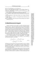
karyogram an ordered array of chromo-
somes of a cell, usually a photograph with
the pairs of chromosomes arranged in
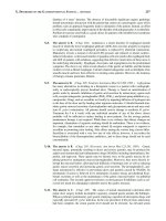
decreasing size (Fig. 49)
karyotype an abbreviated written de-
scription of the karyogram of a cell, clone
K
Figure 49 A karyogram.
A karyogram of a patient with chronic granulocytic leukaemia showing
cytogenetic evolution. The primary abnormality present was
t(9;22)(q34;q11). A secondary abnormality has occurred, t(3;21)(q26;q22).
146
HAE-K 01/13/2005 05:13PM Page 146
knockout mouse 147
kilodalton (kD) 1000 Daltons
Kimura’s disease a chronic inflamma-
tory disease of unknown aetiology, causing
lymphadenopathy with reactive follicular
hyperplasia and infiltration by eosinophils
kinase an enzyme that transfers a phos-
phate group
KIP1 see CDKN1B
KIP2 see CDKN1C
KIT a gene, gene map locus 4q11-34, encod-
ing stem cell factor receptor, a receptor
tyrosine kinase; KIT protein is recog-
nized by CD117 monoclonal antibodies;
KIT is expressed on haemopoietic pro-
genitors, megakaryocytes, mast cells and
a subset of NK cells; it is often overex-
pressed in acute myeloid leukaemia and is
usually mutated, leading to constitutive
activation of KIT, in systemic mastocyto-
sis; mutated in gastrointestinal stromal
tumours, some germ cell tumours and
some sino-nasal NK/T cell lymphoma.
Kleihauer test a cytochemical stain for
demonstrating erythrocytes, including
fetal erythrocytes, with a high concentra-
tion of haemoglobin F
knockin mouse an experimental animal
from which, by means of manipulating
and selecting embryonal stem cells, a
specific gene has been ‘knocked in’ so that
its function can be studied
knockout mouse an experimental
animal from which, by means of mani-
pulating and selecting embryonal stem
cells, a specific gene has been ‘knocked
out’ and replaced by a disabled gene
KEL a locus at 7q33 where there are three
closely linked sets of alleles encoding
antigens of the Kell blood group system
(CD238); the most important of the 25
known antigens and the genes encoding
them are:
•K (KEL1) and k (KEL2)
•Kp
a
(KEL3), Kp
b
(KEL4) and Kp
c
(KEL21)
•Js
a
(KEL6) and Js
b
(KEL7)
Kell a red cell-specific blood group sys-
tem, see also KEL
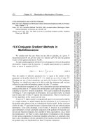
keratocyte a cell with one or two pairs
of spicules, symmetrically arranged
(Fig. 50)
kernicterus damage to the basal ganglia
of the brain by a high concentration of
bilirubin, e.g. in haemolytic disease of the
newborn
Ki-1 a monoclonal antibody that recog-
nizes the CD30 cluster
Ki-1+ lymphoma an earlier designation
of anaplastic large cell lymphoma
Ki-67 a monoclonal antibody that iden-
tifies proliferating cells
KIAA0128 see Septin 2
Kidd a blood group antigen system
including the Jk
a
and Jk
b
antigens (see
also HUT11)
Kiel classification a lymphoma class-
ification which was largely superseded
by the REAL classification (see Revised
European–American Lymphoma classifica-
tion) and then by the WHO classification
killer cell a lymphocyte which can kill
another cell
kilobase (kb) 1000 base pairs of DNA
Figure 50 A keratocyte.
A keratocyte showing two ‘horns’.
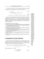
Figure 51 Koilonychia.
Koilonychia in a patient with iron deficiency.
HAE-K 01/13/2005 05:13PM Page 147
neutropenia; a minority of case result
from heterozygosity for a mutation in the
gene encoding the receptor for granulo-
cyte colony-stimulating factor (G-CSFR)
kwashiorkor a form of protein-calorie
malnutrition
148 Knops
Knops a blood group system, the anti-
gens being carried on CD35, a comple-
ment regulatory protein, the C3b/C4b
receptor
koilonychia flat or spoon-shaped nails
as a feature of iron deficiency (Fig. 51)
Kostmann’s syndrome an inherited
abnormality causing severe congenital
HAE-K 01/13/2005 05:13PM Page 148
characterized by the presence of specific
cytoplasmic granules designated Birbeck
granules
Langerhans cell histiocytosis a group
of neoplastic conditions of Langerhans
cells, previously designated histiocytosis
X, further categorized as Letterer–Siwe
disease, Hand–Schüller–Christian disease
and eosinophilic granuloma
Langerhans cell leukaemia an acute
leukaemia of Langerhans cell lineage,
occurring de novo or as the terminal phase
of Langerhans cell histiocytosis
Langhans type giant cell a multi-
nucleated giant cell of monocyte lineage
characterized by peripherally placed nuclei
LAP leucocyte alkaline phosphatase
laparoscopy inspection of the abdom-
inal cavity by means of a laparoscope
inserted through a small incision
laparotomy surgical exploration of the
abdomen for diagnostic or therapeutic
purposes
LARG a gene, Leukaemia-Associated
R
ho Guanine nucleotide exchange factor,
also known as Rh
o Guanine nucleotide
E
xchange Factor 12, RHGEF12; gene
map locus 11q23, encodes a guanine
nucleotide exchange factor (GEF) with
homology to BCR; contributed to a
MLL-LARG fusion gene in a patient with
acute myeloid leukaemia and a complex
karyotype
large cell anaplastic lymphoma a
large cell lymphoma of T lineage with
cells showing strong CD30 positivity and
sometimes aberrant positivity for epithe-
lial membrane antigen
large cell lymphoma a lymphoma of
large lymphoid cells of T, B or NK
lineage
λλ
the Greek letter lambda, one of the
types of light chain found in about 40% of
immunoglobulin molecules
λλ
the lambda immunoglobulin light chain
gene, gene map locus 22q11; transloca-
tions involving this locus can lead to
dysregulation of other genes e.g. MYC
or BCL2
L1, L2, L3 categories in the FAB class-
ification of acute lymphoblastic leukaemia
labelling index the proportion of cells
in which DNA synthesis is occurring,
determined, for example, by incorpora-
tion of tritiated thymidine or bromo-
deoxyuridine
lactate dehydrogenase (LDH) an
enzyme found in many body cells that
catalyses the oxidation of L-lactate to
pyruvate, elevated in haemolytic anaemia
and in ineffective haemopoiesis
lactic acid the end product of anaerobic
metabolism
lactoferrin an iron-binding protein
found in neutrophils (and also milk and
other body fluids) that retards bacterial
proliferation
lactoferrin deficiency see neutrophil
specific granule deficiency
LAF4 a gene, Lymphoid nuclear protein
related to AF4
, gene map locus 2q11.2-
q12, encodes a lymphoid-restricted tran-
scriptional activator that is a homologue
of AF4 and AF5q31; contributed to a
fusion gene in a case of acute lymphoblas-
tic leukaemia
LAK cell lymphokine-activated killer cell
lamellar bone bone with an orderly
structure of concentric layers surround-
ing a central Haversian canal
Langerhans cell a cell of monocyte
lineage, normally found in the skin and
L
149
HAE-L 01/13/2005 05:13PM Page 149
encoding transcriptional coactivators,
p52 and p75, which differ at their carboxy
terminal ends; contributed to a LEDGF-
NUP98 fusion gene in a patient with de
novo acute myeloid leukaemia; the fusion
protein lacks the transactivating domain
of LEDGF
left shift an increased proportion of
immature cells of neutrophil lineage in
either the peripheral blood or the bone
marrow
Leishman–Donovan bodies the rest-
ing stage of Leishmania donovani observed
in tissues including the bone marrow
leishmaniasis infection by the parasite
Leishmania donovani or related species
Lennert’s lymphoma an alternative
designation of lymphoepithelioid lym-
phoma of the Kiel classification, included
in the ‘peripheral T-cell lymphoma,
unspecified’ category of the REAL class-
ification and the WHO classification
lepirudin a recombinant form of hirudin,
a thrombin inhibitor
Lermitte’s syndrome tingling feelings
spreading down the body when the
head is bent forward, which may occur
in vitamin B
12
deficiency and following
irradiation for Hodgkin’s disease
Letterer–Siwe disease a highly aggres-
sive form of Langerhans cell histiocytosis
occurring in infants
leucapheresis removal of white cells from
the peripheral blood, usually either to
relieve hyperviscosity or to obtain cells for
peripheral blood stem cell transplantation
leucine rich repeat (LRR) a hydropho-
bic protein motif believed to participate
in protein-protein interactions
leucine zipper a protein motif char-
acterized by a leucine residue occurring
every 7th base; required for homodimer-
ization and heterodimerization
leucocyte a white blood cell, a term
which includes all the mature peripheral
blood cells of granulocyte, monocyte
or lymphocyte lineage, i.e. neutrophils,
eosinophils, basophils, lymphocytes and
monocytes
leucocyte adhesion deficiency a
genetically heterogeneous group of
conditions in which there is defective
large granular lymphocyte a large
lymphocyte with plentiful cytoplasm and
prominent azurophilic granules
large granular lymphocyte leukaemia
a leukaemia of large granular lympho-
cytes of either T or NK lineage
large unstained cell (LUC) a term used
to describe large, peroxidase-negative
cells detected by Bayer automated
instruments
larynx the part of the throat that con-
tains the vocal cords
LAZ an alternative designation of BCL6
lazy leucocyte syndrome a syndrome
of recurrent infections associated with
defective response of neutrophils to
chemotactic stimuli
LCAT lecithin-cholesterol acyl transferase
LCP1 a gene, Lymphocyte Cytosolic
P
rotein 1, gene map locus 13q14.1, an
IL2-responsive gene that encodes an
actin-binding protein; contributes to the
LCP1-BCL6 fusion gene in B-lineage
non-Hodgkin’s lymphoma associated
with t(3;13)(q27;q14); as a result of the
translocation, the 5′ regulatory regions
of the two genes are exchanged
Leach phenotype an inherited anomaly
of the erythrocyte membrane associated
with loss of the Gerbich antigens and
elliptocytosis
LE cell a neutrophil with a round homo-
geneous inclusion representing altered
nuclear material, characteristic of sys-
temic lupus erythematosus, and demon-
strated by incubating neutrophils with
the patient’s serum
lecithin-cholesterol acyl transferase
deficiency (LCAT deficiency)
an
inherited metabolic disorder associated
with increased target cell formation
lectin any protein other than an
immunoglobulin that binds to a specific
carbohydrate group, may be of plant,
microbial or animal origin; may be used
for identification of antigens (e.g. blood
group antigens) or to stimulate cell
growth and proliferation
Leder stain a histochemical stain to
demonstrate chloroacetate esterase activity
LEDGF a gene, Lens Epithelium-Derived
G
rowth Factor, gene map locus 9p22,
150 large granular lymphocyte
HAE-L 01/13/2005 05:13PM Page 150
light chain the shorter polypeptide
chain of a heterodimer; if not otherwise
specified, the term applies to the light
chain of the immunoglobulin molecule,
each molecule having two heavy chains
and two light chains; light chains may be
kappa or lambda but in a given molecule
are one or the other
light chain-associated amyloidosis
amyloidosis occurring as a complication
of a plasma cell neoplasm, either overt
or occult, previously often designated
‘primary amyloidosis’ when the plasma
cell neoplasm was occult
light chain deposition disease dis-
ease resulting from deposition of a mono-
clonal light chain in tissues, particularly
in the kidneys
LIM domain a zinc-binding protein
motif which provides an interface for
protein interactions
linkage the presence of two or more
genes sufficiently close to each other on a
chromosome that they do not segregate
independently
linkage disequilibrium the association
of particular genes or other DNA
sequences more frequently than would
be expected by chance
lithium a drug used for treating depres-
sion that elevates the neutrophil count
LMAN1 a gene at 18q21.3-q22, encoding
an intracellular mannose-specific protein
that is involved in transport of early pro-
cessed glycoproteins from endoplasmic
reticulum to the Golgi apparatus; muta-
tion can lead to combined factor V and
factor VIII deficiency
locus (plural loci) the specific site on a
chromosome where a gene and its alleles
are located
LOD score a statistical test of linkage;
the higher the LOD score the less the pos-
sibility that the association could occur
by chance
log normal distribution having a
Gaussian or bell-shaped distribution
when the logarithm of the data is plotted
as a histogram (see Fig. 45, p. 128)
LOH loss of heterozygosity
loiasis the disease resulting from infec-
tion by the filarial parasite, Loa loa
adhesion of neutrophils to endothelium,
leading to neutrophilia, a lack of extra-
vascular neutrophils and susceptibility
to infection: type 1 results from muta-
tion in the ITGB2 (integrin β2) gene, the
gene encoding the β chain of the leuco-
cyte integrins, which results in CD18
deficiency; type 2 results from a defect
in carbohydrate fucosylation so that
endothelial sialyl-Lewis
x
is not expressed
and endothelium cannot bind E and P
selectins (see CD62E and CD62P); a
single case has also been reported in
which there was deficiency in RAC2, the
predominant GTPase in neutrophils
leucocyte alkaline phosphatase (LAP)
the alkaline phosphatase isoenzyme in
neutrophils; it should be noted that the
term ‘neutrophil alkaline phosphatase’
and the abbreviation ‘NAP’ are to be
preferred
leucocytosis an increased number of
leucocytes in the peripheral blood
leucoerythroblastic anaemia ana-
emia with both erythroblasts and granu-
locyte precursors in the peripheral
blood
leucopenia a reduced white cell count
leukaemia a myeloid or lymphoid
neoplasm, characterized by circulating
neoplastic cells but also encompassing
similar cases in which there are neoplastic
cells in the bone marrow but not in the
peripheral blood
leukaemic reticuloendotheliosis an
early designation of hairy cell leukaemia,
not in current use
leukaemogenesis the mechanism of
development of leukaemia
leukaemoid reaction a reactive condi-
tion which simulates or closely resembles
leukaemia
Lewis blood group system a blood
group system in which plasma glycosph-
ingolipids, resulting from the expression
of alleles at the FUT1, FUT2, FUT3 and
ABO loci, are adsorbed onto red cells
(see Fig. 30, p. 108)
ligand a molecule that binds specifically
to another molecule, e.g. stem cell factor
is the ligand of c-KIT and thrombopoietin
is the ligand of MPL
loiasis 151
HAE-L 01/13/2005 05:13PM Page 151
by lymphatics and ultimately drains into
the blood stream through the thoracic duct
lymphadenopathy enlargement of the
lymph nodes
lymphangiogram a radiological proce-
dure for demonstrating lymph nodes by
means of radio-opaque dye introduced
into lymphatics on the dorsum of the feet
(Fig. 52)
lymphatic (i) relating to lymphocytes and
lymph nodes (ii) an endothelium-lined
vessel which transports lymph
lymph follicle an organized nodule of
mature B lymphocytes—smaller cells,
designated follicle centre cells or centro-
cytes, and larger cells, designated centrob-
lasts, in a lymph node or other tissue
lymph gland a non-scientific term for
lymph node
lomustine (CCNU) an anti-cancer drug
of the nitrosourea family
long term culture initiating cells
(LTCIC)
cells that are able to give rise to
long term cultures in vitro
loss of heterozygosity (LOH) loss of
one allele at a specific locus
low hyperdiploidy having 47–50 chro-
mosomes in a cell or clone
low molecular weight heparin hep-
arin that has been depolymerized so that
the molecular weight is in the range of
4000 to 5000 daltons, e.g. enoxaparin,
dalteparin, tinzaparin, ardeparin
LPP a gene, LIM domain containing
P
referred translocation Partner in lipoma,
also known as L
ipoma Preferred Partner,
gene map locus 3q28, encodes a putative
cell adhesion molecule that contains a
LIM domain; contributed to MLL-LPP
and LPP-MLL fusion genes in a patient
with secondary M5 acute myeloid
leukaemia with t(3;11)(q28;q23)
LTCIC long term culture initiating cells
LU the gene at 19q13.2 encoding the Lu
a
and Lu
b
antigens of the Lutheran blood
group system
LUC large unstained cell
lupus anticoagulant an antibody that
is an anticoagulant in vitro but is pro-
thrombotic in vivo, occurs in systemic
lupus erythematosus and in the primary
antiphospholipid syndrome; paraproteins
may also be lupus anticoagulants
Lutheran a blood group system, Lutheran
antigens (CD239) being encoded by the
genes at the LU locus
LW a locus at 19p13.3 encoding LW
blood group antigens
LW a blood group antigen system (CD242)
encoded by genes at the LW locus
LYL1 a gene, Lymphoid Leukaemia gene,
gene map locus 19p13, encodes a basic
helix–loop–helix (bHLH) transcription
factor closely related to TAL1, which is
expressed in all haemopoietic cells except
for T cells, dysregulated by proximity to
the TCRB gene at 7q35 in T-lineage acute
lymphoblastic leukaemia associated with
t(7;19)(q35;p13)
lymph a clear or opalescent fluid con-
taining lymphocytes, which is transported
152 lomustine (CCNU)
Figure 52 A lymphangiogram.
A lymphangiogram, showing images of lymph nodes
which have trapped an oily radio-opaque dye that
was injected into lymphatic vessels in the feet; the
normal lymph nodes are small and have a fairly
homogeneous texture whereas abnormal lymph
nodes, identifiable below the left kidney, are large
and heterogeneous in texture; an intravenous
pyelogram has also been performed so that the renal
pelvises and the ureters are outlined, showing that
the left ureter is deviated around a group of
abnormal lymph nodes; the patient had Hodgkin’s
disease.
HAE-L 01/13/2005 05:13PM Page 152
with specific histological features includ-
ing the presence of epithelioid cells, being
recognized as a distinct entity in the Kiel
classification and included in the ‘periph-
eral T-cell lymphoma, unspecified’ cat-
egory of the REAL classification and the
WHO classification
lymphogranulomatosis a lymphopro-
liferative disorder, previously thought
to be reactive but probably a low grade
T-cell neoplasm
lymphoid pertaining to lymphocytes or
lymph nodes
lymphokine any type of molecule,
other than an immunoglobulin mo-
lecule, secreted by a lymphocyte and
influencing the behaviour of other cells
or tissues
lymphoma a neoplasm of lymphocytes
(T, B or natural killer cells); in contrast to
lymphoid leukaemias, lymphomas involve
predominantly tissues other than the bone
marrow, rather than the bone marrow
and blood; in the WHO classification, B-
lineage and T-lineage lymphomas are
lymph node an organized tissue com-
posed mainly of T and B lymphocytes
with some stromal cells and dendritic cells
lymphoblast an immature cell of lym-
phoid lineage, having a high nucleocyto-
plasmic ratio, non-condensed chromatin
and nucleoli (see Fig. 12, p. 29)
lymphoblastic lymphoma a lym-
phoma composed of lymphoblasts, occur-
ring in a tissue other than the bone marrow
lymphocyte a mature cell of lymphoid
lineage
lymphocytic pertaining to lymphocytes
or to lymphoid cells
lymphocytopenia a reduced lympho-
cyte count
lymphocytosis an increased number of
lymphocytes in the peripheral blood
lymphocytotoxin also known as lym-
photoxin and tumour necrosis factor β,
a cytokine secreted by B cells and type 1
helper T cells, which promotes inflamma-
tion, encoded by a gene at 6p21
lymphoepithelioid lymphoma Len-
nert’s lymphoma, a T-cell lymphoma
lymphoma 153
Table 11 WHO classification of T-cell and NK-cell neoplasms.
Precursor T-cell neoplasms
Precursor T lymphoblastic leukaemia/lymphoblastic lymphoma
Mature T-cell and NK-cell neoplasms
Leukaemic/disseminated
T-cell prolymphocytic leukaemia
T-cell large granular lymphocyte leukaemia
Aggressive NK-cell leukaemia
Adult T-cell leukaemia/lymphoma
Cutaneous
Mycosis fungoides
Sezary syndrome
Primary cutaneous anaplastic large cell lymphoma
Lymphomatoid papulosis
Other extra-nodal
Extranodal NK/T-cell lymphoma, nasal type
Enteropathy-type T-cell lymphoma
Hepatosplenic T-cell lymphoma
Subcutaneous panniculitis-like T-cell lymphoma
Nodal
Angioimmunoblastic T-cell lymphoma
Peripheral T-cell lymphoma, unspecified
Anaplastic large cell lymphoma
Neoplasm of uncertain lineage and stage of differentiation
Blastic NK-cell lymphoma
HAE-L 01/13/2005 05:13PM Page 153
transcriptionally active. The inactive X
chromosome is responsible for the
neutrophil drumstick and in other cells
may be detected as a Barr body, a con-
densation of chromatin below the nuclear
membrane
lyonization the process by which one of
the two X chromosomes in each normal
female cell becomes genetically inactive;
this process occurs during early embry-
onic life and is maintained in the progeny
of that cell; it provides the basis of analy-
sis of X-linked polymorphisms for pre-
sumptive establishment of monoclonality
lysis (i) destruction of a cell as a con-
sequence of development of a hole in the
plasma membrane, e.g. destruction of a
red cell following antibody binding and
complement fixation (ii) dissolution of a
thrombus
lysosome a cellular organelle into which
hydrolytic enzymes are secreted, respons-
ible for the degradation of foreign mater-
ial and cellular debris
lysozyme an enzyme secreted by mono-
cytes and, to a lesser extent, neutrophils
lytic lesion an osteolytic lesion, gener-
ally detected radiologically which, if there
is no associated osteosclerosis, is suggest-
ive of multiple myeloma
designated as shown in Tables 10 (p. 137)
and 11
lymphoma of intermediate differen-
tiation
an earlier designation of mantle
cell lymphoma
lymphopenia see lymphocytopenia
lymphoplasmacytoid lymphoma a
lymphoma in which some of the neoplas-
tic cells show plasma cell differentiation
lymphoproliferative disorder a
generic term including all lymphomas and
also some disorders of undefined nature
in which there is abnormal lymphoid pro-
liferation of uncertain nature
lymphoproliferative T-cell deficiency
with autoimmunity syndrome
an
immune deficiency syndrome resulting
from mutation of the IL2 receptor gene at
10p14-15 with resultant CD25 deficiency
lymphosarcoma an outmoded term for
non-Hodgkin’s lymphoma
Lyon hypothesis the hypothesis that
one of the two X chromosomes in the
cells of a normal female becomes inactive,
this process occurring randomly so that,
on average, each X chromosome is
inactive in 50% of cells; it is hypothesized
that the genetic inactivation of one chro-
mosome permits X chromosomal dosage
compensation—no matter how many X
chromosomes a cell has only one remains
154 lymphoma of intermediate differentiation
HAE-L 01/13/2005 05:13PM Page 154
macro-ovalocyte an oval macrocyte
macrophage the end cell of the mono-
cyte lineage, found mainly in tissues and,
being phagocytic, important in protection
against infection; neoplasms of macro-
phage and related lineages are designated
in the WHO classification as shown in
Table 6, p. 127
macrophage colony-stimulating fac-
tor (M-CSF)
a haemopoietic growth
factor, encoded by a gene at 1p13-21
macropolycyte a very large neutrophil,
probably usually tetraploid
macrothrombocyte a large platelet
macule a skin lesion which is not raised
MAD a gene, Max Dimerization protein,
gene map locus 2p13, archetypal mem-
ber of a small family of transcriptional
repressor proteins, which compete with
MYC proteins for binding to MAX
MAF a gene, avian Musculoaponeuro-
tic F
ibrosarcoma gene homologue, gene
map locus 16q23, encodes a basic leucine
zipper transcription factor; archetypal
member of a family of such genes; MAF
is dysregulated when brought into pro-
ximity to the IGH locus in 30–35% of
patients with multiple myeloma and
t(14;16)(q32;q23)
MAFB a gene, avian Musculoaponeurotic
F
ibrosarcoma oncogene homologue B,
gene map locus 20q11, encodes a MAF
family basic region/leucine zipper tran-
scription factor; MAFB is translocated
and overexpressed when t(14;20)(q32;q11)
brings it into proximity to the IGH locus
in some cases of multiple myeloma
magnetic resonance imaging (MRI) or
nuclear magnetic resonance imag-
ing (NMR)
an imaging technique using
the inherent magnetic properties of body
µµ
the Greek letter, mu (i) the heavy chain
of immunoglobulin M (ii) the symbol for
a micron (iii) the symbol for one millionth
part of a unit, as in µg or µl
M the designation of the phase of the cell
cycle when mitosis occurs (see cell cycle)
M0, M1, M2, M3, M4, M5, M6, M7
categories in the FAB classification of
acute myeloid leukaemia (see Table 3, p. 7)
MAC the combined application of Mor-
phology, A
ntibody and Chromosome
techniques to the study of individual cells
McLeod phenotype a syndrome of
acanthocytosis, mild haemolytic anaemia,
myopathy and cardiomyopathy resulting
from deficiency of Kx protein and conse-
quent weak expression or lack of expres-
sion of Kell antigens
macrocyte a large erythrocyte
macrocytic anaemia an anaemia char-
acterized by macrocytosis, e.g. in mega-
loblastic anaemia or the myelodysplastic
syndromes
macrocytosis having large erythrocytes
macroglobulinaemia an increased
plasma concentration of macroglobulins,
specifically IgM
macroglossia a large tongue, may be a
feature of amyloidosis
macronormoblast an erythroblast
which is larger than normal erythroblasts
at the equivalent stage of development
but which does not show the nucleo-
cytoplasmic asynchrony which is charac-
teristic of a megaloblast
macronormoblastic maturation ery-
throid maturation characterized by
haemopoietic cells which are larger than
normal but lack the nucleocytoplasmic
asynchrony which is characteristic of
megaloblastic haemopoiesis
M
155
HAE-M 01/13/2005 05:13PM Page 155
a Golgi-associated membrane protein;
MAL is over-expressed in primary medi-
astinal (thymic) B-cell lymphoma and
contributes to the OTT-MAL fusion gene
in infant M7 acute myeloid leukaemia
associated with t(1;22)(p13;q13)
malabsorption failure to absorb nutri-
ents normally; haematological effects
include deficiency of iron and vitamin B
12
and folic acid and deficiency of vitamin K
leading to coagulation defects
malaria a disease resulting from infec-
tion by protozoan parasites of the Plas-
modium genus
malignant very injurious; in relation to
neoplasms, aggressive and characterized
by local invasion, distant spread (metas-
tasis) and cytological differences from the
equivalent normal tissues
malignant histiocytosis a rare neo-
plasm of monocyte/macrophage lineage
involving predominantly tissues other than
the bone marrow; many patients reported
to have had this condition have actually
had a reactive haemophagocytic syndrome
malignant mastocytosis an aggressive
widespread mast cell neoplasm, more
or less equivalent to aggressive systemic
mastocytosis of the WHO classification
(see Table 12)
malignant melanoma malignant
tumour of cells analogous to melanocytes
but not necessarily pigment-producing
malignant tertian malaria malaria
caused by Plasmodium falciparum
MALT mucosa-associated lymphoid tissue
MALT-lymphoma or MALT-type
lymphoma
a lymphoma of cells ana-
logous to normal mucosa-associated
lymphocytes
MALT a gene, Mucosa-Associated
L
ymphoid Tissue lymphoma transloca-
tion gene 1, also known as MLT; gene
map locus 18q21; encodes a cell surface
adhesion molecule related to CD22, that
is expressed strongly in blood, bone
marrow, thymus and lymph node; con-
tributes to the AP12-MLT fusion gene
in MALT lymphoma associated with
t(11;18)(q21;q21); in the presence of the
API2-MLT fusion protein there is
sequestration of BCL10 in the nucleus
tissues containing free protons; para-
magnetic molecules have their alignment
changed by a combination of a strong
magnetic field and radio frequency
waves; as they regain their original posi-
tion radio frequency waves are emitted
which are then reconstructed into images
(Fig. 53)
MAHA microangiopathic haemolytic
anaemia
MAIPA monoclonal antibody-specific
immobilization of platelet antigens
major histocompatibility complex
(MHC)
a group of polymorphic pro-
teins on the surface of cells, encoded by
the HLA family of genes, which present
modified antigen (as a short peptide in
a surface groove) to T lymphocytes;
molecules are divided into class I and
class II; class I molecules (HLA-A, HLA-
B and HLA-C) are concerned with recog-
nition of antigens expressed on altered
body cells and class II molecules (HLA-
DP, HLA-DQ and HLA-DR) with the
recognition of foreign antigens
MAL a gene, T Lymphocyte Maturation-
A
ssociated protein, also known as
MKL1, gene map locus 22q13, encodes
156 MAHA
Figure 53 A magnetic resonance imaging
(MRI) scan.
A T2 weighted paracoronal magnetic resonance
imaging (MRI) scan of the left shoulder; the humerus
has an irregular articular outline and high and low
signal bands in the subchondral region, typical of
avascular necrosis; such lesions may be seen in
patients with sickle cell disease.
HAE-M 01/13/2005 05:13PM Page 156
marker chromosome (mar) an abnor-
mal chromosome that cannot be charac-
terized by routine cytogenetic techniques
mast cell a tissue cell of myeloid lineage
which is involved in immune responses
and in inflammation
mast cell disease a WHO classification
term for mast cell neoplasms, further
categorized as shown in Table 12
mast cell leukaemia an acute
leukaemia of mast cell lineage
mastocytosis an increase in mast cells,
an alternative generic term encompassing
all mast cell neoplasms used in the WHO
classification being synonymous, in this
context, with ‘mast cell disease’ (see
Table 12)
matched unrelated donor (MUD) a
bone marrow or haemopoietic stem cell
donor who is unrelated to the recipient
but has been matched for histocompat-
ibility antigens (note: the term ‘volunteer
unrelated donor (VUD)’ is now preferred)
matrix metalloproteinases enzymes,
with zinc at the active centre, that can
cleave proteins of the extracellular
matrix; they can be divided into four
main groups—collagenases, gelatinases,
stromeolysins and membrane-type
metalloproteinases
maturation development of the features
characterizing later cells of the same lineage
maturation arrest lack of maturing
cells beyond a certain stage, e.g. arrest of
granulopoiesis at the promyelocyte stage;
usually indicates cell death at an inappro-
priate stage during maturation but an
apparent maturation arrest can also result
from immune destruction of maturing cells
Maurer’s clefts linear inclusions in
erythrocytes in Plasmodium falciparum
malaria
mannose-binding lectin an acute
phase reactant, synthesized by hepato-
cytes, that binds to bacteria, yeasts and
some viruses and parasites, thereby acti-
vating mannose-associated proteases 1
and 2, which initiate the lectin pathway of
complement activation (see complement
system and Fig. 20, p. 81)
mannose-binding lectin deficiency
an inherited condition in which mutation
in the MBL gene leads to reduced plasma
concentration of mannose-binding lectin,
inability to activate the mannose-binding
lectin complement pathway and recur-
rent pyogenic infections
mantle cell lymphoma a lymphoma of
mature B cells analogous to lymphocytes
in the mantle zone of a lymph node
mantle zone the circle of more darkly
staining small lymphocytes that sur-
rounds the paler germinal centre
mar a cytogenetic abbreviation indicat-
ing a marker chromosome
marasmus a form of protein-calorie
malnutrition
marble bone disease see osteopetrosis
march haemoglobinuria haemolytic
anaemia caused by mechanical trauma,
e.g. from long marches on hard earth
marginal zone the zone between the
red and white pulp of the spleen including
the band of lymphocytes that surrounds
the lymphoid follicles of the white
pulp; the marginal zone lymphocytes
are larger and paler than mantle zone
lymphocytes
marginal zone lymphoma a lym-
phoma of mature B lymphocytes analo-
gous to those in the marginal zone of
lymphoid follicles; includes MALT-type
lymphoma, splenic marginal zone lym-
phoma and monocytoid B-cell lymphoma
Maurer’s clefts 157
Table 12 The WHO classification of mast cell disease (mastocytosis).
Cutaneous mastocytosis
Indolent systemic mastocytosis
Systemic mastocytosis with associated clinical, haematological an non-mast-cell lineage disease
Aggressive systemic mastocytosis
Mast cell leukaemia
Extracutaneous mastocytoma
HAE-M 01/13/2005 05:13PM Page 157
tion of haemoglobin in an individual’s
erythrocytes
mean cell volume (MCV) the average
size of an individual’s erythrocytes
median the value that divides a popula-
tion into two numerically equal halves
median disease-free survival the
time when half of a studied population
is still alive and free of relapse
median event-free survival the time
when half of a studied population has
neither died nor suffered relapse nor
changed to alternative treatment
median overall survival the time when
half of a studied population have died
and half are still alive, synonymous with
median survival
median survival the time when half of
a studied population have died and half
are still alive, synonymous with median
overall survival
mediastinum the central part of the
thoracic cavity, between the lungs
Medicines Act 1968 a UK Act of
Parliament which provides the legal
framework governing not only medicines
but also the donation, testing, processing
and issuing of blood components and
products
Medicines Control Agency a UK
organization that, among other duties,
licences transfusion centres to prepare and
provide blood components and products
medulla (i) the central part of a bone
composed of an anastomosing network
of trabecular or cancellous bone (ii) the
central part of the kidney (iii) the central
part of a lymph node (iv) the central part
of the thymus
medullary cavity the spaces between
the trabeculae of medullary bone occu-
pied by fatty or haemopoietic marrow
MEFV the gene encoding pyrin or mareno-
strin, a protein expressed in myeloid
cells and up-regulated during differentia-
tion; strongly expressed in neutrophils
and monocytes and thought to regulate
phagocyte-induced inflammation; muta-
tions in this gene can result in familial
Mediterranean fever
megakaryoblast the earliest recogniz-
able cell of megakaryocyte lineage
MAX a gene, MYC-Associated factor X,
gene map locus 14q23, encodes a
helix–loop–helix leucine zipper protein
which specifically heterodimerizes with
MYC transcriptional activators and
permits their binding to their cognate
DNA binding sites (E-Boxes); also het-
erodimerizes with MAD family proteins
(which encode transcriptional repressors)
and allows them to bind E-boxes, thereby
antagonizing the effects of MYC proteins
MAX a protein that forms heterodimers
with MYC protein
May–Grünwald–Giemsa (MGG) a
Romanowsky type stain used to stain
blood and bone marrow films
May Hegglin anomaly an inherited
anomaly with giant platelets and neu-
trophil inclusions resulting from a muta-
tion in the non-muscle myosin heavy
chain 9 gene (NMMHC-A or MYH9) at
22q11-13 (or 22q12.3-q13.2)
MBL the gene encoding mannose-binding
lectin
McAb monoclonal antibody
MCH mean cell haemoglobin
MCHC mean cell haemoglobin concentra-
tion
M-CSF macrophage colony-stimulating
factor
MCV mean cell volume
MDR multidrug resistance
MDS myelodysplastic syndrome or syn-
dromes
MDS1 a gene, Myelodysplasia Syndrome
1
; gene map locus 3q26; this gene was pre-
viously thought to be an exon of EVI1,
but is widely expressed both as a unique
transcript and as a normal fusion tran-
script with EVI1; encodes a protein of
unknown function; MDS1 can contribute
to an AML1-MDS1 fusion oncogene in
acute myeloid leukaemia, myelodysplas-
tic syndromes and blast crisis of chronic
granulocytic leukaemia associated with
t(3;21)(q26;q22)
mean the average
mean cell haemoglobin (MCH) the
average amount of haemoglobin in an
individual’s erythrocytes
mean cell haemoglobin concentra-
tion (MCHC)
the average concentra-
158 MAX
HAE-M 01/13/2005 05:13PM Page 158
meningeal leukaemia infiltration of
leukaemic cells into the meninges
meninges the membranes covering the
brain and spinal cord
meningitis inflammation of the meninges,
often caused by bacterial or viral infection
menorrhagia heavy menstrual bleeding,
may result from a haemostatic defect and
may lead to iron deficiency anaemia
MER2 a gene at 11p15.5 that encodes
antigens of the MER2 or RAPH blood
group systems
M:E ratio myeloid:erythroid ratio
mercaptopurine an antimetabolite used
in the maintenance treatment of acute
lymphoblastic leukaemia
messenger ribonucleic acid (messen-
ger RNA, mRNA)
ribonucleic acid
that is transcribed in the nucleus, on a
DNA template, and moves to the cyto-
plasm, becoming attached to ribosomes
and serving as a template for synthesis
of proteins
metabolic acidosis acidosis other than
that due to accumulation of carbonic acid
metabolic rate the rate of energy expen-
diture by the body
metabolism the aggregate of chemical
process occurring in a living organism;
includes anabolism, in which complex
molecules and tissues are built up, and
catabolism, in which tissues, cells and
complex molecules are broken down
metachromatic staining uptake of a
dye by a cell component with the dye then
altering its colour
metamyelocyte a late, non-dividing
cell in granulocyte maturation; it is
derived from a myelocyte and matures
to a band cell (see Fig. 25, p. 95)
metaphase the third of the five stages
of mitosis in which the chromosomes
become arranged around the equatorial
plane of the mitotic spindle with their
centromeres being attached to the spindle;
the stage of nuclear division that is opti-
mal for the examination of chromosomes
(see Fig. 6, p. 14)
metarubricyte a name proposed for late
erythroblasts
metastasis (i) the process by which a
tumour spreads to distant parts of the
megakaryocyte a giant bone marrow
cell, generally polyploid, of myeloid line-
age which produces platelets by frag-
mentation of its own cytoplasm
megaloblast an erythroblast of larger
than normal size showing retarded nuclear
maturation in relation to cytoplasmic
maturation
megaloblastic a term indicating abnor-
mal haemopoiesis in which there is
delayed nuclear development leading
to large haemopoietic cells and erythro-
cytes, and dissociation between nuclear
and cytoplasmic maturation (see also
giant metamyelocyte)
megaloblastic anaemia anaemia with
megaloblastic haemopoiesis
meiosis the process in which chromo-
somes of a cell replicate followed by two
nuclear divisions so that the complement
of chromosomes in the resultant cells is
halved; the process by which germ cells
are produced
MEL1 a gene, PR Domain-containing
protein 16
, PRDM16, also known as
M
DS1/EVI1-Like gene 1, gene map locus
1p36.6, encodes a PR zinc finger protein
similar to MDS1, which is transcription-
ally activated, probably by proximity
to the ribophorin 1 gene, RPN1, at 3q21
in myelodysplastic syndrome and acute
myeloid leukaemia associated with
t(1;3)(p36;q21)
melaena black faeces, indicative of the
presence of altered blood following hae-
morrhage into the upper gastrointestinal
tract
melanocyte a pigmented skin cell found
in the lower epidermis
melanoma a tumour of cells analogous
to normal melanocytes; use of this term
is often restricted to malignant tumours
of melanocyte lineage
melphalan an alkylating agent used in
the treatment of multiple myeloma and
certain other tumours
memory cell a long-lived B cell or T cell
that has already been exposed to an anti-
gen and can participate in a second-
ary immune response following further
exposure to the antigen
MEN an alternative name for ELL
metastasis 159
HAE-M 01/13/2005 05:13PM Page 159
of synthetic oligonucleotides arrayed on
a microchip
microcyte an abnormally small
erythrocyte
microcytic anaemia anaemia charac-
terized by microcytosis
microcytosis the presence of abnorm-
ally small erythrocytes
microfilaments 7 nm diameter fila-
ments composed mainly of actin and
actin-binding proteins that form part of
the cytoskeleton of cells
microfilaria the stage of the life cycle of
filaria which is identified by peripheral
blood examination, the prelarval form of
the parasite
microgram (µg) one millionth of a
gram, 1 × 10
–6
of a gram
microhaematocrit a haematocrit deter-
mination performed in a capillary tube
microlitre (
µµ
l) a millionth (10
–6
) of a
litre
micromegakaryocyte a megakaryocyte
no more than 30 microns in diameter
micromole (
µµ
mol) a millionth (10
–6
) of
a mole
micron (
µµ
) a millionth (10
–6
) of a metre
microscope an instrument with a num-
ber of lenses for the visual examination of
small objects
microscopic (i) very small (ii) pertaining
to a microscope
microscopy examination by means of a
microscope
microspherocyte a spherocyte which
is smaller than a normal erythrocyte,
formed by red cell fragmentation or by
removal of parts of the erythrocyte mem-
brane by splenic macrophages
microtubules polymers of tubulin, with
a diameter of about 24 nm, that form part
of the cytoskeleton; among other func-
tions, they form the mitotic spindle
miliary tuberculosis disseminated
tuberculosis with granulomas distributed
through many organs
milligram (mg) one thousandth part
(10
–3
) of a gram
millilitre (ml) one thousandth part (10
–3
)
of a litre
millimole (mmol) one thousandth part
(10
–3
) of a mole
body (ii) a deposit of tumour at a site
distant from the primary tumour
metastatic able to metastasize or having
metastasized
methaemalbumin a breakdown prod-
uct of oxidized haemoglobin that has
been bound to albumin; the Schumm’s
test detects methaemalbumin and a posit-
ive result gives evidence of intravascular
haemolysis
methaemoglobin oxidized haemoglobin
methaemoglobinaemia an increased
proportion of methaemoglobin in
erythrocytes
methotrexate an antimetabolite that
interferes with folic acid metabolism,
used in the maintenance treatment of
acute lymphoblastic leukaemia, in certain
solid tumours and as an immunosuppres-
sive agent
methylene blue a dye or stain which
can be used as a component of a
Romanowsky stain and also for staining
reticulocytes and Heinz bodies; however,
it should be noted that new methylene
blue rather than methylene blue should
be used for staining haemoglobin H
inclusions
methylene tetrahydrofolate reduc-
tase
an enzyme involved in folic acid
metabolism; deficiency, resulting from
homozygosity for a thermolabile variant,
is present in 5% of the population and
is associated with an increased risk of
thrombosis
MGG May–Grünwald–Giemsa (stain)
MGUS monoclonal gammopathy of unde-
termined significance
MHC major histocompatibility complex
MIC the Morphologic-Immunophenotypic-
C
ytogenetic classification of leukaemias
MIC-M the Morphological-Immuno-
phenotypic-C
ytogenetic-Molecular genetic
classification of leukaemias
microangiopathic haemolytic an-
aemia (MAHA)
haemolytic anaemia
caused by pathological processes, either
endothelial damage or fibrin deposition,
operating in small blood vessels
microarray analysis a method of
assessing the RNA expression profile
of a single tissue or cell line by means
160 metastatic
HAE-M 01/13/2005 05:13PM Page 160
mitotic spindle the microtubules to which
chromosomes attach during metaphase
mitozantrone an anti-cancer drug
related to the anthracyclines, used in the
treatment of lymphomas
MKL1 a gene, Megakaryoblastic Leuk-
aemia 1
, also known as MAL, gene map
locus 22q13; encodes a nuclear protein
containing a single SAP DNA-binding
motif; MKL1 contributes to RBM15-
MKL1 and MKL1-RBM15 fusion genes
in acute megakaryoblastic leukaemia of
infants with t(1;22)(p13;q13); it is the
RBM15-MKL1 fusion gene (also known
as OTT-MAL) which is likely to be
oncogenic (see also MAL, OTT ); the
fusion protein retains all functional
motifs encoded by each gene including
an oligomerization domain in MKL1
MLF1 a gene, Myeloid Leukaemia Factor
1
, gene map locus 3q25.1, encodes a
widely expressed protein of unknown
function which has been observed in the
cytoplasm and in the nucleus in discrete
bodies; MLF1 contributes to the NPM-
MLF1 fusion gene in acute myeloid
leukaemia or myelodysplastic syndrome
associated with t(3;5)(q25.1;q34)
MLL a gene, Mixed Lineage Leukaemia,
also known as M
yeloid-Lymphoid
L
eukaemia gene, HRX, ALL-1 and Htrx-
1, gene map locus 11q23, encodes a multi-
domain protein with some homology to
the Drosophila transcriptional regulator,
trithorax, which positively maintains the
expression of multiple homeobox genes
during development; MLL is fused to a
great variety of other genes in acute
myeloid leukaemia, acute lymphoblastic
leukaemia and acute biphenotypic leuk-
aemias; important functional domains
include the AT hook (mediates DNA
binding to AT-rich DNA sequences,
phosphorylated in mitosis), subnuclear
localization domains (SNLs, areas of high
homology to trithorax), CxxC motifs
(mediate interaction with transcriptional
repressors), PHD zinc fingers (mediate
interaction with nuclear cyclophilins),
transactivation domain and SET domains;
fusion proteins always retain the amino
terminus of MLL (which carries the AT
min a cytogenetic abbreviation for a
minute chromosome
minimal residual disease (MRD) the
presence of small numbers of neoplastic
cells, detectable by techniques such as
immunophenotyping or molecular genetic
analysis, in bone marrow or blood sam-
ples in which no such cells are detectable
by microscopic examination; minimal
residual disease can be defined as the
lowest level of residual disease detectable
by available methods
minute chromosome (min) an acen-
tric fragment of a chromosome, smaller
than the width of a single chromatid
(double minute chromosomes are acen-
tric and atelomeric chromatin bodies
composed of circular DNA)
miscarriage a spontaneous abortion
mis-sense mutation a mutation that
results in the encoding of a different
amino acid
mitochondrial pertaining to mitochondria
mitochondrial cytopathy an inherited
disorder transmitted by genes on a cir-
cular mitochondrial chromosome, rather
than by genes located on nuclear
chromosomes
mitochondrion (plural mitochondria)
a rod or oval-shaped organelle enclosed
by two membranes with the inner mem-
brane being folded into cristae which pro-
ject into the matrix of the mitochondrion;
the mitochondrion is the major site of
energy production and has enzymes
responsible for part of the haem synthesis
pathway (see Fig. 34, p. 116)
mitogen an agent that promotes mitosis
mitomycin C an antitumour antibiotic
which can cause microangiopathic haemo-
lytic anaemia
mitosis the process of cell division in
which chromosomes replicate before cell
division; daughter cells therefore have the
same complement of chromosomes as the
parent cell (see Fig. 6, p. 14)
mitotic figure chromosomes visible
during the process of mitosis
mitotic index the proportion of cells in
mitosis
mitotic rate the rate at which cells enter
mitosis
MLL 161
HAE-M 01/13/2005 05:13PM Page 161
with acute myeloid leukaemia associated
with trisomy 11 and in some with normal
cytogenetics and is amplified in some
patients with complex chromosomal
rearrangements—in some of these
patients MLL is found at multiple sites
on the genome and in others the amplified
gene has been located specifically in a ring
chromosome, in a homogeneously stain-
ing region or in double minute chromo-
somes; duplication or amplification of a
non-rearranged MLL gene is common
in therapy-related myelodysplastic syn-
dromes and acute myeloid leukaemia
and is closely related to prior alkylat-
ing agents and to TP53 mutations (which
may be the cause of the duplication/
amplification); partial amino terminal
duplications of MLL due to genomic
rearrangements have been reported in
acute myeloid leukaemia and B-lineage
acute lymphoblastic leukaemia, and carry
a poor prognosis; deletions of exon 8 of
MLL (encoding the first PHD domain)
have been reported in T-lineage acute
lymphoblastic leukaemia
MMSET a gene, Multiple Myeloma SET
domain, officially known as WHSC1—
W
olf-Hirschhorn Syndrome Candidate
gene 1
, gene map locus 4p16.3, encodes
a SET domain protein; is involved,
together with FGFR3, in a cryptic
t(4;14)(p16.3;q32.2) translocation, which
is present in about 20% of cases of
multiple myeloma; the mechanism of
dysregulation is proximity to an intronic
enhancer on the der(4); Wolf–Hirschhorn
syndrome is a congenital malformation
syndrome associated with hemizygous
deletions of 4p—it is unclear whether a
single locus is involved in the phenotype
—WHSC1 is a candidate gene in the region
MN1 a gene, Meningioma 1, gene map
locus 22q12.3-qter, encodes a putative
transcriptional activator, was initially
cloned from a meningioma; MN1 con-
tributes to an MN1-ETV6 fusion gene in
acute myeloid leukaemia associated with
t(12;22)(p13;q11); unusually for leukae-
mogenic fusion proteins involving ETV6,
the MN1-ETV6 fusion protein lacks a
functional ETV6 PNT (oligomerization)
hook, SNL and CxxC motifs), and con-
sistently lose the PHD, transactivation
and SET domains, which are replaced by
sequences of partner genes; MLL trans-
location breakpoints almost invariably
lie within an 8.3 Kb breakpoint cluster
region (bcr) which corresponds to a
nuclear matrix attachment region and is
susceptible to DNA cleavage in response
to topoisomerase II inhibitors (e.g.
etoposide) or apoptosis; there are at least
28 reported partner genes for MLL in
leukaemia; fusion genes to which MLL
contributes include:
• MLL-AF1p in t(1;11)(p32;q23)
• MLL-AF1q in t(1;11)(q21;q23)
• MLL-LAF4 in t(2;11)(p15;p14)
• MLL-LPP and LPP-MLL in
t(3;11)(q28;q23)
• MLL-GMPS in t(3;11)(q25;q23)
• MLL-AF4 in t(4;11)(q21;q23)
• MLL-GRAF in t(5;11)(q31;q23)
• MLL-AF5q31 in ins(5;11)
(q31;q13q23)
• MLL-AF6q21 in t(6;11)(q21;q23)
• MLL-AF6q27 in t(6;11)(q27;q23)
• MLL-AF9 in t(9;11)(p22;q23)
• MLL-AF9q34 in t(9;11)(q34;q22)
• MLL-ABI1 in t(10;11)(p11.2;q23)
• MLL-AF10 in t(10;11)(p12;q23)
• MLL-LARG by interstitial deletion at
11q23
• MLL-FBP17 in ins(11;9)(q23;q34)
inv(11)(q13q23)
• MLL-CALM in inv(q14,2q23.1)
• MLL-GPNH in t(11;14)(q23;q24)
• MLL-AF17 in t(11;17)(q23;q21)
• MLL-RARA in t(11;17)(q23;q12)
• MLL-EEN in t(11;19)(q23;p13)
• MLL-ELL in t(11;19)(q23;p13.1)
• MLL-ENL in t(11;19)(q23;p13.3)
• MLL-CBP in t(11;16)(q23;p13.3)
• MLL-p300 in t(11;22)(q23;q13)
• MLL-hCDCre in t(11;22)(q23;q11.2)
• MLL-AFX in t(X;11)(q13;q23)
• MLL-SEPTIN2 in two cases of AML
with t(X;11;3;11) and ins(X;11)(q24;q23)
respectively
MLL is also rearranged in acute
myeloid leukaemia associated with
t(8;11)(q24;q23) and t(10;11)(q22;q23);
MLL is reduplicated in some patients
162 MMSET
HAE-M 01/13/2005 05:13PM Page 162
monocytopenia a low blood monocyte
count
monocytosis an increase in the number
of monocytes in the blood
monomer a molecule that combines with
other similar molecules to form a polymer
mononuclear cell literally a cell with one
nucleus but in practice this term is used
to indicate a lymphocyte or a monocyte
monosomy the presence in a cell or a
clone of cells of only a single copy of a
chromosome of which there are normally
two copies
monosomy 7 syndrome a myelodys-
plastic syndrome of infancy, now included
in the category juvenile myelomonocytic
leukaemia of the WHO classification
monozygotic arising from a single
zygote, e.g. monozygotic twins
MORF a gene, Monocytic leukaemia zinc
finger protein-R
elated Factor; gene map
locus 10q22; encodes a ubiquitously
expressed histone acetylase related to
MOZ; that contributed to MORF-CBF
and CBF-MORF fusion genes in a child
with M5 acute myeloid leukaemia
morphology the study of the appear-
ance of cells or tissues of higher or lower
organisms
Morquio’s disease an inherited metabolic
disorder, type IV mucopolysaccharidoses
mortality rate the rate of death, usually
expressed as the number of deaths per
100 000 of population per year
morular cell a cell of plasma cell lineage
containing multiple vacuoles, named
because it is considered to resemble a
mulberry; a Mott cell
mosaic an individual with two distinct
cell lines arising from a single zygote
Mott cell a cell of plasma cell line-
age containing multiple vacuoles, see also
morular cell
mouse-rosette forming cell (MRFC)
a lymphocyte able to form rosettes with
mouse erythrocytes, an early test for chronic
lymphocytic leukaemia used before mono-
clonal antibodies became available
MOZ a gene, Monocytic leukaemia Zinc
finger, officially known as Z
inc Finger
protein 220
(ZNF220), gene map locus
8p11; leukaemogenesis mediated by
domain—the mechanism of transforma-
tion is unclear
MNS a red cell-specific blood group sys-
tem, the relevant antigens being encoded
by GYPA (M and N) and GYPB (S and s)
and being expressed on glycophorins A
and B respectively
molar solution a solution containing
one mole per litre
mole the amount of a pure substance
containing the same number of chemical
units as there are atoms of carbon in
exactly 12 grams of carbon
12
(i.e. 6.023 ×
10
23
, Avogadro’s number)
molecular genetic analysis analysis of
DNA or RNA, analysis of genes
molecule the smallest unit of a chemical
substance, formed from atoms
monoblast a blast cell of monocyte
lineage, found in acute myelomono-
cytic leukaemia and acute monocytic/
monoblastic leukaemia (FAB AML cat-
egories M4 and M5)
monoclonal relating to a single clone
monoclonal antibody (McAb) an
antibody produced by a clone of lym-
phoid cells
monoclonal antibody-specific immo-
bilization of platelet antigens
(MAIPA)
a test for anti-platelet
antibodies
monoclonal gammopathy a neoplas-
tic disorder in which a monoclonal
immunoglobulin or part of an immuno-
globulin in synthesized
monoclonal gammopathy of unde-
termined significance (MGUS)
the
presence of a serum paraprotein but with-
out the criteria for a diagnosis of multiple
myeloma being met, previously known as
benign monoclonal gammopathy
monoclonal lymphocytes lymphocyte
derived from a single precursor
monoclonal proliferation increased
numbers of lymphoid (or other) cells
belonging to a single clone, usually indic-
ative of neoplasia
monocyte a mature cell of monocyte/
macrophage lineage, derived from a
promonocyte and maturing into a tissue
macrophage
monocytic pertaining to monocytes
MOZ 163
HAE-M 01/13/2005 05:13PM Page 163
from proximity to the TCRB gene as a
result of t(X;7)(q28;q35)
MTG8 an alternative designation of
ETO
MTG16 a gene, Myeloid Translocation
G
ene on chromosome 16, also known
as C
ore-Binding Factor, Alpha subunit
2
, Translocated to, 3, CBFA2T3, gene
map locus 16q24, a homologue of
MTG8 (ETO); a widely expressed gene,
it encodes 2 proteins which differ at
their amino termini both of which carry
4 nervy zinc finger domains; MTG16
is rearranged and translocated to the
CBFA2 locus in t(16;21)(q24;q22) associ-
ated with therapy-related acute myeloid
leukaemia; analogous to the CBFA2/
MTG8 (ETO) fusion protein, the CBFA2/
MTG16 chimaera comprises the runt
domain of CBFA2 fused to almost the
entire coding region of MTG16
MTS1 see CDKN2A
MUC1 a gene, transmembrane Mucin 1,
also known as P
olymorphic Epithelial
M
ucin (PEM) and Peanut reactive
U
rinary Mucin (PUM), gene map locus
1q21, encodes an epithelium-specific
mucin which is often overexpressed in
carcinoma of the breast and is a target for
the monoclonal antibodies HMFG-1,
HMFG-2 and SM-3, used to detect solid
tumour associated antigens; there is a
series of tandem repeats which consti-
tutes much of the coding region of the
gene, with marked allelic variation due to
variation in their number—consequently,
MUC1 is a locus used for VNTR (vari-
able number of tandem repeats) analysis;
MUC1 is rearranged and over-expressed
in B-cell non-Hodgkin’s lymphoma asso-
ciated with t(1;14)(q21;q32), as a result of
being brought into proximity to the IGH
enhancer; also overexpressed in epithelial
tumours
mucin a mucopolysaccharide secreted
by mucous glands
mucopolysaccharidoses a group of
inherited metabolic disorders in which
there is abnormal accumulation of
mucopolysaccharides
mucosa mucous membrane, lining of the
gastrointestinal, respiratory and geni-
tourinary tracts
fusion gene products may be related to
aberrant chromatin acetylation; MOZ
fuses with:
• part of the CBP oncogene to form
both MOZ-CBP and CBP-MOZ in M5
acute myeloid leukaemia associated
with t(8;16)(p11;p13)
• part of the TIF2 oncogene to form
the MOZ-TIF2 fusion gene in acute
myeloid leukaemia associated with
inv(8)(p11q13)
• part of the p300 gene to form both
MOZ-p300 and p300-MOZ fusion genes
in M5 acute myeloid leukaemia associ-
ated with t(8;22)(p11;q13)
MPD myeloproliferative disorder
MPL a gene, Myeloproliferative Leuk-
aemia virus gene homologue, gene map
locus 1p34, encodes the receptor for
thrombopoietin, a truncated homologue
of v-mpl which is an oncogene of a murine
retrovirus; germline mutations in MPL
are associated with congenital amega-
karyocytic thrombocytopenia
MPO the gene at 17q23.1 encoding neu-
trophil myeloperoxidase
MPO myeloperoxidase
M-protein a largely disused term for a
paraprotein
MRD minimal residual disease
MRFC mouse-rosette forming cell
MRI magnetic resonance imaging
mRNA messenger ribonucleic acid
MSF a gene, MLL Septin-like Fusion
gene, gene map locus 17q25, encoding
a protein of the septin family; MSF
fused to MLL in a child who developed
therapy-related acute myeloid leukaemia
with t(11;17)(q23;q25)
MTCP1 a gene, Mature T-Cell
P
roliferation 1, gene map locus Xq28,
encodes two entirely different proteins,
p8
MTCP1
and p13
MTCP1
, which are gener-
ated by alternative splicing; p13
MTCP1
has
structural homology with TCL-1, which
is more often involved in this type
of leukaemia; p8MTCP1 is a cysteine-
rich mitochondrial protein of uncertain
function; MTCP is juxtaposed to the
TCRAD (αδ) locus at 14q11 in some
cases of T-prolymphocytic leukaemia
with t(X;14)(q28;q11), leading to its over-
expression; activation can also result
164 MPD
HAE-M 01/13/2005 05:13PM Page 164