Báo cáo y học: "Altered expression of circadian clock gene, mPer1, in mouse brain and kidney under morphine dependence and withdrawal" pps
Bạn đang xem bản rút gọn của tài liệu. Xem và tải ngay bản đầy đủ của tài liệu tại đây (545.57 KB, 9 trang )
BioMed Central
Page 1 of 9
(page number not for citation purposes)
Journal of Circadian Rhythms
Open Access
Research
Altered expression of circadian clock gene, mPer1, in mouse brain
and kidney under morphine dependence and withdrawal
Xiaojia Wang
1,2
, Yueqi Wang
1,2,4
, Haoyang Xin
3
, Yanyou Liu
1,2
,
Yuhui Wang
1,2
, Hang Zheng
1,2
, Zhou Jiang
1,2
, Chaomin Wan
1,2
,
Zhengrong Wang*
1,2
and Jian M Ding
4
Address:
1
West China Medical Center, Sichuan University, Chengdu, Sichuan 610041, China,
2
National Laboratory of Biotherapy and
Chronobiology, Public Health Department of China, China,
3
School of Physics, Sichuan University, Chengdu, Sichuan, China and
4
Department
of Physiology, Brody School of Medicine, East Carolina University, Greenville, NC, USA
Email: Xiaojia Wang - ; Yueqi Wang - ; Haoyang Xin - ;
Yanyou Liu - ; Yuhui Wang - ; Hang Zheng - ; Zhou Jiang - ;
Chaomin Wan - ; Zhengrong Wang* - ; Jian M Ding -
* Corresponding author
Abstract
Every physiological function in the human body exhibits some form of circadian rhythmicity. Under
pathological conditions, however, circadian rhythmicity may be dusrupted. Patients infected with
HIV or addicted to drugs of abuse often suffer from sleep disorders and altered circadian rhythms.
Early studies in Drosophila suggested that drug seeking behavior might be related to the expression
of certain circadian clock genes. Our previous research showed that conditioned place preference
with morphine treatment was altered in mice lacking the Period-1 (mPer1) circadian clock gene.
Thus, we sought to investigate whether morphine treatment could alter the expression of mPer1,
especially in brain regions outside the SCN and in peripheral tissues. Our results using Western
blot analysis showed that the mPER1 immunoreactivity exhibited a strong circadian rhythm in the
brains of the control (Con), morphine-dependent (MD), and morphine-withdrawal (MW) mice.
However, the phase of the circadian rhythm of mPER1 expression in the brains of MD mice
significantly differed from that of the Con mice (p < 0.05). In contrast to mPER1 expression in the
brain, the circadian rhythm of mPER1 immunoreactivity in the kidneys was abolished after
morphine administration, whereas the Con mice maintained robust circadian rhythmicity of mPER1
in the kidney. Therefore, the effect of morphine on the circadian clock gene mPer1 may vary among
different organs, resulting in desynchronization of circadian function between the SCN and
peripheral organs.
Introduction
Circadian rhythms are about-daily variations of physio-
logical functions that are found in every living organism
on earth ranging from bacteria to mammals. These daily
rhythms are generated through the integration of the
oscillatory expression of multiple circadian clock genes
[1-3]. In mammals, circadian rhythms are regulated by the
suprachiasmatic nucleus (SCN) of the hypothalamus.
Neurons in the SCN generate self-sustained daily oscilla-
tions of gene expression and electrical activity with a
Published: 22 August 2006
Journal of Circadian Rhythms 2006, 4:9 doi:10.1186/1740-3391-4-9
Received: 04 July 2006
Accepted: 22 August 2006
This article is available from: />© 2006 Wang et al; licensee BioMed Central Ltd.
This is an Open Access article distributed under the terms of the Creative Commons Attribution License ( />),
which permits unrestricted use, distribution, and reproduction in any medium, provided the original work is properly cited.
Journal of Circadian Rhythms 2006, 4:9 />Page 2 of 9
(page number not for citation purposes)
period close to 24 hours [4]. The SCN keeps the circadian
rhythms of different peripheral organs synchronized to
each other as well as to the environmental light-dark cycle
[5]. Although every mammalian cell is believed to express
circadian clock genes, cells outside the SCN cannot main-
tain self-sustained circadian oscillation in the absence of
the SCN [6].
Almost every physiological function in the human body
exhibits some form of circadian rhythmicity. Under path-
ological conditions, however, the normal circadian
rhythm may be disrupted. AIDS patients or frequent users
of recreational drugs often suffer from sleep disorders and
altered circadian rhythms. Drug addicts often doze off
during the day and wander around the street at night. This
altered circadian behavior makes rehabilitation more dif-
ficult as these drug-depended patients cannot keep a
steady daily schedule. It was reported that opioids could
modify light entrainment of the circadian pacemaker via
direct effects on SCN electrical activity and regulation of
the period (Per) genes [7]. An early study found that delta
opioid agonists could modulate light-induced phase
advances in hamsters [8]. In addition, it has been reported
that morphine could shift the circadian rhythm of loco-
motor activity in mice [9]. It is well known that morphine
can induce adaptive changes in the central nervous system
leading to the drug dependence [10]. Although the exact
mechanism underlying morphine dependence is not fully
understood, it has been reported that morphine depend-
ence and morphine withdrawal syndrome are associated
with the alteration of circadian rhythms. Previous studies
in Drosophila indicated that behavioral sensitization to
cocaine might be related to the expression of the clock
genes Period, Clock, Cycle, and Doubletime [11]. Recently,
we reported that conditioned place preference and loco-
motor sensitization for morphine were altered in mice
lacking the Period-1 (mPer1) gene [12,13].
The mammalian Period1 (mPer1) gene is a major partici-
pant in the molecular feedback loop that generates circa-
dian rhythms and plays a role in the resetting of the SCN
by light signals [14]. In sheep, Per1 expression follows cir-
cadian as well as seasonal rhythms, with higher values in
the summer when the day length is longer [15]. In the
mouse SCN, the circadian pacemaker involves a transcrip-
tional feedback loop in which CLOCK and BMAL1 func-
tion as positive regulators, whereas the three Period (mPer)
genes, mPer1, mPer2, and mPer3, are involved in negative
feedback. Moreover, mPer1 expression can be induced in
the SCN by a brief light pulse during the dark phase [16].
The expression of mPer genes is not restricted to the SCN.
The mPer genes are expressed in various other brain
regions and peripheral tissues.
Since drug abuse is known to alter the circadian rhythm of
behavior, we sought to investigate whether morphine
treatment could alter the expression of circadian clock
genes, especially in brain regions outside the SCN and in
peripheral tissues.
Materials and methods
Animals
Male BALB/C mice, 4–6 weeks old, were used in the exper-
iments. Animals were housed under standard conditions
of ambient temperature (22 ± 2°C), humidity (55 ±
10%), and light (12L:12D, lights on at 8:00) and were fed
food and water ad libitum. All efforts were made to mini-
mize the number of animals used and their suffering. All
experiments were performed in accordance with interna-
tional guidelines on the ethical use of animals.
Conditioned place preference (CPP)
The CPP test was carried out in a two-chamber apparatus
(15 cm wide × 30 cm long × 15 cm high) with a sliding
partition that divided the main unit into two equal-sized
chambers. The two chambers differed in floor: one was
white with a textured floor, and the other was black with
a smooth floor. When the sliding partition was raised,
mice could move freely from one chamber to the other.
When CPP measured, the partition was raised to 7 cm
above the floor. Mice were assayed for the time spent in
the two chambers of the apparatus in 15 minutes. The
time that mice spent in the drug-paired chamber was used
as the CPP score. Each mouse had three daily adaptation
sessions followed by CPP training, when it was given a
morphine injection paired with restraint in the white-
floor chamber for 30 min or a saline injection paired with
restraint in the black-floor chamber for 30 min.
Experimental protocol
Mice were randomly divided into three groups of 42 ani-
mals: Control (Con), Morphine-dependent (MD), and
Morphine-withdrawal (MW). During the three adaptation
sessions, the natural preference of the mice (for the white-
floor chamber) was recorded. From the 4
th
day on, all
mice were engaged in the basic CPP training for eight
days. Mice were given morphine (MD and MW, 10 mg/kg)
or saline (Con) subcutaneously at 10:00 and then con-
fined to the white side of the apparatus for 30 min. On the
following day, they were given saline at 10:00 and then
confined to the black section for 30 min. This 2-day pro-
cedure was repeated four times. Measurement of CPP was
conducted at 16:00 each day. On the 12
th
day, the mice in
the Con group and the MD group were sacrificed at 0:00,
4:00, 8:00, 12:00, 16:00, and 20:00 (7 animals per time
point per group). The brains and kidneys of the sacrificed
mice were prepared for later analysis by western blot and
immunohistochemistry. Mice in the MW group under-
went morphine withdrawal for 5 days. On the 6
th
day of
Journal of Circadian Rhythms 2006, 4:9 />Page 3 of 9
(page number not for citation purposes)
withdrawal, the CPP was measured, and 7 mice were sac-
rificed at each of 6 time points (0:00, 4:00, 8:00, 12:00,
16:00, and 20:00). The brains and kidneys of these mice
were prepared for later analysis by western blot and
immunohistochemistry, respectively.
Protein isolation and Western blotting
Brains and kidneys from 5 of the 7 animals in each group
were used for Western blotting. Whole brain and kidney
homogenates were obtained as follows. Tissue samples
were homogenized at 4°C in a solution containing 0.4 M
NaCl, 20 mM HEPES, 1 mM EDTA, 5 mM NaF, 1 mM
dithiothreitol, 0.3% Triton X-100, 5% glycerol, 0.25 mM
phenylmethylsulfonyl fluoride, 10 mg/ml aprotinin, 5
mg/ml leupeptin, and 1 mg/ml pepstatin A. Homogenates
were cleared by centrifugation (twice, 12 min each,
12,000 × g). Proteins were separated by electrophoresis
through 8% polyacrylamide separating gels with 5% poly-
acrylamide stacking gels and then transferred to nitrocel-
lulose membranes. Membranes were blocked with 5%
bovine serum albumin in Tris-buffered saline containing
0.05% Tween 20 and then incubated with affinity-puri-
fied antisera to mPER1 (Santa Cruz Biotechnology, Inc,
USA). Immunoreactive bands were visualized using antig-
oat immunoglobulin G secondary antisera and enhanced
chemiluminescence detection. Signals were then scanned
by a Storm 840 instrument and analyzed by Image-Quant
5.0 software.
Immunohistochemistry
Brains and kidneys from 2 of the 7 animals in each group
were used for immunohistochemistry. The brains and kid-
neys prepared from sacrificed mice were fixed in 10%
paraformaldehyde. Subsequently, they were dehydrated
and blocked in paraffin. Serial sections of 4 nm were cut
and processed for HE staining and immunohistochemis-
try. Sections were cleared of paraffin, and endogenous
peroxidases were blocked by incubation with 3% H
2
O
2
and washed.
Sections of the brains were then incubated with rabbit
serum for 15 min at ambient temperature. Subsequently,
the sections were incubated overnight with a goat polyclo-
nal anti-mPER1 antibody (Santa Cruz Biotechnology, Inc,
USA, 1:100) at 4°C, followed by the addition of bioti-
nylated rabbit anti-goat IgG secondary antibody (Jinshan,
BJ, China).
Sections of the kidneys were incubated overnight with a
rabbit polyclonal anti-mPER1 antibody (Santa Cruz Bio-
technology, Inc, USA, 1:25) at 4°C. Then, the sections
were incubated with horseradish peroxidase (HRP)-con-
jugated secondary antibody directed against the relevant
species (Jinshan, BJ, China).
Immunohistochemistry staining was processed in accord-
ance with the manufacturer's instructions and visualized
by the use of diaminobenzidine (DAB) staining. Immu-
noreactivity was analyzed through image pro plus soft-
ware (Media CY Company). For every section, the integral
optical density (IOD) of every visual field was calculated.
Statistics
Data were analyzed by Student's t-tests for group differ-
ences, by one-way ANOVA for time differences and group
differences separately, and by two-way ANOVA for time
and group differences. The time series data of mPER1 pro-
tein expression, which were obtained by immunohisto-
chemistry analyzed through image pro plus software, were
analyzed for circadian rhythmicity by the cosinor method
[17]. The parameters of the cosinor, i.e. Amplitude (half
the difference between the minimum and maximum of
the fitted cosine function), MESOR (middle value of the
fitted cosine curve representing the rhythm adjusted
mean) and Acrophase (time of peak value of the fitted
cosine function), were tested between the two different
groups separately by the cosinor parameters test designed
by Bingham et al. [18].
Results
CPP
During the three adaptation days, mice of neither group
displayed a preference for the white or black chambers.
After the 8
th
day of morphine injection, MD and MW mice
exhibited a preference for the morphine compartment,
whereas the Con mice exhibited no preference for either
compartments The mean CPPs of Con and MD mice were
significantly different (Figure 1a). The CPP of MW mice
on the 6
th
day of withdrawal did not differ from that on
the 8
th
day of morphine administration (Figure 1b).
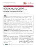
Western blot for mPER1 protein
Western blot analysis of Con, MD and MW mouse brains
and kidneys with anti-mPER1 goat polyclonal antibody
revealed one distinct band at 110 kDa, which corresponds
to mPER1 (Figure 2). Western blot test showed that the
mPER1 protein, which reflects mPer1 gene expression,
exhibited robust circadian rhythmicity in whole brain.
The mPER1 protein expression level in MD mice was
increased between 8:00 and 20:00. In Con and MW mice,
high level of mPER1 protein expression in brains was
observed at 0:00. Therefore, the phase of the circadian
rhythm of mPer1 expression was advanced in mice of the
MD group compared with the Con and MW groups (Fig-
ure 2a). Western blot test also showed that the mPER1
protein exhibited robust circadian variation in the kidneys
of Con mice (Figure 2b). In contrast, there were weak
expressions of mPER1 in the kidneys of the MD and MW
mice.
Journal of Circadian Rhythms 2006, 4:9 />Page 4 of 9
(page number not for citation purposes)
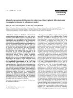
Immunohistochemical analysis of mPER1
Under high power (200×) and viewed with inverted
microscope (Nikon TE 2000-U), mPER1 protein expres-
sion in the brains and kidneys were clearly observed. High
expression of mPER1 is seen as brown-yellow, whereas
low expression is seen as blue in the sections (Figure 3).
Rhythmic expression of mPER1 was analyzed according to
the mPER1 expression data determined by image pro plus
software and shown in Figure 4 and Table 1.
Using immunohistochemistry and image analysis for
expression of mPER1 protein, we found that the expres-
sion of mPER1 protein in the piriform cortex, nucleus
accumbens and gyrus dentatus of the hippocampus fluc-
tuated throughout the 12L:12D cycle (Figure 4a, Table 1).
Circadian rhythmicity of mPER1 expression persisted
after morphine administration, but the circadian pattern
of mPER1 expression in the brains was changed: the
MESOR was elevated and the acrophase (peak time) was
shifted ahead in MD mice as compated to Con and MW
mice. The acrophase of mPER1 expression did not differ
significantly between the Con group (22:54) and the MW
group (23:24). The acrophase was much earlier, however,
in the MD group (17:04), as confirmed by the cosinor test.
Circadian variation of mPER1 protein expression was also
observed in the kidneys of Con group mice, but not of MD
and MW mice (Figure 4b, Table 1). In Con mice, mPER1
protein expression showed a peak at 3:11, whereas the
peak value of mPER1 protein expression was not obvi-
ously noticed after morphine administration. The circa-
dian expression of mPER1 protein was severely damped in
the MD and MW mice compared with Con. The expres-
sion of mPER1 in the kidneys in Con, but not in MD and
MW, showed statistically significant circadian rhythmicity
(Figure 4b, Table 1).
Discussion
Circadian rhythmicity is a highly conserved biological
function that is found in every living organism from bac-
teria to humans. In mammals, circadian rhythms are reg-
ulated by the central circadian pacemaker in the SCN. In
order for the organism to adapt to the environment, the
circadian rhythms must be synchronized to the environ-
mental light-dark cycle. This synchronization process is
known as light entrainment, which occurs through daily
light-induced phase advances and delays of the endog-
enous clock [19]. The SCN receives direct retinal input
through a specialized subpopulation of light-sensitive but
image forming-independent retinal ganglion cells that
contain the photopigment melanopsin [20]. These gan-
glion cells project to the SCN and release glutamate and
the neuropeptide pituitary adenylyl cyclase activating
peptide (PACAP) as the principal neurotransmitters for
light entrainment [21].
In order to optimize the bodily function of different organ
systems, the SCN keeps the circadian rhythms of different
peripheral organs synchronized to each other. For exam-
ple, the catecholamine and the glucocorticoid hormone
levels are high during the day when cardiovascular output
is in high demand. During sleep, circulating lymphocytes
reach the peak level to conduct immune surveillance.
However, under pathological conditions, the circadian
rhythms among different organ systems may not be well
synchronized to each other, or to the environmental light-
dark cycle. The results of the present study indicate that
morphine treatment can abolish the circadian oscillation
of mPER1 protein in the kidney and alter the phase of the
oscillation in the brain. These results strongly suggest that
morphine addiction and withdraw may lead to desyn-
chronization of circadian rhythm between different
organs.
The conditioned place preference (CPP) resultsFigure 1
The conditioned place preference (CPP) results. Data
of CPP in mice are given as mean (± S.E.M.) under the differ-
ent conditions. a: CPP in the Con and MD groups (Con
group after the 8
th
day of saline injection, MD group after the
8
th
day of morphine injection, * p < 0.05 tested by Student's t-
test) b: CPP in the MW group (MW group after the 8
th
day
morphine injection and after 5
th
day of morphine with-
drawal).
Journal of Circadian Rhythms 2006, 4:9 />Page 5 of 9
(page number not for citation purposes)
Besides playing a role in regulating circadian rhythms, the
role of mPer1, if any, in the brain and peripheral tissue is
largely unknown. Our Western blot analysis using the
whole brains revealed that the phase of the circadian
rhythm of mPer1 was advanced in the morphine with-
drawal mice compared to the control mice. In future stud-
ies, we will isolate brain structures that are known to be
involved in drug addiction, including the limbic system,
the dopaminergic neurons in the nucleus accumbens, and
the arcuate nucleus, etc.
The exact role of the circadian clock genes in peripheral
tissues remains unknown. Our results revealed that mor-
phine treatment can abolish the circadian oscillation of
mPER1 protein in the kidney and alter the phase of the
oscillation in the brain. A previous study reported that
morphine and its metabolites were secreted by the kidney
after detoxification in the liver [22]. It was also reported
that opiate addiction could result in renal diseases,
including interstitial nephritis, glomerular epithelial cell
apoptosis, nephrotic syndrome or acute renal failure [23-
26]. Chen et al. [27] reported that urinary water excretion,
sodium excretion and potassium excretion exhibit circa-
dian rhythms in the rats, with peak activity occurring at
night. Our results showed that the expression of mPER1 in
the kidneys was higher at night in the control mice, coin-
ciding with the peak activity of potassium excretion [27].
The SCN may regulate the circadian rhythms of peripheral
organs through diverse pathways. A previous study
reported that circadian rhythms of clock genes including
mPer1 were maintained in the kidneys of SCN-lesioned
mice [28]. In feeding studies, it was found that feeding
schedules could entrain the circadian rhythm of clock
gene expression in the liver independent of the SCN
[29,30]. These findings suggest that the circadian rhythms
of peripheral organs may be synchronized by nutrients or
metabolic products, in addition to the SCN.
In summary, the effects of morphine on the circadian
clock gene, mPer1, seem to be organ specific. In the brain,
morphine increases the level of mPER1 expression and
The mPER1 protein expression levels of mice at the different time pointsFigure 2
The mPER1 protein expression levels of mice at the different time points. Western blot analysis of the brains with
anti-mPER1 polyclonal antibody reveals one distinct band at molecular weight of 110 kDa (Con, MD and MW, respectively). a:
Top: mPER1 protein expressed in brains of mice. Bottom: data for mPER1 protein level were obtained by computerized analysis
of the Western blots. Each value is the mean ± SEM. b: Top: mPER1 protein expressed in the kidneys of mice. Bottom: data for
mPER1 protein level were obtained by computerized analysis of the Western blots. Each value is the mean ± SEM.
Journal of Circadian Rhythms 2006, 4:9 />Page 6 of 9
(page number not for citation purposes)
Immunohistochemical stain for determining mPER1 protein expression in brains and kidneysFigure 3
Immunohistochemical stain for determining mPER1 protein expression in brains and kidneys. a: Positive staining
in the nucleus and cytoplasm are found in brains of Con, MD and MW mice. b: Representative cases show positive staining for
mPER1 in kidneys of Con, MD and MW mice. Original magnification: 200× for all cases.
Journal of Circadian Rhythms 2006, 4:9 />Page 7 of 9
(page number not for citation purposes)
Circadian variation of mPER1 protein expressed in the brains and kidneys of Con, MD and MW miceFigure 4
Circadian variation of mPER1 protein expressed in the brains and kidneys of Con, MD and MW mice. The inte-
gral optical density (IOD) of mPER1 immunoreactivity, an index of mPER1 protein expression level, was analyzed by image pro
plus software. Time point means and SE of protein expression are shown along the 24-hour time scale. The best fitting cosine
curves are shown in these panels. a: The mPER1 protein expression in brains was increased and acrophase of circadian rhythm
was advanced in the MD mice as compared with Con and MW mice, statistically tested by the cosinor parameter test designed
by Bingham et al. [18]. b: The mPER1 protein expression in the kidneys was severely inhibited and the circadian rhythm of
mPER1 protein expression in the MD and MW mice was obliterated by morphine administration. Con mice exhibited robust
rhythmicity in mPER1 expression.
Journal of Circadian Rhythms 2006, 4:9 />Page 8 of 9
(page number not for citation purposes)
advances the phase of the circadian rhythm. In the kidney,
morphine decreases the level of mPER1 expression and
abolishes circadian rhythmicity.
Competing interests
The author(s) declare that they have no competing inter-
ests.
Authors' contributions
XW participated in all of the work and drafted the manu-
script. YW participated in experiment designing. HX par-
ticipated in data analysis. YL and CW participated in the
CPP experiment. YW, HZ and ZJ participated in immuno-
histochemistry and western blot. JMD helped with the
English writing of the paper. ZW directed the study and
wrote the final version of the manuscript. All authors read
and approved the final version of the article.
Acknowledgements
This work was partly supported by the NNSFC (30470623 for Z. Wang and
30570902 for C. Wan) and the NIH (NS047014 for J. M. Ding).
References
1. Harmer SL, Panda S, Kay SA: Molecular bases of circadian
rhythms. Annu Rev Cell Dev Biol 2001, 17:215-253.
2. Dvornyk V, Vinogradova O, Nevo E: Origin and evolution of cir-
cadian clock genes in prokaryotes. Proc Natl Acad Sci USA 2003,
100(5):2495-2500.
3. Merrow M, Spoelstra K, Roenneberg T: The circadian cycle: daily
rhythms from behavior to genes. EMBO Rep 2005,
6(10):930-935.
4. Herzog ED, Schwartz WJ: A neural clockwork for encoding cir-
cadian time. J Appl Physiol 2002, 92(1):401-408.
5. Dardente H, Cermakian N: How many pieces to build a circa-
dian clock? Med Sci (Paris) 2005, 21(1):66-72.
6. Fukuhara C, Tosini G: Peripheral circadian oscillators and their
rhythmic regulation. Front Biosci 2003, 8:d642-651.
7. Vansteensel MJ, Magnone MC, van Oosterhout F, Baeriswyl S, Albre-
cht U, Albus H, Dahan A, Meijer JH: The opioid fentanyl affects
light input, electrical activity and Per gene expression in the
hamster suprachiasmatic nuclei. Eur J Neurosci 2005,
21(11):2958-2966.
8. Tierno A, Fiore P, Gannon RL: Delta opioid inhibition of light-
induced phase advances in hamster circadian activity
rhythms. Brain Res 2002, 937(1–2):66-73.
9. Marchant EG, Mistlberger RE: Morphine phase-shifts circadian
rhythms in mice: role of behavioral activation. Neuroreport
1995, 7(1):209-212.
10. Amini H, Ahmadiani A: In vivo evidence for an increase in
5alpha-reductase activity in the rat central nervous system
following morphine exposure. Int J Dev Neurosci 2005,
23(7):621-626.
11. Andretic R, Chaney S, Hirsh J: Requirement of Circadian Genes
for Cocaine Sensitization in Drosophila. Science 1999,
285:1066.
12. Liu Y, Wang Y, Wan C, Zhou W, Peng T, Liu Y, Wang Z, Li G, Cor-
nelisson G, Halberg F: The role of mPer1 in morphine depend-
ence in mice. Neuroscience 2005, 130(2):383-388.
13. Wang YQ, Zhou W, Liu YY, Liu YH, Peng T, Wang ZR: The role of
circadian gene Period1 in morphine reward in mice. Space
Med Med Eng (Beijing) 2004, 17(5):383-385.
14. Bae K, Jin X, Maywood ES, Hastings MH, Reppert SM, Weaver DR:
Differential functions of mPer1, mPer2, and mPer3 in the
SCN circadian clock. Neuron 2001, 30(2):525-536.
15. Shiromani PJ, Xu M, Winston EM, Shiromani SN, Gerashchenko D,
Weaver DR: Sleep rhythmicity and homeostasis in mice with
targeted disruption of mPeriod genes. Am J Physiol Regul Integr
Comp Physiol 2004, 287(1):R47-R57.
16. von Gall C, Schneider-Huther I, Pfeffer M, Dehghani F, Korf HW,
Stehle JH: Clock gene protein mPER1 is rhythmically synthe-
sized and under cAMP control in the mouse pineal organ. J
Neuroendocrinol 2001, 13(4):313-316.
17. Halberg F: Chronobiology: Methodological problem. Act Med
Rom 1980, 18:399-440.
18. Bingham C, Arbogast B, Cornellissen G, Lee JK, Halberg F: Inferen-
tial statistical methods for estimating and comparing cosi-
nor parameter. Chronobiologia 1981, 9:397-439.
19. Roenneberg T, Daan S, Merrow M: The art of entrainment. J Biol
Rhythms 2003, 18(3):183-94.
20. Van Gelder RN: Nonvisual ocular photoreception in the mam-
mal. Methods Enzymol 2005, 393:746-755.
21. Hannibal J, Moller M, Ottersen OP, Fahrenkrug J: PACAP and
glutamate are co-stored in the retinohypothalamic tract. J
Comp Neurol 2000, 418(2):147-155.
22. Dubs A, Wiedemeier P, Caduff B: Morphine poisoning in chronic
kidney failure. Morphine-6-glucuronide as a pharmacologi-
cally active morphine metabolite. Dtsch Med Wochenschr 1999,
124(30):896-898.
23. Atici S, Cinel I, Cinel L, Doruk N, Eskandari G, Oral U: Liver and
kidney toxicity in chronic use of opioids: an experimental
long term treatment model. J Biosci 2005, 30(2):245-252.
24. Dettmeyer RB, Preuss J, Wollersen H, Madea B: Heroin-associated
nephropathy. Expert Opin Drug Saf 2005, 4(1):19-28.
25. Patel J, Manjappa N, Bhat R, Mehrotra P, Bhaskaran M, Singhal PC:
Role of oxidative stress and heme oxygenase activity in mor-
phine-induced glomerular epithelial cell growth. Am J Physiol
Renal Physiol 2003, 285(5):F861-F869.
Table 1: Cosinor analysis of mPER1 expression.
Group P MESOR ± SE (IOD) Amplitude ± SE (IOD) Acrophase (95 %CL) Hour
Expression of m PER1 in the brains
Con < 0.001 1010.8 ± 47.2 728.6 ± 66.8 -343.5° (-333, -354) 22:54
MD < 0.001 1609.5 ± 149.9* 1263.1 ± 212.0* -256.1° (-237, -275)* 17:04
MW < 0.001 1221.6 ± 66.6 832.6 ± 94.3 -348.6° (-335, 0) 23:24
Expression of mPER1 in the kidneys
Con < 0.001 2559.2 ± 110.3 1368.4 ± 156.1 -47.7° (-34, -60) 03:11
MD 0.538 113.1 ± 15.9# 25.4 ± 22.5# -148.5° (0, 0)# 09:54
MW 0.602 396.8 ± 45.9# 66.0 ± 64.9# -350.5° (0, 0)# 23:22
* p < 0.05 compared with control or MW groups, separately, tested by the parameters of cosinor designed by Bingham et al. [18].
#
p < 0.05
compared with control group, tested by the parameters of cosinor. The mPER1 protein expression level is represented by the IOD, which was the
value of immunoreactivity of mPER1 in tissues reacting with mPER1 antibody determined by image pro plus software.
Con: Control; MD: Morphine-dependent; MW: Morphine-withdrawal. P in the table is the p-value of circadian rhythm coming from cosine function
fitting. The hour in the table is the time of clock hour for the acrophase of the fitted cosine function.
Publish with Bio Med Central and every
scientist can read your work free of charge
"BioMed Central will be the most significant development for
disseminating the results of biomedical research in our lifetime."
Sir Paul Nurse, Cancer Research UK
Your research papers will be:
available free of charge to the entire biomedical community
peer reviewed and published immediately upon acceptance
cited in PubMed and archived on PubMed Central
yours — you keep the copyright
Submit your manuscript here:
/>BioMedcentral
Journal of Circadian Rhythms 2006, 4:9 />Page 9 of 9
(page number not for citation purposes)
26. Stratta P, Canavese C, Messina M, Colla L, Dogliani M, Vercellone A:
Postpartum acute renal failure in a drug addict. Drug Alcohol
Depend 1986, 17(4):377-380.
27. Chen LG, Wang ZR, Wan CM, Xiao J, Guo L, Guo HL, Cornelissen
G, Halberg F: Circadian renal rhythm influenced by implanted
encapsulated hANP-producing cells in Goldblatt hyperten-
sion rats. Gene Ther 2004, 11(20):1516-1522.
28. Guo H, Brewer JM, Champhekar A, Harris RB, Bittman EL: Differen-
tial control of peripheral circadian rhythms by suprachias-
matic-dependent neural signals. Proc Natl Acad Sci USA 2005,
102:3111-3116.
29. Stokkam KA, Yamazaki S, Tei H, Sakaki Y, Menaker M: Entrainment
of the circadian clock in the liver by feeding. Science 2001,
291:490-493.
30. Oishi K, Kasamatsu M, Ishida N: Gene- and tissue-specific altera-
tions of circadian clock gene expression in streptozotocin-
induced diabetic mice under restricted feeding. Biochem Bio-
phys Res Commun 2004, 317(2):330-334.