Báo cáo y học: "Structural insights into the function of the core-circadian factor TIMING OF CAB2 EXPRESSION 1 (TOC1)" ppt
Bạn đang xem bản rút gọn của tài liệu. Xem và tải ngay bản đầy đủ của tài liệu tại đây (3.79 MB, 12 trang )
BioMed Central
Page 1 of 12
(page number not for citation purposes)
Journal of Circadian Rhythms
Open Access
Research
Structural insights into the function of the core-circadian factor
TIMING OF CAB2 EXPRESSION 1 (TOC1)
Elsebeth Kolmos, Heiko Schoof, Michael Plümer and Seth J Davis*
Address: Max Planck Institute for Plant Breeding Research, Carl-von-Linné-Weg 10, D-50829 Cologne, Germany
Email: Elsebeth Kolmos - ; Heiko Schoof - ; Michael Plümer - pluemer@mpiz-
koeln.mpg.de; Seth J Davis* -
* Corresponding author
Abstract
Background: The plant circadian clock has at its core a feedback loop that includes TIMING OF
CAB2 EXPRESSION 1 (TOC1). This protein has an as of yet unknown biochemical activity. It has
been noted that the extreme amino-terminus of this protein is distantly related in sequence to
response regulators (RR), and thus TOC1 is a member of the so-called pseudo response regulator
(PRR) family. As well, the extreme carboxy-terminus has a small sequence stretch related to the
other PRRs and CONSTANS (CO)-like proteins, and this peptide stretch has been termed the
CCT (for C
ONSTANS, CONSTANS-LIKE, TOC1) domain.
Methods: To extend further our understanding of the TOC1 protein, we performed a ROSETTA
structural prediction on TOC1 orthologues from four plant species. Phylogenetic interpretations
assisted in model construction.
Results: From our models, we suggest that TOC1 is a three-domain protein: TOC1 has an amino-
terminal signaling-domain related to response receivers, a carboxy-terminal domain that could
participate both in metal binding and in transcriptional regulation, and a linker domain that connects
the two.
Conclusion: The models we present should prove useful in future hypothesis-driven biochemical
analyses to test the predictions that TOC1 is a multi-domain signaling component of the plant
circadian clock.
Background
Circadian clocks are prevalent timing mechanisms used to
predict the daily changes present in the 24-h day-night
cycle. In plants, this clock regulates several developmental
and metabolic processes. Dominant outputs include the
oscillation of free-cytosolic calcium (Ca
2+
) [1], which are
generated from cADPR-derived signals [2], and the rhyth-
mic accumulation of around 10% of all transcripts [2-6].
In particular, transcription factors are over-represented as
cycling gene products [3,7]. In this way, the circadian
timer drives numerous molecular outputs in the establish-
ment of fitness in physiological processes and develop-
mental timing. This fitness benefit has been confirmed
[8]. The current aims on studies of the mechanism of the
plant clock are to define the factors that contribute to
rhythm-generating properties of the oscillator.
Published: 25 February 2008
Journal of Circadian Rhythms 2008, 6:3 doi:10.1186/1740-3391-6-3
Received: 23 December 2007
Accepted: 25 February 2008
This article is available from: />© 2008 Kolmos et al; licensee BioMed Central Ltd.
This is an Open Access article distributed under the terms of the Creative Commons Attribution License ( />),
which permits unrestricted use, distribution, and reproduction in any medium, provided the original work is properly cited.
Journal of Circadian Rhythms 2008, 6:3 />Page 2 of 12
(page number not for citation purposes)
Molecular-genetic analyses have lead to a framework
understanding of the core elements that make up the cir-
cadian clock. Mutants of Arabidopsis thaliana that are clock
defective have been used to identify loci critical for nor-
mal rhythmicity. TIMING OF CAB2 EXPRESSION 1
(TOC1) was the first such locus identified [9], and TOC1
continues to be placed central within the clock mecha-
nism [10-14]. Extending from these studies, many clock
genes are reciprocally regulated, and thus the transcrip-
tional components that drive the clock are themselves
clock controlled. Using this analytical approach, with a
focus on molecular-expression analyses in clock mutants,
the first model that partially explained mutant behavior
was described [15]. In this model, TOC1 serves as an
evening-expressed positive factor that regulates the morn-
ing expression of CIRCADIAN CLOCK ASSOCIATED 1
(CCA1) and LATE ELONGATED HYPOCOTYL (LHY) [15-
18]. The central role of TOC1 has been genetically con-
firmed [10,11], but although TOC1 is unquestionably
important for the circadian clock, lack of functional bio-
chemical understanding has hampered characterization
of its functional role within the oscillator.
Multiple regions of the TOC1 coding region are suscepti-
ble to mutagenesis. Weak mutations, such as the toc1-1
and toc1-3 alleles (both A562V changes within the car-
boxy-terminal portion) result in clock-specific defects. As
well, missense mutations in the amino terminus of TOC1
have been isolated from direct circadian screens [toc1-5
(P124S); toc1-8 (P96L)] [19,20]. In contrast, null mutants,
such as toc1-2 (splice site mutation that leads to N-termi-
nal 1–59 aa fragment) and toc1-21 (a null allele derived
from a T-DNA insertion), have defects both in circadian
properties and in light signaling [10,21,22]. Thus, TOC1
can have multiple physiological roles that can be geneti-
cally separated.
To date, the only defined activity within any region of the
TOC1 polypeptide is a nuclear-trafficking signal estab-
lished by the CCT motif (for C
ONSTANS, CONSTANS-
LIKE, T
OC1) in the carboxy-terminus [22,23]. It has been
previously noted that the amino-terminal domain resem-
bles in its primary structure sequence conservation with
bacterial-type response regulators (RR) [23]. This domain
in TOC1 thus places it as a founding member of the
pseudo-response-regulator (PRR) protein family. The
function of the pseudo-receiver domain is unknown,
because results of in vitro experiments confirm that the
PRR domain does not undergo phosphorylation, as sus-
pected, due to a lack of a conserved Asp within the
response-receiver [23]. One collective interpretation pro-
posed here, which incorporates these diverse experiments,
is that TOC1 is a multi-domain protein. TOC1 thus inte-
grates signal inputs that bridge multiple physiological
responses [24]. That weak mutations can be uncovered
which only display a subset of phenotypes [15,22] sup-
port our hypothesis of multiple signaling functions of
TOC1.
Diurnal calcium (Ca
2+
) rhythms are evident in the plant
cell. The daily rise and fall of free-cytosolic calcium has
been proposed to encode a photoperiodic signal [25-27].
The signaling nature of the encoded rhythmic Ca
2+
is an
active area of investigation [25,27,28], and the receptor
for this Ca
2+
-derived signal is as of yet unknown. One
point of note is that the phase of calcium increase is coin-
cident with that seen with TOC1 protein levels, as both
occur around dusk [26,29]. Therefore, it would be of
interest to define whether evening factors such as TOC1
comprise part of a decoding mechanism of the Ca
2+
signal.
In this work we used modeling and phylogenetic
approaches to further dissect the TOC1 protein sequence.
Several TOC1 polypeptides were detected in sequence
databases. These TOC1 proteins appear to contain three
distinct modules. Computational approaches using the
ROSETTA suite of programs lead to the development of
structural models of the TOC1 modules. One interpreta-
tion of these structures is the implication that TOC1 func-
tions as a signaling protein that in part works to process
calcium information in the induction of transcriptional
responses.
Methods
Defining TOC1 orthologous sequences
To assess putative structures of TOC1, as it relates to dif-
ferences with the PRR related sequences, we searched pub-
lic sequence databases for genes that encode full-length
proteins. The following Genbank accessions were used:
AtTOC1 (NM_125531
), AtPRR3 (NM_125403), AtPRR5
(NM_122355
), AtPRR7 (NM_120359), AtPRR9
(NM_201974
), OsTOC1 (AB189038), OsPRR37
(AB189039
), OsPRR73 (AB189040), OsPRR95
(AB189041
), OsPRR59 (ABA91559), CsTOC1
(AY611028
), LjTOC1 (AP004931), McTOC1
(AY371288
), PtTOC1 (NW_001492741), and VvTOC1
(CAO64513
)
For phylogenetic confirmation of TOC1 sequence identi-
fication, polypeptides where clustered using CLUSTALW
[30], and this was used to generate a tree using UPGMA,
where CLC FREE WORKBENCH (CLC bio, Aarhus, Den-
mark) facilitated these efforts.
Modeling and model comparisons
The ROSETTA software suite was generously supplied by
the Baker Laboratory (University of Washington, Seattle,
USA) and it was used to model the three modules of four
selected TOC1 polypeptides; each were modeled 500
times. These models were clustered, and up to 10 consen-
Journal of Circadian Rhythms 2008, 6:3 />Page 3 of 12
(page number not for citation purposes)
sus structures for all four given domains were compared
by SARF2 [31]. From this, those structures most related
were taken forward for comparisons. These 12 structures
are available as supplemental files in PDB format (see
Additional files 1, 2, 3, 4, 5, 6, 7, 8, 9, 10, 11, 12). The
three-dimensional domains were aligned and visually
presented using MACPYMOL 0.99 (DeLano Scientific
LLC, Palo Alto, USA). Related structures were found with
SSM [32]. Calcium was fit using the GG method [33]. The
bacterial response regulators were CheY (PDB code 1E6K
)
and SPO0F (PDB code 1SRR
). A PDF file of the CCT
domain of CONSTANS was provided by Dr. Coupland.
Results and discussion
Phylogenetics
We sought to detect TOC1-related sequences from various
plants as a phylogenetic starting tool for structural predic-
tions. For this, AtTOC1 [22] and OsTOC1 [34] were used
to search genome-sequence databases. Full-length pre-
dicted proteins were found for Castanea sativa, Lotus japon-
icus, and Mesembryanthemum crystallinum, and more
recently, Vitis vinifera and Populus trichocarpa. These full-
length sequences were chosen as they were reported to
exhibit the architecture typical to TOC1, as was defined
previously by the Mizuno group [35]. Out-group
sequences were the paralogues of the PRR family, which
are PRR3/5/7/9 from Arabidopsis, and from rice (Oryza
sativa), OsPRR37 and OsPRR73, OsPRR59 and OsPRR95
(rice PRR5 and PRR9 have not yet been phylogenetically
resolved from each other, nor have rice PRR3 and PRR7)
[23,34].
We generated a phylogenetic tree using UNWEIGHTED
PAIR GROUP METHOD WITH ARITHMETIC MEAN
(UPGMA) clustering and a bootstrap replicate number of
10,000 to confirm that the encoded proteins isolated from
databases were the orthologues of TOC1 and paralogous
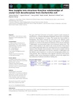
to the other PRRs. As can be seen in Figure 1, the
sequences CsTOC1, LjTOC1, McTOC1, PtTOC1, and
VvTOC1 all clustered with the rice and Arabidopsis TOC1
proteins, as expected. Because it would have been compu-
tationally too intense to model all TOC1 polypeptides, a
selection of four was taken forward. These representatives
were AtTOC1, CsTOC1, LjTOC1, and McTOC1; noted in
red in Figure 1. We further reasoned that the use of four
structural models of orthologous sequences would pro-
vide a template to assign the relatedness of any one given
structure.
Model predictions of TOC1
We sought to infer tertiary structure of TOC1 using ab ini-
tio approaches through the ROSETTA software suite. This
suite provides one strategy towards understanding poten-
tial folds of a target protein starting simply with the pri-
mary amino-acid sequence [36,37]. The TOC1 sequences
are computationally too large for complete structural
solution by ROSETTA as a single polypeptide chain [36],
thus putative folding modules within the sequences were
required to be defined. Here, a folding module is defined
as a unit within the polypeptide required for a given bio-
chemical activity. To define modules, the full set of above
defined TOC1 proteins were aligned (Figure 2) and the
transition areas in the lineup where sequence conserva-
tion moves to non-conservation was noted (color points
to these transitions is indicated in Figure 2). These infor-
matic "cut sites" are estimates of folding modules [38]. By
this approach, TOC1 could be dissected into three
domain modules (Figure 2). With respect to the AtTOC1
protein, these modules were from amino-acid positions
1–189, 190–412, and 413–618, respectively. As four
TOC1 sequences were to be applied to ROSETTA, with
three modules each, we therefore proceeded with predict-
ing structures for twelve separate polypeptide domains.
Each module was edited from the four respective full-
length proteins and modeled separately. A family of 500
models of each module was generated and these were
TOC1 and PRR phylogenyFigure 1
TOC1 and PRR phylogeny. UPGMA phylogenetic tree of
TOC1/PRR proteins. The groupings are strongly supported,
as indicated by high bootstrap values (>70%). The scale bar
represents 0.05 estimated amino-acid change per sequence
position. Sequences in red were selected for further analysis
in this study. Pt, Populus trichocarpa; Cs, Castanea sativa; At,
Arabidopsis thaliana; Vv, Vitis vinifera Lj, Lotus japonicus; Mc,
Mesembryanthemum crystallinum; Os, Oryza sativa. Sequence
origin can be found in the Methods section.
Journal of Circadian Rhythms 2008, 6:3 />Page 4 of 12
(page number not for citation purposes)
clustered based on the free-energy landscape within these,
leading to groups of up to 10 related structural families. In
these clusters, the structure centered within a given cluster
was selected as the representative of said cluster. For this,
ROSETTA determines an all-atom energy axis and plots
this against an axis of the ROOT MEAN SQUARE DEVIA-
TION (RMSD) of the resultant structures [36]. From there,
each of the related four proteins of each module was proc-
essed on SPATIAL ARRANGEMENT OF BACKBONE
FRAGMENTS 2 (SARF2) [31] as an approach to define
those structures within clusters that most resembled like-
ness to orthologous structural domains. We note that
Global alignment of selected TOC1 sequencesFigure 2
Global alignment of selected TOC1 sequences. ClustalW multiple alignment of TOC1 amino-acid sequences chosen
based on the phylogenetic analysis in Figure 1. The three colors (green, red and blue) represent the modular domains for the
four TOC1 sequences that were selected for further analysis by defining regions in sequence that move from conservation to
non-conservation. The conservation block highlights the percentage identity of amino-acids in the lineup. Note that for module
I and module III, there is far more identity than in module II. Abbreviations refer to: At, Arabidopsis thaliana; Cs, Castanea sativa;
Lj, Lotus japonicus; Mc, Mesembryanthemum crystallinum; Os, Oryza sativa; Pt, Populus trichocarpa; Vv, Vitis vinifera
Journal of Circadian Rhythms 2008, 6:3 />Page 5 of 12
(page number not for citation purposes)
SARF2 was developed as a clustering approach that detects
ensembles of secondary-structure elements that form sim-
ilar spatial arrangements, whilst accepting different possi-
ble topological connections [31]. With this approach, we
found within the identified structural clusters the subc-
lade with the best statistical fit, as assessed by RMSD, for
a given structural module. Combining the representative
clustering of ROSETTA to the relatedness clusters of SARF2
lead to one choice for each module within a given
sequence. The resultant structures from this method were
thus selected as the most representative of a given struc-
tural protein module. What follows is a description of
each model and our discussion of the implications for
that particular module.
Models of module I
We first generated protein models for the amino-terminal
third of the TOC1 polypeptides (Table 1, Figure 3). These
models were highly related in structure to each other (Fig-
ure 3). Using a query of the generated structures against all
known protein structures at the Protein Data Bank, via the
use of the software SECONDARY STRUCTURE MATCH-
ING (SSM) [32], we found that all models were predicted
to fold similarly to bacterial RR proteins (data not shown;
see below for discussion and Figure 4 for representative
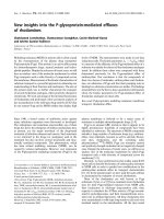
example) [39,40]. Generally, all module I structures have
a core of five alpha helices interdigited with alternating
beta sheets. This resembles the canonical fold of all RR
structures. As well, an alpha-helical tail extends from the
RR-like portion of the structure.
The mutations toc1-5 (P124S) and toc1-8 (P96L) lay
within module I, and the AtTOC1 structure allows exami-
nation of where this mutation would perturb function.
Amino acid 96 is in a predicted beta sheet that bridges
helix three and four. This proline mutation might disrupt
folding activity as a structural mutation. Amino-acid posi-
tion 124 is in a loop between helix four and five. Whilst
this could be a structural mutation, this position does not
lie within an obvious folding pattern. The P124S muta-
tion might affect TOC1 binding to a putative associated
molecule (see "additional files" to retrieve the PDB files to
expand a view on these, and all other, structures).
The RR class of proteins mediates phospho-relay signaling
in bacteria and plants [41,42]. That the amino terminus of
TOC1 was predicted to fold like an RR is not a surprise, as
the primary sequence of this domain is detected by BASIC
LOCAL ALIGNMENT SEARCH TOOL (BLAST) [43] as
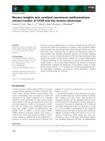
resembling an RR. We found that a superimposition of the
Arabidopsis model on two bona fide RR crystal structures
(Escherichia coli CheY and Bacillus subtilis SPO0F [44-46])
reveals an excellent structural fit (Figure 4). We note that
there is an amino- and carboxy-terminal extension of the
first domain of TOC1 relative to the two bacterial proteins
tested.
A structure resembling an RR implicates an origin of func-
tion for the amino-terminal module of TOC1. This further
supports the phylogeny relations of the amino-terminal
module of PRR to genuine RRs [40]. In each of the four
TOC1 modules, an Ala is present at what is the Asp site of
phosphorylation in a bona fide RR. In the illustrated mod-
els for module I (Figure 3), this Ala is predicted to be
within the center of the five alpha-helical borders. This is
all consistent with the previous hypothesis that TOC1 is
not a substrate of a histidine kinase [22]. As the structures
generated all resemble an RR (Figures 3 and 4; and data
not shown), we conclude that these models are likely to
resemble the "true" fold of this domain module.
Models of module IFigure 3
Models of module I. Structural models of module I (left)
and aligned with the Arabidopsis domain I (right). For the
images at the left, the colors from blue to red represent
sequence length from an amino- to carboxy-terminal direc-
tion. For the aligned figures at the right, the Arabidopsis
module I is colored green in contrast to a red color for the
compared alignment.
$OLJQ
$W
&V
/M
0F
Journal of Circadian Rhythms 2008, 6:3 />Page 6 of 12
(page number not for citation purposes)
What could be the function of an RR-domain-type fold
within module I of TOC1, particularly as it appears inca-
pable of functioning as a true RR? Several possibilities
exist. For one, this domain could be a protein-binding site
incorporating, via a scaffold function, the activities of
other clock proteins, as for example, transcription factors.
Specifically, TOC1 is known to bind members of the
bHLH transcription factor family (e.g. PIL1, PIF3, PIF4,
PIL6) [47,48]. However, in these studies, the RR domain
was shown not to be required for binding of PIF4 or PIL6
[49]. PRR proteins can also form dimers, and in case of
TOC1 binding to PRR9, PRR9 was found to interact with
TOC1 through the RR domain [49]. Furthermore, an
important role of the RR domain in protein-protein inter-
action was found for PRR3 when defined as a substrate of
the kinase WNK1 [50,51]. In addition, it is not yet estab-
lished if the ZEITLUPE (ZTL) or the PRR3 binding sites
associate with the RR domain [13,29]; both ZTL and PRR3
are confirmed protein interactors to TOC1. It is also plau-
sible that the RR-type domain/module could be a redox-
responsive site, as was hypothesized by the work of the
Golden group [52,53]. What appears clear is that identifi-
cation of interacting molecules to the amino-terminal
module will likely define a biochemical function.
Models of module II
Our next efforts were to model the middle third of the
four TOC1 modules. These predictions were found to be
structurally unrelated to each other (Figure 5, Table 1).
This is of interest as the primary amino-acid composition
Comparison of module I to response regulators from bacteriaFigure 4
Comparison of module I to response regulators from bacteria. (A) Multiple alignment of module I from plants and
response regulators from bacteria. Ec, Escherichia coli CheY; Bs, Bacillus subtilis SPO0F. The lineup is as described in Figure 2. (B)
Structures of the Arabidopsis model for module I and published structures for two response regulators (left) and aligned with
to Arabidopsis module I (right). Coloration is as shown.
$W
(F
%V
$OLJQ
!"
Journal of Circadian Rhythms 2008, 6:3 />Page 7 of 12
(page number not for citation purposes)
of the middle third is the most distinct (Figure 2). We note
that this is true for the other PRR proteins as well [54]. The
lack of a consensus structure within the middle third of
the polypeptide (Figure 5) prohibits us from making any
structural conclusions. As well, this module lacks relations
to other structural features bioinformatically character-
ized. One small amino-acid stretch is conserved in the sec-
ond module; respective to AtTOC1 module II, the
sequence is KKSRLKIGESSAFFTYVKST. Examination of
this stretch within module II of the four predicted struc-
tures revealed no fold consensus. It is thus difficult for us
to predict the reliability of the presented models of the
middle module.
What could be the function of this middle module? As
this region is poorly predicted, and no structural elements
were found to resemble the folds of known proteins (data
not shown), we present the hypothesis that this part of the
protein functions as a linker domain. This is supported by
the sequence dissimilarity in this region of the protein
(Figure 2). In addition, the previously defined direct-
repeat within AtTOC1 (position 275–369) is not present
in orthologous TOC1 proteins. Thus, amino-acid compo-
sition of module II appears to be under rapid divergence.
We note that a linker is a known feature in separating pro-
tein modules, as for example, this is seen in cullin [55]
and calmodulin [56]. In each case, linker spacing is critical
[57,58]. The sequence degeneration of a putative linker
within TOC1 might imply that the PRR polypeptides have
dissimilar folds in their middle third. It is also plausible
that module II is a native unfolded domain. Perhaps pro-
tein length here is more important than a particular struc-
ture or amino acid composition.
Models of module III
Our final structural efforts targeted the carboxy-termini of
the four described TOC1 proteins (Figure 6, Table 1).
Unlike module II, each of these was predicted to generate
a fold family. All four structures contain two alpha-helices
towards the extreme terminus of the protein. This serves
to center alignments and represents the CCT sub-domain.
This CCT was always found to consist of a small alpha-
helical interphase, and in all cases this predicted fold was
similar (Figure 6). The overall folding of these structures
was found to be predominantly alpha-helical with inter
bundle-to-bundle interactions and folded substructures
that lack prolonged secondary structure (Figure 6). We
further note that module III of TOC1 contains a primary
amino-acid composition that does not lend to a detecta-
ble primary architecture of known factors. Given the relat-
edness of the four module III structures, we conclude that
the predicted structures could contain structural elements
that resemble the true fold.
Models of module IIFigure 5
Models of module II. Structural models of module II. The
colors from blue to red represent sequence length from the
amino- to carboxy-terminal direction.
Table 1: The table summarizes the number of selected cluster-
center modules chosen from the starting point of 500 generated
ROSETTA structures (see Methods).
Module I Module II Module III
AtTOC1 3 10 10
CsTOC1 4 8 3
LjTOC1 5 9 9
McTOC1 3 10 8
Journal of Circadian Rhythms 2008, 6:3 />Page 8 of 12
(page number not for citation purposes)
The presented fold of module III implicates the carboxy
terminus of TOC1 in metal binding and also associations
to DNA-binding proteins (see below). One interesting fea-
ture of the four carboxy-terminal modules is that in struc-
tural searches against the three-dimensional folds we
generated, each of these four TOC1 modules was found to
be in a fold most similar to that present in various metal-
binding proteins. Interestingly, the primary amino-acid
composition of these domains is unlike that of other
metal-binding domains, such as an EF-hand [59]. As the
primary and secondary structures of the terminal domain
of TOC1 did not detect such relations, we suspect that a
structural-folding pattern was required to detect structural
elements that relate to biochemical function.
Each TOC1 module III might be related to a metal-bind-
ing protein. By SSM searches, we found that the AtTOC1
structure was most related to calmodulin-sensitive ade-
nylate cyclase (a protein known to be regulated by cal-
cium) [60]; CsTOC1 was most related to calmodulin (a
known calcium-binding protein) [61,62]; LtTOC1 was
also most related to calmodulin; and McTOC1 was most
related to the zinc-bound form of cell filamentation pro-
tein (Structure 2f6s in The Protein Data Bank). Based on
the obvious implication that module III could participate
in Ca
2+
binding, we tried to detect such a binding pocket
by a computational approach. Here, we were successful in
our ability to fit each of these structures with a bound cal-
cium ion using the GG computational approach [33]. In
each case, we could detect that the amino-terminal region
of module III harbors a site that could accept the place-
ment of a calcium ion (Figure 6). Note that this is distant
from the CCT domain in each case (Figure 6). We thus
propose that the third module of TOC1 can be implicated
in aspects of metal signaling. This computational finding
provides a testable hypothesis for the future.
We found that the CCT domain within this third of TOC1
was predicted to fold in a similar manner as the CCT
domain from CONSTANS (CO) (Figure 7) [63]. As CO is
a bona fide interactor to HEME ACTIVATOR PROTEIN
(HAP) transcription factors [63], it is intriguing that TOC1
could also associate with this class of DNA-binding fac-
tors. Two mutant alleles map to the CCT subdomain of
module III, and we can thus view the location of these
changes. The toc1-1 and toc1-3 mutations (A562V) both
map to an alpha-helical fold within the CCT subdomain,
and we note that this Ala residue is conserved in all
sequences. The A562V mutation could affect the ability of
the CCT to fold into a helix. This would impair its ability
to bind target proteins, such as HAP factors. If the hypoth-
esis that the CCT subdomain of TOC1 is a binding inter-
face of HAP factors were true, this would directly implicate
TOC1 as a co-regulator of transcription. As TOC1 geneti-
cally functions to promote CCA1 and LHY transcription
Models of module III in predictive complex with calciumFigure 6
Models of module III in predictive complex with cal-
cium. Structural models of module III. The colors from blue
to red represent sequence length from the amino- to car-
boxy-terminal direction. Note that alpha-helical clusters in
the carboxy terminus center these structures, and that a cal-
cium ion can be fit into all four structures in an amino-termi-
nal position within all structures. The red arrow points to
the fit calcium, which is colored as a gray sphere.
Journal of Circadian Rhythms 2008, 6:3 />Page 9 of 12
(page number not for citation purposes)
[10,15-18,24], it is an exciting hypothesis that TOC1 func-
tions as a transcriptional co-activator in a multi-protein
complex on promoters of clock-regulated genes.
What could be the function of module III in TOC1? It is
intriguing that the concentration of cytosolic Ca
2+
oscil-
lates with an evening peak close to the time that TOC1 is
most abundant [26,29]. cAMPR drives both the circadian
oscillations of cytosolic calcium and the rhythmic expres-
sion of many clock genes, however not TOC1 [2]. It might
be that Ca
2+
interacts with TOC1 posttranslationally, an
idea that is consistent with the fact that calcium rhythms
are unaffected in the toc1-1 mutant [27]. This calcium
interaction would drive the ability of TOC1 protein to reg-
ulate its targets. One could thus hypothesize TOC1 to be
a component of decoding the Ca
2+
signal. If true, TOC1
could generate this function by direct interaction with
Ca
2+
. A direct test of Ca
2+
-binding to TOC1 seems a plau-
sible experiment to implicate this protein as a sensor for
the circadian levels of Ca
2+
. From there, it would be of
interest to test TOC1 binding to HAP factors, and test the
role of Ca
2+
(or another metal) in supporting or attenuat-
ing this binding.
General considerations of the models and implications of
a unified TOC1
How likely are the TOC1 models we present to be correct?
This is difficult to assess. In fact, the community standard
to answer this question requires the actual structure to be
determined [64]. In the absence of an experimentally
derived TOC1 structure, we believe that modeling could
be useful for predictive biochemistry and to direct further
experimentation. We also note that in various bench-
marks, ROSETTA correctly predicted protein structures
approximately half of the time [36]. We thus conclude
that aspects of the model presented here are likely to have
useful structural information, but that major structural
features could be flawed. Certainly, minor features of the
models, such as side-chain directionality, are unlikely to
be correct.
An over-riding theme generated from our models is the
hypothesis that TOC1 acts as a signal adapter that senses
a small ligand (e.g. Ca
2+
or a redox signal) and that this is
part of a transcription complex (Figure 8). This multifac-
eted hypothesis is intriguing given that the plant clock is
modulated by small-molecule signaling [65]. For exam-
ple, redox levels change in response to light [53,66]. Thus,
as predicted by Golden and colleagues, the amino-termi-
nus of TOC1 could be involved in metabolite sensing to
mediate entrainment. Also, Ca
2+
levels coincide with that
of TOC1 [26,29]. The scaffold principles implicated from
the amino- and carboxy-modules could support a mecha-
nism for TOC1 as a transcriptional mediator that func-
tions in response to signal integration from distinct
signaling pathways. This scaffold hypothesis defines the
middle module as a tether that links modules I and III.
The high degeneration of amino-acid composition in this
middle module would support a spacer function rather
than a scaffold or enzymatic activity. What is clear is that
a biochemical hypothesis now exists to describe how
TOC1 leads to transcriptional induction of CCA1 and
LHY.
Competing interests
The author(s) declare that they have no competing inter-
ests.
Authors' contributions
EK, HS, MP and SJD performed the work. EK and SJD
wrote the paper.
Schematic representation of a TOC1 structural modelFigure 8
Schematic representation of a TOC1 structural
model. I PRR domain – this resembles bona fide response
regulators. II Linker domain – a putative bridge between
modules I and III. III Calcium-binding domain – a potential
sensor for a metal. IIIb Protein-binding domain – a potential
interaction motif for HAP DNA-binding factors.
Comparison of CCT sub-module structuresFigure 7
Comparison of CCT sub-module structures. From left
to right, the predicted structures of the CCT sub-module of
CO and AtTOC1, and their alignment match when aligned.
The colors from blue to red represent sequence length from
the amino- to carboxy-terminal direction.
Journal of Circadian Rhythms 2008, 6:3 />Page 10 of 12
(page number not for citation purposes)
Additional material
Acknowledgements
We are especially thankful to David Baker, Dylan Chivian, Phil Bradley, and
Andrew Wollacott for supplying ROSETTA and their extensive assistance
in its use. The PDB file of the CCT domain of CONSTANS supplied by
George Coupland is acknowledged. We thank Amanda M. Davis for per-
forming the SSM searches, and Ulrike Göbel and Anika Jöcker for compu-
tational assistance. This work was supported in the SJD group by the Max
Planck Society and the German-Israeli Project Cooperation (DIP project
H3.1) and in the HS group by the Max Planck Society.
References
1. Dodd AN, Love J, Webb AA: The plant clock shows its metal:
circadian regulation of cytosolic free Ca(2+). Trends Plant Sci
2005, 10:15-21.
2. Dodd AN, Gardner MJ, Hotta CT, Hubbard KE, Dalchau N, Love J,
Assie JM, Robertson FC, Jakobsen MK, Goncalves J, et al.: The Ara-
bidopsis circadian clock incorporates a cADPR-based feed-
back loop. Science 2007, 318:1789-1792.
3. Edwards KD, Anderson PE, Hall A, Salathia NS, Locke JC, Lynn JR,
Straume M, Smith JQ, Millar AJ: FLOWERING LOCUS C Medi-
ates Natural Variation in the High-Temperature Response
of the Arabidopsis Circadian Clock. Plant Cell 2006, 18:639-650.
4. Harmer SL, Hogenesch JB, Straume M, Chang HS, Han B, Zhu T,
Wang X, Kreps JA, Kay SA: Orchestrated transcription of key
pathways in Arabidopsis by the circadian clock. Science 2000,
290:2110-2113.
5. Davis SJ, Millar AJ: Watching the hands of the Arabidopsis bio-
logical clock. Genome Biol 2001, 2:REVIEWS1008.
6. Michael TP, Mockler TC, Breton G, McEntee C, Byer A, Trout JD,
Hazen SP, Shen R, Priest HD, Sullivan CM, et al.: Network discovery
pipeline elucidates conserved time of day specific cis-regula-
tory modules. PLoS Genetics 2008. preprint:e14.eor
7. Hanano S, Stracke R, Jakoby M, Merkle T, Domagalska M, Weisshaar
B, Davis SJ: A systematic survey in Arabidopsis thaliana of
transcription factors that modulate circadian parameters.
BMC Genomics 2008, , 9:182.
8. Dodd AN, Salathia N, Hall A, Kevei E, Toth R, Nagy F, Hibberd JM,
Millar AJ, Webb AA: Plant circadian clocks increase photosyn-
thesis, growth, survival, and competitive advantage. Science
2005, 309:630-633.
9. Millar AJ, Carre IA, Strayer CA, Chua NH, Kay SA: Circadian clock
mutants in Arabidopsis identified by luciferase imaging. Sci-
ence 1995, 267:1161-1163.
10. Ding Z, Doyle MR, Amasino RM, Davis SJ: A Complex Genetic
Interaction Between Arabidopsis thaliana TOC1 and CCA1/
LHY in Driving the Circadian Clock and in Output Regula-
tion. Genetics 2007, 176:1501-1510.
11. Kolmos E, Davis SJ: ELF4 as a central gene in the circadian
clock. Plant Signal Behavior 2007, 2:370-372.
12. Martin-Tryon EL, Kreps JA, Harmer SL: GIGANTEA acts in blue
light signaling and has biochemically separable roles in circa-
dian clock and flowering time regulation. Plant Physiol 2007,
143:473-486.
Additional file 1
Structural file. Structure of AtTOC1_dom1
Click here for file
[ />3391-6-3-S1.pdb]
Additional file 2
Structural file. Structure of AtTOC1_dom2
Click here for file
[ />3391-6-3-S2.pdb]
Additional file 3
Structural file. Structure of AtTOC1_dom3
Click here for file
[ />3391-6-3-S3.pdb]
Additional file 4
Structural file. Structure of CsTOC1_dom1
Click here for file
[ />3391-6-3-S4.pdb]
Additional file 5
Structural file. Structure of CsTOC1_dom2
Click here for file
[ />3391-6-3-S5.pdb]
Additional file 6
Structural file. Structure of CsTOC1_dom3
Click here for file
[ />3391-6-3-S6.pdb]
Additional file 7
Structural file. Structure of LjTOC1_dom1
Click here for file
[ />3391-6-3-S7.pdb]
Additional file 8
Structural file. Structure of LjTOC1_dom2
Click here for file
[ />3391-6-3-S8.pdb]
Additional file 9
Structural file. Structure of LjTOC1_dom3
Click here for file
[ />3391-6-3-S9.pdb]
Additional file 10
Structural file. Structure of McTOC1_dom1
Click here for file
[ />3391-6-3-S10.pdb]
Additional file 11
Structural file. Structure of McTOC1_dom2
Click here for file
[ />3391-6-3-S11.pdb]
Additional file 12
Structural file. Structure of McTOC1_dom3
Click here for file
[ />3391-6-3-S12.pdb]
Journal of Circadian Rhythms 2008, 6:3 />Page 11 of 12
(page number not for citation purposes)
13. Para A, Farre EM, Imaizumi T, Pruneda-Paz JL, Harmon FG, Kay SA:
PRR3 Is a Vascular Regulator of TOC1 Stability in the Arabi-
dopsis Circadian Clock. Plant Cell 2007.
14. Perales M, Mas P: A Functional Link between Rhythmic
Changes in Chromatin Structure and the Arabidopsis Bio-
logical Clock. Plant Cell 2007, 19:2111-2123.
15. Alabadi D, Oyama T, Yanovsky MJ, Harmon FG, Mas P, Kay SA:
Reciprocal regulation between TOC1 and LHY/CCA1 within
the Arabidopsis circadian clock. Science 2001, 293:880-883.
16. Locke JCW, Southern MM, Kozma-Bognar L, Hibberd V, Brown PE,
Turner MS, Millar AJ: Extension of a genetic network model by
iterative experimentation and mathematical analysis. Mol
Syst Biol 2005. doi:10.1038/msb4100018
17. Locke JCW, Kozma-Bognar L, Gould PD, Feher B, Kevei E, Nagy F,
Turner MS, Hall A, Millar AJ: Experimental validation of a pre-
dicted feedback loop in the multi-oscillator clock of Arabi-
dopsis thaliana. Mol Syst Biol 2006, 2:59.
18. Zeilinger MN, Farre EM, Taylor SR, Kay SA, Doyle FJ: A novel com-
putational model of the circadian clock in Arabidopsis that
incorporates PRR7 and PRR9. Mol Syst Biol 2006, 2:58.
19. Hazen SP, Borevitz JO, Harmon FG, Pruneda-Paz JL, Schultz TF,
Yanovsky MJ, Liljegren SJ, Ecker JR, Kay SA: Rapid array mapping
of circadian clock and developmental mutations in Arabi-
dopsis. Plant Physiol 2005, 138:990-997.
20. Kevei E, Gyula P, Hall A, Kozma-Bognar L, Kim WY, Eriksson ME,
Toth R, Hanano S, Feher B, Southern MM, et al.: Forward Genetic
Analysis of the Circadian Clock Separates the Multiple Func-
tions of ZEITLUPE. Plant Physiol 2006, 140:933-945.
21. Hall A, Bastow RM, Davis SJ, Hanano S, McWatters HG, Hibberd V,
Doyle MR, Sung S, Halliday KJ, Amasino RM, Millar AJ: The TIME
FOR COFFEE gene maintains the amplitude and timing of
Arabidopsis circadian clocks. Plant Cell 2003, 15:2719-2729.
22. Strayer C, Oyama T, Schultz TF, Raman R, Somers DE, Mas P, Panda
S, Kreps JA, Kay SA: Cloning of the Arabidopsis clock gene
TOC1, an autoregulatory response regulator homolog. Sci-
ence
2000, 289:768-771.
23. Makino S, Kiba T, Imamura A, Hanaki N, Nakamura A, Suzuki T, Tan-
iguchi M, Ueguchi C, Sugiyama T, Mizuno T: Genes encoding
pseudo-response regulators: Insight into His-to-Asp phos-
phorelay and circadian rhythm in Arabidopsis thaliana. Plant
Cell Physiol 2000, 41:791-803.
24. Mas P, Alabadi D, Yanovsky MJ, Oyama T, Kay SA: Dual role of
TOC1 in the control of circadian and photomorphogenic
responses in Arabidopsis. Plant Cell 2003, 15:223-236.
25. Dodd AN, Jakobsen MK, Baker AJ, Telzerow A, Hou SW, Laplaze L,
Barrot L, Poethig RS, Haseloff J, Webb AAR: Time of day modu-
lates low-temperature Ca2+ signals in Arabidopsis. Plant J
2006, 48:962-973.
26. Love J, Dodd AN, Webb AAR: Circadian and diurnal calcium
oscillations encode photoperiodic information in Arabidop-
sis. Plant Cell 2004, 16:956-966.
27. Xu X, Hotta CT, Dodd AN, Love J, Sharrock R, Lee YW, Xie Q, John-
son CH, Webb AA: Distinct Light and Clock Modulation of
Cytosolic Free Ca2+ Oscillations and Rhythmic CHLORO-
PHYLL A/B BINDING PROTEIN2 Promoter Activity in Ara-
bidopsis. Plant Cell 2007, 19:3474-3490.
28. Tang RH, Han S, Zheng H, Cook CW, Choi CS, Woerner TE, Jackson
RB, Pei ZM: Coupling diurnal cytosolic Ca2+ oscillations to the
CAS-IP3 pathway in Arabidopsis. Science 2007, 315:1423-1426.
29. Mas P, Kim WY, Somers DE, Kay SA: Targeted degradation of
TOC1 by ZTL modulates circadian function in Arabidopsis
thaliana. Nature 2003, 426:567-570.
30. Thompson JD, Higgins DG, Gibson TJ: CLUSTAL W: improving
the sensitivity of progressive multiple sequence alignment
through sequence weighting, position-specific gap penalties
and weight matrix choice. Nucleic Acids Res 1994, 22:4673-4680.
31. Alexandrov NN: SARFing the PDB. Protein Eng 1996, 9:727-732.
32. Krissinel E, Henrick K: Secondary-structure matching (SSM), a
new tool for fast protein structure alignment in three dimen-
sions. Acta Crystallogr D Biol Crystallogr 2004, 60:2256-2268.
33. Deng H, Chen G, Yang W, Yang JJ: Predicting calcium-binding
sites in proteins – a graph theory and geometry approach.
Proteins 2006, 64:34-42.
34. Murakami M, Ashikari M, Miura K, Yamashino T, Mizuno T: The evo-
lutionarily conserved OsPRR quintet: rice pseudo-response
regulators implicated in circadian rhythm. Plant Cell Physiol
2003, 44:1229-1236.
35. Mizuno T, Nakamichi N: Pseudo-Response Regulators (PRRs)
or True Oscillator Components (TOCs). Plant Cell Physiol 2005,
46:677-685.
36. Bradley P, Misura KM, Baker D: Toward high-resolution de novo
structure prediction for small proteins. Science 2005,
309:1868-1871.
37. Rohl CA: Protein structure estimation from minimal
restraints using Rosetta. Methods Enzymol 2005, 394:244-260.
38. Kim DE, Chivian D, Malmstrom L, Baker D: Automated prediction
of domain boundaries in CASP6 targets using Ginzu and
RosettaDOM. Proteins 2005, 61(Suppl 7):193-200.
39. Saier MH Jr: Bacterial sensor kinase/response regulator sys-
tems: an introduction. Res Microbiol 1994, 145:349-355.
40. West AH, Stock AM: Histidine kinases and response regulator
proteins in two-component signaling systems. Trends Biochem
Sci 2001, 26:369-376.
41. Hwang I, Chen HC, Sheen J: Two-component signal transduc-
tion pathways in Arabidopsis. Plant Physiol 2002, 129:500-515.
42. Varughese KI: Molecular recognition of bacterial phosphorelay
proteins. Curr Opin Microbiol 2002, 5:142-148.
43. Altschul SF, Gish W, Miller W, Myers EW, Lipman DJ: Basic local
alignment search tool.
J Mol Biol 1990, 215:403-410.
44. Madhusudan , Zapf J, Whiteley JM, Hoch JA, Xuong NH, Varughese
KI: Crystal structure of a phosphatase-resistant mutant of
sporulation response regulator Spo0F from Bacillus subtilis.
Structure 1996, 4:679-690.
45. Sola M, Lopez-Hernandez E, Cronet P, Lacroix E, Serrano L, Coll M,
Parraga A: Towards understanding a molecular switch mech-
anism: thermodynamic and crystallographic studies of the
signal transduction protein CheY. J Mol Biol 2000, 303:213-225.
46. Stock AM, Mottonen JM, Stock JB, Schutt CE: Three-dimensional
structure of CheY, the response regulator of bacterial chem-
otaxis. Nature 1989, 337:745-749.
47. Makino S, Matsushika A, Kojima M, Yamashino T, Mizuno T: The
APRR1/TOC1 quintet implicated in circadian rhythms of
Arabidopsis thaliana: I. Characterization with APRR1-over-
expressing plants. Plant Cell Physiol 2002, 43:58-69.
48. Yamashino T, Matsushika A, Fujimori T, Sato S, Kato T, Tabata S,
Mizuno T: A link between circadian-controlled bHLH factors
and the APRR1/TOC1 quintet in Arabidopsis thaliana. Plant
Cell Physiol 2003, 44:619-629.
49. Ito S, Matsushika A, Yamada H, Sato S, Kato T, Tabata S, Yamashino
T, Mizuno T: Characterization of the APRR9 pseudo-response
regulator belonging to the APRR1/TOC1 quintet in Arabi-
dopsis thaliana. Plant Cell Physiol 2003, 44:1237-1245.
50. Murakami-Kojima M, Nakamichi N, Yamashino T, Mizuno T: The
APRR3 component of the clock-associated APRR1/TOC1
quintet is phosphorylated by a novel protein kinase belong-
ing to the WNK family, the gene for which is also transcribed
rhythmically in Arabidopsis thaliana. Plant Cell Physiol 2002,
43:675-683.
51. Nakamichi N, Murakami-Kojima M, Sato E, Kishi Y, Yamashino T,
Mizuno T: Compilation and characterization of a novel WNK
family of protein kinases in Arabiodpsis thaliana with refer-
ence to circadian rhythms. Biosci Biotech Biochem 2002,
66(11):2429-2436.
52. Ivleva NB, Bramlett MR, Lindahl PA, Golden SS:
LdpA: a compo-
nent of the circadian clock senses redox state of the cell.
Embo J 2005, 24:1202-1210.
53. Ivleva NB, Gao T, LiWang AC, Golden SS: Quinone sensing by the
circadian input kinase of the cyanobacterial circadian clock.
Proc Natl Acad Sci USA 2006, 103:17468-17473.
54. Matsushika A, Makino S, Kojima M, Mizuno T: Circadian waves of
expression of the APRR1/TOC1 family of pseudo-response
regulators in Arabidopsis thaliana: insight into the plant cir-
cadian clock. Plant Cell Physiol 2000, 41:1002-1012.
55. Thornton BR, Toczyski DP: Precise destruction: an emerging
picture of the APC. Genes Dev 2006, 20:3069-3078.
56. Snedden WA, Fromm H: Calmodulin as a versatile calcium sig-
nal transducer in plants. New Phytologist 2001, 151:35-66.
57. Strynadka NCJ, James MNG: Crystal Structures of the Helix-
Loop-Helix Calcium-Binding Proteins. Annu Rev Biochem 1989,
58:951-999.
Publish with BioMed Central and every
scientist can read your work free of charge
"BioMed Central will be the most significant development for
disseminating the results of biomedical research in our lifetime."
Sir Paul Nurse, Cancer Research UK
Your research papers will be:
available free of charge to the entire biomedical community
peer reviewed and published immediately upon acceptance
cited in PubMed and archived on PubMed Central
yours — you keep the copyright
Submit your manuscript here:
/>BioMedcentral
Journal of Circadian Rhythms 2008, 6:3 />Page 12 of 12
(page number not for citation purposes)
58. Zheng N, Schulman BA, Song L, Miller JJ, Jeffrey PD, Wang P, Chu C,
Koepp DM, Elledge SJ, Pagano M, et al.: Structure of the Cul1-
Rbx1-Skp1-F box Skp2 SCF ubiquitin ligase complex. Nature
2002, 416:703-709.
59. Nelson MR, Chazin WJ: Structures of EF-hand Ca 2+-binding
proteins: Diversity in the organization, packing and response
to Ca 2+ Binding. BioMetals 1998, 11:297-318.
60. Masure HR, Oldenburg DJ, Donovan MG, Shattuck RL, Storm DR:
The interaction of Ca2+ with the calmodulin-sensitive ade-
nylate cyclase from Bordetella pertussis. J Biol Chem 1988,
263:6933-6940.
61. Chou JJ, Li S, Klee CB, Bax A: Solution structure of Ca(2+)-cal-
modulin reveals flexible hand-like properties of its domains.
Nat Struct Biol 2001, 8:990-997.
62. McCormack E, Tsai YC, Braam J: Handling calcium signaling:
Arabidopsis CaMs and CMLs. Trends Plant Sci 2005, 10:383-389.
63. Wenkel S, Turck F, Singer K, Gissot L, Le Gourrierec J, Samach A,
Coupland G: CONSTANS and the CCAAT box binding com-
plex share a functionally important domain and interact to
regulate flowering of Arabidopsis. Plant Cell 2006,
18:2971-2984.
64. Zhang Y: Template-based modeling and free modeling by I-
TASSER in CASP7. Proteins 2007, 69(Suppl 8):108-117.
65. Hanano S, Domagalska MA, Nagy F, Davis SJ: Multiple phytohor-
mones influence distinct parameters of the plant circadian
clock. Genes Cells 2006, 11:1381-1392.
66. Kim BH, von Arnim AG: The early dark-response in Arabidopsis
thaliana revealed by cDNA microarray analysis. Plant Mol Biol
2006, 60:321-342.