Báo cáo y học: "Hazards of tube thoracostomy in patients on a ventilator" doc
Bạn đang xem bản rút gọn của tài liệu. Xem và tải ngay bản đầy đủ của tài liệu tại đây (625.39 KB, 2 trang )
CAS E REP O R T Open Access
Hazards of tube thoracostomy in patients on a
ventilator
Kasra Shaikhrezai
*
and Vipin Zamvar
Abstract
A patient with post-pneumonia empyema complicated by type-2 respiratory failure required mechanical ventilation
as part of his therapy. A pneumothorax was noted on his chest radiograph. This was treated with an intercostal
chest drain (ICD). Unfortunately, he was still hypoxic, his subcutaneous emphysema was worsening and the ICD
was bubbling. A computed tomography (CT) scan of chest demonstrated that the ICD has penetrated the right
upper lobe parenchyma. A new ICD was inserted and the previous one was removed. Although both hypoxia and
subcutaneous emphysema improved, the patient chronically remained on mechanical ventilation.
Background
Tube thoracostomy is a common procedure to drain
fluids and/or air from the pleural space via an ICD. The
British Thoracic Society (BTS) has published a guideline
[1] for ICD insertion which in many i nstitutions h as
been deployed as a standard approach to tube thoracost-
omy in both practice and training programs. Recently
there is an increasing concern regarding the t raining of
doctors with regard to preci se and methodological ICD
insertion [2,3]. Harris et al [4] conducted a national sur-
vey among chest physicians in the UK recording their
experiences regardi ng complications and serious harms
following ICD insertion. The study revealed 67% of
NHS trusts have experienced major complications of
ICD insertion.
Case presentation
A 5 1-year-old man with history of chronic obstructive
pulmonary disease (COPD) and cigarette smoking pre-
sented with a shortness of breath, chronic pneumonia
and empyema involving the right side of his chest. Soon
after admission his conditi on deteriorated developing
type-2 respiratory failure necessitating intubation and
commencement of mechanical ventilation. Patient
required positive end-expiratory pressure (PEEP) of
10 mmHg and 80% fraction of inspired oxygen (FiO2)
to maintain the oxygen saturation of 91% with PCO2
(partial pressure of carbon dioxide) and PO2 (partial
pressure of oxygen) of 7.1 and 8.2 kPa respectively.
Following central line insertion a pneumothorax was
noted on his chest radiograph. Under aseptic technique
and blunt dissection a large bore ICD was inserted ante-
rolaterally into the right chest preceded by the introduc-
tion of index finger and sweeping manoeuvre explained
by the BTS guidelines [1]. It is imperative to appreciate
that a diseased hyperventilated lung with a high PEEP is
very prone to perforation by any instruments penetrat-
ing the chest wall and pleura. Shortly after tube
thoracostomy the pati ent started to develop a large sub-
cutaneous emphysema originating in the right moving
towar ds the left side of the chest wall. Unfortuna tely his
hypoxic state became worse requiring augmentation o f
mechanical ventilation. In the interim ICD was bubbling
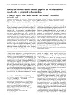
constantly. A CT scan of chest demonstrated that the
ICD has penetrated the right upper lobe parenchyma
(Figure 1). As a result patient was urgently transferred
to our institute for further management.
A new ICD was inserted with the same technique
whilst the ventilator was briefly disconnected. When it
was proved that the new ICD is in the appropriat e posi-
tion with a characteristic swing of column of water, the
previous ICD was removed.
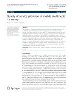
Subsequent chest CT scan revealed the right upper
lobe laceration containing gas communicating with the
anterior chest wall. This was accompanied by massive
subcutaneous emphysema (Figure 2).
Although following the new ICD both hypoxia and
subcutaneous emp hysema improved the patient was
chronically remained on ventilation.
* Correspondence:
Department of Cardiothoracic Surgery, Royal Infirmary of Edinburgh,
Edinburgh, UK
Shaikhrezai and Zamvar Journal of Cardiothoracic Surgery 2011, 6:39
/>© 2011 Shaikhrezai a nd Zamvar; licensee BioMed Central Ltd. This is a n Open Access article distributed under t he terms of the Creative
Commons Attribu tion License ( which permits unrestricted use, distribution , and
reproduction in any medium, provided the original work is pro perly cited.
Conclusion
Previously the risks of ICD insertion in patients on
mechanical ve ntilation has been desc ribed [5] however
we presented the above case due to frequent referral of
patients on mechanic al ventilation to us with harmful
complications of tube tho racostomy. Prior to ICD in ser-
tion in a patient on mechanical ventilation, the PEEP
must b e turned off and the ventilator must be discon-
nected briefly during the introduction of the ICD. In
ICD i nsertion deploying Seldinger technique the same
steps need to be taken for introducing the guide wire as
well as the chest tube. Any ICD breaching the lung par-
enchyma s hould b e removed after insertion of another
ICD in the pleural space.
We believe the BTS guidelines [1] require a new revi-
sion with the view to including the mechanical ventila-
tion as a hazardous clinical setting in “pre-drainage risk
assessment” section. Furthermore ICD insertion needs
to be explained separately in self- and mechanical-
ventilating patients along with considering the clinical
settings as well as the specialty demands.
For instance efficient drainage of left-sided pleural
effusion in a post-CABG (coronary artery bypass graft
surge ry) patient requires a tube thoracost omy below t he
triangle of safety; or fine bore ICD insertion under Sel-
dinger technique for the treatment of pneumothorax is
a well e stablished procedure deployed by respiratory
physicianswhileinthoracicsurgeryalargeboreICD
with conventional insertion technique is favourable.
The royal college of surgeons has intro duce d S-DOP S
(direct observation of procedural skills in surgery) via
intercollegiate surgical curriculum programme (ISCP)
[6]. We recommend a unified usage of surgical DOPS in
all specialties to sign off junior doctors’ competency in
tube thoracostomy in self- and mechanical-ventilating
patients.
Consent
Written informed consent was obtained from the patient
for publication of this case report and accompanying
images. A copy of the written consent is available for
review by the Editor-in-Chief of this journal.
Authors’ contributions
KS performed the procedure; VZ admitted the patient under his care,
instructed and supervised the procedure. All authors read and approved the
final manuscript.
Competing interests
The authors declare that they have no competing interests.
Received: 14 December 2010 Accepted: 29 March 2011
Published: 29 March 2011
References
1. Laws D, Neville E, Duffy J: BTS guidelines for the insertion of a chest
drain. Thorax 2003, 58(Suppl II):ii53-ii59.
2. Elsayed H, Roberts R, Emadi M, Whittle I, Shackcloth M: Chest drain
insertion is not a harmless procedure - are we doing it safely? Interact
CardioVasc Thorac 2010, 11:745-748.
3. Guidance for the implementation of local trust policies for the safe
insertion of chest drains for pleural effusions in adults, following the
NPSA Rapid Response Report. British Thoracic Society , NPSA/2008/RRR003.
4. Harris A, O’Driscoll BR, Turkington PM: Survey of major complications of
intercostal chest drain insertion in the UK. Postgrad Med 2010,
86(1012):68-72.
5. Peek GJ, Firmin RK, Arsiwala S: Chest tube insertion in the ventilated
patient. Injury 1995, 26(6):425-6.
6. Intercollegiate Surgical Curriculum Programme. [ />home/assessment_sdops.aspx], Accessed on 12 December 2010.
doi:10.1186/1749-8090-6-39
Cite this article as: Shaikhrezai and Zamvar: Hazards of tube
thoracostomy in patients on a ventilator. Journal of Cardiothoracic Surgery
2011 6:39.
Figure 1 ICD (arrows) penetrating the lung parenchyma.
Figure 2 Right upper lobe laceration (arrow) conta ining gas
communicating with the anterior chest wall (post ICD removal).
Shaikhrezai and Zamvar Journal of Cardiothoracic Surgery 2011, 6:39
/>Page 2 of 2