Báo cáo y học: "LV reverse remodeling imparted by aortic valve replacement for severe aortic stenosis; is it durable? A cardiovascular MRI study sponsored by the American Heart Association" pot
Bạn đang xem bản rút gọn của tài liệu. Xem và tải ngay bản đầy đủ của tài liệu tại đây (1.82 MB, 8 trang )
RESEARCH ARTIC LE Open Access
LV reverse remodeling imparted by aortic valve
replacement for severe aortic stenosis; is it
durable? A cardiovascular MRI study sponsored
by the American Heart Association
Robert WW Biederman
1*
, James A Magovern
3
, Saundra B Grant
1
, Ronald B Williams
1
, June A Yamrozik
1
,
Diane A Vido
1
, Vikas K Rathi
1
, Geetha Rayarao
1
, Ketheswaram Caruppannan
1,2
and Mark Doyle
1
Abstract
Background: In patients with severe aortic stenosis (AS), long-term data tracking surgically induced effects of
afterload reduction on reverse LV remodeling are not available. Echocardiographic data is available short term, but
in limited fashion beyond one year. Cardiovascular MRI (CMR) offers the ability to serially track changes in LV
metrics with small numbers due to its inherent high spatial resolution and low variability.
Hypothesis: We hypothesize that changes in LV structure and function following aortic valve replacement (AVR)
are detectable by CMR and once triggered by AVR, continue for an extended period.
Methods: Tweny-four patients of which ten (67 ± 12 years, 6 female) with severe, but compensated AS underwent
CMR pre-AVR, 6 months, 1 year and up to 4 years post-AVR. 3D LV mass index, volumetrics, LV geometry, and EF
were measured.
Results: All patients survived AVR and underwent CMR 4 serial CMR’s. LVMI markedly decreased by 6 months (157
± 42 to 134 ± 32 g/m
2
, p < 0.005) and continued trending downwards through 4 years (127 ± 32 g/m
2
). Similarly,
EF increased pre to post-AVR (55 ± 22 to 65 ± 11%,(p < 0.05)) and continued trending upwards, rema ining stable
through years 1-4 (66 ± 11 vs. 65 ± 9%). LVEDVI, initially high pre-AVR, decreased post-AVR (83 ± 30 to 68 ± 11 ml/
m2, p < 0.05) trending even lower by year 4 (66 ± 10 ml/m
2
). LV stroke volume increased rapidly from pre to post-
AVR (40 ± 11 to 44 ± 7 ml, p < 0.05) continuing to increase non-significantly through 4 years (49 ± 14 ml) with
these LV metrics paralleling improvements in NYHA. However, LVmass/volume, a 3D measure of LV geometry,
remained unchanged over 4 years.
Conclusion: After initial beneficial effects imparted by AVR in severe AS patients, there are, as expected, marked
improvements in LV reverse remodeling. Via CMR, surgically induced benefits to LV structure and function are
durable and, unexpectedly express continued, albeit markedly incomplete improvement through 4 years post-AVR
concordant with sustained improved clinical status. This supports down-regulation of both mRNA and MMP activity
acutely with robust suppression long term.
* Correspondence:
1
Center for Cardiovascular Magnetic Resonance Imaging, The Gerald
McGinnis Cardiovascular Institute, Department of Medicine, Division of
Cardiology, Allegheny General Hospital, Drexel University College of
Medicine, Pittsburgh, Pennsylvania, USA
Full list of author information is available at the end of the article
Biederman et al. Journal of Cardiothoracic Surgery 2011, 6:53
/>© 2011 Biederman et al; licensee BioMed Central Ltd. This is an Open Access article distributed under the terms of the Creative
Commons Attribution License ( which permits unrestricte d use, distribution , and
reproduction in any medium, provided the ori ginal work is properly cited.
Introduction
In patients with severe aortic stenosis (AS), compensa-
tory left ventricular hypertrophy (LVH) is the predomi-
nate mechanism manifest to attempt to normalize the
markedly elevated afterload imposed at the aortic valve
level [1]. Overtime this initially beneficial response leads
to deleterious downstream effects not limited to mis-
matched neovascularization relative to the extent of l eft
ventricular (LV) hypertrophy, supranormal LV perfor-
mance likely due to geometic remodeling and marked
interstial fibrosis due to collagen deposition that even-
tually leads to codominant explanations for the often
pronounced hypertrophy often seen in late stage AS
[2-5]. It is for thes e reasons that the goal of aortic valve
replacemen t (AVR) is aimed. AVR is designed to relieve
valvular afterload but with the cardinal physiologic effect
directed at inducing regression of the excessive LVH. In
this manner it has long been known that there is a sur-
vival advantage in those who receive AVR as compared
to those who, for other reasons, fail to undergo correc-
tive surgery. However, the l ong-term data tracking the
surgically induced beneficial effects of afterload reduc-
tion on reverse LV remo deling are available only in lim-
ited fashion. Moreover, the majority of the available data
exists in echocardiographic literature, is pertinent to
remodeling concepts is available short term [6,7], but
only in limited fashion beyond one year [8-12].
Cardiac magnetic resonance imaging (CMR) is the
‘gold standard’ for measuring cardiac volumetrics LV
mass and offers the ability to track changes in LV
metrics with innordinantly small numbers due to its
inherent high spatial resolution and low intraobserver
variability [13]. Indeed, as compared to echocardiogra-
phy, Bottini et al demonstrated that if one wished to be
able to detect a 10 gram regression in LV mass with an
alphaof0.05andabetaof0.80itwouldrequire550
patients, whereas only 17 patients were necessary by
CMR [14]. This represents over a log-fold reduction in
the number of patients required in order to detect a
beneficia l effect by CMR over the more commonly used
modality, echocardiography. Thus, the pattern and tem-
poral manner in which LVH regresses, currently
unknown, conceivably should be discernable over a long
period of time pre and post-AVR non-invasively via
CMR in a small number of pati ents providing answers
as to the completeness and durability of LVH regression
following AVR.
Hypothesis
We hypothesize that progressive LV reverse remodeling
changes following AVR are detectable by CMR and
changes in LV structure and function, once triggered by
AVR, continue for an extended period.
Methods
Population
Patients referred for AVR were enrolled after institu-
tional review board (IRB) approval and signed consent
obtained. All patients were identified via standard clini-
cal metrics independent of CMR evaluation chiefly
through cardiac catheterization and/or echocardiogra-
phy. To provide homogeneity in the pathology of AS,
patients were excluded if there was aortic or mitral
regurgitation assessed by echocardiographic imaging as
greater than moderate (>2+), mitral stenosis, prior valve
replacement, myocardial infarction, history of hyperten-
sion, coronary artery bypass grafting (CABG) or angio-
plasty. Specific contraindications to CMR were presence
of a pacemaker, defibrillator, history of metal fragments,
implants, cerebrovascular clips or claustrophobia.
CMR Imaging
The 3D CMR methodology has been described else-
where [15,16]. Briefly, using a General Electric (Milwau-
kee, Wisconsin) 1.5T Excite EKG-triggered CMR system
(50 mT/m maximum gradient strength, 150 mT/m/ms
maximum slew rate), scout images were obtained to
plan double-oblique views in horizontal and vertical
long- axis views from which short-axis contiguous 8 mm
slices traversing the mitral valve plane through LV apex
were ac quired using a steady-state free p recession
(FIESTA) cine sequence with a field of view 38 cm
2
,
matrix 256 × 192, flip angle 45°. The temporal resolu-
tion was 30 ± 3 ms,100% phase FOV and 0.75 NEX, TR
3.2 ms and TE 1.4 ms. From the short-axis images, LV
end-diastolic v olume (LVEDV), LV end-systolic volume
(LVESV), LV stroke volume (LVSV), LV ejection frac-
tion (EF), and LV mass were me asured and indexed to
BSA. LV mass was derived via Simpson’s method multi-
plied by the specific gravity of myocardium (1.055 g/ml).
Image acquisition was kept constant to include LV basal
plane-registration throughout the study and between
patients to minimize variability in measurements.
Phase velocity mapping (PVM) was employed to quan-
titate 3D peak and mean aortic tra nsvalvular gradients
in the through and in-plane slices. Velocity encoding
was set at 350-550 cm/sec with encoding in the x, y and
z directions. PVM was resolved into 60 phases/cardiac
cycle achieving high temporal resolution(19 ± 3 ms).
ROI’sweremanuallydrawnencirclingtheentiresupra-
valvular plane for complete interrogation of all veloci-
ties, as opposed to the ‘ice-pick’ view employed by
echocardiography. 2D transthoracic and/or transesopha-
geal echocardiography was also performed for indepen-
dent clinical assessment of AS.
All images were analyzed offline on semi-automatic
MASS Plus and Flow programs (Medis, The Netherlands).
Biederman et al. Journal of Cardiothoracic Surgery 2011, 6:53
/>Page 2 of 8
CMR imaging was performed (5 ± 3 days) prior to AVR, 6
month and 1 year and up to 4 years post-AVR. An inde-
pendent comparison of AS degree assessed by each modal-
ity (CMR and echocardiography) was performed for future
reference and was recorded, see image (Figure 1). All data
was analyzed by a single dedicated CMR technologist (JAY
or RW) throughout the study period to minimize interob-
server variability with all images blindly over-read by a
dedicated cardiologist (RWWB or VR). The mean imaging
time for the patients was 54 ± 15 minutes.
Mitral regurgitation was retrospectively semiquantita-
tivly assesed as a function of the intervoxel dephasing
artifact from the vertical and horizontal long-axis using
the steady s tate free-precession (FIESTA) dynamic cine
sequence at each time point. Measurements of the
mitral annulus, valve tenting angle and valve tenting
area were meaured using standard approaches in 2D
from the vertical and horizontal long-axis.
Statistics
Continuous variables were reported as mean ± 1 SD.
Categorical variables were reported as percentages with
95 percent confidence intervals. Serial comparisons pre-
to post-AVR were performed by the paired t-test. Effects
across groups were analyzed using one-way analysis of
variance (ANOVA) and repeated-measures ANOVA was
performed for comparisons over time. Statistical ana-
lyses were performed using S PSS for Windows, version
11.0 (SPSS, Inc., Chicago). All statistical comparisons
were performed using two-tailed significance tests with
a ‘p’ value of < 0.05 considered statistically significant.
Results
Twenty-four patients underwent pre-AVR CMR. A
random subset of patients who were imaged at the 6
month and 1 year time point were specifically invited
back to be imaged at a fourth very late time point and
underwent post-AVR imaging at 6 ± 2mo and 1 yr ±
2mo and up to 4 years (one patient imaged at 3.5
years) for 40 total time points. Thus, ten patients (67
± 12 years, 6 female) with severe, but reasonably well
compensated AS, underwent CMR pre-AVR and 3
subsequent time points post-AVR. Two patients were
classified as NYHA class III, all others were < NYHA
II. Four patie nts had concomitant CAD but were with-
out significant differences in their peak and mean
transvalvular gradient by either echocardiography or
CMR. There w as no significant diffe rence between the
CMR derived mean and peak transvalvula r gradients
(47 ± 12 and 7 0 ± 24 mmHg, respectivly) vs. the mean
and peak gradients as measured by echocardiography
(42 ± 10 and 68 ± 21, respectively) though CMR velo-
cities tended to be higher, p=NS). Stated alternatively,
there was no difference i n the number of patients with
>4 m/s p eak transvalvular gradient as measured by
CMR and echocardiography (7 vs. 7 patients). The
mean NYHA pre-AVR was 2.5 ± 1.2.
All patients had severe LVH prior to undergoi ng AVR.
Following AVR, LVMI markedly decreased at 6 months
(157±42to134±32g/m
2
, p < 0.005) and continued to
further trend downward at 4 years (127 ± 32 g/m
2
;p=
NS), see Figures 2 and 3. Similarly, EF increased pre to
post AVR (55 ± 22 to 65 ± 11%, (p < 0.05)) and continued
trending upward, however remaining statistically stable at
years 1-4 (66 ± 11 vs. 65 ± 9%). LVEDV index, initially
high pre-AVR, declined post-AVR (83 ± 30 to68 ± 11 ml/
m
2
, p < 0.05) trending even lower by year 4 but again
remaining statistically insignificant (66 ± 10 ml/m
2
).
LV stroke volume index increased rapidly from pre to
post-AVR (40 ± 11 to 44 ± 7 ml/m
2
, p < 0.05) trending
to increase at 4 years (49 ± 14 ml/m
2
) but also remain-
ing statistically insignifi cant as compared to the 6
month time period.
However, despite the relatively long term follow-up
there remained incomplete LV mass regression, failing
to return to historic age-matched control level (59 ±
11 g/m
2
) [17], see Figure 4. Likewise, LVEDVI did not
normalize, remaining above historic age-matched con-
trols [17].
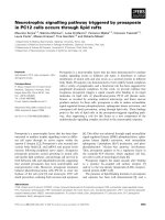
Figure 1 A coronal view from a steady-state free precession
acquisition demonstrating the heavily calcified (arrow) and
restricted aortic valve leaflets with a intervoxel dephasing
defect as depicted by the systolic turbulence (bifid arrow)
radiating into the proximal ascending aorta. In itself, this is
indictative of a highly velocity jet consistant with severe AS. Using
phase velocity mapping to formally quantitate the mean and peak
transvalvular gradients, they were 53 and 78 mmHg, respectively;
severe AS.
Biederman et al. Journal of Cardiothoracic Surgery 2011, 6:53
/>Page 3 of 8
The 3D CMR equivalent to echocardiographic relative
wall thickness (RWT), an indicator of 1D LV geometry,
is the mass/volume ratio. As a 3D metric, the mass/
volume ratio has obvious advantages over any 1D mea-
surement and a ccordingly is used to more definitively
relate changes in LV geometry over time. The mass/
volume ratio demonstrated no change initially (1.9 to
2.0 at 6 months) remaining unchanged at 1 year (2.0)
and out to 4 years (1.9), p = NS between all.
While all metrics except for EF were markedly ele-
vated as compared to normals, despite substantial metric
approaching within 2 standards deviations of normal.
LV mass index specifically remained >5 standard devia-
tions above normal.
The temporal pattern for regression for all stand-alone
metrics including EF demonstrated that a minimum of
nearly50%ofthechangethatwastobeevidentby4
years occurred within the first 6 months. For instance,
for LVMI, 76% of the mass that regressed by year 4 did
so in the first 6 months while for LVEDV, 88% of the
reduction occurred within the first 6 months. Likewise,
nearly all (91%) of the final EF achieved was present
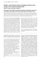
Figure 2 Serial cardiovascular MRI mid short-axis images in diastole (top row) and systole (bottom row) in a 76 WM taken the day
prior to AVR, 6 months, one year and 4 years following AVR. The LV mass decreased from 186 to 154 g over the first 6 months to only
regress to 132 g over the next 3 1/2 years demonstrating the early-rapid and late-slow pattern of LVH regression. Similarly, LVEF markedly
improved after afterload relief from 54% to 60% in the first 6 months with no further improvements over the ensuing 3 1/2 years (62%).
Figure 3 Demonstrating that, despite marked afterload mismatch in a 5 5YOWM with an LVEF 2 3% and LV mass of 251 g, surgical
relief of afterload in a patient with demonstrated myocardial reserve (mean/peak gradients of 52 and 33 mmHg, respectively) can
ensue with striking improvements in LVEF and LV mass (57%EF and 197 g at 6 months post-AVR) with minimal change by year 4
(LVEF 56% and LV mass 158 g). The initial improvements in morphometrics and volumetrics paralled marked improvements in the patients
clinical response, again most evident within the first 6 months post-AVR.
Biederman et al. Journal of Cardiothoracic Surgery 2011, 6:53
/>Page 4 of 8
within the first 6 months with no signific ant changes
appare nt afterwards. Due to the near parallel changes in
LVMI and LVEDVI, by definition, there would be no
discernable temporal pattern in the mass/volume ratio
over the entire 4 years.
Mitral Regurgitation
It should be noted that the primary objective of the
study was to interrogate a pure human pressure after-
load model of AS induced concentric LVH pathology,
such that any significant amount of potential eccentric
LVH due to volume overload was aprioriexcluded.
Nevertheless, a biolo gic signal to assess whether the
degree of mitral regurgitation (MR) could be favorably
influenced might be deducible from this population.
Pre-AVR, the grade of MR was ‘0’ through ‘2+’ (moder-
ate MR). Post-AVR the MR remained stable or
decreased late in 80% and increased in two patients (0-
trace in one patient and trace to 2+ another patient
(both in patients who had the least amount of reverse
remodeling), see Figure 5. The favorable changes in LV
mass and LV EDVI post-AVR were highly correlated
with MR improvement (r = 0.51 and 0.60, respectively).
While EF increased, it was not well correlated (r = 0.31)
with MR reduction post-AVR, while LV sphericity (r/h)
just failed to reach statistical significance with the
improvement in mitral regurgitation.
Clinical Sequelae
Paralleling improvements in CMR d erived LV volu-
metrics and mo rphometrics including mitral regurgita-
tion, there were concordant improvements in NYHA
class. Pre-AVR NYHA was 2.5 ± 1.2 and rapidly
improved to 1.6 ± 0.9 at 6 months and 1.6 ± 0.9 at 1
year but remained statistically insignifica ntly improved
out to 4 years as compared to the interim time points
(1.4 ± 1.1). However, as compared to pre-AVR, there
was an important significant difference over time by 4
years (p < 0.05).
Discussion
Due to excessive afterload imposed on the LV from the
markedly restricted valvular narrowing in patients with
severe but compensated AS, substantial LVH is typically
Figure 4 Plot s of the temporal nature of the pattern of LVH
regression serially out to 4 years. Note the immediate LVH
regression sparked by the massive afterload relief by AVR. However,
the trajectory of initial regression at 6 months would have predicted
a far greater mass reduction then evident at 4 years.
Figure 5 (Fig A, B, C) Change in mitral regurgitation that
ensues upon the relief of afterload by AVR. All but 2 patients
had CMR defineable reduction in their MR grade (defined herein as
0 through 7 representing no (absent) through 2+ (moderate) MR. In
those 2 patients the least amount of LV remodeling was present
suggesting that effective mass/volume normalization is an
important mechanism towards stabilizng and eventual MR relief as
it is in its initiating pathophysiology. (Note, superimposition
prevents all 10 patients from being displayed).
Biederman et al. Journal of Cardiothoracic Surgery 2011, 6:53
/>Page 5 of 8
apparent. While initially a favorable compensatory
response to the often extraordinary intraventricular pres-
sure, left unchecked, LVH heralds a slow inexorable dete-
rioration in cardiac function promulgated by further
changes at the myocardial and interstitial level. To the
extent that these now pathologic process are reversible is
unclear. To be sure, it is well known that the epidemiolo-
gical post-surgical effect is extremely favorable nearly
restoring survival by actuarials back to the pre-morbid
state. However, the nature, extent and temporal pattern of
these surgically induced reverse remodeling effects are
much less clear. Limited attempts to track LVH regression
after AVR have been perfor med by 2D echocardiography
but generally over short periods of time, often under one
year post-AVR. To our knowledge this is the first attempt
to apply the long known reference standard CMR, interro-
gatingLVvolume,EFandLVmass,incorporatinglong-
term remodeling to this issue.
CMR
CMR has an ability to detect exceedingly small aliquots
of myocardial mass change (intraobserver variability of
2.5 g) while detecting changes in volumetri cs such that
EF changes of 1.5%, while at lower limits of intraobserver
variability, are discernable and relevant. This provides for
an unparalleled ability for CMR to be used to interrogate
pre and post-AVR changes in a reliable and clinical ly
relevant manner. As described above, CMR retains the
ability to discriminate such findings in historically smal-
ler populations then previously considered via other
modalities due to its ultra high spatial resolution often
leading to log-fold less patient requirements to achieve
statistical significance yet retaining preserved power
14
.
LV Metrics after AVR
In this study, after the initial beneficial effects imparted
by afterload relief by AVR in severe AS patients, there
are as expected, marked improvements in LV reverse
remodeling. We have shown, via CMR, that surgically
induced benefits to LV structure and function, including
favorable alterations in LV geometry, are definable, dur-
able and, unexpec tedly, show continued improvement
up to 4 years concordant with sustained improvement
in clinical status. That these finding have awaited recog-
nition and substantiation for decades detracts nothing
from the expected, even predicatable reasoning that they
would be present since there is a clear survival advan-
tage for those that do undergo AVR as compared to
those that choose no t to, (depite being equivalent in all
other demographic and pathological characteristics).
However, the observed pattern of reverse remodeling
has never been defined before in this patient population
and was unexpected in its temporal trajectory. Fully 75%
of the LV mass regressi on that was to occur did so
within the first 6 months following AVR. In fact, nearly
90% of the change in volumetrics (LVEDVI and LVEF)
were completed in the first 6 months with clinically
insignifcant changes detected subsequently. In that the
first oportunity to detect the changes was by protocol
definedat6months,itisconceivablethatoneormore
of these metrics had their improvement at an even ear-
lier time course.
Incomplete LVH Regression after AVR
The most striking finding in this study was not the
extent of LV reverse remodeling that was found but
that, despite serial follow-up up to 4 years, there is a
distinct failure to normalize LV mass. LV mass
remained >5 standard deviations above normal for >85%
of the population without explanations on the basis of
age, sex, CAD, and pre-AVR metrics such as gradient,
valve type, cross-clamp time via multivariate analysis as
they were unable to account for the failure of LVH
regression. Should t his be surprising to us? Are there
inferences in the literature that might guide us to this
conclusion? Several avenues of support for this finding
are available as well as some that require a more consid-
ered approach.
First off, AVR itself does not restore the transvalvular
gradient to normal. Despite the advent of increasingly
lower profile aortic valves, to include the Toronto SPV
(used in 40% of this patient group), residual gradients
exist and to the extent that they remain, invariably con-
tribute to residual afterload and obligatorily thwart com-
plete LV mass regression. In most cases, however, the
ratio of residual to initial gradient is likely to be low ( <
20%) thereby having only modest impairment of even-
tual LV mass regression.
Secondly, at the same time the afterload is surgically
relieved at the valve level, supravalvular afterload is
likely to be increasing due to aortic and peripheral
changes in compliance and arterial inelasticity due to
aging. The surgically induced relief of a fterload may be
counterbalanced by the resultant increase another type
of afterload; arterial hypertension [18].
Another mechanism thwarting regression of LVH is
less obvious. Classically, the hypertrophic process is
thought to be composed chiefly of sarcomeres being laid
down in parallel resulting in concentric hypertrophy.
This process is governed mostly by m RNA expression.
Naturally, LVH regression therefore would be thought
as a reve rsal of this process following AVR. What has
become clear however is that the pathologic perturba-
tion in AS is not confined at the ventricular level only
to the myocyte [19]. The extracellular matrix, primarily
composed of collagen deposition as a response to the
pressure overload and probably due to increased peri-
mysial fibers to translate the generated myocardial
Biederman et al. Journal of Cardiothoracic Surgery 2011, 6:53
/>Page 6 of 8
deformation, expands to become a very significant pro-
portion of the total LV mass [20,21]. Its regulation and
subsequent regression is governed principally by metal
metalloproteinase (MMP’s) and by the tissue inhibitors
of MMP’s(TIMP’ s) [22,23]. In several studies the pro-
portion of collagen in AS can be as much as 30-60%
21
.
Thus, in advanced AS, pure myocyte hypertrophy is not
the only pathology that must be accounted for and con-
sequently regress post-AVR. Were both sarcomere
hypertrophy and collagen expression to be finely gov-
erned by a common pathway, coordinate regression of
both would be evident [24]. However, the signaling
pathway presiding over myocyte and sarcomeres appears
distinct and expressed at dissimilar rates resulting in
asymmetrical LVH normalization post-AVR. mRNA sig-
naling following abrupt relief of afterload is halted
within 4-6 hours in stark contrast to MMP activity
which, inhibited by TIMP’ s, is activated late and then
incompletely [25]. The resultant effect is ‘accelerated”
myocyte atrophy but with a more preserved interstitial
composition that serves in toto to ameliorate the
expected regression of LVH.
Clinical Perspective
Put into perspective, the surgeon who replaces the aortic
valve now has a number of expla nations to account for
the lack of adequate LVH regression following AVR.
Even in those admirable cases in which the post-AVR
gradient is reduced to < 15-20 mmHg, substantial
mechanisms are operative serving to thwart the other-
wise expected beneficial effects of AVR at the level of
the myocardium. In short, surgical success or failure to
trigger LVH regression should no longer be placed in
the surgeon’s prerogative.
Regarding concomit ant mitral regurgitation (up to 2+;
moderate) that often is associated with AS, AVR
achieves improvements in MR in severe AS that are
detectable by CMR and rema ins stable in up to 4 years
of follow-up. Favorable changes appear attributable to
LV and mitral valvular/annular geometry, LVH regres-
sion, less so on improved EF. Since considerable mor-
bidity and mortality exists for sim ultaneous AVR and
MVR, CMR suggests that AVR without MVR may be
indicated in such patients.
Conclusion
Patients with advanced AS upon surgical relief of valvu-
lar afterload, undergo rapid regression of LVH with cor-
responding improvements in many LV metrics
measurable by CMR that is in conjunction with
improvements in clinical sequelae. However, the pre-
ponderance of the surgical benefits appear early, almost
truncated within the first 6 months and while durable,
only minimally continue long-term out to 4 years. The
long-term expected reverse remodeling appears thwarted
by a myriad of s o-named factors rendering incomplete
the otherwise beneficial post-AVR effects. From a surgi-
cal perspective, it would seem initially apparent that any
‘less then complete’ normalization of LV mass after such
an extended fol low-up would be perceived potentially as
a shortcoming of the surgical techniq ue. From this data
we can provide substantial evidence to support that this
is an incorrect supposition. Whether longer-term fol-
low-up would eventually reveal a normalized trajectory
on course with historic controls is unknown but worthy
of further investigation.
Acknowledgements
RWWB is the recipient of American Heart Association National Scientist
Development Grant (02350226N); MD is supported in part by National Heart,
Lung and Blood Institutes, No.5 R01HL72317 for which RWWB is an
investigator.
We are grateful for the conversations over the years with Dr. Blase A.
Carabello, Nathaniel Reichek and thankful for the support of Dr. George
Magovern, Jr. and Srinivas Murali.
Presented at the American Heart Association in Orlando, Florida at the
Surgical Sessions, November 2007, Circ 2007.116;16(suppII):543 and, in part,
the Society of Cardiovascular Magnetic Resonance in Orlando, FL, February
2007, J Cardiovasc Mag Res 2007. 9;2:260-261.
This work was supported in part from a grant from the American Heart
Association: National Scientist Development Grant (0235026N) and the
National Heart, Lung and Blood Institutes, No. 5 RO1 HL72317.
Author details
1
Center for Cardiovascular Magnetic Resonance Imaging, The Gerald
McGinnis Cardiovascular Institute, Department of Medicine, Division of
Cardiology, Allegheny General Hospital, Drexel University College of
Medicine, Pittsburgh, Pennsylvania, USA.
2
Division of Internal Medicine,
Allegheny General Hospital, Pittsburgh, Pennsylvania, USA.
3
Department of
Surgery, Division of Cardiothoracic Surgery, Allegheny General Hospital,
Pittsburgh, Pennsylvania, USA.
Authors’ contributions
RB conceived, designed coordinated and analyzed primary data, assisted in
recruitment, IRB issues as well as wrote the manuscript. JM discussed the
design of the study and performed the majority of the aortic valve
replacements. SG was the nurse coordinator, recruited patients, coordinated
follow-up CMR exams and all the IRB/HIPPA requirements as well as partially
conceived of the secondary 4 year follow-up coordination principal study.
RW performed the CMR exams and data analysis. JY performed the CMR
exams and data analysis. DV statistical analysis. VR helped interpret CMR
exams served as the second cardiologist on the study. GR assisted in
primary data analysis and was the software engineer for the study. KC
participated in the study as the cardiology fellow and separately analyzed
mitral regurgitation data. MD helped to implement design, analysis and
performance of the study as well as implemented optimization of the RF
tissue-tagging sequence, critical discussions of the study results, critical
analysis of the various drafts of the manuscript and review/approval of its’
final draft. All authors read and approved the final manuscript.
Competing interests
The authors declare that they have no competing interests.
Received: 7 January 2011 Accepted: 14 April 2011
Published: 14 April 2011
References
Lorell, BH, and BA Carabello. 2000. Left ventricular hypertrophy: pathogenesis,
detection, and prognosis. Circ 25(4): 470–9, 102.
Biederman et al. Journal of Cardiothoracic Surgery 2011, 6:53
/>Page 7 of 8
Smucker, ML, CL Tedesco, SB Manning, RM Owen, and MD Feldman. 1988.
Demonstration of an imbalance between coronary perfusion and excessive
load as a mechanism of ischemia during stress in patients with aortic
stenosis. Circ 78(3): 573–82.
Bishop, SP, PC Powell, N Hasebe, YT Shen, TA Patrick, L Hittinger, and SF Vatner.
1996. Coronary vascular morphology in pressure-overload left ventricular
hypertrophy. J Mol Cell Cardiol 28(1): 141–54. doi:10.1006/jmcc.1996.0014.
Nakano, K, WJ Corin, JF Spann, RWW Biederman, S Denslow, and BA Carabello.
1989. Abnormal subendocardial blood flow in pressure overload hypertrophy
is associated with pacing-induced subendocardial dysfunction. Circ Res 65(6):
1555–64.
Weber, KT, Y Sun, and SC Tyagi, et al. 1994. Collagen network of the
myocardium: function, structural remodeling and regulatory mechanisms. J
Mol Cell Cardiol 26: 279–292. doi:10.1006/jmcc.1994.1036.
Rao, L, S Mohr-Kahaly, S Geil, M Dahm, and J Meyer. 1999. Left ventricular
remodeling after aortic valve replacement. Z Kardiol 88(4): 283–9.
doi:10.1007/s003920050287.
Djavidani, B, FX Schmid, A Keyser, B Butz, J Seitz, A Luchner, K Debl, S Feuerbach,
and WR Nitz. 2004. Early regression of left ventricular hypertrophy after aortic
valve replacement by the Ross procedure detected by cine MRI. J Cardiovasc
Magn Reson 6(1): 1–8. doi:10.1081/JCMR-120027799.
Niwaya, K, RC Elkins, CJ Knott-Craig, KL Santangelo, MB Cannon, and MM Lane.
1999. Normalization of left ventricular dimensions after Ross operation with
aortic annular reduction. Ann Thorac Surg 68(3): 812–8. doi:10.1016/S0003-
4975(99)00765-1.
Dalmau, MJ, J María González-Santos, J López-Rodríguez, M Bueno, A Arribas,
and F Nieto. 2007. One year hemodynamic performance of the Perimount
Magna pericardial xenograft and the Medtronic Mosaic bioprosthesis in the
aortic position: a prospective randomized study. Interact Cardiovasc Thorac
Surg 6(3): 345–9, Epub 2007. doi:10.1510/icvts.2006.144196.
Takeda, S, and H Rimington. 1998. Chambers J. How often do we operate too
late in aortic stenosis? J Heart Valve Dis , 4: 428–30.
Kato, Y, S Suehiro, T Shibata, Y Sasaki, and H Hirai. 2007. Impact of valve
prosthesis-patient mismatch on long-term survival and left ventricular mass
regression after aortic valve replacement for aortic stenosis. J Card Surg
22(4): 314–9. doi:10.1111/j.1540-8191.2007.00414.x.
Perez de Arenaza, D, B Lees, M Flather, F Nugara, T Husebye, M Jasinski, M
Cisowski, M Khan, M Henein, J Gaer, L Guvendik, A Bochenek, S Wos, M Lie,
G Van Nooten, D Pennell, and J Pepper. 2005. ASSERT (Aortic Stentless versus
Stented valve assessed by Echocardiography Randomized Trial) Investigators.
Randomized comparison of stentless versus stented valves for aortic stenosis:
effects on left ventricular mass. Circ 112(17): 2696–702, Epub 2005.
doi:10.1161/CIRCULATIONAHA.104.521161.
Biederman, RWW, M Aldrich, W Rogers, S Mankad, J Ripple, J Yamrozik, K
Simpson, J Magovern, and N Reichek. 2002. Does the Adage, “There is Safety
in Numbers” Lead Us Astray? An MRI Remodeling Study. J Cardiovasc Mag
Reson 4(1): 177, Abst.
Bottini, PB, AA Carr, LM Prisant, FW Flickinger, JD Allison, and JS Gottdiener. 1995.
Magnetic resonance imaging compared to echocardiography to assess left
ventricular mass in the hypertensive patient. Am J Hypertens 8(3): 221–8.
doi:10.1016/0895-7061(94)00178-E.
Biederman, RWW, M Doyle, J Yamrozik, RB Williams, VK Rathi, D Vido, K
Caruppannan, N Osman, V Bress, G Rayarao, C Biederman, S Mankad, J
Magovern, and N Reichek. 2005. Physiologic Compensation is Supranormal in
Compensated Aortic Stenosis: Does it Return to Normal after Aortic Valve
Replacement or is it Blunted by Coexistent Coronary Artery Disease? Circ
112(suppl I): I-429-I-436.
Biederman, RWW, JA Magovern, SB Grant, Williams Ronald B, JA Yamrozik, DA
Vido, VK Rathi, K Rayarao, K Caruppannan, and M Doyle. 2007. LV Reverse
Remodeling Imparted by Aortic Valve Replacement for Severe Aortic
Stenosis; Is it Durable? A Cardiovascular MRI Study sponsored by the
American Heart Association. Circ 116(suppII): 543, 16 abst.
Hudsmith, LE, SE Petersen, JM Francis, MD Robson, and S Neubauer. 2005.
Normal human left and right ventricular and left atrial dimensions using
steady state free precession magnetic resonance imaging. Cardiovasc Magn
Reson 7(5): 775–82. doi:10.1080/10976640500295516.
Imanaka, K, O Kohmoto, S Nishimura, Y Yokote, and S Kyo. 2005. Impact of
postoperative blood pressure control on regression of left ventricular mass
following valve replacement for aortic stenosis. Eur J Cardiothorac Surg 27(6):
994–9, Epub 2005. doi:10.1016/j.ejcts.2005.02.034.
Swynghedauw, B. 1999. Molecular Mechanisms of myocardial remodeling. Physio
R 79: 21–261.
Tagawa, H, M Koide, H Sato, MR Zile, BA Carabello, and G Cooper. 1998.
Cytoskeletal role in the transition from compensated to decompensated
hypertrophy during adult canine left ventricular pressure overloading. Circ
Res 82(7): 751–61.
Villari, BM, SE Campbell, and OM Hess. 1993. Influence of collagen network on
left ventricular systolic and diastolic function in aortic valve disease. J Am
Coll Cardiol 22: 1477–1484. doi:10.1016/0735-1097(93)90560-N.
Fielitz, J, M Leuschner, HR Zurbrügg, B Hannack, R Pregla, R Hetzer, and V Regitz-
Zagrosek. 2004. Regulation of matrix metalloproteinases and their inhibitors
in the left ventricular myocardium of patients with aortic stenosis. J Mol Med
82(12): 809–20, Epub 2004. doi:10.1007/s00109-004-0606-4.
Polyakova, V, S Hein, S Kostin, T Ziegelhoeffer, and J Schaper. 2004. Matrix
metalloproteinases and their tissue inhibitors in pressure-overloaded human
myocardium during heart failure progression. J Am Coll Cardiol 44(8):
1609–18. doi:10.1016/j.jacc.2004.07.023.
Ahmed, SH, LL Clark, WR Pennington, CS Webb, DD Bonnema, AH Leonardi, CD
McClure, FG Spinale, and MR Zile. 2006. Matrix metalloproteinases/tissue
inhibitors of metalloproteinases: relationship between changes in proteolytic
determinants of matrix composition and structural, functional, and clinical
manifestations of hypertensive heart disease. Circ 113(17): 2089–96, Epub
2006 doi:10.1161/CIRCULATIONAHA.105.573865.
Nagatomo, Y, BA Carabello, ML Coker, PJ McDermott, S Nemoto, M Hamawaki,
and FG Spinale. 2000. Differential effects of pressure or volume overload on
myocardial MMP levels and inhibitory control. Am J Physiol Heart Circ
Physiol 278(1): H151–61.
doi:10.1186/1749-8090-6-53
Cite this article as: Biederman et al.: LV reverse remodeling imparted by
aortic valve replacement for severe aortic stenosis; is it durable? A
cardiovascular MRI study sponsored by the American Heart Association.
Journal of Cardiothoracic Surgery 2011 6:53.
Submit your next manuscript to BioMed Central
and take full advantage of:
• Convenient online submission
• Thorough peer review
• No space constraints or color figure charges
• Immediate publication on acceptance
• Inclusion in PubMed, CAS, Scopus and Google Scholar
• Research which is freely available for redistribution
Submit your manuscript at
www.biomedcentral.com/submit
Biederman et al. Journal of Cardiothoracic Surgery 2011, 6:53
/>Page 8 of 8