Báo cáo y học: "Leukotriene biosynthesis inhibition ameliorates acute lung injury following hemorrhagic shock in rats" pdf
Bạn đang xem bản rút gọn của tài liệu. Xem và tải ngay bản đầy đủ của tài liệu tại đây (4.94 MB, 10 trang )
RESEARCH ARTICLE Open Access
Leukotriene biosynthesis inhibition ameliorates
acute lung injury following hemorrhagic shock in
rats
Fadhil G Al-Amran
1*
, Najah R Hadi
2
and Ali M Hashim
2
Abstract
Background: Hemorrhagic shock followed by resuscitation is conceived as an insult frequently induces a systemic
inflammatory response syndrome and oxidative stress that results in multiple-organ dysf unction syndrome
including acute lung injury. MK-886 is a leukotriene biosynthesis in hibitor exerts an anti inflammatory and
antioxidant activity.
Objectives: The objective of present study was to assess the possible protective effect of MK-886 against
hemorrhagic shock-induced acute lung injury via interfering with inflammatory and oxidative pathways.
Materials and methods: Eighteen adult Albino rats were assigned t o three groups each containing six rats:
group I, sham group, rats underwent all surgical instrumentation but neither hemorrhagic shock nor
resuscitation was done; group II, Rats underwent h emorrhagic shock (HS) for 1 hr then resuscitated with
Ringer’s lactate (1 hr) (induced untreated group, HS); group III, HS + MK-886 (0.6 mg/kg i.p. injection 30 min
before the induction of HS, and the same dose was repeated just before reperfusion period). At the end of
experiment (2 hr after completion of resuscitation), blood samples were collected for measurement of serum
tumor necrosis factor-a (TNF-a) and interleukin-6 (IL-6). The trach ea was then isolated and bronchoalveolar
lavage fluid (BALF) was carried out for measurement of leukotriene B
4
(LTB
4
), leukotriene C
4
(LTC
4
)andtotal
protein. The lungs were harvested, excised and the left lung was homogenized for measurement of
malondialdehyde (MDA) and reduced glutathione (GSH) a nd the right lung was fixed in 10% formalin for
histological examination.
Results: MK-886 treatment significantly reduced the total lung injury score compared with the HS group (P < 0.05).
MK-886 also significantly decreased serum TNF-a & IL-6; lung MDA; BALF LTB
4
, LTC
4
& total protein compared with
the HS group (P < 0.05). MK-886 treatment significantly prevented the decrease in the lung GSH levels compared
with the HS group (P < 0.05).
Conclusions: The results of the present study reveal that MK-886 may ameliorate lung injury in shocked rats via
interfering with inflammatory and oxidative pathways implicating the role of leukotrienes in the pathogenesis of
hemorrhagic shock-induced lung inflammation.
Keywords: MK-886 hemorrhagic shock, acute lung injury, oxidative stress, inflammatory markers
* Correspondence:
1
Department of Surgery, Colorado Denver university, Box C-320 12700 E 19
th
Avenue, Aurora, CO 80045 USA
Full list of author information is available at the end of the article
Al-Amran et al. Journal of Cardiothoracic Surgery 2011, 6:81
/>© 2011 Al-Amran et al; licensee BioMed Central Ltd. This is an Open Access article distributed under the terms of the Creative
Commons Attribution License ( y/2.0), which permits unrestricted use, dist ribution, and
reproduction in any medium, provided the original work is properly cited.
1. Introduction
Hemorrhagic shock (HS) is a commonly encountered
complication within a blunt traumatic or surgical injury.
Hemorrhagic shock followed by resuscitation (HSR) is
conceived as an insult frequently induces a s ystemic
inflammatory response syndrome (SIRS) that results in
multiple-organ dysfunction syndrome (MODS) [1,2]
including acute lung injury (ALI), which is a major clin-
ical problem, leading to significant mortality and mor-
bidity [1,3]. The mechanism of pathogenesis of SIRS in
the field of HS is complex and a variety of mechanisms
are implicated. The most widely recognized mechanisms
are ischemia and reperfusion (I/R) and stimulation of
cells of the innate immune system [4]. Ischemia and
reperfusion is mainly participating in oxidative stress
and SIRS arising during post-ischemic resuscitation. I/R
injury is, by itself, a potent inflammatory trigger,
increasing cytokine release, reactive oxygen species gen-
eration, and endothelial activation, with consequent
nitric oxide production and expression of adhesion
molecules [5]. Neutrophils are the major cel lular ele-
ments involved in acute lung inflammation after resusci-
tated hemorrhagic shock [6]. Studies have shown that
neutroph ils are activated following HS [7] and that lung
injury is associated with an increased neutrophils accu-
mulation in the lungs after HS [8]. The activated neu-
trophils appear to infiltrate the injured lung in parallel
with increased expression of adhesion molecules on
endothelial cells and elevated local chemokines/cyto-
kines levels following HS [7].
MK-886 (investigational compound) is a highly potent
inhibitor of leukotriene formationinvivoandinvitro
[9]. This compound inhibits leukotriene biosynthesis
indirectly by a mechanism through the binding of a
membrane bound 5-lipoxygenase-activating protein
(FLAP), thereby inhibiting the translocation and activa-
tion of 5-lipoxygenase [10,11]. The 5-li poxygenase inhi-
bition by MK-886 prevents stimulated neutrophil
adherence and chemotaxis and neutrophil mediated
lung injury in vitro [12]. MK-886 has been shown to
reduce t he extravasation of plasma [13] and prevent the
leukocyte adhesion to the endothelium [14] in experi-
mental animals. MK-886 was found to be effective in
prevention of liver and intestine in jury by reducing
apoptosis and oxidative stress in a hepatic I/R model.
Anti-inflammatory properties and inhibition of lipid per-
oxidation by MK-886 could be protective for these
organs in I/R injury [15]. MK-886 significantly reduces
acute colonic mucosal inflammation in animals wit h
colitis when the treatment is performed during the early
phase of the inflammatory response [16]. Recently, treat-
ment of mice with MK-886 significantly abolished the
increase in the BALF total protein level in a model of
acute lung injury following hemorrhagic shock [17].
2. Materials and methods
2.1. Animals and Study Design
A total of eighteen adult male Albino rats weighing 150-
220 g were purchased from Ani mal Resource C enter,
the Institute of embryo research and treatment of infer-
tility, Al-Nahrain University. They were housed in the
animal house of Kufa College of Medicine in a tempera-
ture-controlled (25°C) room with alternating 12-h light/
12-h dark cycles and were allowed free access to water
and chow diet until th e start of experiments. All experi-
men ts were approved by the Animal Car e and Research
Committee of the University of Colorado Denver, and
this investigation conforms with the Guide for the Care
and Use of Laboratory Animals (National Research
Council, revised 1996).
After the 1
st
week of acclimatization the rats were ran-
domized into three groups as follow:
I. Sham group: this group consisted of 6 rats; rats
underwent the same anesthetic and surgical procedures
for an identical period of time as shock animals, but
neither hemorrhage nor fluid resuscitation was
performed.
II. Control group: (induced untreated group): this
group consisted of six rats; rats underwent hemorrhagic
shock (for 1 hr) the n resuscitated with Ringer’s lactate
(RL) (for 1 hr), and left until the end of the experiment.
III. MK-886 treated group: this group consisted of 6
rats; Rats received MK-886 0.6 mg/kg i.p. injection 30
min before the induction of HS, and the same dose was
repeated just before reperfusion period.
❖Both sham and induced untreated rats received the
same volume of the vehicle.
The drug was purchased from (Cayman chemic al,
USA) and prepared immediately before use as a homo-
genizedsolutionin2%ethanol[15].Ethanolwasused
to form a homogenized drug. Each dose was homoge-
nized in 1ml ethanol and injected via i.p [15].
2.2. Hemorrhagic Shock Protocol
Animals were intraperitoneally anesthetized with 80 mg/
kg ketamine and 8 mg/kg xylazine [18] and subjected to a
50% blood loss (30 ml/kg) via intracardiac puncture from
the left side of the chest over 2 min and left in shock state
for 1 hr. The animals were then resuscitated with two
times blood loss (60 ml/kg) using i.v lactated Ringers via
tail over 1 hr [19]
.
The sham group underwent all instru-
mentation procedures, but neither hemorrhage nor resus-
citation was carried out. Animals were allowed to breathe
spontaneously throughout the experiment. Two hour after
the completion of resuscitation, rats were again anesthe-
tized and sacrificed by exsanguinations, where the chest
cavity was opened and blood samples were taken directly
Al-Amran et al. Journal of Cardiothoracic Surgery 2011, 6:81
/>Page 2 of 10
from the heart. The trachea was then isolated and bronch-
oalveolar lavage fluid (BALF) was carried out. The lungs
were harvested, excised and the left lung was homogenized
and stored unt il use for the study and the right lung was
fixed in 10% formalin for histological examination.
2.3. Preparation of Blood Samples and Cytokine Assays
About 3 ml of blood was collected from the heart of
each rat. The blood sampling was done at the end of
the experiment (2hr after the completion of resuscita-
tion). The blood samples wer e allowed to clot at 37°C
and then centrifuged at 3000 rpm for 15 min; Sera were
removed, and analyzed for determination of serum
TNF-a and IL-6. Serum TNF-a and IL-6 were qu antified
according to the manufactur er’sinstructionsandguide-
lines using enzyme-linked immunosorbent assay
(ELISA) kits (IMMUNOTECH. France).
2.4. Preparation of Bronchoalveolar Lavage Fluid and
determination of leukotrienes and total protein
The trachea was then isolated, and bronchoalveolar lavage
fluid was obtained by washing the airways four times with
5 ml of phosphate buffered saline. The bronchoalveolar
lavage fluid was centrifuged at 1200 × g for 10 min at 4°C.
The supernatant was collected and stored at -70°C until
analyzed for LTB
4
,LTC
4
and total protein [20]. The BALF
levels of LTB
4
and LTC
4
were quantified according to the
manufacturer’s instructions and guidelines using ELISA
kits (USBiological. USA). Cell free BALF was evaluated for
total protein content using Biuret method (photometric
colorimetric test total proteins) [21].
2.5. Tissue Preparation for Oxidative Stress Measurement
The lung specimens were homogenized with a high
intensity ultrasonic liquid processor and sonicated in
phosphate buffered saline containing 0.1mmol/L EDTA
(pH7.4) (10%). The homogenate was centrifuged at 10
000 rpm for 15 min at 4°C a nd supernatant was used
for determination of GSH and MDA [18]. The MDA
levels were assayed for prod ucts of lipid peroxidation by
monitoring thiobarbituric acid reactive substanc e forma-
tion according to the method of Buege and Aust in
1978 [22] . Lipid perox idation was expressed in terms of
MDA equivalents using an extinction coefficient of 1. 56
×10
5
M
− 1
cm
− 1
and results were expressed as nmol
MDA/g tissue. GSH m easurements were performed
using a colorimetric method at 412nm (BioAssay Sys-
tems’ QuantiChrom™ Glutathione Assay Kit).
2.6. Tissue Sampling for Histopathology
At the end o f the experiment, rats were sacrificed and
the lung was harvested. All specimens were immediatel y
fixed in 10% buffered formalin. After fixation they were
processed in usual manner. The sections we re examined
by microscope then the histological changes were
determined.
The degree of lung injury was assessed using the scor-
ing system described by Matute-Bello et al. that graded
congestion of alveolar septae, intra-alveolar cell infil-
trates, and alveolar hemorrhage [23]. Each parameter
was graded on a scale of 0-3, as follows: alveolar septae,
0: septae thin and delicate, 1: congested alveolar septae
in < 1/3 of the field, 2: congested alveolar septae in 1/3-
2/3 of the fie ld, 3: congested alveolar septae in > 2/3 of
the field; intra-alveolar cell infiltrates, 0: < 5 intra-alveo-
lar cells per field, 1: 5 to 10 intra-alveolar cells per field,
2: 10 to 20 intra-alveolar cells per field, 3: > 20 intra-
alveolar cells per field; Al veolar hemorr hage, 0: no
hemorrhage, 1: at least 5 erythrocytes per alveolus in 1
to 5 alveoli, 2: at least 5 erythrocytes in 5 to 10 alveoli,
3: at least 5 erythrocytes in > 10 alveoli. The total lung
injury score was calculated be adding the individual
scores for each category and lung injury was categorized
according to the sum of the score to normal (0), mild
(1-3), moderate (4-6) and severe injury (7-9). The histo-
logical sections were evaluated by a pathologist without
prior knowledge of the treatment given to the animals.
2.7. Statistical Analysis
Statistical analy ses were performed u sing SPSS 12.0 for
windows.lnc. Data were expressed as mean ± SEM. Ana-
lysis of Variance (ANOVA) was used for the multiple
comparisons among all groups followed by post-hoc
tests using LSD method. The histopathological grading
of lung changes is a non-normally distributed variable
measured on an ordinal level of measurement; therefore
non-parametric tests were used to assess t he statistical
significance involving this variable. The statistical signifi-
cance of difference in total score between more than 2
groups was assessed by Kruskal-Wallis test, while
Mann-Whitney U test was used for the difference
between 2 groups. In all tests, P < 0.05 was considered
to be statistically significant.
3. Results
3.1. Effect on Proinflammatory Cytokines (TNF-a and IL-6)
At the end of the experiment, the serum TNF-a and IL-
6 levels were significantly higher in the HS group w hen
compared with the sham group (P < 0.05). Treatment
with MK-886 si gnific antly decreased the se rum TNF-a
and IL-6 levels when compared with the HS gro up (P <
0.05). The TNF-a and IL-6 values for the different
groups are shown in table 1 and Figures 1&2.
3.2. Effect on Lung MDA and GSH Levels
The MDA levels, measured as a major degradation pro-
duct of lipid peroxidation in the pulmonary tissue, were
found to be significantly higher in HS group as
Al-Amran et al. Journal of Cardiothoracic Surgery 2011, 6:81
/>Page 3 of 10
compared to those of the sham group (P <0.05),while
treatment with MK-886 abolished these elevations (P <
0.05). The HS caused a significant decrease in lung GSH
level (P < 0.05) when compared with the sham group,
while in the MK-886 treated group, the lung GSH level
was found to be pr eserved (P < 0.05) and n ot signifi-
cantly different from that of the sham group. The MDA
and GSH values for the different groups are shown in
table 2 and Figure 3, 4.
3.3. Effect on Leukotrienes (LTB
4
& LTC
4
)
At the end of the experiment; the LTB
4
and LTC
4
levels
in the BALF were significantly increased in the HS
groupascomparedwiththeshamgroup(P <0.05).
Treatment with MK-886 significantly decreased the
BALF LTB
4
and LTC
4
levels when compared with the
HS group (P < 0.05). The LTB
4
and LTC
4
values for the
different groups are shown in table 3 and Figure 5, 6.
3.4. Effect on BALF Total Protein
At the end of the experiment; the total protein level o f
the BALF was significantly increased in HS group as
comparedwithshamgroup(P < 0.05). Treatment with
MK-886 significantly decreased the B ALF total protein
levels when compared with the HS group (P <0.05).
The total protein values for the different gr oups are
shown in table 4 and Figure 7.
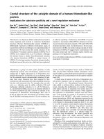
3.5. Histological finding
A cross section of sham rat’s lung showed the normal
appearance of all three parameters (thin and delicate
alveolar septae, no intra-alveolar cell infiltrates and no
alveolar hemorrhage) Figure 8. All rats in this group
showed normal lung appearance (100%) as shown in
table 5.
There was statistically significant difference between
induced untreated (HS) group and sham group (P <
0.05) and the total scor e mean of the HS group showed
mod erate lung injury. 66.7% of t he group had m oderate
lung injury and 33.3% had severe lung injury as shown
in table 5, 6 and Figures 9, 10.
Treatment of rats with MK-886 ameliorated the lung
injury significantly (P < 0.05) as compared with induced
untreated group and the total score mean of this group
showed mild lung injury (Figure 11). 16.7% of the gro up
had normal histopathological appearance and 83.3% of
the group had mild lung injury as shown in table 5.
Discussion
The present study demonstrates that HS causes ALI, as
evidenced by biochemical and histologic changes. MK-
886 prevented the biochemical changes a nd protected
the lung mor phology after HS. Although leukotriene-
shave been known to be associated with the I/R injury
in other tissues, including intestine [24]kidney [25],
myocardium [26] and liver [27], there are only a few
Table 1 Serum TNF-a and IL-6 levels (pg/ml) of the three
experimental groups at the end of the experiment
Group TNF-a (pg/ml) IL-6 (pg/ml)
1. Sham 19.4 ± 2.12 21.16 ± 2.61
2. Control (HS) 93.3 ± 6.48* 44.84 ± 2.33*
3. MK-886 treated group 49.4 ± 3.81
†
29.78 ± 1.27
†
The data expressed as mean ± SEM (N = 6 in each group).
• P < 0.05 vs. sham group,
†
P < 0.05 vs. HS (induced untreated) group
^ĞƌƵŵdE&Ͳɲ;ƉŐͬŵůͿ
Ϭ
ϮϬ
ϰϬ
ϲϬ
ϴϬ
ϭϬϬ
ϭϮϬ
ϭ͘^ŚĂŵ Ϯ͘ŽŶƚƌŽů ϯ͘D<ͲϴϴϲƚƌĞĂƚĞĚŐƌŽƵƉ
Figure 1 The mean of serum TNF-a level (pg/ml) in the three
experimental groups at the end of the experiment.
^ĞƌƵŵ/>Ͳϲ;ƉŐͬŵůͿ
Ϭ
ϱ
ϭϬ
ϭϱ
ϮϬ
Ϯϱ
ϯϬ
ϯϱ
ϰϬ
ϰϱ
ϱϬ
ϭ͘^ŚĂŵ Ϯ͘ŽŶƚƌŽů ϯ͘D<ͲϴϴϲƚƌĞĂƚĞĚŐƌŽƵƉ
Figure 2 The mean of serum IL-6 level (pg/ml) in the three
experimental groups at the end of the experiment.
Table 2 Lung MDA and GSH levels of the three
experimental groups at the end of the experiment
Group Lung MDA (nmol/g) Lung GSH (μmol/g)
1. Sham 95 ± 2.78 4.36 ± 0.27
2. Control (HS) 157 ± 6.15* 2.12 ± 0.25*
3. MK-886 treated group 107.2 ± 3.76
†
3.7 ± 0.35
†
The data expressed as mean ± SEM (N = 6 in each group).
• P < 0.05 vs. sham group,
†
P < 0.05 vs. HS (induced untreated) group
Al-Amran et al. Journal of Cardiothoracic Surgery 2011, 6:81
/>Page 4 of 10
studies describing the correlation b etween hemorrhagic
shock-induced lung injury and 5-lipoxygenase pathway
products, where two studies demonstrated that the 5-
lipoxygenase pathway products meditate acute lung
injury following hemorrhagic shock [28,29]. And it has
been demonstrated that LTB4 levels were significantly
increased in the rat lungs following T/HS [30]. Studies
in humans confirm el evated levels of LTB
4
,LTC
4
,LTD
4
in BAL, pulmonary edema fluid, and plasma in patients
with ALI compared with “ at-risk” group or those with
hydrostatic edema [31,32]. In the present study a signifi-
cant increase i n BALF leukotriene (LTB
4
<C
4
) levels
were found in the shocked rats as compared with sham
group. The incre ased leukotriene level in shocked rats
might be due to the associated splanchnic I/R, which
activates gut PLA
2
-mediated release of AA into the
lymph w here it is delivered to the lungs [33]. Arachido-
nic acid is a biologically active lipid released from the
cellular membrane by PLA
2
that can engage the LTB
4
receptor and initiate LTB
4
production with autocrine
effects [34]. Arachidonic acid also promotes 5-lipoxy-
genase translocation to the nucleus, a key step in leuko-
trienes production [35]. Additionally, it is known that
ischemia elevates cytosolic calcium concentration, which
in turn elevates PLA
2
and lipoxygenase activity, generat-
ing leukotrienes. Furthermore, increased leukotriene
level might be due to the leukocytes accumulated in the
lungs as observed in the histological section of the
shocked rat lung where activated neutr ophils following
hemorrhagic shock are capable of releasing cytotoxic
products including leukotrienes, and the i ntrinsic 5-
lipoxygenase activity is required for neutrophil adher-
ence and chemotaxis and neutrophil-mediated lung
injury [36]. In addition to neu trophils, alveolar macro-
phages and circulating macrophages aggravate lung
injury and alveolar neutrophil sequestration in hemor-
rhagic shock [37] and might contribute to further
release of leukotrienes. In this study we have demon-
strated that treatment with MK-886 appeared to have a
significant decrease in BALF leukotrienes (LTB
4
&
LTC
4
) level in the shocked rats in comparison with the
induced untreated group. It is reported that selective
inhibition of leukotriene biosynthesis by MK-886 pre-
vents postischemic leukotrienes accumulation and the
microcirculatory changes after I/R in the striated muscle
in vivo [14]. Furthermore, MK-886 was found to be a
potent and specific inhibitor of both LTB
4
and LTC
4
synthesis in human phagocytes [9,38].
Hemorrhagic shock is considered as an insult fre-
quently leading to systemic inflammatory response syn-
drome including the systemic release of proinflammatory
cytokines which is central in t he inflammatory response.
Previous studies have shown that levels of IL-6 and TNF-
a significantly increased following trauma-hemorrhage
and remain elevated for several hours [39]. The results in
present study are consistent with that reported by Vin-
cenzi et al. [40] Who found that a significant increase in
the TNF-a and IL-6 levels in shocked rats in comparison
>ƵŶŐD;ŶŵŽůͬŐͿ
Ϭ
ϮϬ
ϰϬ
ϲϬ
ϴϬ
ϭϬϬ
ϭϮϬ
ϭϰϬ
ϭϲϬ
ϭϴϬ
ϭ͘^ŚĂŵ Ϯ͘ŽŶƚƌŽů ϯ͘D<ͲϴϴϲƚƌĞĂƚĞĚŐƌŽƵƉ
Figure 3 The mean of lung MDA level (nmol/g) in the three
experimental groups at the end of the experiment.
>ƵŶŐ'^,;ʅŵŽůͬŐͿ
Ϭ
Ϭ͘ϱ
ϭ
ϭ͘ϱ
Ϯ
Ϯ͘ϱ
ϯ
ϯ͘ϱ
ϰ
ϰ͘ϱ
ϱ
ϭ͘^ŚĂŵ Ϯ͘ŽŶƚƌŽů ϯ͘D<ͲϴϴϲƚƌĞĂƚĞĚŐƌŽƵƉ
Figure 4 The mean of lung GSH level (μmol/g) in the three
experimental groups at the end of the experiment.
Table 3 BALF LTB
4
and LTC
4
level (pg/ml) of the three
experimental groups at the end of the experiment
Group BALF LTB
4
(pg/ml) BALF LTC
4
(pg/ml)
1. Sham 0.42 ± 0.02 0.33 ± 0.05
2. Control (HS) 1.84 ± 0.03* 8.64 ± 0.31*
3. MK-886 treated group 0.37 ± 0.04
†
0.28 ± 0.05
†
The data expressed as mean ± SEM (N = 6 in each group).
• P < 0.05 vs. sham group,
†
P < 0.05 vs. HS (induced untreated) group
Table 4 BALF total protein level (g/l) of the three
experimental groups, at the end of the experiment
Group BALF total protein (g/l)
1. Sham 7.2 ± 0.5
2. Control (HS) 14.7 ± 0.57*
3. MK-886 treated group 8 ± 0.3
†
The data expressed as mean ± SEM (N = 6 in each group).
• P < 0.05 vs. sham group,
†
P < 0.05 vs. HS (induced untreated) group
Al-Amran et al. Journal of Cardiothoracic Surgery 2011, 6:81
/>Page 5 of 10
with sham group. Activated inflammatory cells, especially
macrophages a nd neutrophils have been shown to play a
pivotal role in the propagation of SIRS following resusci-
tated shock and could be considered the main source of
inflammatory cytokines including TNF-a and IL-6. In
this study MK-886 significantly reduced the elevation of
IL-6 and TNF-a levelintheshockedratsascompared
withinduceduntreatedgroupsuggestingthatMK-886
has protective effect in hemorrhagic shock-induced acute
>&>dϰ;ƉŐͬŵůͿ
Ϭ
Ϭ͘Ϯ
Ϭ͘ϰ
Ϭ͘ϲ
Ϭ͘ϴ
ϭ
ϭ͘Ϯ
ϭ͘ϰ
ϭ͘ϲ
ϭ͘ϴ
Ϯ
ϭ͘^ŚĂŵ Ϯ͘ŽŶƚƌŽů ϯ͘D<ͲϴϴϲƚƌĞĂƚĞĚŐƌŽƵƉ
Figure 5 The mean of BALF LTB
4
level (pg/ml) in the three
experimental groups at the end of the experiment.
>&>dϰ;ƉŐͬŵůͿ
Ϭ
ϭ
Ϯ
ϯ
ϰ
ϱ
ϲ
ϳ
ϴ
ϵ
ϭϬ
ϭ͘^ŚĂŵ Ϯ͘ŽŶƚƌŽů ϯ͘D<ͲϴϴϲƚƌĞĂƚĞĚŐƌŽƵƉ
Figure 6 The mean of BALF LTC
4
level (pg/ml) in the three
experimental groups at the end of the experiment.
>&ƚŽƚĂůƉƌŽƚĞŝŶ;ŐͬůͿ
Ϭ
Ϯ
ϰ
ϲ
ϴ
ϭϬ
ϭϮ
ϭϰ
ϭϲ
ϭϴ
ϭ͘^ŚĂŵ Ϯ͘ŽŶƚƌŽů ϯ͘DŬͲϴϴϲƚƌĞĂƚĞĚŐƌŽƵƉ
Figure 7 The mean of BALF total protein level (g/l) in the three
experimental groups at the end of the experiment.
Figure 8 Photomicrograph of lung section of normal rats
shows the normal architecture. The section stained with
Haematoxylin and Eosin (X 10).
Table 5 The differences in histopathological grading of
abnormal lung changes among the three experimental
groups
Histopathological grading Study group
Sham Control (HS) MK-886
N% N % N %
Normal 6 100 0 0 1 16.7
Mild 0 0 0 0 5 83.3
Moderate 0 0 4 66.7 0 0
Severe 0 0 2 33.3 0 0
Total 6 100 6 100 6 100
Figure 9 Photomicrograph of lung section with moderate
injury. The section stained with Haematoxylin and Eosin (X 10).
Al-Amran et al. Journal of Cardiothoracic Surgery 2011, 6:81
/>Page 6 of 10
lung injury. Inhibition of end ogenous CysLT production
by MK-886 significantly attenuated the generation of
TNF-a by mast cells activated by FcεRI cross-linkage
[41]. MK-886 pretreatment attenuated subsequent pul-
monary expression of TNF- a in a mouse model of bron-
chial inflammation and hyperreactivity [42]. LTB
4
augments IL-6 production in human monocytes by
increasing both IL-6 gene transcription and mRNA stabi-
lization [43,44]. activation of NF-BandNF-IL-6tran-
scriptional factors may be important in this enhancement
of IL-6 release [44]. Furthermore, TNF-a production is
enhanced by LT C
4
and LTD
4
[45]. So that, inhibition of
LTB
4
and CysLTs synthesis by MK-886 might result in
lowering TNF-a and IL-6 levels.
Through examination of meta bolic processes, GSH has
been shown to be important in host defenses against oxi-
dative stress [46]. Another important agent showing oxi-
dative stress is MDA, a marker of free radical activity [4].
It was reported that oxidative stress significantly elevated
MDA levels and reduce d GSH levels [47]. Oxidative
stress has been implicated as an important cause of HSR
pathogenesis [2,46]. The result in present study are con-
sistent with other study who found that a significant
increase in lung MDA level and significant decrease in
lung GSH level were found in hemorrhagic shock group
as compared to sham group in a rat model of hemorrha-
gic shock-induced acute lung injury [18]. In this study
MK-886 significantly reduced the elevation of lung MDA
level and significantly elevates the lung GSH level in the
shocked rats as compared with induced untreated group
suggesting that MK-886 has protective effect in hemor-
rhagic shock-induced oxidative i njury of the lung. There
is no data available about the effect of MK-886 on oxida-
tive lung injury in HS. But they found that MK-886 sig-
nificantly reduces hepatic and intestinal MDA level and
elevates GSH level in these organs in rats that underwent
hepatic I/R model and anti-inflammatory properties and
inhibition of lip id peroxidation by MK-886 could be pro-
tective for these organs in I/R injury [18]. The antioxi-
dant effect of MK-886 might be largely due to its
inhibitory action on leukotrienes synthesis.
In the present study a significant increase in the BALF
total protein level was found in the shocked rats as
compared with sham group, suggesting t hat hemorrha-
gic shock induces lung injury in rats. Increased protein
concentration in BALF is an important marker of
damage to the alveolar-capillary barrier of lung. Further-
more, the increase in BALF total protein concentration
may be due to increased lu ng permeabi lity and lung
edema during acute lung injury [48]
The acute phase of ALI and ARDS is characterized by
the influx of protein-rich edema fluid into the air spaces
as a consequence of increased permeability of the alveo-
lar-capillary barrier [49]. As previously reported, T/HS
Table 6 Acute lung injury score
Study group Congestion of alveolar septae Intra-alveolar cell infiltrates Alveolar hemorrhage Total score Total score grade
Sham 0 0 0 0 Normal
HS 1.5 ± 0.34 2.5 ± 0.22 1.83 ± 0.16 5.83 ± 0.60* Moderate
MK-886 treated group 0.5 ± 0.22 0.66 ± 0.21 0.17 ± 0.16 1.33 ± 0.42
†
Mild
The data expressed as means ± SEM.
* P < 0.05 vs. sham group,
†
P < 0.0 5 vs. HS (induced untreated) group
Figure 10 Photomicrograph of lung section with severe injury.
The section stained with Haematoxylin and Eosin (X 40).
Figure 11 Photomicrograph of lung section with mild injury.
The section stained with Haematoxylin and Eosin (X 40).
Al-Amran et al. Journal of Cardiothoracic Surgery 2011, 6:81
/>Page 7 of 10
caused lung injury as reflected in increased permeability
to Evans blue dye, BALF protein levels and the BALF to
plasma protein ratio [50,51]. Two studies showed that
hemorrhagic shock significantly increases BALF total pro-
tein in the rats and mice [20,29]. CysLTs mediate
increased permeability leading to leukocyte extravasation,
plasma exudation and edema[52, 53, and 54]. Further-
more, LTB
4
increases the expression of CD11b/CD18 b2-
integrin (Mac-1) on neutrophils, which can facilitate neu-
trophil adherence and migration [55] and enhanced leuko-
cyte adhesivity accounts for capillary obstruction after I/R
[56]. T/HS lymph induces an increase in endothelia l per-
meability by triggering the release of IL-6 [57]. It has been
demonstrated that IL-6 is an important autocrine factor
produced by endothelial cells that contributes to the
increase in endothelial permeability during hypoxia [58].
Free radicals are implicated to damage biomembranes,
thereby compromising cell integrity and function [59].
Besides increasing pulmo nary arterial pressure [60], the
free radical production under hypoxic environment may
cause oxidative injury of the endothelium [61], resulting in
increased pulmonary capillary permeability. In this study
treatment with MK-886 appeared to have a significant
decrease in BALF total protein level in the shocked rats in
comparison with the induced untreated group. MK-886
has been shown to reduce the extravasation of plasma [13]
and prevent the leukocyte adhesion to the endothelium
[14] in experimental animals. It was demonstrated that
treatment of mice with MK-886 significantly abolished the
increase in the BALF total protein level in acute lung
injury following hemorrhagic shock [29].
Morphologically, there was a statistically significant dif-
ference between induced untreated group and sham
group and the total score mean of the HS group shows
moderate lung injury. 66.7% of the HS group had moder-
ate lung injury and 33.3% had severe lung injury. Treat-
ment of rats with MK-886 ameliorates the lung injury
significantly as compared w ith induced untreated group
and the total score mean of the control group shows
mild lung injury. Although there is no data available
about the protective effect of MK-886 on the lung par-
enchyma in HS rats, but they found that MK-886 signifi-
cantly reduces the histological changes in the liver and
small intestine of rats that underwent hepatic I/R model
(15). M K-886 was able to reduce the cortical infarct size
by 30% in a model of focal cerebral ischemia in rats [62].
Furthermore, a separate research work found that treat-
ment of rats with MK-886 reduces brain lesion volume in
experimental traumatic brain injury model [63].
Author details
1
Department of Surgery, Colorado Denver university, Box C-320 12700 E 19
th
Avenue, Aurora, CO 80045 USA.
2
Department of pharmacy, Kufa university,
Najaf kufa street, Najaf, Iraq.
Authors’ contributions
FG carried out the surgical experimental work and gives the outline of
research. NR participated in the design of the study and performed the
statistical analysis and supervised main skeleton. AM participated in the
sequence alignment and drafted the manuscript and did all the biochemical
and histopathological tests.
All authors read and approved the final manuscript.
Competing interests
The authors participated in the design of the study and performed the
statistical analysis declare that they have no competing interests.
Received: 21 February 2011 Accepted: 7 June 2011
Published: 7 June 2011
References
1. Bhatia M, Moochhala S: Role of inflammatory mediators in the
pathophysiology of acute respiratory distress syndrome. J Pathol 2004,
202:145-56.
2. Jarrar D, Chaudry IH, Wang P: Organ dysfunction following hemorrhage
and sepsis: mechanisms and therapeutic approaches. Int J Mol Med 1999,
4:575-583.
3. Hudson LD, Milberg JA, Anardi D, Maunder RJ: Clinical risks for
development of the acute respiratory distress syndrome. Am J Respir Crit
Care Med 1995, 151:293-301.
4. Keel M, Trentz O: Pathophysiology of polytrauma. Injury 2005, 36:691-709.
5. Anaya-Prado R, Toledo-Pereyra LH, Lentsch AB, Ward PA: Ischemia/
reperfusion injury. J Surg Res 2002, 105:248-258.
6. Rizoli SB, Kapus A, Fan J, Li YH, Marshall JC, Rotstein OD:
Immunomodulatory effects of hypertonic resuscitation on the
development of lung inflammation following hemorrhagic shock. J
Immunol 1998, 161:6288-6296.
7. Yu HP, Shimizu T, Hsieh YC, Suzuki T, Choudhry MA, Schwacha MG, et al:
Tissue specific expression and their role in the regulation of neutrophil
infiltration in various organs following trauma-hemorrhage. J Leukoc Biol
2006, 79:963-970.
8. Yu HP, Hsieh YC, Suzuki T, Shimizu T, Choudhry MA, Schwacha MG, et al:
Salutary effects of estrogen receptor-β agonist on lung injury after
trauma-hemorrhage. Am J Physiol Lung Cell Mol Physiol 2006, 290:
L1004-L1009.
9. Gillard J, Ford-Hutchinson AW, Chan C, Charleson S, Denis D, Foster A, et al:
Full-size imageL-663,536 (MK-886) (3-1-(4-chlorobenzyl)-3-t-butyl-thio-5-
isopropylindol-2-yl) 2,2-dimethylpro-panoic acid), a novel, orally active
leukotriene biosynthesis inhibitor. Can J Physiol Pharmacol 1989,
67(5):456-464.
10. Dixon RAF, Diehl RE, Opas E, Rands E, Vickers PJ, Evans JF, et al:
Requirement of a 5-lipoxygenase activating protein for leukotriene
synthesis. Nature 1990, 343:282-284.
11. Rouzer CA, Ford-Hutchinson AW, Morton HE, Gillard JW: MK-886, a potent
and specific leukotriene biosynthesis inhibitor, blocks and reverses the
membrane association of 5-lipooxygenase in ionophore challenged
leucocytes. J Biol Chem 1990, 265:1436-1442.
12. Guidot DM, Repine MJ, Westcott JY, Repine JE: Intrinsic 5-lipoxygenase
activity is required for neutrophil responsivity. Proc Natl Acad Sci USA
1994, 91:8156-8159.
13. Fernandez-gallardo S, Gijon MA, Garcia C, Furio V, Ciu FT, Crespo SM: The
role of platelet activating factor and peptidoleukotrienes in the vascular
changes of rat passive anaphylaxis. Br J Pharmacol 1992, 105:119-125.
14. Lehr HA, Guhlmann A, Nolte D, Keppler D, Messmers K:
Leukotrienes as
mediators
in ischemia-reperfusion injury in a microcirculation model in
the hamster. J Clin Invest 1991, 87:2036.
15. Daglar G, Karaca T, Yuksek YN, Gozalan U, Akbiyik F, Sokmensuer C, et al:
Effect of Montelukast and MK-886 on Hepatic Ischemia-Reperfusion
Injury in Rats. Journal of surgical research 2009, 153(1):31-38.
16. Wallace JL, Keenan CM: An orally active inhibitor of leukotriene synthesis
accelerates healing in rat model of colitis. Am J Physiol 1990, 258:
G527-G534.
17. Eun JC, Moore EE, Mauchley DC, Meng X, Banerjee A: The 5-Lipoxygenase
Pathway Meditates Acute Lung Injury Following Hemorrhagic Shock.
Journal of Surgical Research 2010, 158(2):215-216.
Al-Amran et al. Journal of Cardiothoracic Surgery 2011, 6:81
/>Page 8 of 10
18. Kilicoglu B, Eroglu E, Kilicoglu SS, Kismet K, Eroglu F: Effect of abdominal
trauma on hemorrhagic shock induced acute lung injury in rats. World J
Gastroenterol 2006, 12(22):3593-3596.
19. Rhee P, Waxman K, Clark L, Kaupke CJ, Vaziri ND, Tominaga G, et al: Tumor
necrosis factor and monocytes are released during hemorrhagic shock.
Resuscitation 1993, 25(3):249-255.
20. Yu HP, Hsieh PW, Chang YJ, Chung PJ, Kuo LM, Hwang TL: DSM-RX78, a
new phosphodiesterase inhibitor, suppresses superoxide anion
production in activated human neutrophils and attenuates hemorrhagic
shock-induced lung injury in rats. Biochemical pharmacology 2009,
78(8):983-992.
21. Josephson B, Gyllenswärd C: Scand J Clin Lab Invest 1957, 9:29.
22. Beuge JA, Aust SD: Microsomal lipid peroxidation. Meth Enzymol 1978,
52:302-311.
23. Matute-Bello G, Winn RK, Jonas M, Chi EY, Martin TR, Liles WC: Fas (CD95)
induces alveolar epithelial cell apoptosis in vivo: Implications for acute
pulmonary inflammation. Am J Pathol 2001, 158:153.
24. Souza DG, Coutinho SF, Silveira MR, Cara DC, Teixeira MM: Effects of a BLT
receptor antagonist on local and remote reperfusion injuries after
transient ischemia of the superior mesenteric artery in rats. Eur J
Pharmacol 2000, 403:121.
25. Noiri E, Yokomizo T, Nakao A, Izumi T, Fujita T, Kimura S, Shimizu T: An in vivo
approach showing the chemotactic activity of leukotriene B (4) in acute
renal ischemic-reperfusion injury. Proc Natl Acad Sci USA 2000, 97:823.
26. Rossoni G, Sala A, Berti F, Testa T, Buccellati C, Molta C, et al: Myocardial
protection by the leukotriene synthesis inhibitor BAY X1005: Importance
of transcellular biosynthesis of cysteinyl-leukotrienes. J Pharmacol Exp
Ther 1996, 276:335.
27. Takamatsu Y, Shimada K, Chijiiwa K, Kuroki S, Yamaguchi K, Tanaka M: Role
of leukotrienes on hepatic ischemia/reperfusion injury in rats. Journal of
Surgical Research 2004, 119(1):14-20.
28. Eun JC, Moore EE, Jordan JR, Peltz ED, Banerjee A: Products of the 5-
lipoxygenase pathway are critical for the development of acute lung
injury following hemorrhagic shock. Journal Of the American College of
Surgeons 2009, 209(3):S35, Suppl 1.
29. Eun JC, Moore EE, Mauchley DC, Meng X, Banerjee A: The 5-Lipoxygenase
Pathway Meditates Acute Lung Injury Following Hemorrhagic Shock.
Journal of Surgical Research 2010, 158(2):215-216.
30. Jordan JR, Moore EE, Damle SS, Kashuk SB, Silliman CC, et al: Arachidonic
acid in postshock mesenteric lymph induces pulmonary synthesis of
leukotriene B
4.
. J Appl Physiol 2008, 104:1161-1166.
31. Amat M, Barcons M, Mancebo J, Mateo J, Oliver A, Mayoral JF, et al:
Evolution of leukotriene B4, peptide leukotrienes, and interleukin-8
plasma concentrations in patients at risk of acute respiratory distress
syndrome and with acute respiratory distress syndrome: mortality
prognostic study. Crit Care Med 2000, 28:262-263.
32. Matthay MA, Eschenbacher WL, Goetzl EJ: Elevated concentrations of
leukotriene D
4
in pulmonary edema fluid of patients with the adult
respiratory distress syndrome. J Clin Immunol 1984, 4:479-483.
33. Partrick D, Moore EE, Moore FA, Barnett CC, Silliman CC: Lipid mediators
up-regulate cd11b and prime for concordant superoxide and elastase
release in human neutrophils. J Trauma 1997, 43:297-303.
34. Surette ME, Krump E, Picard S, Borgeat P: Activation of leukotriene
synthesis in human neutrophils by exogenous arachidonic acid:
inhibition by adenosine A2a receptor agonists and crucial role of
autocrine activation by leukotriene B4. Mol Pharmacol 1999, 56:1055-1062.
35. Murphy RC, Gijon MA: Biosynthesis and metabolism of leukotrienes.
Biochem J 2007, 405:379-395.
36. Guidot DM, Repine MJ, Westcott JY, Repine JE: Intrinsic 5-lipoxygenase
activity is required for neutrophil responsivity. Proc Natl Acad Sci USA
1994, 91:8156-8159.
37. Fan J, Marshall JC, Jimenez M, Shek PN, Zagorski J, Rotstein OD:
Hemorrhagic shock primes for increased expression of cytokine-induced
neutrophil chemoattractant in the lung: role in pulmonary inflammation
following lipopolysaccharide. J Immunol 1998, 161(1):440-447.
38. Menard L, Pilote S, Naccache PH, Laviolette M, Borgeat P: Inhibitory effects
of MK-886 on arachidonic acid metabolism in human phagocytes. Br J
Pharmacol 1990, 100:15-20.
39. Ayala A, Wang P, Ba ZF, Perrin MM, Ertel W, Chaudry IH: Differential
alterations in plasma IL-6 and TNF levels after trauma and hemorrhage.
Am J Physiol 1991, 260:R167-R171.
40. Vincenzi R, Cepeda LA, Pirani WM, Sannomyia P, Rocha-e-Silva M, Cruz RJ Jr:
Small volume resuscitation with 3% hypertonic saline solution decrease
inflammatory response and attenuates end organ damage after
controlled hemorrhagic shock. The American Journal of Surgery 2009,
198(3):407-414.
41. Mellor EA, Austen KF, Boyce JA: Cysteinyl leukotrienes and uridine
diphosphate induce cytokine generation by human mast cells through
an interleukin 4-regulated pathway that is inhibited by leukotriene
receptor antagonists. J Exp Med 2002, 195:583.
42. Oliveira SH, Hogaboam CM, Berlin A, Lukacs NW: SCF-induced airway
hyperreactivity is dependent on leukotriene production. Am J Physiol
Lung Cell Mol Physiol 2001, 280:L1242-1249.
43. Rola-Pleszczynski M, Stankova J: Leukotriene B
4
enhances interleukin-6 (IL-
6) production and IL-6 messenger RNA accumulation in human
monocytes in vitro: transcriptional and posttranscriptional mechanisms.
Blood 1992, 80:1004-1011.
44. Brach MA, de Vos S, Arnold C, Gruss HJ, Mertelsmann R, Herrmann F:
Leukotriene B
4
transcriptionally activates interleukin-6 expression
involving NK-κB and NF-IL6. Eur J Immunol 1992, 22:2705-2711.
45. Ben-Efraim B, Bonta IL: Modulation of antitumour activity of macrophages
by regulation of eicosanoids and cytokine production. Int J
Immunopharmacol 1994, 16:397-399.
46. Szabo C: The pathophysiological role of peroxynitrite in shock,
inflammation, and ischemia-reperfusion injury. Shock 1996, 6:79-88.
47. Johnson KJ, Fantone JC, Kaplan J, Ward PA: In vivo damage of rat lungs
by oxygen metabolites. J Clin Invest 1981, 67:983-993.
48. Lum H, Roebuck KA: Oxidant stress and endothelial dysfunction. Am J
Physiol Cell Physiol 2001, 280:C719-C741.
49. Pugin J, Verghese G, Widmer M-C, Matthay MA: The alveolar space is the
site of intense inflammatory and profibrotic reactions in the early phase
of acute respiratory distress syndrome. Crit Care Med 1999, 27:304-312.
50. Magnotti LJ, Upperman JS, Xu DZ, Lu Deitch EA Q: Gut-derived mesenteric
lymph but not portal blood increases endothelial cell permeability and
promotes lung injury after hemorrhagic shock. Ann Surg 1998,
228:518-527.
51. Deitch EA, Adams C, Lu Q, Xu DZ: A time course study of the protective
effect of mesenteric lymph duct ligation on hemorrhagic shock-induced
pulmonary injury and the toxic effects of shocked rats on endothelial
cell monolayer permeability. Surgery 2001, 129:39-47.
52. Funk CD: Prostaglandins and leukotrienes: advances in eicosanoid
biology. Science 2001, 294:1871-1875.
53. Dahlén SE: Treatment of asthma with antileukotrienes: first line or last
resort therapy? Eur J Pharmacol 2006, 533:40-56.
54. Ogawa Y, Calhoun WJ: The role of leukotrienes in airway inflammation. J
Allergy Clin Immunol 2006, 118:789-798.
55. Crooks SW, Stockley RA: Leukotriene B4. Int J Biochem Cell Biol 1998,
30(2):173-178.
56. Schmid-Schonbein GW: Capillary plugging by granulocytes and the no-
reflow phenomenon in the microcirculation. Fed Proc 1987, 46:2397-2401.
57. Dayal SD, Haskó G, Lu Q, Xu DZ, Caruso JM, Sambol JT, et al: Trauma/
Hemorrhagic Shock Mesenteric Lymph Upregulates Adhesion Molecule
Expression and IL-6 Production in Human Umbilical Vein Endothelial
Cells. Shock 2002, 17(6):491-495.
58. Ali MH, Schlidt SA, Chandel NS, Hynes KL, Schumacker PT, Gewertz BL:
Endothelial
permeability and IL-6 production during hypoxia: role of
ROS in signal transduction. Am J Physiol 1999, 277:L1057-L1065.
59. Vanita G, Asheesh G, Shalini S, Harish MD, Grover SK, Ratan K: Anti-stress
and adaptogenic activity of L-arginine supplementation. eCAM 2005,
2:93-97.
60. Hoshikawa Y, Sadafumi O, Satoshi S, Tatsuo T, Masayuki C, Chun S, et al:
Generation of oxidative stress contributes to the development of
pulmonary hypertension induced by hypoxia. J Appl Physiol 2001,
90:1299-1306.
61. Herget J, Wilhelm J, Novotna J, Eckhardt A, Vytasek R, Mrazkova L, et al: A
possible role of the oxidant tissue injury in the development of hypoxic
pulmonary hypertension. Physiol Res 2000, 49:493-501.
Al-Amran et al. Journal of Cardiothoracic Surgery 2011, 6:81
/>Page 9 of 10
62. Ciceri P, Rabuffetti M, Monopoli A, Nicosia S: Production of leukotrienes in
a model of focal cerebral ischemia in the rat. Br J Pharmacol 2001,
133:1323.
63. Farias S, Frey LC, Murphy RC, Heidenreich KA: Injury-Related Production of
Cysteinyl Leukotrienes Contributes to Brain Damage following
Experimental Traumatic Brain Injury. Journal of Neurotrauma 2009,
26(11):1977-1986.
doi:10.1186/1749-8090-6-81
Cite this article as: Al-Amran et al .: Leukotriene biosy nthesis inhibition
ameliorates acute lung injury following hemorrhagic shock in rats.
Journal of Cardiothoracic Surgery 2011 6:81.
Submit your next manuscript to BioMed Central
and take full advantage of:
• Convenient online submission
• Thorough peer review
• No space constraints or color figure charges
• Immediate publication on acceptance
• Inclusion in PubMed, CAS, Scopus and Google Scholar
• Research which is freely available for redistribution
Submit your manuscript at
www.biomedcentral.com/submit
Al-Amran et al. Journal of Cardiothoracic Surgery 2011, 6:81
/>Page 10 of 10