Báo cáo y học: "Cor triatriatum presenting as heart failure with reduced ejection fraction: a case report" pptx
Bạn đang xem bản rút gọn của tài liệu. Xem và tải ngay bản đầy đủ của tài liệu tại đây (1.56 MB, 4 trang )
CAS E REP O R T Open Access
Cor triatriatum presenting as heart failure with
reduced ejection fraction: a case report
John Kokotsakis
1
, Vania Anagnostakou
2*
, George Almpanis
3
, Ioannis Paralikas
1
, Ioannis Nenekidis
1
,
Theodoros Kratimenos
2
, Efi Prapa
1
, Nikolitsa Tragotsalou
3
, Achilleas Lioulias
1
and Andreas Mazarakis
3
Abstract
Cor triatriatum is a rare congenital cardiac malformation and it usually refers to the left atrium. We report an
unusual case of cor triatriatum in a 33 - year old woman pres ented with congestive heart failure caused by left
ventricular systolic dysfunction.
Background
Cor triatriatum is a rare congenital cardiac malforma-
tion with an estimated incidence of 0,1% of all congeni-
tal heart disease and it usually refers to the left atrium
(cor triatriatum sinister). In cor triatriatum sinist er the
left atrium is divided by a fibromuscular membrane into
two distinct chamb ers: a posterior - superior chamber
receiving the four pulmonary veins and an anterior -
inferior chamber ( true left atrium ) that connects to the
left ventricle by means of the mitral valve [1]. In the
majority of cases it is diagnosed in neonatal p eriod or
early infancy, w hereas adult cases are very rare. We
report an unusual case of cor tria triatum in a 33 - year
old woman presented with congestive heart failure
caused by left ventricular systolic dysfunction.
Case presentation
A 33 - year old woman presented to o ur cardiology ser-
vice with signs and symptoms of congestive heart fail-
ure. Her medical history was unremarkable, however a
year ago and soon after her third child delivery, she had
been admitted in another hospital for acute pulmonary
oedema after labor. Cor triatriatum with obstructive
behavior causing pulmonary hypertension had bee diag-
nosed, while the left ventricle was structurally and func-
tionally intact. The patient at that time denied surgey
and had been discharged on medical therapy. At present
admission the patient presented with NYHA functional
class III, symptoms of heart failure and palpittions as a
result of persistent atrial flutter. On physical examina-
tion a loud pulmonary component of the 2
nd
heart
sound and a diastolic murmur was heard in the mitral
area. Signs of right-sided heart failure were absent.
A transthoracic echocardiography revealed a moder-
ately dilated left ventricle (LV), globally hypokinetic,
with severely impaired systolic function (EF estimated
≥30%). Left atrium (LA) was dilated, with a mobile,
membrane-like echogenic structure into it.
Transesophageal echocardiogram (TEE ) documented a
fibromuscular membrane across the LA, dividing it into
two compartments, a proximal one receiving the pul-
monary venous flow and a distal one containing the left
atrial appendage (LAA). The two chambers communi-
cated via a non-restrictive orifice, but the membrane pro-
lapsed towards the mitral valve inflow causing severe
obstruction. Mitral valve appeared normal, with mild
regurgitation. Patent foramen ovale (PFO), atrial septal
defect (ASD) and anomalous venous connections were
ruled out and the diagnosis of cor triatriatum was recon-
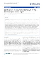
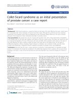
firmed. Magnetic resonance imaging ( MRI) of the heart
also revealed the fibromuscular septum into the left
atrium and the low left ventricular ejection fraction
[(LVEF) 30%, cardiac index 1,6 L/min/m
2
, cardiac output
2,7 L/min] (figure 1). Coronary angiography showed nor-
mal coronary arteries. With these findings the patient
was scheduled for surgery.
Anesthetic induction was achieved with standar d tech-
nique including administration of sodium pentothal,
sevofluorane, fentanyl and muscle relaxant. Invasive
monitoring included the use of right radial arterial lines,
a pulmonary artery catheter and a foley catheter with
temperature probe to measure bladder temperature as an
* Correspondence:
2
Radiology Department, Evaggelismos General Hospital, Athens, Greece
Full list of author information is available at the end of the article
Kokotsakis et al. Journal of Cardiothoracic Surgery 2011, 6:83
/>© 2011 Ko kotsakis et al; licensee BioMed Central Ltd. This is an Open Access article distri buted under the terms of the Creative
Commons Attributio n License (http://creative commons.org/licenses/by/2.0), which permits unre stricted use, distribu tion, and
reproduction in any medium, provided the original work is properly cited.
indicator of core body temperature. Transesophageal
echocardiography (TEE) was also instituted. Surgery was
performed through a median sternotomy. Connection to
cardiopulmonary bypas s (CPB) was achieved by standard
ascending aorta and bicaval cannulation. Mildly
hypothermic (32°C) CPB was established. Cold blood car-
dioplegia was administered in an antegrade fashion
through the aortic root after clamping the aorta. The
interatrial groove was developed and the common pul-
monary venous chamber of the left atrium was opened
through a vertical incision anterior to the right pulmon-
ary veins, exactly as for mitral valve surgery. After inser-
tion of a self-retaining retractor to facilitate exposure, the
diaphragm was exposed and the central hole in it was
identified. A preliminary incision out from the hole
improved exposure for the definitive excision. Orifi ces of
the pulmonary veins on both sides were located. Position
of the atrial septum was also identified by a small open-
ing in the right atrium and by inserting a curved clamp
to displace the s eptum into the common pulmonary
venous chamber of the left atrium. There was no atrial
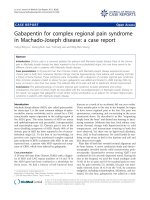
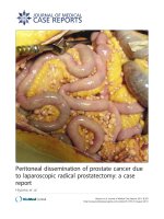
septal defect or patent foramen ovale. The diaphragm
was then easily completely excised exposing the mitral
valve (figure 2). The left atrial appendage was closed
from inside the left atrium using a running 3-0 polypro-
pylene suture to prevent future thrombus formation. The
atriotomy incisions were closed, the heart having been
filled with blood before the last few sutures were placed.
The patient was rewarmed, the aortic cross-clamp was
removed and additional de-airing was carried out in the
usual manner. CPB was terminated with minimal inotro-
pic support, invo lving milrinone and levophed with good
hemodynamics.
The p ostoperative course was uneventful and the
patient was extubated after 12 hours and discharged
from the hospital on the fifth postoperative day. At 3
months follow-up, the patient was asymptomatic
(NYHA class I), in sinus rhythm. TTE and MRI revealed
a mildly dilated LV with great improvement in systolic
function and an estimated LVEF of 50%.
Discussion
Cor triatriatum is a rare co ngenital anomaly with a ratio
of men to women of 1.5:1 [2]. In cor triatriatum the
right and left pulmonary veins can be considered as not
joining the left atrium but rather as entering a chamber
posterior and a little superior or medial to the l eft
atrium that is analogous to the common pulmonary
Figure 1 MR of the heart cine 4-chamber view (left) showing a f ibro muscular septum into the left atrium dividing it into two
compartments which communicate via a central orifice (left). Mid-esophageal (ME) 4-chamber view (right) showing the membrane coursing
transversely into the left atrium (right).
Figure 2 Surgical image of the membrane in the left atrium with an eccentric opening (left). Completely resected membrane (right).
Kokotsakis et al. Journal of Cardiothoracic Surgery 2011, 6:83
/>Page 2 of 4
venous sinus found in patients with total or partial
anomalous pulmonary connection. However the pul-
monary veins in c or triatriatum are incorporated into
the structure of the left atrium, wh ereas in total anoma-
lous pulmonary venous connection the pulmonary veins
connect to sites separate from the left atrium. O ther
associated anomalies are the unrooted coronary sinus
with a left superior vena cava joining the left atrium,
ventricular septal defect, coarctation of the aorta, atrio-
ventricular septal defect, tetra logy of Fallot and rarely
asplenia and polysplenia. No genetic predisposition has
been linked to this particular anomaly. The clinical fea-
tures on presentation can mimic those of mitral steno-
sis, supravalvular mitral ring, left atrial thombus or
pulmonary venous stenosis, since these entities share a
common haemodynamic pathophysiology of flow
obstruction between the pulmonary venous system and
the left ventricle. The most com mon presenting s ymp-
toms in adults are dyspnea, hemoptysis, orthopnea as a
result of the obstructive function of the intra-atrial
membrane [3]. Several techniques have been used for
diagnosis establishment such as TTE, TEE, CT, MRI.
The use of CT bares the risk of radiation, while TEE the
discomfort of scope intubation. MR imaging when com-
pared with ec hocardiography and ca rdiac angiography
was found to have a higher detection rate [4]. In addi-
tion MR fast gradient-recalled echo imaging of the car-
diac cycle has been shown to be of better benefit in the
asse ssment of cardiac function and has been established
asthemodalityofchoisefortheassessmentofLVEF
[5]. According to Loeffler’s classification of the lesion,
group 3 lesions have large openings in the membrane,
leading to little or no obstruction [6]. Patients with
group 3 lesions can survive into adulthood with minor
or no symptoms at all, as in the case of our patient.
Late clinical presentations and conversion to a sympto-
matic state may be due to fibrosis and calcification of
the orifice of the sept um, onset of atrial flutter and
fibrillation with rapid ventricular response, development
of mitral regurgitation. Asymptomatic patients with an
incidental diagnosis and a non-restrictive opening of the
intra-atrial diaphragm, can be observed and followed-up
regularly by TTE or MRI [7]. For symptomatic patients,
surgical excision is the definite treatment, eventhough
successful balloon catheter dilatation of the communica-
tion between the two chambers has been described [8].
Our p atient had two previous uneventful pregnancies
and experienced acute heart failure symptoms in the
early postpartum period of her third normal pregnancy.
The increased demands of pregnancy induce an even
greater pressure gradient between the left cardiac cham-
bers and thus a greater elevation of left atrial pressure,
causing a decompensation of the patient’spreviously
compensated cardiac function. However, s evere systolic
dysfunction causing symptomatic heart failure, to the
best of our knowledge, has never been reported in
patients with cor triatriatum.
Conclusion
The presence of normal coronary anatomy and the
exclusion of cardiomyopathies, using CMR, combined
with the rapid recovery after surgical correction, leads
us to believe that there is a causal relationship among
these entities. Pronounced preload mismatch due to
severe membrane prolapse in the LV inflow, combined
with the sequential volume changes during pregnancies,
leaded to decompensation and s ystolic dysfunction.
Membrane surgical excursion le aded to rapid recover y.
Peripartum cardiomyopathy seems highly unlikely, due
to late onset, and rapid postoperative recovery.
Consent
Written informed consent was obtained from the patient
for publication of this case report and any accompany-
ing images. A co py of the written consent is available
for review by the Editor-in-Chief of this journal.
Author details
1
Cardiac Surgery Department, Evaggelismos General Hospital, Athens,
Greece.
2
Radiology Department, Evaggelismos General Hospital, Athens,
Greece.
3
1
st
Cardiology Department, Agios Andreas General Hospital, Patra,
Greece.
Authors’ contributions
All authors have made substantial contributions to conception and design,
or acquisition of data, or analysis and interpretation of data and have been
involved in drafting the manuscript or revising it critically for important
intellectual content. All authors read and approved the final manuscript. JK,
VA, IN: Manuscript Preparation, Study Design, Data Interpretation, Literature
Search; GA, IP, NT, EP, TK: Manuscript Preparation, Literature Search, Data
Acquisition; AL, AM: Manuscript Preparation, Study Design and coordination.
Competing interests
The authors declare that they have no competing interests.
Received: 8 October 2010 Accepted: 14 June 2011
Published: 14 June 2011
References
1. Rorie M, Xie GY, Miles H, Smith MD: Diagnosis and surgical correction of
cor triatriatum in an adult: combined use of transesophageal
echocardiography and cathetirazation. Cathet Cardiovasc Interv 2000,
51:83-6.
2. Chieh-Shou Su, Tsai I-Chen, Wei-Wen Lin, Tain Lee, Ting Chih-Tai, Kae-
Woei Liang: Usefulness of multidetector-row computed tomography in
evaluating adult cor triatriatum. Tex Heart Inst J 2008, 35:349-51.
3. Krasermann Z, Scheld HH, Tjan TD, Krasermann T: Cor-triartriatum review.
Hertz 2007, 32:506-510.
4. Masui T, Seelos KC, Kersting-Sommerhoff BA, Higgins CB: Abnormalities of
the pulmonary veins: Evaluation with MR imaging and comparison with
cardiac angiography and echocardiography. Radiology 1991, 181:645-649.
5. Pennell DJ, Sechtem UP, Higgins CB, Manning WJ, Pohost GM,
Rademakers FE, van Possum AC, Shaw LJ, Yucel EK: Clinical indications for
cardiovascular magnetic resonance (CMR): consensus panel report. Eur
Heart J 2004, 25(21):1940-65.
6. Loeffler E: Unusual malformation of the left atrium; pulmonary sinus.
Arch Pathol (Chic) 1949, 48:371-6.
Kokotsakis et al. Journal of Cardiothoracic Surgery 2011, 6:83
/>Page 3 of 4
7. Bucholtz S, Jenni R: Doppler echocardiographic findings in 2 identical
variants of a rare cardiac anomaly “subtotal” cor triatriatum: a critical
review of the literature. J Am Soc Echocardiogr 2001, 14:846-49.
8. Papagiannis J, Harrison JK, Hermiller JB, Harding MB, Armstrong BE,
Ungerleider RM, Bashore TM: Use of balloon occlusion to improve
visualization of anomalous pulmonary venous return in an adult with
cor triatriatum. Cathet Cardiovasc Diagn 1992, 25:323-6.
doi:10.1186/1749-8090-6-83
Cite this article as: Kokotsakis et al.: Cor triatriatum presenting as heart
failure with reduced ejection fraction: a case report. Journal of
Cardiothoracic Surgery 2011 6:83.
Submit your next manuscript to BioMed Central
and take full advantage of:
• Convenient online submission
• Thorough peer review
• No space constraints or color figure charges
• Immediate publication on acceptance
• Inclusion in PubMed, CAS, Scopus and Google Scholar
• Research which is freely available for redistribution
Submit your manuscript at
www.biomedcentral.com/submit
Kokotsakis et al. Journal of Cardiothoracic Surgery 2011, 6:83
/>Page 4 of 4