Báo cáo y học: "A silent gigantic solitary fibrous tumor of the pleura: case report." potx
Bạn đang xem bản rút gọn của tài liệu. Xem và tải ngay bản đầy đủ của tài liệu tại đây (1.84 MB, 4 trang )
CAS E REP O R T Open Access
A silent gigantic solitary fibrous tumor of the
pleura: case report
Nobuyuki Furukawa
1*
, Bert Hansky
1
, Jost Niedermeyer
2
, Jan Gummert
1
and Andre Renner
1
Abstract
Solitary fibrous tumor of the pleura is a rare mesenchymal tumor, representing less than 5% of all neoplasms
associated with the pleura. A 57-year-old man had general malaise without chest symptoms for 1 month. A chest
roentgenogram and computed tomography showed a giant mass in the left thorax. Although the tumor
compressed the descending aorta and other mediastinal structures strongly, thereby shifting them to the right
side, the patient had no symptoms except malaise. The tumor was successfully resected via two separate
thoracotomies. The tumor was measured (20 cm × 19 cm × 15 cm) and weighed (2150 g). The tumor was
histologically and immunohistochemically diagnosed as benign. Although SFT is benign, a long follow-up period is
essential as even patients with complete resection are at risk of recurrence many years after surgery.
Background
Solitary fibrous tumors (SFT) of the pleura are rare
intrathoracic neoplasm. Immunohistoche mical analysi s
has confirmed that SFTs originate from mesenchyme
underlying the mesothelial layer of the pleura. Although
they are usually asymptomatic, larger tumors occupying a
large space in the thoracic cavity, present more commonly
with symptoms such as dyspnea, chest pain and malaise.
Although the tumor was large enough to push the des-
cending aorta and other m ediastinal structures to the
right, our patient displayed no symptoms other than
malaise. We successfully resected the huge tumor via two
separate thoracotomies. One year later, the patient is in
good health without tumor recurrence.
Case presentation
A 57-year-old man was referred to a hospital because of
progressive general malaise for a month. His medical his-
tory was unremarkable and he had no history of exposure
to asbestos. At physical examination, breath sounds were
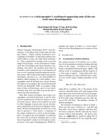
absent on the left lower region. A roentgenogram showed
a giant tumor in the left thorax (Figure 1A). The heart
appeared to be compressed towards the right side. He had
no other chest complaints, such as cough, chest pain, and
dyspnea. Computed tomography (CT) revealed a well-
circumscribed homogeneous mass, which compressed the
descending aorta (Figure 1B). The hematological and bio-
chemical findings were normal. Bronchofiberoscopy
showed stenosis of the left lower lobar bronchus from
extraluminal compression. Bronchoscopic cytology
revealed no abnormal findings and no evidence of bron-
chitis. CT-guided biopsy demonstrated fibrotic soft tissue
without evidence of malignancy but the appearance of the
specimen did not have enough diagnostic strength. Spiro-
metry showed the following results: vital capacity, 2.4 L
(49% of predicted); forced expiratory volume in a second,
1.7 L (42% of predicted). Results of blood gas a nalysis
were also within normal limits. The patient was referred
to our institution for surgical treatment of a suspected
SFT.
Left posterolateral thoracotomy through the fifth and
eighth intercostal spaces was performed for the resect ion
ofthetumor.Wechoosethefifthintercostalspaceas
our initial Thoracotomy site. Upon entering the pleura
we could easily visualize the encapsulated circumscribed
gigantic tumor. The tumor was large (20 cm × 19 cm ×
15 cm), extended from the thoracic aperture to the dia-
phragm, and caused atelectasis of the lower lobe of the
left lung. An additional incision through the eighth inter-
costal space was made to dissect the tumor away from
the diaphragm. Because the tumor had strongly a ttached
to the lingula of the left lung, atypical wedge resection of
the lingula was performed. The main vascular pedicle of
the tumor was identified in the hilum of the lung. There
* Correspondence:
1
Department of Cardiothoracic Surgery, Heart and Diabetes Center North
Rhine-Westphalia, Georgstr. 11, 32545 Bad Oeynhausen, Germany
Full list of author information is available at the end of the article
Furukawa et al. Journal of Cardiothoracic Surgery 2011, 6:122
/>© 2011 Furukawa et al; licensee BioMed Central Ltd. This is an Open Access article distributed under the terms of the Creative
Commons Attribution License (http:/ /creativecommons.org/l icenses/by/2.0), which permits unrestricted use, distribution, and
reprodu ction in any medium, provided the original work is properly cited.
were several small feeder vessels from the diaphragm.
The tumor was fixed to the diaphragm, and we dissected
it precisely either by ligation or occlusion with diathermy.
The main pedicle from the hilum was ligated with nonab-
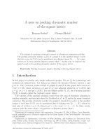
sorbable ties. The tumor weighed 2150 g, and appeared
smooth surfaced and well-circumscribed on macroscopic
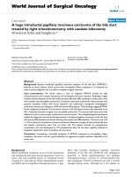
examination (Figure 2). Histologically, the tumor
appeared to be composed of a varying proportion of spin-
dle-shaped cells and collagen. The neoplastic cells dis-
played vesicular nuclei with demarcated nuclear
membranes, and dispersed chromatin. Mitoses were rare,
and immunoreactivity to vimentin, CD34, and Bcl2 were
positive; cytokeratin was n egative (Figure 3). The tumor
was pathologically diagnosed as benign localized fibrous
tumor of the pleura. The left lung expanded completely
and pulmonary function recovered to the normal level
after removal of the giant tumor. The postoperative
course was uneventful and the patient was discharged 12
days after the operation.
Discussion
Although diffuse pleural tumors or mesothelioma are
common, solitary fibrous pleu ral tumors are rare. SFT
represents less than 5% of pleural tumors [1] and occurs
most often in the visceral (80%) and parietal pleura (20%)
[2]. It has been recently considered to originate from the
mesenchymal cells of the s ubmesothelial connective tis-
sue of the pleura. Ac cording to immunohis tochemical
analysis, SFT of the pleura is positive for vimentin, CD34,
CD99, and Bcl2, which are markers of mesenchymal
cells; but it is negative for cytokeratin, w hich is found in
mesotheliomas. These results indicate that SFT originates
from mesenchymal cells rather than mesothe lial cells [1].
England et al. listed classical criteria of malignant SFT,
which is also useful for diagnosis, as follows: more than 4
mitotic activity in 10 high-powered fields, necrosis, high
cellularity, and pleomorphism [3].
The common presentations are relatively small tumors
less than 10 cm in diameter in an asymptomatic patient,
discovered incidentally on chest roentgenograms. For
tumors larger than 10 cm, occupying a large space and
compressing other thoracic structures may cause symp-
toms such as dyspnea, chest pain, cough, and fatigue.
Uncommonly hypertrophic pulmonary osteoarthropathy
and hypoglycemia are also caused. Hypertrophic osteoar-
thropathy, called Pierre Marie-Bamberge r syndrome, is
associated with the abnormal production of hyaluronic
acid by the tumors. Hypoglycemia is caused by the insulin-
like growth factor 2, which i s secreted by the tumors [2].
In our case, the gigantic tumor weighed 2150 g. Large
tumors, heavier than 2 kg, have been rarely reported
Figure 1 Chest radiography and CT scan images. (A) Initial chest radiography revealed a large well-circumscribed mass in the left thorax. (B)
Initial contrast-enhanced computed tomography showed a huge homogeneous, sharply defined mass compressing the aorta.
Figure 2 The gigantic encapsulated solitary fibrous tumor of
the pleura, weighed 2150 g and measured 20 cm × 19 cm ×
15 cm.
Furukawa et al. Journal of Cardiothoracic Surgery 2011, 6:122
/>Page 2 of 4
[4,5]. Larger tumors are more likely to be malignant and
are associated with the worst prognosis [3,5,6]. The pre-
sence of symptoms and pleural effusion, which are also
reported as factors associated with malignancy, are more
likely in patients with large tumors [3,7,8]. This indi-
cates that the prognosis depends on the complete
resectability o f the tumor and on the diagnosis o f
malignancy.
Occasional recurrences have been reported not only in
malignant cases but also in benign cases, even though it
is small percentage (1.4%) [7]. In our case, postoperative
adjuvant chemotherapy was not performed, because his-
tologically the tumor was identified as a benign SFT, and
surgical margins revealed no residual tumor. The role of
adjuvant chemotherapy in SFTs remains uncertain.
Although complete resection was achieved, close follow-
up is indicated because of the possibility of recurrence.
Conclusion
Wereportacaseofapatientwithagiganticsolitary
fibrous tumor (SFT) of the pleura. Although the tumor
compressed the lung, the desce nding aorta and other
mediastinal structures strongly, the patient had no symp-
toms except malaise and had normally worked as a furni-
ture remover. We successfully resected the huge solitary
fibrous tumor of the pleura via two separate thoraco-
tomies. Although SFT is beni gn, a long follow-up period
is essential as even patients with complete resection are
at risk of recurrence many years after surgery.
Consent
Written informed consent was obtained from the patient
for publication of this case report and any accompany-
ing images. A copy of the writ ten consent is available
for review by the Editor-in-Chief of this journal.
Abbreviations
SFT: solitary fibrous tumor; CT: computed tomography.
Author details
1
Department of Cardiothoracic Surgery, Heart and Diabetes Center North
Rhine-Westphalia, Georgstr. 11, 32545 Bad Oeynhausen, Germany.
2
Department of Pulmonology, Krankenhaus Bad Oeynhausen, Wielandstr. 28,
32545 Bad Oeynhausen, Germany.
Authors’ contributions
NF carried out the manuscript and collected references. JN and JG helped
to revise the manuscript. BH and AR underwent the operation. All Authors
read and approved the final manuscript.
Competing interests
The authors declare that they have no competing interests.
Received: 31 July 2011 Accepted: 29 September 2011
Published: 29 September 2011
References
1. Harrison-Phipps KM, Nichols FC, Schleck CS, Deschamps C, Cassivi SD,
Schipper PH, Allen MS, Wigle DA, Pairolero PC: Solitary fibrous tumors of
Figure 3 Microscopic examination of solitary fibrous tumor of the pleura. (A, B) Micr oscopic specimen of the tumor shows solid
proliferation of spindle-shaped fibroblastic cells in a patternless pattern. (Hematoxylin and eosin; magnification 40× and 200×) (C, D) Spindle-
shaped tumor cells show strong positivities for immunohistochemical staining with CD34 (C) and BCL2 (D).
Furukawa et al. Journal of Cardiothoracic Surgery 2011, 6:122
/>Page 3 of 4
pleura: Results of surgical treatment and long-term prognosis. J Thorac
Cardiovasc Surg 2009, 138:19-25.
2. Shaker W, Meatchi T, Dusser D, Riquet M: An unusual presentation of
solitary fibrous tumor of the pleura: right atrium and inferior vene cava
compression. Eur J Cardiothorac Surg 2002, 22:640-2.
3. Cardillo G, Carbone L, Carleo F, Masala N, Graziano P, Bray A, Martelli M:
Solitary fibrous tumors of the pleura: An analysis of 110 patients treated
in a single institution. Ann Thorac Surg 2009, 88:1632-7.
4. Hu CK, Chang YL, Lin WC, Lee YC: Resection of giant thoracic solitary
fibrous tumor through two separate thoracotomies. J Thorac Cardiovasc
Surg 2008, 136:1077-9.
5. Altinok T, Topcu S, Tastepe I, Yazici Y, Cetin G: Localized fibrous tumors of
the pleura: Clinical and surgical evaluation. Ann Thorac Surg 2003,
76:892-5.
6. Parrot M, Kurt AM, Robert JH, Borisch B, Spiliopoulos A: Clinical behavior of
solitary fibrous tumors of the pleura. Ann Thorac Surg 1999, 67:1456-9.
7. Magdeleinat P, Alifano M, Petino A, Rochais JP, Dulmet E, Galateau F,
Icard P, Regnard JF: Solitary fibrous tumors of the pleura: clinical
characteristics, surgical treatment and outcome. Eur J Cardiothorac Surg
2002, 21:1087-93.
8. Nonaka M, Kadokura M, Takaba T: Benign solitary fibrous tumors of the
parietal pleura which invaded the intercostal muscle. Lung Cancer 2001,
31:325-9.
doi:10.1186/1749-8090-6-122
Cite this article as: Furukawa et al.: A silent gigantic solitary fibrous
tumor of the pleura: case report. Journal of Cardiothoracic Surgery 2011
6:122.
Submit your next manuscript to BioMed Central
and take full advantage of:
• Convenient online submission
• Thorough peer review
• No space constraints or color figure charges
• Immediate publication on acceptance
• Inclusion in PubMed, CAS, Scopus and Google Scholar
• Research which is freely available for redistribution
Submit your manuscript at
www.biomedcentral.com/submit
Furukawa et al. Journal of Cardiothoracic Surgery 2011, 6:122
/>Page 4 of 4