Báo cáo y học: "Technical phosphoproteomic and bioinformatic tools useful in cancer research" potx
Bạn đang xem bản rút gọn của tài liệu. Xem và tải ngay bản đầy đủ của tài liệu tại đây (1.8 MB, 14 trang )
REVIE W Open Access
Technical phosphoproteomic and bioinformatic
tools useful in cancer research
Elena López
1*
, Jan-Jaap Wesselink
2,3
, Isabel López
4
, Jesús Mendieta
2,3
, Paulino Gómez-Puertas
2†
and
Sarbelio Rodríguez Muñoz
5*†
Abstract
Reversible protein phosphorylation is one of the most important forms of cellular regulation. Thus,
phosphoproteomic analysis of protein phosphorylation in cells is a powerful tool to evaluate cell functional status.
The importance of protein kinase-regulated signal transduction pathways in human cancer has led to the
development of drugs that inhibit protein kinases at the apex or intermediary levels of these pathways.
Phosphoproteomic analysis of these signalling pathways will provide important insights for operation and
connectivity of these pathways to facilitate identification of the best targets for cancer therapies. Enrichment of
phosphorylated proteins or peptides from tissue or bodily fluid samples is required. The application of technologies
such as phosphoenrichments, mass spectrometry (MS) coupled to bioinformatics tools is crucial for the
identification and quantification of protein pho sphorylation sites for advancing in such relevant clinical research. A
combination of different phospho peptide enrichments, quantitative techniques and bioinformatic tools is necessary
to achieve good phospho-regulation data and good structural analysis of protein studies. The current and most
useful pro teomics and bioinformatics techniques will be explained with research examples. Our aim in this article is
to be helpful for cancer research via detailing proteomics and bioinformatic tools.
Introduction
Phosphoproteomics plays an important role in our
understanding of how phosphorylation participates in
translating distinct signals into the normal and or
abnormal physiological responses, and has shifted
research towards screening for potential therapies for
diseases and in-depth analysis o f phosphoproteomes.
These issues can also be studied by structural analysis of
proteins and bioinformatic tools. Specific domains dis-
criminate between the phosphorylated vs. the non-phos-
phorylated state of proteins, based on the
conformational changes induced by the presence of a
negatively-cha rged phosphate group in the basal state of
the phosphopeptide [1]
Phosphorylated proteins, chemically quite stable, are
prone to enzymatic modification, so that when tissues
or cells are lysed, it is very likely that further enzymatic
reactions will occur [2]. Good sample preparation is the
key to successful analysis. These will generally be sna p-
frozen and treated with phosphatase inhibitors to avoid
modifying phosphopeptides during sample work-up
[3,4]. Also, it is critical to avoid salts and detergents,
which can decrease the recovery of phosphopeptides or
interfere with subsequent analysis [5]. Phosphopeptides
generally make up a small portion of the peptides in a
given protein sample, making detection difficult. Their
enrichment [e.g. via Immobilised metal ion affinity chro-
matography (IMAC), Titanium dioxide metal-based
chromatography (TiO
2
), Zirconium dioxi de (ZrO
2
),
Sequential elution from IMAC (SIMAC) or Calcium
phosphate precipitation] helps to combat this problem.
When combining the previ ously mentioned phos-
phoenrichments with Strong cation and anion exchange
(SCX and SAX) or Hydrophilic interaction chromatogra-
phy (HILIC), large-scale phosphoproteomic studies o f
interest can be carried out successfully [6]. If the goal of
the research study in cludes quantification of phosphory-
lated proteins, there are several useful techniques [e.g.
Stable Isotope Labelling with Amino acids in cell
* Correspondence: ;
† Contributed equally
1
Centro de Investigación i+12 del Hospital Universitario 12 de Octubre, Avda
de Córdoba s/n Madrid, 28041, Spain
5
Servicio de Digestivo, Hospital Universitario 12 Octubre, Avda de Córdoba
s/n Madrid, 28041, Spain
Full list of author information is available at the end of the article
López et al. Journal of Clinical Bioinformatics 2011, 1:26
/>JOURNAL OF
CLINICAL BIOINFORMATICS
© 2011 López et al; license e BioMed Central Ltd. This is a n Open Access article distributed under the terms of the Creative Commons
Attribution License ( which permits unr estricted use, distribution, and reproduction in
any medium, provided the original work is properly cited.
Culture (SILAC), Isobaric Tag for Relative and Absolute
(iTRAQ), Absolute Quantitation (AQUA), Multiple
Reaction Monitoring (MRM), or Label-free quantifica-
tion], which allow important large-scale phosphoproteo-
mic studies [7-19]
Once the phosphorylation state of a protein, consti-
tutive or associated to cancer disorders has been estab-
lished by proteomics methods, a range of
bioinformatics methods permits deeper study of its
properties and contacts. Using sequence analysis,
sequence comparison, virtual approaches of protein-
protein, protein-ligand interaction or molecular
dynamics simulations, initial physical information can
be applied for the potential development of persona-
lized approaches, aimed at the concept of personalized
medicine. Bioinformatics covers a w ide spectrum of
techniques for the generation and use of beneficial
information from structure, sequence or relationships
among biological items (DNA, RNA, proteins, macro-
molecular complexes, etc) [20,21]. From all these
methods, those most useful in clinical cancer studies
are: Ascore, PhosphoScore, data analysis from Next-
Generation Sequencing, studies of sequence compari-
son and sequence–structure relationship, homology
modelling and the more sophisticated rational drug
design and molecular dynamics techniques. Using
phosphoproteomics together with structural analysis of
proteins and bioinformatic tools, important biological
understanding of malignant diseases can be ach ieved.
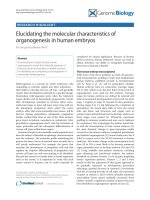
A prototypica l proteomics coup led to bioinforma tics
pipe-line useful for clinical cancer research is illu-
strated ( Figure 1)
Current MS-based resins to isolate phosphoproteins-
phosphopeptides useful for cancer research
Immobilised metal ion affinity chromatography (IMAC),
Titanium dioxide metal-based chromatography (TiO
2
),
Sequential elution from IMAC (SIMAC) and Zirconium
dioxide (ZrO
2
)
TiO
2
and IMAC are capable ofbindingnegatively
charged phosphate groups from aqueous solutions. Sim-
ple and complex samples containing phosphopeptides
andnon-phosphorylatedpeptidesaredissolvedinan
acidic solution to reduce the n on-specific binding of
acidic peptides (e.g. those con taining aspartic acid an d
glutamic acid), and to stimulate the electrostatic interac-
tions between the negatively charged peptides, mainly
phosphopeptides, and the metal ions. The phosphopep-
tides isolated are eluted from the stationary phase using
alkaline buffers [22]
Both resins (TiO
2
and IMAC) have the drawback of
binding a cidic non-phosphorylated peptides (negatively
charged peptides). Peptides containing acidic amino acid
residues, glutamic acid and aspartic acid, can also bind
to the metal ions. Ficarro et al (2002) circumvented this
difficulty with IMAC (Fe
3+
)byconvertingacidicamino
acid residues to methyl esters [23-29]. Heck et al [27]
suggested esterification of the acidic residues prior to
the MS analysis, as they observed a number of non-
phosphorylated peptides in their analysis. Larsen et al
[34] achieved higher specificity and yield compared to
IMAC (Fe
3+
) for the selective enrichment of phosphory-
lated peptides from model proteins when using 2,5-dihy-
droxybenzoic acid (DHB) with TiO
2
. In addition, more
phosphopeptides are bound to the metal ions and more
phosphopeptides can be eluted by using ammonium
hydroxide as the eluent by use of glycolic aci d in the
loading buffer of TiO
2
[30-35]
SIMAC allows enrichment of mono and multiply-
phosphopeptides in a single experiment, and, from com-
plex biological samples. Mono-phosphorylated peptides
mainly elute from IMAC (Fe
3+
) under acidic conditions
whereas multi-phosphorylated peptides elute at high
basic pH. Following SIMAC protocol, TiO
2
allows cap-
ture of the unbound mono-phosphorylated peptides in
the combined IMAC flow-through and washing steps
[35,36]
ZrO
2
, like the phosphoenrichments previously men-
tioned, is very useful for phosphopeptide isolation prior
to MS analysis. The strong affinity of ZrO
2
nanoparti-
cles to phosphopeptides enables the specific enrichment
of phosphopeptides from a complex peptide mixture in
which the abundance of phosphopeptides is two orders
of magnitude lower than that of n onphosphopeptides
[37,38]
Calcium phosphate precipitation (CPP), Strong cation and
anion exchange (SCX and SAX) and Hydrophilic interaction
chromatography (HILIC)
CPP consists of a pre-fractionation step in order to sim-
plify and enr ich phosphopeptides from complex sam-
ples. CPP coupled to two step IMAC (Fe
3+
) procedure
resulted in the observation of a higher number of phos-
phopeptides recovered. Phosphopeptides are precipitated
by adding 0.5 M NaHPO
4
and 2 M NH
3
OH to the pep-
tide-mixture followed by 2 M CaCl
2
. The washed pellet
(with 80 mM CaCl
2
) is dissolved in 5% of formic acid.
Before isolating the phosphopeptides by IMAC (Fe
3+
),
the resulting peptide-mixture is desalted via reversed
phase chromatography (RP) [39]
A positively charged analyte is attracted to a negatively
charged solid-support, and a negatively charged analyte
is attracted to a positively charged solid-support during
SCX and SAX operations respectively. SCX and SAX
has been successfully combined with IMAC and resulted
in greater recovery and identification by MS of interest-
ing phosphorylated peptides originating from yeast pher-
omone signalling pathway and membrane proteins
respectively [28,40]
López et al. Journal of Clinical Bioinformatics 2011, 1:26
/>Page 2 of 14
HILIC consist of a liquid/liquid extraction system
between the mobile and stationary phase. A water-rich
layer on the surface of the stationary phase (polar) is
formed; therefore a distributio n of the analytes betwee n
these two layers will occur. Weak electrostatic mechan-
isms as well as hydrogen donor interactions between
neutral polar molecules under high organic elution con-
diti ons occur during HILIC operat ions. Moreover, more
polar compounds have stronger interaction with the sta-
tionary aqueous layer than less polar compounds, result-
ing in a stronger retention [41]
Pros and Cons of Phosphoproteomic tools
Using IMAC, TiO
2
and ZrO
2
, the negatively charged
phosphopeptides are purified by their affinity to posi-
tively charged metal ions. However, some of these meth-
ods experience the problem of binding acidic, non-
phosphorylated peptides. Ficarro et al [29] bypassed this
problem on IMAC (Fe
3+
) b y converting acidic peptides
to methyl esters but increased the spectra complexit y
and required lyophilization of the sample, causing
adsorptive losses of phosphopeptides in particular. TiO
2
chromatography using DHB was introduced as a pro-
mising strateg y by Larsen et al [34]. Ti O
2
/DHB resulted
in higher specificity and yield compared to IMAC (Fe
3+
)
for the selective enrichment of pho sphorylated peptides
from model proteins (e.g. lactoglobulin bovine, casein
bovine). TiO
2
offers increased capacity compared to
IMAC resins in order to b ind and elute mono-phos-
phorylated peptides. TiO
2
exploits the same pr inciple as
IMAC, and is similarly prone to nonspecific retention of
acidic nonphosphorylated peptides. However, when
loading peptides in DHB, glycolic and phthalic acids,
nonspec ific binding to TiO
2
is r educed, thereby improv-
ing phosphope ptide enrichment without chemical mod i-
fication of the sample. SIMAC appeared as a
phosphopeptide enrichment tool which exploits t he
Figure 1 A prototypical proteomics pipe-line coupled to bioinformatics useful for clinical research. Depending on the application,
different samples processed and fed into the proteomics pipeline yield different results. The pipeline’s several steps are listed in the different
panels: (1) proteolytic digest, (2) the separation and ionization of peptides, (3) their analysis by mass spectrometry, (4) fragmentation of selected
peptides and analysis of the resulting MS/MS spectra and, (5) (6) data-computer bioinformatic-analysis, which mainly includes: Conversion-data
format, Spectrum identification with a search engine, Validation of identifications, Protein inference, Organization in local data managements
systems, Interpretation and classification of the protein lists, Transfer to public data repositories, Identification and Classification of proteins,
Quantification, Structural Analysis of proteins, PTM analysis and Cellular composition.
López et al. Journal of Clinical Bioinformatics 2011, 1:26
/>Page 3 of 14
properties of IMAC coupled to TiO
2
, thus facilit ating
more refined studies [36]
Another phosphopeptide enrichment prior to mass
spectrometric analysis i s ZrO
2
[37] and its principle is
based on metal affinity chromatography like IMAC and
TiO
2
.ZrO
2
permits the isolation of single phosphory-
lated peptides in a more selective manner t han TiO
2
[30]
Strategies which consist of fractionating and subse-
quently enriching phosphopeptides on a proteome wide
scale are based on SCX/SAX and HILIC interaction
chromatography. Calcium phosphate precipitation is
also a useful pre-fractionation step to simplify and
enrich phosphopeptides from complex samples which
can be coupled to IMAC and TiO
2
[13]. Mainly those
phosphopeptides from highly expressed proteins within
cells can be purified, while those from phosphorylated
proteins with low level expression (e.g . kinases) do not
bind so well to those resins. This is an important limita-
tion concerning phosphoenrichment methods and is due
to the low proportion of this kind of protein, or, their
available amount binds to metal ions although not su ffi-
ciently so as to be detected by MS.
The combination of SCX with IMAC has been proven,
resulting in a huge number of phosphorylated residues
identified (over 700 including Fus3p kinase). Although
more than 100 signalling proteins and functional phos-
phorylation sites, including receptors, kinases and tran-
scription factors, have been identified, it is clear that
only a fraction of the phosphoproteome has been
revealed [7,40]
Combinations of HILIC with IMAC have been proven
in clinical studies (e.g. HeLa samples), with the result of
the identification of a large number o f phosphorylated
residues (around 1000) [41]
Improvement in methodologies to enrich for phos-
phorylated residues from kinases is clearly necessary.
However, this is not straightforward for several reasons:
the low abundance of those signalling molecules within
cells, the stress/stimulation time-duration, as only a
small fraction of phosphorylated kinases are available at
any given time as a result of a stimulus and the time
adaptation over signalling pathways [5]
Current phosphoproteomic MS-based quantitative
strategies presently used for cancer research
Stable Isotope Labelling with Amino acids in cell Culture
(SILAC), Isobaric Tag for Relative and Absolute (iTRAQ),
Absolute Quantitation (AQUA), Multiple Reaction
Monitoring (MRM) and
18
O labelling
SILAC is a technique based on MS that detects differ-
ences in protein abundance among samples using non-
radioactive isotopic labelling. Two populations of cells
are cultivated in cell culture. One of the cell populations
is fed with growth medium containing normal amino
acids. The second population is fed with growth med-
ium containing amino acids labelled with stable (non-
radioactive) heavy isotopes. For example, the medium
can contain arginine labelled with six carbon-13 atoms
(
13
C) instead of the normal carbon-12 (
12
C). W hen the
cells are growing in this medium, they incorporate the
heavy arginine into all of their proteins. All of the argi-
nine containing peptides are now 6 Da heavier than
their normal counterparts. The trick is that the proteins
from both cell populations can be combined and ana-
lyzed together by MS. Pairs of chemically identical pep-
tides of different stable-isotope composition can be
differentiated via MS owing to their mass difference
[42-45]
iTRAQ uses isotope-coded covalent tags and is based
on the covalent labelling of the N-terminus and side
chain amines of peptides from protein digestions with
tags of varying mass. There are currently two mainly
used reagents: 4-plex and 8-plex, which can be used to
label all peptides from different samples/treatments.
These samples are then pooled and usually fractionated
by nano liquid chromatography and analyzed by tandem
MS (MS/MS). The fragmentation of the attached tag
generates a low molecular mass reporter io n that can be
used to relatively quantify the peptides and the proteins
fromwhichtheyoriginated.Thesignalsofthereporter
ions of each MS/MS spectrum allow for calculating the
relative abundance (ratio) of the peptide(s) identified by
this spectrum. In contrast t o SILAC and AQUA
(described below), it is during MS/MS experiments, that
relative quantification of peptides takes place [46-50]
AQUA was developed for the precise determination of
protein expression and post-translational modification
(PTM) levels. A peptide from a protein is constructed
synthetically containing stable isotopes, and the AQUA
peptide is the isotopically labelled synthetic peptide. The
synthetic peptides can be synthesized with PTMs. The
stable isotopes are incorporated into the AQUA peptide
by using isotopically “heavy” amino acids during the
synthesis p rocess of the peptide of interest (native pep-
tide). The synthetic peptide has a mass increase of e.g.
10Dal tons, due to the incorporation of a
13
C
6
and
15
N
4
-
arginine into the synthetic peptide, compared to the
native peptide. The mass difference between the native
and the synthetic peptide allows the mass spectrometer
to differentiate between the two forms - both forms
have the same chemical properties - resulting in the
same chromatographic retention, ionization efficiency,
and fragmentation distribution [51-53]
MRM r equires that knowledge of the sequence of the
protein be known in order to calculate precursor and
fragment ion values, which can be used to trigger
dependent ion scans in a qTRAP (hybrid triple
López et al. Journal of Clinical Bioinformatics 2011, 1:26
/>Page 4 of 14
quadrupole linear ion trap mass spectrometer). It can
also be used to perform a precursor ion and neutral loss
scan, to identify unknown phosphopeptides from a com-
plex mixture, and is a powerful method for the identifi-
cation and quantification of PTMs in proteins. Indeed,
MRM has been used by White et al to identify and
quantify tyrosine phosphorylated kinases for hundreds
of nodes within a signalling network and across multiple
experimental conditions. White et al.; Cox et al.,and
other relevant scientists [48,49,54,55] applied this strat-
egy for phospho quantitative analysis of signalling net-
works, identifying and quantifying a high number of
tyrosine phosphorylated peptides, obtaining an extre-
mely high percentage of signalling nodes covered.
18
O labelling is a label-free strategy that incorporates a
stable isotope
18
O-labelled ″universal″ reference sample
as a comprehensive set of internal standards for analyz-
ing large sample sets quantitatively. As a pooled sample,
the
18
O-labelled ″universal″ reference sample is spiked
into each individually processed unlabelled biological
sample and the peptide/protein abundances are quanti-
fied based on
16
O/
18
O i sotopic peptide pair abundance
ratios that compare each unlabelled sample to the iden-
tical reference sample. This approach also allows for the
direct application of label-free quantitation across the
sample set simultaneously along with the labelling-
approach (e.g ., dual-quantitation) since each b iological
sample is unlabelled except for the labelled reference
sample t hat is used as internal standard. The effective-
ness of this approach for large-scale quantitative proteo-
mics has been demonstrated by Qian et al 20 09; Wong
et al 2008 and other important scientists, giving relevant
clues for malignant diseases [56,57]
Some examples of phosphorylated proteins involved in
relevant clinical diseases explaining how useful
phosphoproteomic tools are for those clinical
investigations
Some drugs that bind to microtubules and block mitosis
are ineffective in cancer treatment; others show inexplic-
able focal efficacy. The vinca alkaloids are useful for
treating lymphoma, neuroblastoma and nephroblasto-
mas, whereas taxol is useful for advanced breast cancer
and ovarian cancer. It is not known why these drugs are
not all equally effective n or is it known why they have
different therapeutic value against different cancers.
Steen et al [58] examined the role of phosphorylation
on the dynamics of the anaphase promoting complex
(APC), observing distinct phosphorylation states of the
APC in response to different antimitotic drugs and sug-
gest that they may explain some of these differences.
Cells from different tissues or with different mutations,
or cells under different physiological stresses such as
hypoxia, may differ in their response to spindle poisons
and would reflect those differences in different sites of
phosphorylation.
Differences in spindle checkpoint phosphorylation may
reveal new features of the mitotic state. The ability to
characterise drug candidates based o n the spectrum of
APC phosphorylations may facilitate the discrimination
of the response of tumours to drugs and the identifica-
tion of new means of checkpoint control.
The authors suggested that the results of their study
indicate that the term mitotic arrest is a misnomer:
arrest is a dynamic state in which some cells enter
apoptosis and other cells revert to interphase. The abil-
ity to observe biochemical events during arrest could be
very important fo r understanding antiproliferative
treatments.
Exploring the dynamics of phosphorylation makes
great demands on the accuracy of quantitation. Most
MS-based quantitative approaches including SILAC and
iTRAQ give relative data, meaning that one state of
phosphor ylation is determ ined relative to another phos-
phorylation state. These data can help to establish the
kinetics of a pathway. These approaches allowed the
measurement of specific quantitative changes in APC
phosphorylation in cells arrested in nocodazole for vary-
ing periods. If these dynamics can be correlated with
the proc ess by which the arrested state is resolved, they
may provide us with new tools to understand the mito-
tic process and to find more effective drug targets in
cancer [59-61]
Development of drugs for specific biological pathways
with inc reased specificity and reduced toxicity has vali-
dated the long-held belief in the cancer research com-
munity that a precise molecular understanding of cancer
can result in cancer therapy.
An example of cancer-specific drugs is the develop-
ment of Herceptin - a monoclonal antibody against the
HER2 receptor for breast cancer therapy. HER2 is an
important target in cancer. HER2 overexpression
increases tumour cell proliferation, invasiveness and pre-
dicts poor prognosis. Wolf-Yadlin and other scientists
[48,49,58-61] have used phosphoproteomics and MS to
investigate the role of phosphorylation in the effects of
HER2 overexpression on EGF- and HRG-mediated sig-
nalling of erbB receptors. They identified specific combi-
nations of phosphorylation sites that correlate with cell
proliferation and migration and that potentially repre-
sent targets for therapeutic intervention. 68 out of 322
phosphorylation sites could be analysed kinetically and
it marks an important breakthrough in the characterisa-
tion of the erbB receptor signalling network in tumours
and illustrates the importance of understanding protein
phosphorylation.
Mitochondria play a central role in energy metabolism
and cellular survival and consequently mitochondrial
López et al. Journal of Clinical Bioinformatics 2011, 1:26
/>Page 5 of 14
dysfunction is associated with a number of human
pathologies. Mitochondrial dysfunction is linked to insu-
lin resistance in humans with obesity and type 2 dia-
betes. Zhao et al (2011) [62] studied the
phosphoproteome of the mitochondria isolated from
human skeletal muscle. They revealed extensive phos-
phorylation of inner membrane protein complexes and
enzymes combining TiO
2
with reve rse phase chromato-
graphy coup led to MS analysis. 155 distinct phosphory-
lation sites i n 77 mitochondrial phosphoproteins
including 116 phosphoserine, 23 phosphothreonine and
16 phosphotyrosine residues were identified. They also
assigned phosphorylation sites in mitochondrial proteins
involved in amino acid degradation, importers and
transporters, calcium homeostasis and apoptosis. Many
of these mitochondrial phosphoproteins a re substrates
for protein kinase A, protein kinase C, casein kinase II
and DNA-dependent protein kinase. The high number
of phosphotyrosine residues suggests an important role
for tyrosine phosphorylation in mitochondria l signalling.
Many of the mitochondr ial phosphoproteins are
involved in oxidative phosphorylation, tricarboxylic acid
cycle and lipid metabolism e.g. processes proposed to be
involved in insulin resistance [63].
In this study [64] the most prevalent form of cellular
protein post-translational modifications (PTMs) reversi-
ble phosphorylation is emerging as a central mechanism
in the regulation of mitochondrial functions [64-71].
Boja et al (2009) [50] successfully monitored phosphory-
lation sites of mitochondrial proteins including adenine
nucleotide transl ocase, malate dehydrogenase and mito-
chondrial creatine kinase. Among them, four proteins
exhibited phos phorylation changes with these physiolo-
gical stimuli: BCKDH-E1a subunit increased phosphory-
lation at Ser337 with DCA and de-energization,
apoptosis-inducing factor phosphorylation was elevated
at Ser345 with calcium, ATP synthase F1 comple x a
subunit and mitofilin dephosphorylated at Ser65 and
Ser264 upon de-ener gization. This screening validated
the iTRAQ technology as a method for functional quan-
titation of mitochondrial protein phosphorylation as
well as providing insights into the regulation of mito-
chondria via phosphorylation [69-71]
White et al [48,49] applied iTRAQ and MRM for
phosphor-quantitative analysis of signalling networks
identifying and quantifying 222 tyrosine phosphorylated
peptides, obtaining an extremely high percentage of sig-
nalling nodes covered. Ziwei Yu et al (2007) using
AQUA as a novel system of in situ quantitative protein
analysis, studied the protein expression levels of phos-
phorylated Akt (p-Akt). Activation of Akt in tumours is
mediated via several mechanisms including activation of
cell membrane receptor tyrosine kinases such as EGFR
and loss of phosphatase PTEN with dephosphorylation
of phosphoinositol triphosphate. Ziwei et al discovered
tha t Akt activatio n in oropharyngeal squamous cell car-
cinoma (OSCC) is associated with adverse patient out-
come, indicating that Akt is a promising molecular
target in oropharyngeal squamous cell carcinoma [53]
White et al [59,61] defined the mechanisms by which
EGFRvIII protein alters cell physiology, as it is one of
the most commonly mutated proteins in GBM and has
been linked to radiation and chemot herapeutic resis-
tance. They performed a phosphoproteomic analysis of
EGFRvIII signalling networks in GBM cells. They pro-
vided important insights into the biology of this mutated
receptor including oncogene dose effects and differential
utilization of signalling pathways. Clustering of the
phosphoproteomic data set revealed a previously unde-
scribed crosstalk between EGFRvIII and the c-Met
receptor. They observed that treatment of the cells with
a c ombination employing both EGFR and c-Met kinase
inhibitors dramatically decreased cell viability in vitro.
Hoffert et al [72] carried out quantitative phosphopro-
teomic analysis of vasopressin-sensitive renal cells of rat
inner medullary collecting duct cells by using IMAC
and p hosphorylation-site identification by MS combin-
ing label-free quantitation.
They identified 714 phosphorylation sites on 223
unique phosphoproteins from inner medullary collecting
duct samples treated short term with either calyculin A
or vasopressin. Rinschen et al [73] studied vasopressin’s
actionin renal cells related to the fact that the regulation
of water transport depends on protein phosphorylatio n.
Using SILAC with two treatment groups (0.1 nM
dDAVP or vehicle for 30 min), they carried out quantifi-
cation of 2884 phosphopeptides. The majority of quanti-
fied phosphopeptides did not change in abundance in
response to dDAVP. Analysis of the 273 phosphopep-
tides increased by dDAVP showed a predominance of
so-called “basophilic” motifs consistent with activation
of kinases of the AGC family. Increases in phosphoryla-
tion of several known protein kinase A targets were
found. Increased phosphorylation of targets of the cal-
mod ulin-dependent kinase family was also seen, includ-
ing autophosphorylation of calmodulin-dependent
kinase 2 at T286. Analysis of the 254 ph osphopeptides
decreased in abundance by dDAVP showed a predomi-
nance of so called “proline-directed ” motifs, consistent
with down-regulation of mitogen-activated or cyclin-
dependent kinases. dDAVP decreased phosphorylation
of both JNK1/2 (T183/Y185) and ERK1/2 (T183/Y185;
T203/Y205), consistent with a decrease in activation of
these proline-directed kinases in response to dDAVP.
Both ERK and JNK were able to phosphorylate residue
S261 of aquaporin-2 i n vitro, a site showing a decrease
in phosphorylation in response to dDAVP in vivo. Their
data support roles for multiple vasopressin V2-receptor-
López et al. Journal of Clinical Bioinformatics 2011, 1:26
/>Page 6 of 14
dependent signalling pathways in the vasopressin signal-
ling network of collecting duct cells, involving several
kinases not generally accepted to regulate collecting
duct function. We should remark that Hoffert and co-
workers carried out a very interesting research study, via
a label-free quantitation strategy that measures phos-
phopeptide precursor ion abundances from extracted
ion chromatograms (XIC).
The comparison of cellular phosphorylation levels for
control, epidermal growth factor stimulus and growth
fact or combined with kinase inhibitors has been studied
by Mann et al [74] using triple labelling SILAC coupled
to SCX and TiO
2
.
They evaluated the effects of kinase inhibitors on the
entire cell signalling network. From tho usands of phos-
phopeptides, less than 10% had a response pattern indi-
cative of targets of U0126 and SB202190, two widely
used MAPK inhibitors. They found that the 83% of the
growth factor-induced phosphorylation events were
affected by either or both inhibitors, showing quantita-
tively that early signalling processes are predominantly
transmitted through the MAPK cascades. In contrast to
MAPK inhibitors, dasatinib, a clinical drug directed
against BCR-ABL, which is the cause of chronic myelo-
genous leukemia, affected nearly 1,000 phosphopeptides.
Their assay is streamlined and could become a useful
tool in kinase drug development.
Knowlton et al [45] conducted quantitative mass spec-
trometry via SILAC and immunoaffinity purification of
tyrosine phosphorylated peptides to profile candidate
SRC-substrates induced by the CSF-1R tyrosine k inase
by comparing the phosphotyrosine-containing peptides
from cells expressing either CSF-1R or a mutant form
of this RTK that is unable to bind to SFKs.
They identified uncharacterized changes in tyrosine
phosphorylation i nduced by CSF -1R in mammary
epithelial cells as well as a set of candidate substrates
dependent on SRC recruitment to CSF-1R. Many of
these candidates may be direct SRC targets as the amino
acids flanking the phosphorylation sites in these proteins
are similar to known SRC kinase phosphorylation
motifs. Their collection of substrates includes proteins
involved in multiple cellular processes including cell-cell
adhesion, endocytosis and signal transd uct ion. Analyses
of phosphoproteomic data from breast and lung cancer
patient samples identified a subset of the SRC-depen-
dent phosphorylation sites as being strongly correlated
with SRC activatio n, which represent candidate markers
of SRC activation downstream of receptor tyrosine
kinases in human tumours.
Integrins interact with extracellular matrix (ECM) and
deliver intracellular signalling for cell proliferation, sur-
vival and motility. During tumour metastasis, integrin-
mediated cell adhesion and migration on the ECM
proteins are required for cancer cell survival and adapta-
tion to the new microenvironment.
Chen Y et al [75] using SILAC, IMAC and MS pro-
filed the phosphoproteomic changes induced by the
interactions of cell integrins with type I collagen, the
most common ECM substratum. The authors depicted
an integrin-modulated phosphorylation network during
cell-ECM protein interactions and revealed novel regula-
tors for cell adhesion and migration, discovering that
integrin-ECM interactio ns modulate phosphorylation of
517 serine, threonine or tyrosine residues in 513 pep-
tides, corresponding to 357 proteins. Among these pro-
teins, 33 key signalling mediators with kinase or
phosphatase activity were subjected to siRNA-based
functional screening. In their study, three integrin-regu-
lated kinases, DBF4, PAK2 and GRK6, were identif ied
for thei r critical role in cell adhesion and migration pos-
sibly through their regulat ion of actin cytoskeleton
arrangement.
Current Bioinformatics Tools useful for Phosphoproteomic
Research in Cancer studies
PhosphoScore
Correct phosphorylation site assignment is a critical
aspect of phosphoproteomic analysis. Large-scale phos-
phopeptide data sets that are generated through liquid
chromatography-coupled tandem MS often contain hun-
dreds or thousands of phosphorylation sites that require
validation.
PhosphoScore is an open-s ource assignment program
that is compatible wit h phosphopeptide data from mul-
tiple MS levels (MSn). It consists of an algorithm which
takes into account the match quality and the normalized
intensity of observed spectral peaks compared to a theo-
retical spectrum. It has been demonstrated by Rutten-
berg et al [76] that PhosphoScore produces > 95%
correct MS2 assignments from known synthetic data, >
98% agreement with an established MS2 assignment
algorithm (Ascore), and > 92% agreement with visual
inspection of MS3 and MS4 spectra. It was successfully
used for the isolation of phosphopeptides from rat liver.
The resulting phosphopeptides were enriched via IMAC
and analized by MS allowing important data of phos-
phorylated proteins from rat liver.
Ascore
Ascore consists of a statistical algorithm that measures
the probability of correct phosphorylation site localiza-
tion based on the presence and intensity of site-deter-
mining ions in MS2 spectra. Phosphorylation sites with
an Ascore ≥ 19 (corresponding t o > 99% certainty) ar e
usua lly considered unambiguously assigned. The Ascore
algorithm is compatible with MS2 spectra and phos-
phorylation sites from phosphopeptides found only at
the MS3 level are assigned by manual examination of
López et al. Journal of Clinical Bioinformatics 2011, 1:26
/>Page 7 of 14
the spectra (http://ascor e.med.harvard.edu/ascor e.php).
To distinguish the correct site(s) of phosphorylation for
each phosphopeptide, automated site assignment is per-
formed on MS2 data using the Ascore algorithm. It was
used for an interesting research study of the phospho-
protein aquaporin-2 (AQP2) that was also quantified.
This particular AQP2 peptide was identified from an
MS3 spectrum and contained three unambiguously
assigned phosphorylation sites: Ser-256, Ser-261, and
Ser-264. A previous phosph oproteomic study by the
samegroupincludedMS-basedquantificationofAQP2
at Ser-256 and Ser-261. The dramatic increase in abun-
dance of this phosphopeptide in vasopressin-treated
samples was consistent with increased phosphorylation
of AQP2 at Ser-256 in response to vasopressin [77]
Next Generation Sequencing
Next Generation Sequencing (NGS) has been recently
used in a detailed study of genes involved in Colorectal
Cancer (CRC) [78]. As a main conclusion of the study,
the authors stated that sequencing of whole tumour
exomes allowed predic tion of the microsatellite status of
CGC, facilitating, at the same time, the putative finding
of relevant mutations. In addition, NGS can be applied
to formalin-fixed and paraffin embedded material, allow-
ing the renewed study of all the ancient mater ial stored
in the pathology departments [79].
Sequence-to-sequence and sequence-to-structure
comparisons (MSA: multiple sequence analysis)
Once mutations or phosphorylation of modified residues
have been found in sequencing or p roteomics studies,
routine sequence-to-sequence and sequence-to-structure
comparisons (MSA: multiple sequence a nalysis) are
applied to obtain valuable information on the nature of
the functional implications of the mutated residues in
the protein context. Multiple alignments of proteins,
and mainly those based on the comparison of experi-
mentally obtained-three dimensional atomic structures
(structural alignments), are a very valuable source of
information related to the evolutionary strategies fol-
lowed by the different members of a family of proteins
to conserve or modify their f unctions and structures
[80]
The analysis of structural alignments allows the detec-
tion of at least three types of regions or multiple align-
ment positions according to conservation:
1. Conserved positions, usually key for function or
structure maintenance.
2. Tree-determinant residues, conserved only in pro-
tein subfamilies and related to family-specific active
sites, substrate binding sites or protein-protein interac-
tion surfaces. These sites contain essential information
for the design of family-specific activator or inhibitor
drugs [81].
3. Positions that correspond to compensatory muta-
tions that s tabilize the mutations in one protein with
changes in the other (co rrelated mut ations). These sites
are very effective for the detection of protein-protein
interaction contacts [82], as they allow for the selection
of the correct structural arrangement of two proteins
based on the accumulation of signals in the proximity of
interacting surfaces.
Homology modelling methods
As a consequence of the sequence-to-structure compari-
son, and in absence of experimental crystal structures,
the homology modelling meth ods, can develop a 3D
model from a protein sequence based on the structures
of a crystallized homologous protein. The method can
only be applied to proteins having a common evolution-
ary origin, as only for proteins that are hypothesized to
be homologous, this assertion implies that their three-
dimensional structures are conserved to a greater extent
than their primary structures. For cases where a good
homology hypothesis cannot be supported, alternative
methods can be applied in order to obtain a putative 3D
structure. These procedures, known as “far-homolo gy
modelling” or “threading” methods, provid e structures
with lower confidence compared to those generated
using homology modelling methods.
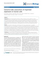
Routine pipe-line for structural bioinformatics techni-
ques used from structure identification to Molecular
Dynamics analysis of the phosphorylated forms is sum-
marized in Figure 2.
The 3D structure of the active centre of a protein of interest
Information on the 3D structure of the active centre of
a protein of interest and/or its natural ligands can be
used as a basis for the design of effective drugs. This
rational drug design is usually performed using multiple
docking experiments in the active centre of the said pro-
tein, requiring the use of advanced software such as
Autodock-4 [83], that a llows the evaluation of not only
the docking to a rigid model of the active centre, but
also a certain mobility of the side chain of enzyme resi-
dues to the ligand shape. Typically, all the calculated
binding conformations to the target protein obtained in
every docking run are clustered according to scoring cri-
teria (as “lowest binding energy model” or “lowest
energy model representative of the most-populated clus-
ter”) and sorted according to their estimated free energy
of binding. These computer procedures are a useful
cost-reducing tool to prospect and model new molecules
with potential inhibiting properties or even successful
future drugs. Recently, rational drug design approach
has been used in the case of putative cancer therapie s,
focused on the pharmacological reactivation of mutant
p53 [84]. T his promising str ategy implies t he simulta-
neous use of several approaches for the identification of
López et al. Journal of Clinical Bioinformatics 2011, 1:26
/>Page 8 of 14
small molecules that target mutant p53, including “de
novo” design and screening of chemical libraries.
Molecular dynamics (MD) techniques
Finally, molecular dynamics (MD) techniques are com-
monly used to obtain refined models for protein struc-
ture, protein-protein and protein-ligand interactions.
Molecular dynamics is a computational simulation
technique in which atoms within molecules are allowed
to interact for a period of time according to the princi-
ples of physics. In the case of proteins, the relevant
forces taken into account are the electrostatic
interactions (attractive or repulsive), Van der Waals
interactions, and the properties of the covalent bond
(length, angle, and dihedral angle). In general, simula-
tion times for macromolecular protein complexes are up
to 20 ns and the number of atoms of the simulated sys-
tems is in the order of up to 250,000, including solvent
molecules. MD techniques have been used to simulate
the individual behaviour of small p rotein or peptides
[85], protein-protein interfaces and ligand-protein rela-
tionship in catalytic macromolecular complexes with
GTPase activity [86,87] or kinases involved in cell
Figure 2 Routine pipe-line for structural bioinformatics analysis of protein phosphorylated states. Once t he protein is identified, a
sequence-based search (1) in the Protein Data Bank ( structure database is done to download a 3D structure suitable
to be used in computational simulation studies. In the case that the protein is not present in the database, bioinformatics modelling methods
are used to generate an approximate model of the desired structures (2). Next step consists of the generation of the 3D model for the single
protein or the interacting pair of proteins both in the unphosphorylated (basal) or the phosphorylated states (3). Finally, a Molecular Dynamics
approach is used to compare the behaviour of the two states. RMSD (root mean square distance) values are collected for several nanoseconds
in order to obtain a quantitative measure of the differences (4).
López et al. Journal of Clinical Bioinformatics 2011, 1:26
/>Page 9 of 14
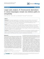
Figure 3 Case study. Analysis of the structural inte ractions of GRK2 [Swiss-Prot: P21146], Gaq [Swiss-Prot: P21279]andGbg proteins
[Swiss-Prot: P62871and Swiss-Prot: P63212] according to the crystallized structure of the macromolecular complex [PDB: 2BCJ].A.
Crystallized structure of the complex of GRK2, Gaq and Gbg polypeptides. Position of a GTP molecule in Gaq active centre is indicated. B.
Computer model of the electrostatic interaction between a putative phosphorylated GRK2-Ser121 residue and Arg214 of Gaq. C: Surface models
for GRK2 protein in the vicinity of Ser121 residue. Left: Unphosphorylated Ser121; centre: model for the putative phosphorylated state of Ser121.
Right: complementarity between the positively Arg214 and negative pSer121charged residues patched in both protein surfaces, probably
implicated in the stabilization of the complex. D. Root mean square deviation (RMSD) plots of the protein domains implicated in the GRK2-Gaq
interaction in presence (green) or absence (red) of phosphorylated Ser121 during a simulation of molecular dynamics. Plots are presented solely
to illustrate the putative stabilization of the complex after Ser121 phosphorylation. Figure plots were generated using PyMOL Molecular Graphics
System, Schrödinger, LLC.
López et al. Journal of Clinical Bioinformatics 2011, 1:26
/>Page 10 of 14
signalling pathways (e.g. Src ty rosine kinase [88] or pro-
tein kinase B/Akt [89])
Figure 3 shows, as an example, the bioinformatics ana-
lysis of the crystallized macromolecular complex of acti-
vated G proteins [90], composed of, GaqandGbg
proteins. GRK2 has been implied in the inhibition of
WNT signalling [91], a pathway that plays a central role
in the etiology of colorectal cancer. GRK2 plays a pivotal
role in the G protein-coupled receptor (GPCR) desensi-
tization and re-sensitization processes. The increasing
complexity of the GRK2 “interact ome” implies this
kinase in several cardiovascular, inflammatory or
tumour pathologies [92-94]
Using the crystallized structure of the GRK2-Gaq-Gbg
complex as initial template (Figure 3A), and homology
modelling procedures, a model was generated illustrat-
ing the putative interaction between Arg214 in the Gaq
chain and a putative phosphorylated Ser121 in the
GRK2 chain (Figure 3B). As expected, the main qualita-
tive changes in surface electrostatic properties corre-
spond to an increase in the surface electro-negativity
caused by the presence of an extra phosphate group in
pSer212. This added negative charge complements the
positive charge of Arg214, stabilizing the protein contact
(Figure 3C). T o obtain a quantitative comparison
between both phospho- and unphosphorylated states of
Ser121, a simulated molecular dynamics procedure was
applied for 10 nanoseconds. The variation in the inter-
action complex was evaluated by continuous measuring
of root-mean square deviation (rmsd) values with
respect to the initial crystallized structure. The result,
shown in Figure 3D, indicates that the presence of a
phosphate group associ ated to Se r121 results in more
stable interaction.
From a clinical perspec tive, this result would indicate
that the presence of a mutated Ser121 residue in GRK2
will produce different effects depending on the nature of
the new residue. A c onservative mutation (e.g. S121A)
will not cause important changes in the overall 3D
structure of GRK2, but a consolidation of the “unpho-
sphorylated” state, thus disturbing the p rotein-pro tein
contact at this level. However, putative mutations such
as S121D or S121E would generate a “constitutively
phos phorylated-like state”, stabilizing a reinforce d inter-
action between the two polypeptides.
All these results can be also extrapolated to all mem-
bers of the same family of proteins. Sequence analysis
reveals high similarity values, indicative of close homol-
ogy. Structure in Figure 2 corresponds to the bovine
GRK2 protein. Human close homologues are: GRK2,
GRK6, GRK5, GRK4 and GRK7. Sequence similarity
between these proteins will allow comparative studies of
the putative effect of Ser/Thr phosphorylation in the
interaction of all these kinases with their respective G
proteins.
Conclusions
Aberrant activation of kinase signalling pathways is com-
monly associated with several t ypes of can cer. Recent devel-
opments in phosphoprotein/pho sphopeptide enrichment
strategies, quantitative m ass spectrometry and bioinformatic
tools have resulted in robust pipelines for high-throughput
characterization of ph osphorylation in a global fashion.
It is possible to profile site-specific phosphorylation
events on thousands of proteins in a single experiment.
Chemical proteomic strategies have been used to unra-
vel targets of kinase inhibitors, which are otherwise diffi-
cult to charac terize. This approach’s poten tial is already
being used to characterize signalling pathways that gov-
ern oncogenesis. We summarized various approaches
used for the analysis of the phosphoproteome in general
and protein kinases in particular, highlighting key cancer
phosphoproteomic studies.
Different proteomic and bioinformatic strategies need
to be combined to achieve good phosphopeptide quanti-
tative-protein studies. From the point of view of the so-
calle d “personalized medicine”, bioinformatics studies of
reversible phosphorylation in proteins will allow the
gene ration of models for protein-protein contacts at the
atomic level taking into account each particular protein
sequence. Molecular dynamic analysis of those contacts,
be it in healthy people or in cancer studies, will allow
the modification of the 3D computer models obtaining
virtual structures tailored to individual patients. The
next step in the future of drug development will be the
generation of drugs specifically designed to each particu-
lar patient. It is necessary that clinicians, proteomics and
bioinformatics work together in order to improve thera-
pies and drug candidates development.
List of Abbreviations
Note: These abbreviations are useful proteomic abbreviations; some of them are
mentioned and described in this Review, and they are also described in the
References of this article.
AQUA: Absolute Quantitation; CID: Collision-Induced Dissociation; Da:
Dalton (molecular mass); DIGE 2-D: Fluorescence Difference Gel
Electrophoresis; ECD: Electron Capture Dissociation; ESI: Electron Spray
Ionization; ETD: Electron Transfer Dissociation; FT-ICR: Fourier transform-Ion
Cyclotron Resonance; HILIC: Hydrophilic interaction chromatography; HPLC:
High-performance liquid chromatography or high-pressure liquid
chromatography; H
3
PO
4
Phosphoric acid; ICR: Ion Cyclotron Resonance;
IMAC: Immobilized Metal Affinity Capture; IT: Ion Trap; iTRAQ: Isobaric Tag
for Relative and Absolute Quantitation; kDa: kilodalton (molecular mass); LC:
Liquid Chromatography; MALDI: Matrix-Assisted Laser Desorption/Ionization;
MD: Molecular Dynamics; MOAC: Metal Oxide Affinity Chromatog raphy; Mr:
Relative molecular mass (dimensionless); MRM: Multiple reaction monitoring;
MS: Mass Spectrometry; MSA: MultiStage Activation; MS/MS: tandem mass
spectrometry; m/z: Mass to charge ratio; PID: Primary Immunodeficiencies;
PTM: Post-Translational Modification; SILAC: Stable Isotope Labelling with
Amino acid in cell Culture; SIMAC: Sequential Elution from IMAC; TiO
2
Titanium dioxide; TOF: Time Of Flight; ZrO
2
: Zirconium dioxide
López et al. Journal of Clinical Bioinformatics 2011, 1:26
/>Page 11 of 14
Acknowledgements
EL is a recipient of a Post-doctoral fellowship of Ministerio de Ciencia e
Innovación de España. IL is a recipient of a FLL (Fundación Leucemia y
Linfoma) grant. SRM holds a tenured position at Spanish National Hospital
12 de Octubre. This study was supported by: the Spanish Ministerio de
Ciencia e Innovación through grants SAF2007-61926 (to PGP) and the
European Commission through grant FP7 HEALTH-F3-2009-223431 (to PGP).
Biomol-Informatics was financed by the European Social Fund. Support from
the “Fundación Ramón Areces” is acknowledged. We also thank the Centro
de Computación Científica-UAM for computational support. Special thanks
Prof. Ernest Feytmans (Honorary Director at Swiss Institute of Bioinformatics
-Location
Geneva Area, Switzerland) and Prof. Shabaz Mohammed (Theme
Leader at the Netherlands Proteomics Centre, Lecturer Utrecht University)
who contributed to the publication of this article.
Author details
1
Centro de Investigación i+12 del Hospital Universitario 12 de Octubre, Avda
de Córdoba s/n Madrid, 28041, Spain.
2
Centro de Biología Molecular “Severo
Ochoa” (CSIC-UAM) Campus de Cantoblanco, c/Nicolás Cabrera, 1, 28049
Madrid, Spain.
3
Biomol-Informatics, S.L., Parque Científico de Madrid, Campus
de Cantoblanco, c/Faraday 7, 28049 Madrid, Spain.
4
Servicio de Hematología
Hospital QUIRÓN, Madrid, Diego de Velázquez 1 28223, Pozuelo Madrid
Spain.
5
Servicio de Digestivo, Hospital Universitario 12 Octubre, Avda de
Córdoba s/n Madrid, 28041, Spain.
Authors’ contributions
EL carried out the proteomics, phosphoproteomics and mass spectrometry
studies for this review. JJW, JM and PGP carried out the bioinformatic
studies for this review. IL and SMR carried out the clinical studies for this
review. EL, JJW, IS, JM, PGP and SMR carried out these complementary
studies in order to develop Clinical Phosphoproteomic-Bioinformatic
research and publish this article. All authors read and approved the final
manuscript.
Competing interests
The authors declare that they have no competing interests.
Received: 9 June 2011 Accepted: 3 October 2011
Published: 3 October 2011
References
1. López E, López I, Sequi J, Ferreira A: Discovering and validating unknown
phosphosites from p38 and HuR protein kinases in vitro by
Phosphoproteomic and Bioinformatic tools. Journal of Clinical
Bioinformatics 2011, 1(1):16[ />16].
2. Virshup DM, Shenolikar S: From promiscuity to precision: protein
phosphatases get a makeover. Mol Cell 2009, 33(5):537-45.
3. Grønborg M, Kristiansen TZ, Stensballe A, Andersen JS, Ohara O, Mann M,
Jensen ON, Pandey A: A mass spectrometry-based proteomic approach
for identification of serine/threonine-phosphorylated proteins by
enrichment with phospho-specific antibodies: identification of a novel
protein, Frigg, as a protein kinase A substrate. Mol Cell Proteomics 2002,
1(7):517-27.
4. Zhang ZY: Functional studies of protein tyrosine phosphatases with
chemical on different phosphopeptide enrichment techniques. Biochim
Biophys Acta 2005, 1754(1-2):100-7.
5. Jensen SS, Larsen MR: Evaluation of the impact of some experimental
procedures on different phosphopeptide enrichment techniques. Rapid
Commun Mass Spectrom 2007, 21(22):3635-45.
6. White FM: Quantitative phosphoproteomic analysis of signalling network
dynamics. Curr Opin Biotechnol 2008, 19(4):404-9.
7. Schmelzle K, White FM: Phosphoproteomic approaches to elucidate
cellular signalling networks. Curr Opin Biotechnol 2006, 17(4):406-14.
8. Springer WR: A method for quantifying radioactivity associated with
protein in silverstained polyacrylamide gels. Anal Biochem 1991,
195(1):172-6.
9. Wyttenbach A, Tolkovsky AM: Differential phosphoprotein labeling
(DIPPL), a method for comparing live cell phosphoproteomes using
simultaneous analysis of (33)P- and (32)P-labeled proteins. Mol Cell
Proteomics 2006, 5(3):553-9.
10. Ong SE, Mann M: Mass spectrometry based proteomics turns
quantitative. Nat Chem Biol 2005, 1(5):252-62.
11. Blaukat A: Identification of G-protein-coupled receptor phosphorylation
sites by 2D phosphopeptide mapping. Methods Mol Biol 2004, 259:283-97.
12. Gafken PR, Lampe PD: Methodologies for characterizing phosphoproteins
by mass spectrometry. Cell Commun Adhes 2006, 13(5-6):249-62.
13. Lopez E, Lopez I, Ferreira A, Sequi J: Clinical and Technical
Phosphoproteomic Research. Proteome Sci 2011, 9(1):27.
14. Zhang H, Zha X, Tan Y, Hornbeck PV, Mastrangelo AJ, Alessi DR,
Polakiewicz RD, Comb MJ: Phosphoprotein analysis using antibodies
broadly reactive against phosphorylated motifs.
J Biol Chem 2002,
277(42):39379-87.
15.
Rush J, Moritz A, Lee KA, Guo A, Goss VL, Spek EJ, Zhang H, Zha XM,
Polakiewicz RD, Comb MJ: Immunoaffinity profiling of tyrosine
phosphorylation in cancer cells. Nat Biotechnol 2005, 23(1):94-101.
16. Di Vizio D, Solomon KR, Freeman MR: Cholesterol and cholesterol-rich
membranes in prostate cancer: an update. Tumori 2008, 94(5):633-9.
17. Dietz A, Boehm A, Mozet C, Wichmann G, Giannis A: Current aspects of
targeted therapy in head and neck tumors. Eur Arch Otorhinolaryngol
2008, 265(Suppl 1):S3-12.
18. Huang F, Gu H: Negative regulation of lymphocyte development and
function by the Cbl family of proteins. Immunol Rev 2008, 224:229-38.
19. Shah NP: Advanced CML: therapeutic options for patients in accelerated
and blast phases. J Natl Compr Canc Netw 2008, 6(Suppl 2):S31-S36.
20. Wang X, Liotta L: Clinical bioinformatics: a new emerging science. Journal
of Clinical Bioinformatics 2011, 1(1):1[ />content/1/1/1].
21. Baumgartner C, Osl M, Netzer M, Baumgartner D: Bioinformatic-driven
search for metabolic biomarkers in disease. Journal of Clinical
Bioinformatics 2011, 1(1):2[ />2].
22. Andersson L, Porath J: Isolation of phosphoproteins by immobilized
metal (Fe3+) affinity chromatography. Anal Biochem 1986, 154(1):250-4.
23. Neville DC, Rozanas CR, Price EM, Gruis DB, Verkman AS, Townsend RR:
Evidence for phosphorylation of serine 753 in CFTR using a novel metal-
ion affinity resin and matrix-assisted laser desorption mass
spectrometry. Protein Sci 1997, 6(11):2436-45.
24. Figeys D, Gygi SP, McKinnon G, Aebersold R: An integrated microfluidics-
tandem mass spectrometry system for automated protein analysis. Anal
Chem 1998, 70(18):3728-34.
25. Li S, Dass C: Iron (III)-immobilized metal ion affinity chromatography and
mass spectrometry for the purification and characterization of synthetic
phosphopeptides. Anal Biochem 1999, 270(1):9-14.
26. Posewitz MC, Tempst P: Immobilized gallium(III) affinity chromatography
of phosphopeptides. Anal Chem 1999, 71(14):2883-92.
27. Pinkse MW, Uitto PM, Hilhorst MJ, Ooms B, Heck AJ: Selective isolation at
the femtomole level of phosphopeptides from proteolytic digests using
2D-NanoLC-ESI-MS/MS and titanium oxide precolumns. Anal Chem 2004,
76(14):3935-43.
28. Nuhse TS, Stensballe A, Jensen ON, Peck SC: Large-scale analysis of in vivo
phosphorylated membrane proteins by immobilized metal ion affinity
chromatography
and mass spectrometry. Mol Cell Proteomics 2003,
2(11):1234-43.
29. Ficarro SB, McCleland ML, Stukenberg PT, Burke DJ, Ross MM,
Shabanowitz J, Hunt DF, White FM: Phosphoproteome analysis by mass
spectrometry and its application to Saccharomyces cerevisiae. Nat
Biotechnol 2002, 20(3):301-5.
30. Ashman K, Villar EL: Phosphoproteomics and cancer research. Clin Transl
Oncol 2009, 11(6):356-62.
31. López E, Matthiesen R, López I, Ashman K, Mendieta J, Wesselink JJ, Gómez-
Puertas P, Ferreira A: Functional phosphoproteomics tools for current
immunological disorders research. Journal of Integrated OMICS 2011,
1(1):1-16[].
32. Connor PA, Dobson KD, McQuillan J: Infrared Spectroscopy of the TiO2/
Aqueous Solution Interface. Langmuir 1999, 15:2402.
33. Connor PA, McQuillan A: Phosphate adsorption onto TiO2 from aqueous
solutions: an in situ internal reflection infrared spectroscopic study.
Langmuir 1999, 15:2916.
34. Larsen MR, Thingholm TE, Jensen ON, Roepstorff P, Jørgensen TJ: Highly
selective enrichment of phosphorylated peptides from peptide mixtures
López et al. Journal of Clinical Bioinformatics 2011, 1:26
/>Page 12 of 14
using titanium dioxide microcolumns. Mol Cell Proteomics 2005,
4(7):873-86.
35. Thingholm TE, Jensen ON, Larsen MR: Enrichment and separation of
mono- and multiply phosphorylated peptides using sequential elution
from IMAC prior to mass spectrometric analysis. Methods Mol Biol 2009,
527:67-78.
36. Thingholm TE, Jensen ON, Robinson PJ, Larsen MR: SIMAC (sequential
elution from IMAC), a phosphoproteomics strategy for the rapid
separation of monophosphorylated from multiply phosphorylated
peptides. Mol Cell Proteomics 2008, 7(4):661-71.
37. Kweon HK, Hakansson K: Selective zirconium dioxide-based enrichment
of phosphorylated peptides for mass spectrometric analysis. Anal Chem
2006, 78(6):1743-9.
38. Zhou H, Tian R, Ye M, Xu S, Feng S, Pan C, Jiang X, Li X, Zou H: Highly
specific enrichment of phosphopeptides by zirconium dioxide
nanoparticles for phosphoproteome analysis. Electrophoresis 2007,
28(13):2201-15.
39. Zhang X, Ye J, Jensen ON, Roepstorff P: Highly Efficient Phosphopeptide
Enrichment by Calcium Phosphate Precipitation Combined with
Subsequent IMAC Enrichment. Mol Cell Proteomics 2007, 6(11):2032-42.
40. Gruhler A, Olsen JV, Mohammed S, Mortensen P, Faergeman NJ, Mann M,
Jensen ON: Quantitative phosphoproteomics applied to the yeast
pheromone signalling pathway. Mol Cell Proteomics 2005, 4(3):310-27.
41. McNulty DE, Annan RS: Hydrophilic interaction chromatography reduces
the complexity of the phosphoproteome and improves global
phosphopeptide isolation and detection. Mol Cell Proteomics 2008,
7(5):971-80.
42. Ong SE, Blagoev B, Kratchmarova I, Kristensen DB, Steen H, Pandey A,
Mann M: Stable isotope labeling by amino acids in cell culture, SILAC, as
a simple and accurate approach to expression proteomics. Mol Cell
Proteomics 2002, 1(5):376-86.
43. Gruhler S, Kratchmarova I: Stable isotope labeling by amino acids in cell
culture (SILAC). Methods Mol Biol 2008, 424:101-11.
44. Ballif BA, Roux PP, Gerber SA, MacKeigan JP, Blenis J, Gygi SP: Quantitative
phosphorylation profiling of the ERK/p90 ribosomal S6 kinase-signalling
cassette and its targets, the tuberous sclerosis tumor suppressors. Proc
Natl Acad Sci USA 2005, 102(3):667-72.
45. Knowlton ML, Selfors LM, Wrobel CN, Gu TL, Ballif BA, Gygi SP,
Polakiewicz R, Brugge JS: Profiling. Y561-dependent and -independent
substrates of CSF-1R in epithelial cells. PLoS One 2010, 26;5(10).
46. Ross PL, Huang YN, Marchese JN, Williamson B, Parker K, Hattan S,
Khainovski N, Pillai S, Dey S, Daniels S, Purkayastha S, Juhasz P, Martin S,
Bartlet-Jones M, He F, Jacobson A, Pappin DJ: Multiplexed protein
quantitation in Saccharomyces cerevisiae using amine-reactive isobaric
tagging reagents. Mol Cell Proteomics 2004, 3(12):1154-69.
47. Sachon E, Mohammed S, Bache N, Jensen ON: Phosphopeptide
quantitation using amine-reactive isobaric tagging reagents and tandem
mass spectrometry: application to proteins isolated by gel
electrophoresis. Rapid Commun Mass Spectrom
2006, 20(7):1127-34.
48.
Zhang Y, Wolf-Yadlin A, White FM: Quantitative proteomic analysis of
phosphotyrosinemediated cellular signalling networks. Methods Mol Biol
2007, 359:203-12.
49. Wolf-Yadlin A, Hautaniemi S, Lauffenburger DA, White FM: Multiple
reaction monitoring for robust quantitative proteomic analysis of
cellular signalling networks. Proc Natl Acad Sci USA 2007, 104(14):5860-5.
50. Boja ES, Phillips D, French SA, Harris RA, Balaban RS: Quantitative
mitochondrial phosphoproteomics using iTRAQ on an LTQ-Orbitrap with
high energy collision dissociation. J Proteome Res 2009, 8(10):4665-75.
51. Kirkpatrick DS, Gerber SA, Gygi SP: The absolute quantification strategy: a
general procedure for the quantification of proteins and post-
translational modifications. Methods 2005, 35(3):265-73.
52. Wang G, Wu WW, Zeng W, Chou CL, Shen RF: Label-free protein
quantification using LC-coupled ion trap or FT mass spectrometry:
Reproducibility, linearity, and application with complex proteomes. J
Proteome Res 2006, 5(5):1214-23.
53. Yu Z, Weinberger PM, Sasaki C, Egleston BL, Speier WF, Haffty B, Kowalski D,
Camp R, Rimm D, Vairaktaris E, Burtness B, Psyrri A: Phosphorylation of Akt
(Ser473) predicts poor clinical outcome in oropharyngeal squamous cell
cancer. Cancer Epidemiol Biomarkers Prev 2007, 16(3):553-8.
54. Cox DM, Zhong F, Du M, Duchoslav E, Sakuma T, McDermott JC: Multiple
reaction monitoring as a method for identifying protein
posttranslational modifications. J Biomol Tech 2005, 16(2):83-90.
55. Williamson BL, Marchese J, Morrice NA: Automated identification and
quantification of protein phosphorylation sites by LC/MS on a hybrid
triple quadrupole linear ion trap mass spectrometer. Mol Cell Proteomics
2006, 5(2):337-46.
56. Qian WJ, Liu T, Petyuk VA, Gritsenko MA, Petritis BO, Polpitiya AD,
Kaushal A, Xiao W, Finnerty CC, Jeschke MG, Jaitly N, Monroe ME, Moore RJ,
Moldawer LL, Davis RW, Tompkins RG, Herndon DN, Camp DG, Smith RD,
Inflammation and the Host Response to Injury Large Scale Collaborative
Research Program: Large-scale multiplexed quantitative discovery
proteomics enabled by the use of an (18)O-labeled “universal” reference
sample. J Proteome Res 2009, 8(1):290-9.
57. Wong JW, Sullivan MJ, Cagney G: Computational methods for the
comparative quantification of proteins in label-free LCn-MS experiments.
Brief Bioinform 2008, , 2: 156-65.
58. Steen JA, Steen H, Georgi A, Parker K, Springer M, Kirchner M, Hamprecht F,
Kirschner MW: Different phosphorylation states of the anaphase
promoting complex in response to antimitotic drugs: a quantitative
proteomic analysis. Proc Natl Acad Sci USA 2008, 105(16):6069-74.
59. Zhang Y, Wolf-Yadlin A, Ross PL, Pappin DJ, Rush J, Lauffenburger DA,
White FM: Time-resolved mass spectrometry of tyrosine phosphorylation
sites in the epidermal growth factor receptor signalling network reveals
dynamic modules. Mol Cell Proteomics 2005, 4(9):1240-50.
60. Olsen JV, Blagoev B, Gnad F, Macek B, Kumar C, Mortensen P, Mann M:
Global,
in vivo, and site-specific phosphorylation dynamics in signalling
networks. Cell 2006, 127(3):635-48.
61. Wolf-Yadlin A, Kumar N, Zhang Y, Hautaniemi S, Zaman M, Kim HD,
Grantcharova V, Lauffenburger DA, White FM: Effects of HER2
overexpression on cell signalling networks governing proliferation and
migration. Mol Syst Biol 2006, 2:54.
62. Zhao X, León IR, Bak S, Mogensen M, Wrzesinski K, Højlund K, Jensen ON:
Phosphoproteome analysis of functional mitochondria isolated from
resting human muscle reveals extensive phosphorylation of inner
membrane protein complexes and enzymes. Mol Cell Proteomics 2011,
10(1).
63. Green DR, Kroemer G: The pathophysiology of mitochondrial cell death.
Science 2004, 305(5684):626-9.
64. Cohen P: The origins of protein phosphorylation. Nat Cell Biol 2002, 4(5):
E127-30.
65. Boneh A: Regulation of mitochondrial oxidative phosphorylation by
second messenger-mediated signal transduction mechanisms. Cell Mol
Life Sci 2006, 63(11):1236-48.
66. Hunter T: Signalling–2000 and beyond. Cell 2000, 100(1):113-27.
67. Pagliarini DJ, Dixon JEDixon: Mitochondrial modulation: reversible
phosphorylation takes center stage? Trends Biochem Sci 2006, 31(1):26-34.
68. Hüttemann M, Lee I, Samavati L, Yu H, Doan JW: Regulation of
mitochondrial oxidative phosphorylation through cell signalling. Biochim
Biophys Acta 2007, 1773(12):1701-20.
69. Goldenthal MJ, Marín-García J: Mitochondrial signalling pathways: a
receiver/integrator organelle. Mol Cell Biochem 2004, 262(1-2):1-16.
70. Horbinski C, Chu CT: Kinase signalling cascades in the mitochondrion: a
matter of life or death. Free Radic Biol Med 2005, 1;38(1):2-11.
71. Thomson M: Evidence of undiscovered cell regulatory mechanisms:
phosphoproteins and protein kinases in mitochondria. Cell Mol Life Sci
2002, 59(2):213-9.
72. Hoffert JD, Pisitkun T, Wang G, Shen RF, Knepper MA: Quantitative
phosphoproteomics of vasopressin-sensitive renal cells: regulation of
aquaporin-2 phosphorylation at two sites. Proc Natl Acad Sci USA 2006,
103(18):7159-64.
73. Rinschen MM, Yu MJ, Wang G, Boja ES, Hoffert JD, Pisitkun T, Knepper MA:
Quantitative phosphoproteomic analysis reveals vasopressin V2-
receptor-dependent signalling pathways in renal collecting duct cells.
Proc Natl Acad Sci USA 2010, 107(8):3882-7.
74. Pan C, Olsen JV, Daub H, Mann M:
Global effects of kinase inhibitors on
signalling
networks revealed by quantitative phosphoproteomics. Mol
Cell Proteomics 2009, , 12: 2796-808.
75. Chen Y, Lu B, Yang Q, Fearns C, Yates JR, Lee JD: Combined integrin
phosphoproteomic analyses and small interfering RNA–based functional
López et al. Journal of Clinical Bioinformatics 2011, 1:26
/>Page 13 of 14
screening identify key regulators for cancer cell adhesion and migration.
Cancer Res 2009, 69(8):3713-20.
76. Ruttenberg BE, Pisitkun T, Knepper MA, Hoffert JD: PhosphoScore: an
open-source phosphorylation site assignment tool for MSn data. J
Proteome Res 2008, , 7: 3054-9.
77. Hoffert JD, Wang G, Pisitkun T, Shen RF, Knepper MA: An automated
platform for analysis of phosphoproteomic datasets: application to
kidney collecting duct phosphoproteins. J Proteome Res 2007, , 9: 3501-8.
78. Timmermann B, Kerick M, Roehr C, Fischer A, Isau M, Boerno ST,
Wunderlich A, Barmeyer C, Seemann P, Koenig J, et al: Somatic mutation
profiles of MSI and MSS colorectal cancer identified by whole exome
next generation sequencing and bioinformatics analysis. PLoS One 2010,
5:e15661.
79. Schweiger MR, Kerick M, Timmermann B, Albrecht MW, Borodina T,
Parkhomchuk D, Zatloukal K, Lehrach H: Genome-wide massively parallel
sequencing of formaldehyde fixed-paraffin embedded (FFPE) tumor
tissues for copy-number- and mutation-analysis. PLoS One 2009, 4:e5548.
80. Zuckerkandl E, Pauling L: Molecules as documents of evolutionary history.
J Theor Biol 1965, 8:357-366.
81. López-Romero P, Gómez MJ, Gómez-Puertas P, Valencia A: Prediction of
functional sites in proteins by evolutionary methods. In Principles and
practice Methods in proteome and protein analysis. Edited by: Kamp RM,
Calvete J, Choli-Papadopoulou T. Berlin Heidelberg: Springer-Verlag;
2004:319-340.
82. Carettoni D, Gomez-Puertas P, Yim L, Mingorance J, Massidda O, Vicente M,
Valencia A, Domenici E, Anderluzzi D: Phage-display and correlated
mutations identify an essential region of subdomain 1C involved in
homodimerization of Escherichia coli FtsA. Proteins 2003, 50:192-206.
83. Huey R, Morris GM, Olson AJ, Goodsell DS: A semiempirical free energy
force field with charge-based desolvation. J Comput Chem 2007,
28:1145-1152.
84. Wiman KG: Pharmacological reactivation of mutant p53: from protein
structure to the cancer patient. Oncogene 2010, 29:4245-4252.
85. Mendieta J, Fuertes MA, Kunjishapatham R, Santa-Maria I, Moreno FJ,
Alonso C, Gago F, Munoz V, Avila J, Hernandez F: Phosphorylation
modulates the alpha-helical structure and polymerization of a peptide
from the third tau microtubule-binding repeat. Biochim Biophys Acta
2005, 1721:16-26.
86. Mendieta J, Rico AI, Lopez-Vinas E, Vicente M, Mingorance J, Gomez-
Puertas P: Structural and functional model for ionic (K(+)/Na(+)) and pH
dependence of GTPase activity and polymerization of FtsZ, the
prokaryotic ortholog of tubulin. J Mol Biol 2009, 390:17-25.
87. Mingorance J, Rivas G, Velez M, Gomez-Puertas P, Vicente M: Strong FtsZ is
with the force: mechanisms to constrict bacteria. Trends Microbiol 2010,
18:348-356.
88. Mendieta J, Gago F: In silico activation of Src tyrosine kinase reveals the
molecular basis for intramolecular autophosphorylation.
J Mol Graph
Model 2004, 23:189-198.
89. Calleja V, Laguerre M, Larijani B: 3-D structure and dynamics of protein
kinase B-new mechanism for the allosteric regulation of an AGC kinase.
J Chem Biol 2009, 2:11-25.
90. Tesmer VM, Kawano T, Shankaranarayanan A, Kozasa T, Tesmer JJ: Snapshot
of activated G proteins at the membrane: the Galphaq-GRK2-
Gbetagamma complex. Science 2005, 310:1686-1690.
91. Wang L, Gesty-Palmer D, Fields TA, Spurney RF: Inhibition of WNT
signalling by G protein-coupled receptor (GPCR) kinase 2 (GRK2). Mol
Endocrinol 2009, 23:1455-1465.
92. Bienz M, Clevers H: Linking colorectal cancer to Wnt signalling. Cell 2000,
103:311-320.
93. Aragay AM, Ruiz-Gomez A, Penela P, Sarnago S, Elorza A, Jimenez-Sainz MC,
Mayor F Jr: G protein-coupled receptor kinase 2 (GRK2): mechanisms of
regulation and physiological functions. FEBS Lett 1998, 430:37-40.
94. Penela P, Murga C, Ribas C, Lafarga V, Mayor F Jr: The complex G protein-
coupled receptor kinase 2 (GRK2) interactome unveils new
physiopathological targets. Br J Pharmacol 2010, 160:821-832.
doi:10.1186/2043-9113-1-26
Cite this article as: López et al.: Technical phosphoproteomic and
bioinformatic tools useful in cancer research. Journal of Clinical
Bioinformatics 2011 1:26.
Submit your next manuscript to BioMed Central
and take full advantage of:
• Convenient online submission
• Thorough peer review
• No space constraints or color figure charges
• Immediate publication on acceptance
• Inclusion in PubMed, CAS, Scopus and Google Scholar
• Research which is freely available for redistribution
Submit your manuscript at
www.biomedcentral.com/submit
López et al. Journal of Clinical Bioinformatics 2011, 1:26
/>Page 14 of 14