Báo cáo y học: "Multislice computed tomography is useful for evaluating partial anomalous pulmonary venous connectio" pdf
Bạn đang xem bản rút gọn của tài liệu. Xem và tải ngay bản đầy đủ của tài liệu tại đây (573.97 KB, 3 trang )
Kasahara et al. Journal of Cardiothoracic Surgery 2010, 5:40
/>Open Access
COMMENTARY
BioMed Central
© 2010 Kasahara et al; licensee BioMed Central Ltd. This is an Open Access article distributed under the terms of the Creative Commons
Attribution License ( which permits unrestricted use, distribution, and reproduction in
any medium, provided the original work is properly cited.
Commentary
Multislice computed tomography is useful for
evaluating partial anomalous pulmonary venous
connection
Hirofumi Kasahara
1
, Ryo Aeba*
1
, Yutaka Tanami
2
and Ryohei Yozu
1
Abstract
Volume-rendered images, derived from multidetector-row computed tomography (MDCT), can facilitate assessment
of the morphology of partial anomalous pulmonary venous connection and are thus useful in pre-operative planning
to prevent surgical morbidity and assist post-operative evaluations.
Introduction
Partial anomalous pulmonary venous connection
(PAPVC) is usually diagnosed by echocardiography, and
catheter-based angiographies are often performed for
confirmation. However, echocardiography occasionally
provides insufficient information due to its small field of
view, insufficient resolution to identify individual pulmo-
nary veins, and the difficulty of confirming its penetra-
tion into the connection site of the systemic venous
system, especially around the hilar [1,2]. Conventional
angiography remains the standard diagnostic tool,
despite its inherent risks and occasional insufficient reso-
lution for detecting faint images of the pulmonary vein in
the late phase [2,3]. Post-operative evaluations of recon-
structed vessels are often problematic in certain diseases.
High-slice multidetector-row computed tomography
(MDCT) angiography can provide more precise morpho-
logic delineation due to its non-invasive nature and high
spatial and temporal resolution. We herein report our
current experience of applying MDCT angiography for
pre- and post-operative evaluations in patients with
PAPVC.
Case Reports
A 10-year-old girl was diagnosed with a sinus venous sep-
tal defect with PAPVC of the right upper lobe vein to the
superior vena cava (SVC). Echocardiography and cathe-
ter angiography demonstrated a large defect with a signif-
icant left-to-right shunt. A pre-operative MDCT
angiogram revealed a higher SVC site connection (Fig.
1A), not detected in other examinations, and this child
had a persistent left SVC; hence the decision to avoid the
complex technique of patch placement within the rela-
tively small right-side SVC. During the operation, the
SVC was dissected extensively to beyond the high con-
nected pulmonary vein under the guidance of the MDCT
image. Caval division and atriocaval anastomosis
described by Warden [4] was performed according to
pre-operative planning.
A 4-year-old girl had anomalous drainage of the right
upper pulmonary vein to the SVC, underwent a repair
operation. During the operation, a pair of right upper
pulmonary veins draining into the proximal site of the
SVC was found following extensive dissection around the
atriocaval junction, but a pre-operative MDCT angio-
gram indicated an additional higher SVC site connection
(Fig. 1B). The higher one was erroneously considered to
be the azygos vein without MDCT angiography due to its
high connection onto the posterior aspect of the SVC in
operative findings. The connection site of the azygos vein
was found to be slightly distal following additional dissec-
tion around the SVC (Fig. 1C).
A 28 year-old woman had anomalous drainage of the
left upper pulmonary vein as a vertical vein into the bra-
chiocephalic vein, and underwent surgical repair. Post-
operative enhanced images of the target vessels, derived
from MDCT, demonstrated the patent pulmonary vein
anastomosed to the left atrial appendage (Fig. 1D), which
had been missed on echocardiography.
* Correspondence:
1
Division of Cardiovascular Surgery, Keio University, Tokyo, Japan
Full list of author information is available at the end of the article
Kasahara et al. Journal of Cardiothoracic Surgery 2010, 5:40
/>Page 2 of 3
Discussion
Having consecutively applied MDCT angiography to 12
patients with PAPVC, we note that the high quality of the
images facilitates precise pre-operative planning, making
MDCT useful for evaluating surgical morbidity post-
operatively with only minor failures.
Pre-operative information obtainable via MDCT
angiography concerning the number and sizes of pulmo-
nary veins into the SVC, the precise connection sites, and
the spatial relations between the PAPVC, azygos vein and
cavoatrial junction, is useful for surgical planning and
preventing surgical complications, as well as diagnosing
various types of PAPVC. Our experience suggests there is
a possibility of missing reconstruction of higher con-
nected pulmonary veins without sufficient preoperative
information because surgeons are likely not to extend dis-
section around the SVC because it is associated with a
certain risk of injury to the phrenic nerve unless adequate
preoperative information is available. To prevent cavoa-
trial stenosis in the Warden procedure, cavoatrial anasto-
mosis must be accomplished without tension, requiring
extensive dissection around the SVC toward the brachio-
cephalic vein and the jugulo-subclavian junction [5]. The
three-dimensional images from MDCT angiography
clearly reveal the morphology of pulmonary venous con-
nection without any need for mental reconstruction,
facilitating precise planning of the operation.
MDCT angiography is useful for post-operative evalua-
tions to clearly demonstrate patency or occlusion of the
connected pulmonary veins and SVC. No symptoms are
likely to emerge in a patient with bilateral SVC, such as
the present case (a 10-year-old girl), even with the recon-
structed vena cava totally occluded or complicated by
severe stenosis [5]. Occlusion of the reconstructed pul-
monary vein is also likely to be symptom-free, if its drain-
age area is limited. Post-operative transthoracic
echocardiography occasionally provides a poor image,
especially when using intracardiac patch baffling, making
it impossible to rule out significant surgical morbidity.
Catheter angiography may reveal reconstructed vessels
but is relatively invasive soon after the operation espe-
cially for small patients
Advanced magnetic resonance (MR) angiography
devices have potential as new standard tools for visualiz-
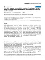
Figure 1 Volume-rendered image multidetector-row computed tomography (MDCT). A - Blue-colored vessels return to the superior vena cava
(SVC). The arrow indicates a high connected pulmonary vein. The arrowhead indicates the left persistent SVC. B, C - Target vessel-enhanced image
derived from MDCT. The arrow indicates the highest SVC site connection, which without this image is at risk of being mistaken for the azygos vein.
The arrowhead indicates the azygos vein. D - A target component-enhanced image derived from MDCT. The arrow indicates the anastomosis site of
the left atrium appendage with the vertical vein. The asterisk (green) indicates the left atrium.
*
DA
B C
Kasahara et al. Journal of Cardiothoracic Surgery 2010, 5:40
/>Page 3 of 3
ing pulmonary vein pathologies, since they allow non-
invasive assessment of anatomy and pathophysiology at
the same time [6]. When it comes to surgical planning,
however, surgeons are likely to prefer high resolution
MDCT angiography, since it offers a superior spatial res-
olution with clear morphology [2,7]. Although there are
concerns regarding the higher degree of patient exposure
to radiation than with MRI, its faster scanning and
reduced sedation requirements are beneficial, especially
in ill, uncooperative or small patients [7], and its wider
availability is also an advantage.
Conclusion
MDCT angiograms that facilitate improved assessment
of the morphology of PAPVC are useful in pre-operative
planning to prevent surgical complications, and assist
post-operative evaluations.
Consent
Written informed consent was obtained from the patient
for publication of this case-report and any accompanying
images. A copy of the written consent is available for
review by the Editor-in-Chief of this journal
Competing interests
The authors declare that they have no competing interests.
Authors' contributions
All authors have read and approved the final manuscript. HK performed the
operation and has been involved in drafting the manuscript. RA performed the
operation, has been involved in drafting the manuscript. YT performed analysis
of CT scan images, and has been involved in drafting the manuscript. RY per-
formed the operation, and has given the final approval to publish the manu-
script.
Author Details
1
Division of Cardiovascular Surgery, Keio University, Tokyo, Japan and
2
Department of Radiology, Keio University, Tokyo, Japan
References
1. Ucar T, Fitoz S, Tutar E, Atalay S, Uysalel A: Diagnostic tools in the
preoperative evaluation of children with anomalous pulmonary
venous connections. Int J Cardiovasc Imaging 2008, 24(2):229-35.
2. Khatri S, Varma SK, Khatri P, Kumar RS: 64-slice multidetector-row
computed tomographic angiography for evaluating congenital heart
disease. Pediatr Cardiol 2008, 29(4):755-62.
3. Sungur M, Ceyhan M, Baysal K: Partial anomalous pulmonary venous
connection of left pulmonary veins to innominate vein evaluated by
multislice CT. Heart 2007, 93(10):1292.
4. Warden HE, Gustafson RA, Tarnay TJ, Neal WA: An alternative method for
repair of partial anomalous pulmonary venous connection to the
superior vena cava. Ann Thorac Surg 1984, 38(6):601-5.
5. Nakahira A, Yagihara T, Kagisaki K, Hagino I, Ishizaka T, Koh M: Partial
anomalous pulmonary venous connection to the superior vena cava.
Ann Thorac Surg 2006, 82(3):978-82.
6. Grosse-Wortmann L, Al-Otay A, Goo HW, Macgowan CK, Coles JG, Benson
LN: Anatomical and functional evaluation of pulmonary veins in
children by magnetic resonance imaging. J Am Coll Cardiol 2007,
49(9):993-1002.
7. Moral S, Ortuno P, Aboal J: Multislice CT in congenital heart disease:
Partial anomalous pulmonary venous connection. Pediatr Cardiol 2008,
29(6):1120-1.
doi: 10.1186/1749-8090-5-40
Cite this article as: Kasahara et al., Multislice computed tomography is use-
ful for evaluating partial anomalous pulmonary venous connection Journal
of Cardiothoracic Surgery 2010, 5:40
Received: 10 December 2009 Accepted: 18 May 2010
Published: 18 May 2010
This article is available fro m: http://www. cardiothoracics urgery.org/con tent/5/1/40© 2010 Kasahara et al; licensee BioMed Central Ltd. This is an Open Access article distributed under the terms of the Creative Commons Attribution License ( ), which permits unrestricted use, distribution, and reproduction in any medium, provided the original work is properly cited.Journal of Cardiothoracic Surgery 2010, 5:40